
Effects of Intermediate Frequency (150 kHz) Electromagnetic Radiation on the Vital Organs of Female Sprague Dawley Rats
 ,
,  , , ,
, , ,  and
and
Simple Summary
Abstract
1. Introduction
2. Materials and Methods
2.1. Animal Care and Husbandry
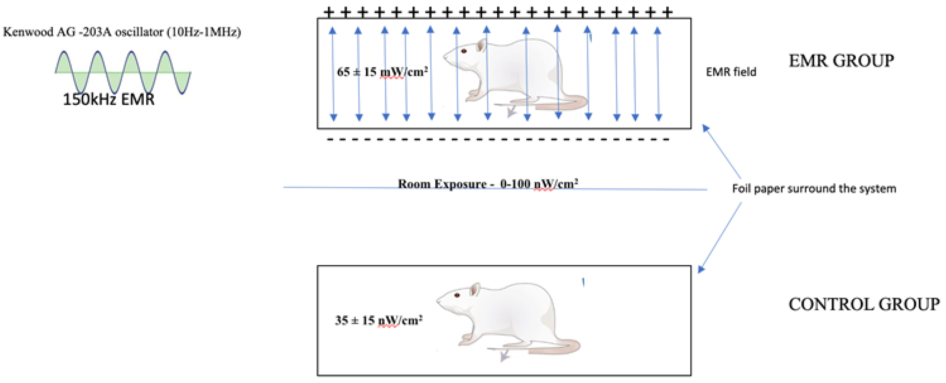
2.2. Experimental Setup and EMR-Generation System
2.3. After 2 Months of Exposure to EMR
2.4. Haematological and Biochemical Analysis
2.5. Gross and Histopathological Analysis
2.6. Statistical Analysis
3. Results
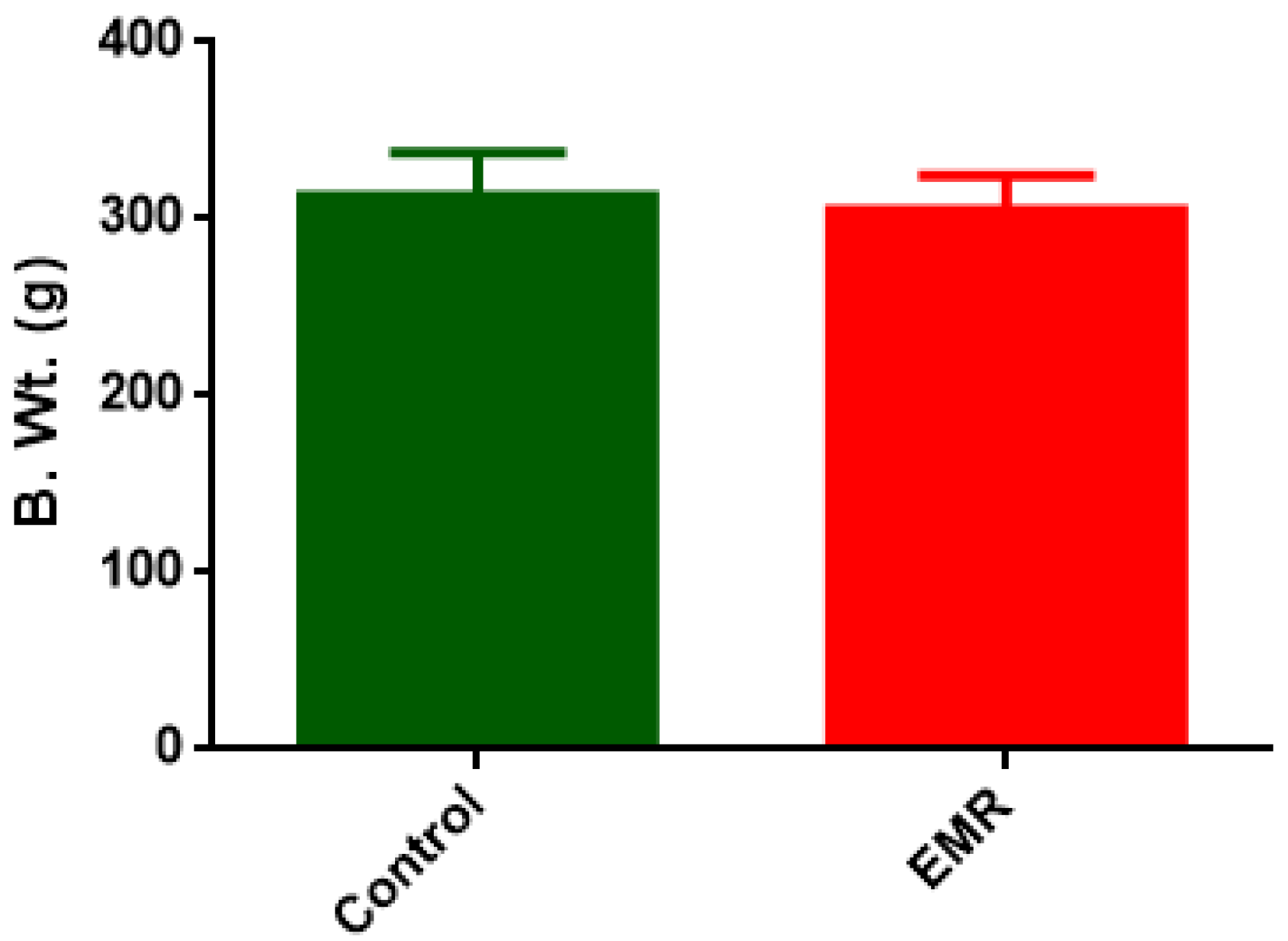
3.1. Body Weight
3.2. Haematological Analysis
3.3. Serum Biochemical Analysis
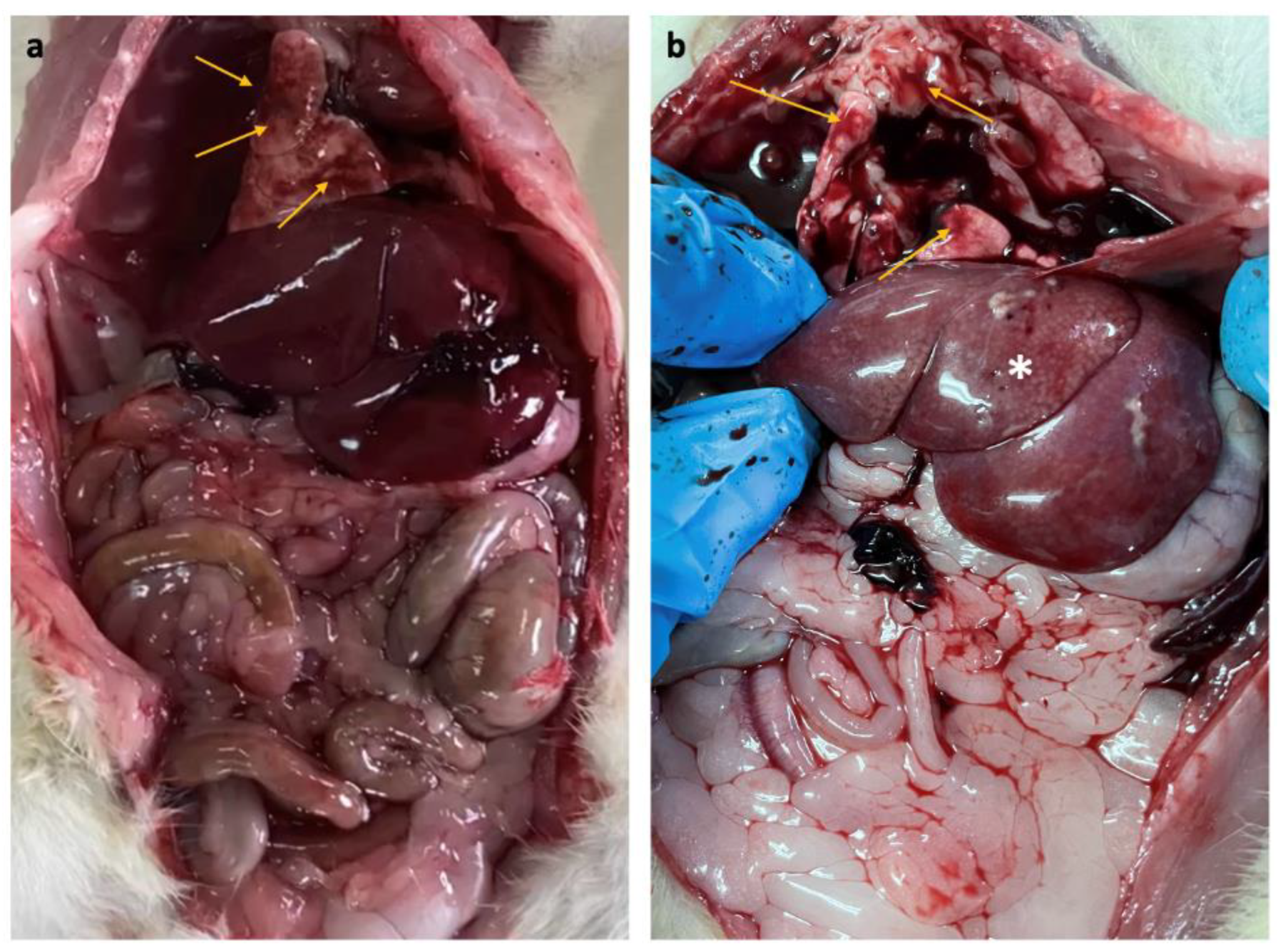
3.4. Gross Pathology
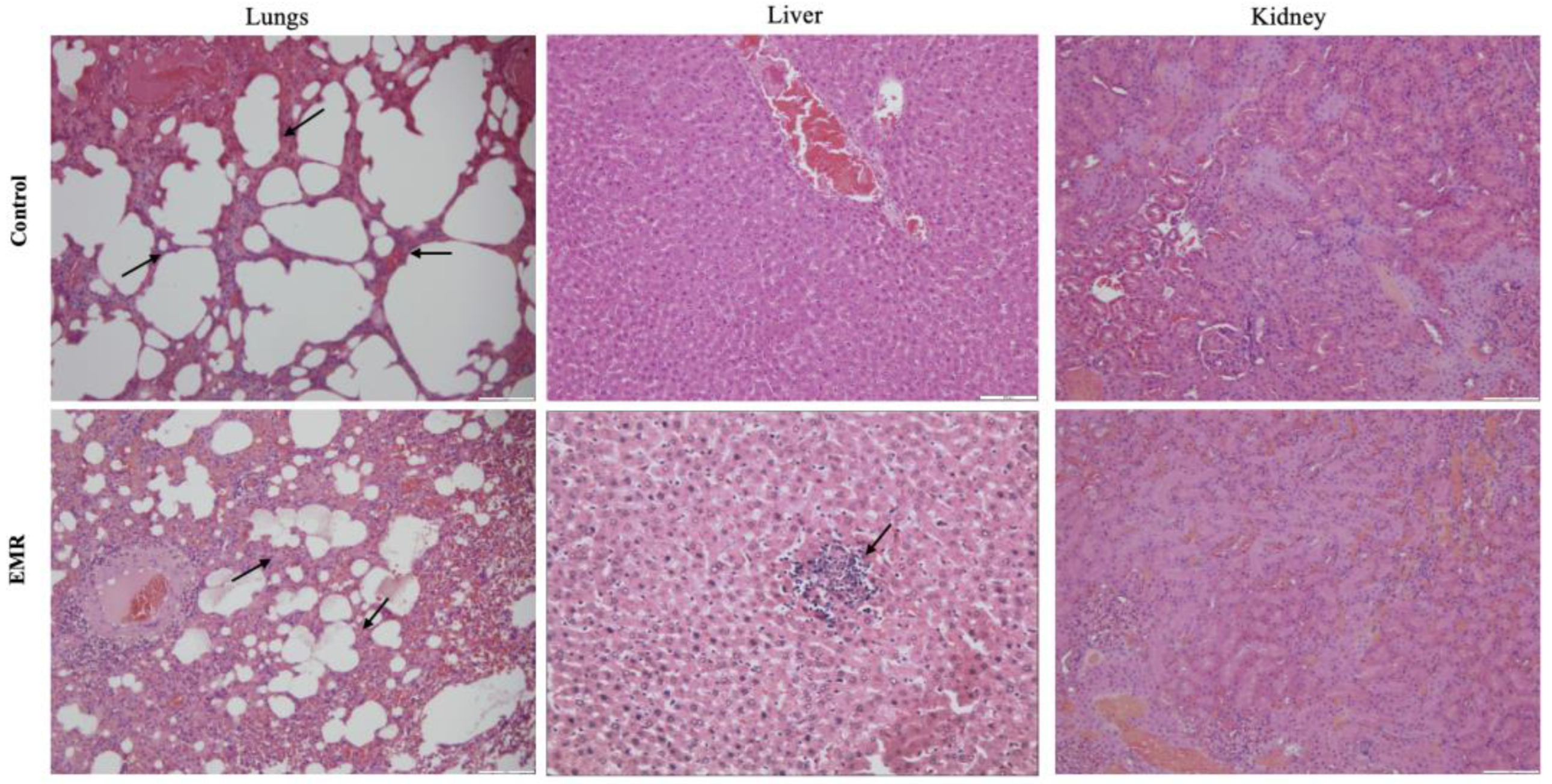
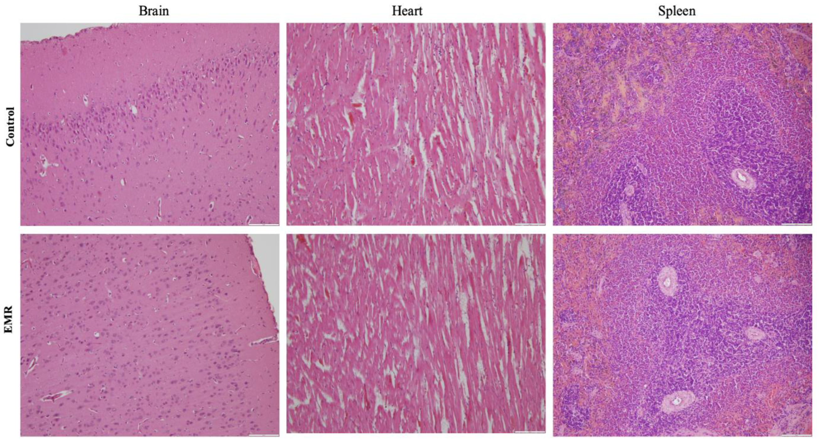
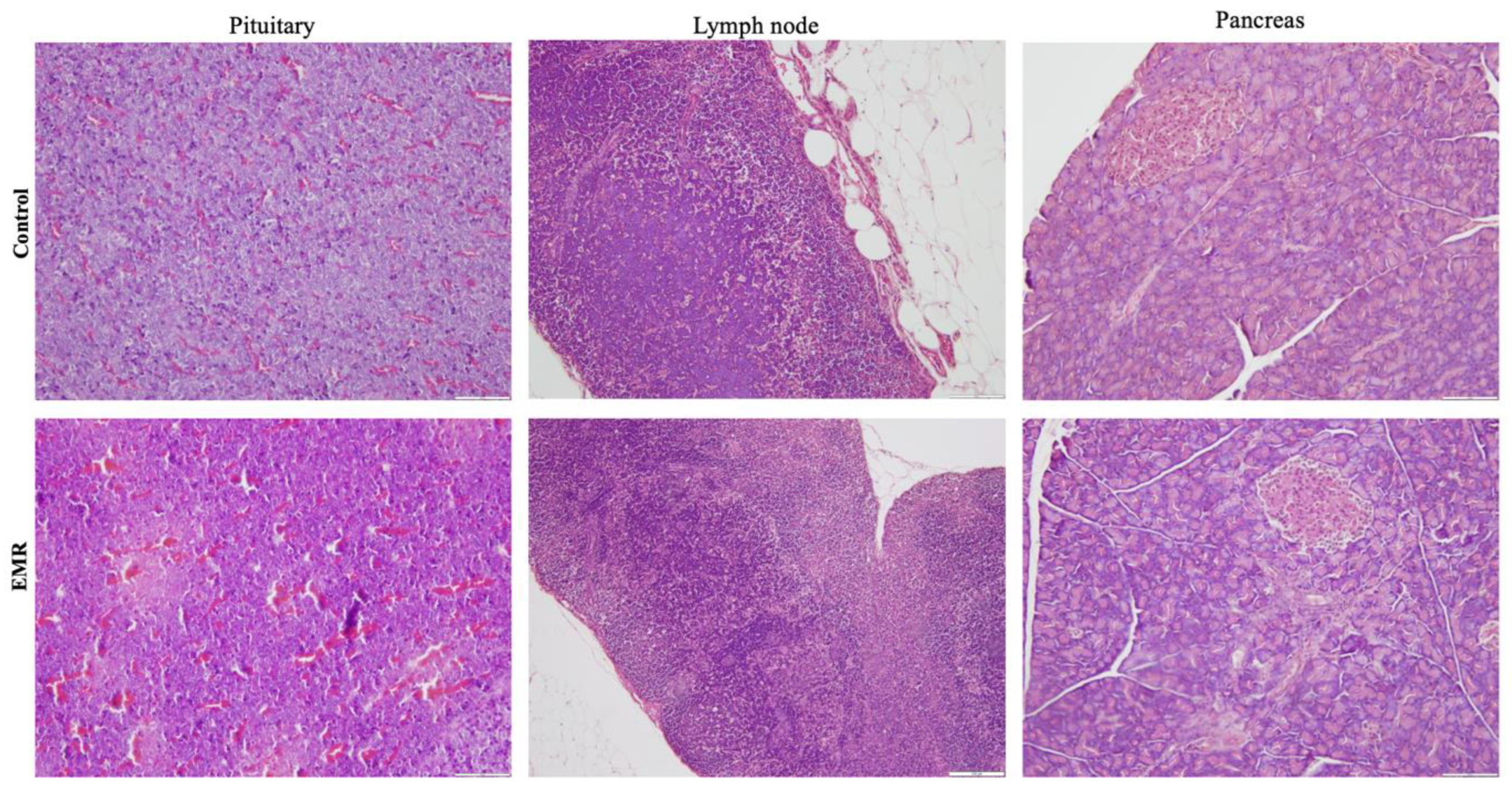
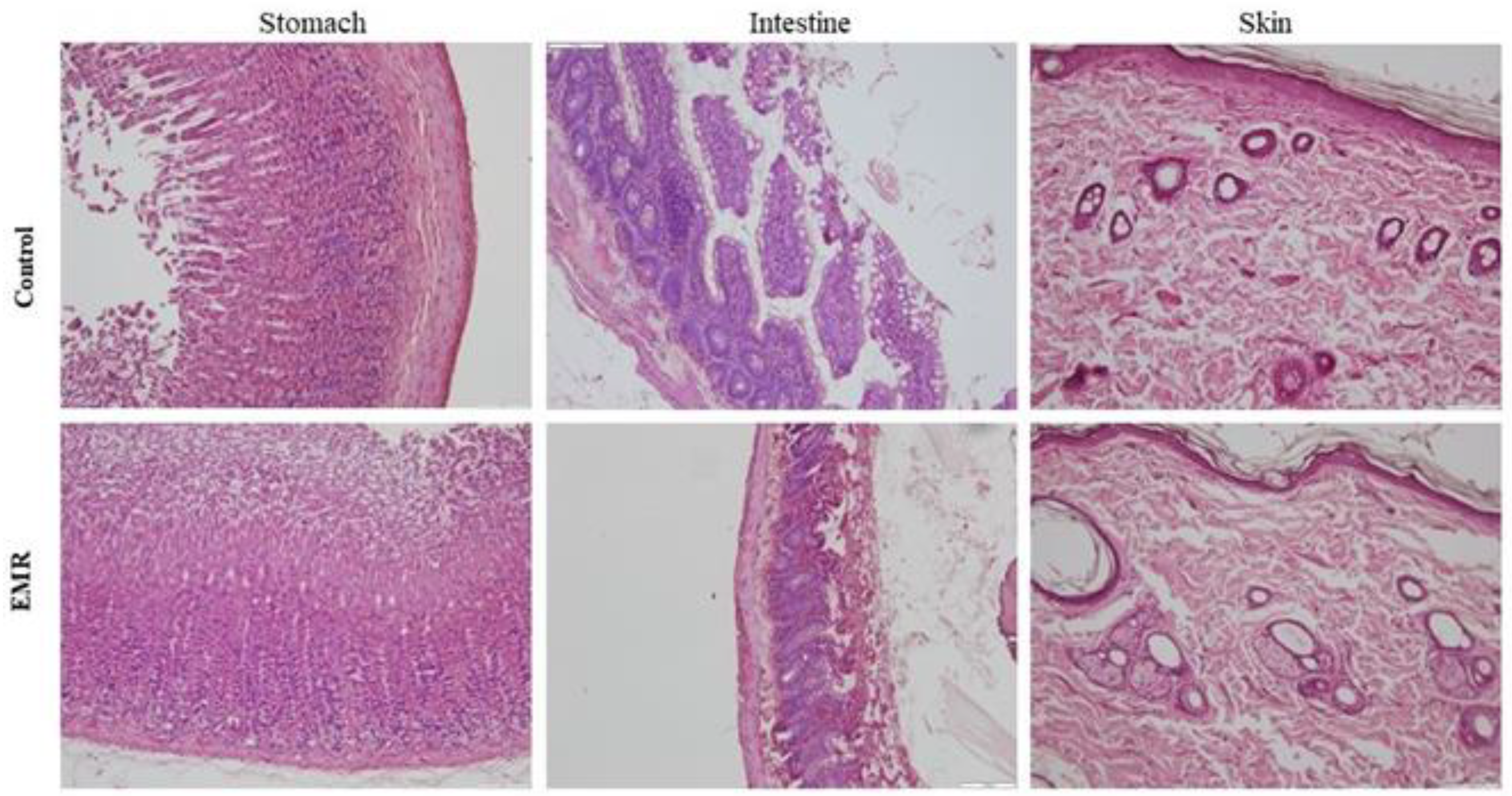
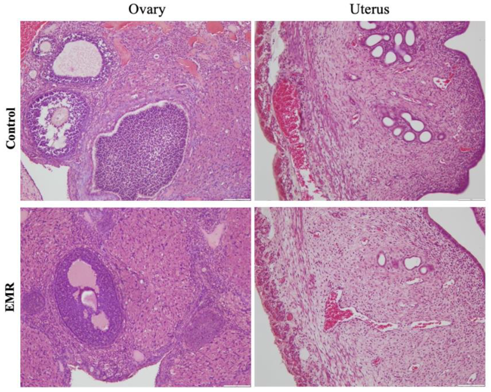
3.5. Histopathological Analysis
4. Discussion
5. Conclusions
Author Contributions
Funding
Institutional Review Board Statement
Informed Consent Statement
Data Availability Statement
Acknowledgments
Conflicts of Interest
References
- International Commission on Non-Ionizing Radiation Protection. Guidelines for limiting exposure to time-varying electric, magnetic, and electromagnetic fields (up to 300 GHz). Health Phys. 2020, 74, 494–522. [Google Scholar]
- World Health Organization. Electromagnetic Fields and Public Health Intermediate Frequencies (IF), International EMF Project Information Sheet 1–4. 2005. Available online: https://www.who.int/pehemf/publications/facts/intermediatefrequencies_infosheet.pdf (accessed on 14 March 2022).
- Bodewein, L.; Schmiedchen, K.; Dechent, D.; Stunder, D.; Graefrath, D.; Winter, L.; Kraus, T.; Driessen, S. Systematic review on the bio-logical effects of electric, magnetic and electromagnetic fields in the intermediate frequency range (300 Hz to 1 MHz). Environ. Res. 2019, 171, 247–259. [Google Scholar] [CrossRef] [PubMed]
- Yamazaki, K.; Taki, M.; Ohkubo, C. Safety assessment of human exposure to intermediate frequency electromagnetic fields. Electr. Eng. Jpn. 2016, 197, 3–11. [Google Scholar] [CrossRef]
- Roivainen, P.; Eskeline, T.; Jokela, K.; Juutilainen, J. Occupational exposure to intermediate frequency and extremely low magnetic fields among personnel working near electronic article surveillance systems. Bioelectromagnetics 2014, 35, 245–250. [Google Scholar] [CrossRef] [PubMed]
- Scientific Committee on Emerging and Newly Identified Health Risks. Potential Health Effects of Exposure to Electro-Magnetic Fields (EMF). 2015. Available online: https://ec.europa.eu/health/scientific_committees/emerging/docs/scenihr_o_04.1.pdf (accessed on 14 March 2022).
- World Health Organization. Extremely Low Frequency Fields; World Health Organization: Geneva, Switzerland, 2007; Available online: https://www.who.int/peh-emf/publications/ELF%20EHC%20No238%20full.pdf (accessed on 14 March 2022).
- International Commission on Non-ionizing Radiation Protection. Guidelines for limiting exposure to time-varying electric and magnetic fields (1 Hz to 100 kHz). Health Phys. 2010, 99, 818–836. [Google Scholar] [CrossRef] [PubMed]
- Joseph, W.; Vermeeren, G.; Verloock, L.; Goeminne, F. In situ magnetic field exposure and ICNIRP-based safety distances for electronic article surveillance systems. Radiat. Prot. Dosim. 2012, 148, 420–427. [Google Scholar] [CrossRef] [PubMed]
- BIO Intelligent Service. 2016. Promoting Healthy Environments with a Focus on the Impact of Actions on Electromagnetic Fields; Final Report for Health and Consumer. Available online: http://ec.europa.eu/health/electromagnetic_fields/docs/bio_frep_en.pdf (accessed on 14 March 2022).
- Topal, Z.; Hanci, H.; Mercantepe, T.; Erol, H.S.; Keleş, O.N.; Kaya, H.; Mungan, S.; Odaci, E. The effects of prenatal long-duration exposure to 900-MHz electromagnetic field on the 21-day-old newborn male rat liver. Turk. J. Med. Sci. 2015, 45, 291–297. [Google Scholar] [CrossRef] [PubMed]
- Challis, L.J. Mechanisms for interaction between RF fields and biological tissue. Bioelectromagnetics 2005, 26 (Suppl. 7), S98–S106. [Google Scholar] [CrossRef]
- Tkalec, M.; Malaric, K.; Pevalek-Kozlina, B. Exposure to radiofrequency radiation induces oxidative stress in duck week Lemna minor L. Sci. Total Environ. 2007, 388, 78–89. [Google Scholar] [CrossRef] [PubMed]
- Sharma, S.; Sharma, A.; Shukla, S. Electromagnetic radiation on vital organs in rats. Octa J. Biosci. 2017, 5, 1–8. [Google Scholar]
- Hao, Y.H.; Zhao, L.; Peng, R.Y. Effects of microwave radiation on brain energy metabolism and related mechanisms. Mil. Med. Res. 2015, 2, 4. [Google Scholar] [CrossRef]
- Deniz, O.G.; Kıvrak, E.G.; Kaplan, A.A.; Altunkaynak, B.Z. Effects of folic acid on rat kidney exposed to 900MHz electromagnetic radiation. J. Microsc. Ultrastruct. 2017, 5, 198–200. [Google Scholar] [CrossRef] [PubMed]
- Sepehrimanesh, M.; Kazemipour, N.; Saeb, M.; Nazifi, S.; Davis, D.L. Proteomic analysis of continuous 900-MHz radiofrequency electromagnetic field exposure in testicular tissue: A rat model of human cell phone exposure. Environ. Sci. Pollut. Res. Int. 2017, 24, 13666–13673. [Google Scholar] [CrossRef]
- Goldhaber, M.K.; Polen, M.R.; Hiatt, R.A. The risk of miscarriage and birth defects among women who are visual display terminals during pregnancy. Am. J. Ind. Med. 1988, 13, 695–706. [Google Scholar] [CrossRef] [PubMed]
- Khan, M.W.; Roivainen, P.; Herrala, M.; Tikkaja, M.; Sallmen, M.; Hietanen, M.; Jukka Juutilainen, J. A pilot study on the reproductive risks of maternal exposure to magnetic fields from electronic article surveillance systems. Int. J. Radiat. Biol. 2018, 94, 902–908. [Google Scholar] [CrossRef] [PubMed]
- Shigemitsu, T.; Yamazaki, K.; Nakasono, S.; Kakikawa, M. A review of the biological effects of electromagnetic fields in the intermediate frequency range. IEEJ Trans. Electr. Electron. Eng. 2007, 2, 405–412. [Google Scholar] [CrossRef]
- Sundaram, V.; Mohammed, S.; Zyuzikov, N. Effects of 150 kHz intermediate frequency electromagnetic radiation on fertility indicators in male rats. Heliyon 2002, 8, e12228. [Google Scholar] [CrossRef]
- Stupp, R.; Taphoorn, M.; Driven, L.; Tailibert, S.; Honnorat, J.; Chen, T.C.; Sroubek, J.; Paek, S.; Escuder, J.; Easaw, J.; et al. Tumor Treating Fields (TTFields)-A Novel Cancer Treatment Modality: Translating Preclinical Evidence and Engineering into a Survival Benefit with Delayed Decline in Quality of Life. Int. J. Radiat. Oncol. Biol. Phys. 2017, 99, 1316–1317. [Google Scholar] [CrossRef]
- Mohammed, S.; Sundaram, V.; Rao, C.A.V.; Zyuzikov, N. Polycystic ovary rat model exposure to 150 kHz electromagnetic frequency: Hypothalamic-pituitary-ovarian axis at the receptor, cellular, tissue, and hormone levels. J. Ovarian Res. 2021, 14, 173. [Google Scholar] [CrossRef]
- Mohammed, S.; Sundaram, V.; Zyuzikov, N. Effect of 150 kHz electromagnetic radiation on the development of polycystic ovaries induced by Estradiol Valverate in Sprague Dawley rats. J. Ovarian Res. 2021, 14, 26. [Google Scholar] [CrossRef]
- Zamanian, A.; Hardiman, C.J.H.F.E. Electromagnetic radiation and human health: A review of sources and effects. High Freq. Electron. 2005, 4, 16–26. [Google Scholar]
- National Research Council (US) Committee for the Update of the Guide for the Care and Use of Laboratory Animals. Guide for the Care and use of Laboratory Animals, 8th ed.; National Academies Press (US): Washington, DC, USA, 2011. Available online: https://www.ncbi.nlm.nih.gov/books/NBK54050/ (accessed on 14 March 2022).
- Jameus, A.; Kennedy, A.E.; Thome, C. Haematological Changes Following Low Dose Radiation Therapy and Comparison to Current Standard of Care Cancer Treatments. Dose Response 2021, 19, 15593258211056196. [Google Scholar] [CrossRef] [PubMed]
- Olson, H.; Betton, G.; Robinson, D.; Thomas, K.; Monro, A.; Kolaja, G.; Lilly, P.; Sanders, J.; Sipes, G.; Bracken, W.; et al. Concordance of the toxicity of pharmaceuticals in humans and animals. Regul. Toxicol. Pharmacol. 2000, 32, 56–67. [Google Scholar] [CrossRef] [PubMed]
- He, Q.; Su, G.; Liu, K.; Zhang, F.; Jiang, Y.; Gao, J.; Liu, L.; Jiang, Z.; Jin, M.; Xie, H. Sex-specific reference intervals of hematologic and biochemical analytes in Sprague-Dawley rats using the nonparametric rank percentile method. PLoS ONE 2017, 20, 12. [Google Scholar] [CrossRef] [PubMed]
- Lillie, L.E.; Temple, N.J.; Florence, L.Z. Reference values for young normal Sprague-Dawley rats: Weight gain, hematology and clinical chemistry. Hum. Exp. Toxicol. 1996, 15, 612–616. [Google Scholar] [CrossRef]
- Abdolmaleki, A.; Sangimabadi, F.; Rajabi, A.; Saberi, R. Effect of electromagnetic wave on blood parameters. Int. J. Heamtol. Oncol. Stem Cell Res. 2012, 6, 13–16. [Google Scholar]
- Peng, W.J.; Xin, R.H.; Luo, Y.J.; Liang, G.; Ren, L.H.; Liu, Y.; Wang, G.B.; Zheng, J.F. Evaluation of the acute and sub chronic toxicity of Aster tataricus L. F. Afr. J. Tradit. Complement. Altern. Med. 2016, 13, 38–53. [Google Scholar] [CrossRef] [PubMed]
- Traesel, G.K.; Menegati, S.E.L.T.; dos Santos, A.C.; Carvalho Souza, R.I.; Villas Boas, G.R.; Justi, P.N.; Kassuya, C.A.L.; Sanjinez Argandona, E.J.; Oesterreich, S.A. Oral acute and subchronic toxicity studies of the oil extracted from pequi (Caryocar brasiliense, Camb.) pulp in rats. Food Chem. Toxicol. 2016, 97, 224–231. [Google Scholar] [CrossRef] [PubMed]
- Josef, O.; Ratner, M.; Shaw, M.; Bailey, W.; Schomaker, S. The current state of serum biomarkers of hepatotoxicity. Toxicology 2008, 245, 194–205. [Google Scholar]
- Utohnedosa, A.U.; Akah, P.A.; Okoye, T.C.; Okoli, C.O. Evaluation of the toxic effects of dihydroartemisinin on the vital organs of Wistar albino rats. Am. J. Pharmacol. Toxicol. 2009, 485, 169–173. [Google Scholar]
- Kadhum, E.H. Histological and Biochemical Study on the Effects of Electromagnetic Radiation on Males and Females Rats (Rattus norvegicus). Masters’Thesis, College of Science, University of Thi-Qar, Nasiriyah, Iraq, 2015; pp. 1–105. [Google Scholar]
- Adebayo, E.A.; Adeeyo, A.O.; Ogundiran, M.A.; Olabisi, O. Bio-physical effects of radiofrequency electromagnetic radiation (RF-EMR) on blood parameters, spermatozoa, liver, kidney and heart of albino rats. J. King Saud Univ.-Sci. 2019, 31, 813–821. [Google Scholar] [CrossRef]
- Sani, A.; Labaran, M.M.; Dayyabu, B. Effects of electromagnetic radiation of mobile phones on hematological and biochemical parameters in male albino rats. Eur. Exp. Biol. 2018, 8, 11. [Google Scholar] [CrossRef]
- Usikalu, M.R.; Aweda, M.A.; Babatunde, E.B.; Awobajo, F.O. Low level microwave exposure decreases the number of male germ cells and affect vital organs of Sprague Dawley rats. Am. J. Sci. Ind. Res. 2010, 1, 410–420. [Google Scholar] [CrossRef][Green Version]
- Forgacs, Z.; Somosy, Z.; Kubinyi, G.; Sinay, H.; Bakos, J.; Thuroczy, G.; Surjan, A.; Hudak, A.; Olajos, F.; Lazar, P. Effects of whole-body 50-Hz magnetic field exposure on mouse leydig cells. Sci. World J. 2004, 4, 83–90. [Google Scholar] [CrossRef]
- Buckus, R.; Strukčinskienė, B.; Raistenskis, J.; Stukas, R.; Šidlauskiene, A.; Čerkauskienė, R.; Isopescu, D.N.; Stabryla, J.; Cretescu, I. A technical approach to the evaluation of radiofrequency radiation emissions from mobile telephony base stations. Int. J. Environ. Res. Public Health 2017, 14, 244–254. [Google Scholar] [CrossRef]
- Forgacs, Z.; Somosy, Z.; Kubinyi, G.; Bakos, J.; Hudak, A.; Surjan, A.; Thuroczy, G. Effect of whole-body 1800 MHz GSM-like microwave exposure on testicular steroidogenesis and histology in mice. Reprod. Toxicol. 2006, 22, 111–117. [Google Scholar] [CrossRef] [PubMed]
- El-Bedwi, A.B.; El-Kott, A.F.; Saad, M.; Eid, E. Effects of electromagnetic radiation produced by mobile phone on some visceral organs of rat. J. Med. Sci. 2011, 11, 256–260. [Google Scholar] [CrossRef]
- Ezeja, M.I.; Anaga, A.O.; Asuzu, I.U. Acute and sub-chronic toxicity profile of methanol leaf extract of Gouania longipetala in rats. J. Ethnopharmacol. 2014, 151, 1155–1164. [Google Scholar] [CrossRef]
- DeJong, C.A.; Van den Brink, W.; Harteveld, F.M.; van der Wielen, E.G.M. Personality disorders in alcoholics and drug addicts. Compr. Psychiatry 1993, 34, 87–94. [Google Scholar] [CrossRef]
- Borzoueisileh, S.; Shabestani Monfared, A.; Ghorbani, H.; Mortazavi, S.M.J.; Zabihi, E.; Pouramir, M.; Shafiee, M.; Niksirat, F. Combined Effects of Radiofrequency Electromagnetic Fields and X-Ray in Renal Tissue and Function. Res. Rep. Urol. 2020, 12, 527–532. [Google Scholar] [CrossRef]
- Vecchio, F.; Tombini, M.; Buffo, P.; Assenza, G.; Pellegrino, G.; Benvenga, A.; Babiloni, C.; Rossini, P.M. Mobile phone emission increases inter-hemispheric functional coupling of electroencephalographic α rhythms in epileptic patients. Int. J. Psychophysiol. 2012, 84, 164–171. [Google Scholar] [CrossRef] [PubMed]
- Valentini, E.; Ferrara, M.; Presaghi, F.; De Gennaro, L.; Curcio, G. Systematic review and meta-analysis of psychomotor effects of mobile phone electromagnetic fields. Occup. Environ. Med. 2010, 67, 708–716. [Google Scholar] [CrossRef]
- Appleton, J.; Lee, K.M.; Sawicka Kapusta, K.; Damek, M.; Cooke, M. The heavy metal content of the teeth of the bank vole (Clethrionomys glareolus) as an exposure marker of environmental pollution in Poland. Environ. Pollut. 2010, 110, 441–449. [Google Scholar] [CrossRef] [PubMed]
- Shrimanker, I.; Bhattarai, S. Electrolytes. In Stat Pearls; Stat Pearls Publishing: Treasure Island, FL, USA, 2022. [Google Scholar]
- Kunt, H.; Şentürk, I.; Gönül, Y.; Korkmaz, M.; Ahsen, A.; Hazman, O.; Bal, A.; Genc, A.; Songur, A. Effects of electromagnetic radiation exposure on bone mineral density, thyroid, and oxidative stress index in electrical workers. Onco Targets Ther. 2016, 9, 745. [Google Scholar]
- Chang, K.; Chang, W.H.S.; Huang, S.; Huang, S.; Shih, C. Pulsed electromagnetic fields stimulation affects osteoclast formation by modulation of osteoprotegerin. RANK ligand and macrophage colony-stimulating factor. J. Orthop. Res. 2005, 23, 1308–1314. [Google Scholar] [CrossRef] [PubMed]
- Lee, H.-H.; Jin, H.; Ahn, Y.H.; Kim, N.; Pack, J.K.; Choi, H.-Y.; Lee, Y.S. Effects of intermediate frequency electromagnetic fields: A review of animal studies. Int. J. Radiat. Biol. 2022, 99, 166–182. [Google Scholar] [CrossRef] [PubMed]
- Mohamed, A.K. The possible rescue effect of vitamin E or Silymarin on lung tissue of male albino rats exposed to electro-magnetic field. Egypt. J. Hosp. Med. 2014, 57, 470–481. [Google Scholar] [CrossRef]
- Ibrahim, R.A.; Ali, A.H.; Khamis, N.H.; Mohammed, H.H. Effect of Exposure to Wi-Fi Router Radiation on The Lung of Adult Male Albino Rats: Histological and Immunohistochemical Study. Egypt. J. Histol. 2019, 42, 1059–1069. [Google Scholar]
- Al-Glaib, B.; Al-Dardfi, M.; Al-Tuhami, A.; Elgenaidi, A.; Dkhil, M. A technical report on the effect of electromagnetic radiation from a mobile phone on mice organs. Libyan J. Med. 2008, 3, 8–9. [Google Scholar] [CrossRef]


| Parameters | Control (Median) | EMR (Median) | Mann-Whitney p-Value | Statistical Significance | Control (Mean ± Standard Deviation) | EMR (Mean ± Standard Deviation) |
|---|---|---|---|---|---|---|
| WBC (×109/L) | 8.6 | 4.3 | 0.1 | NS | 8.1 ± 1.75 | 4.4 ± 1.33 |
| RBC (×1012/L) | 7.5 | 7.9 | 0.2 | NS | 7.5 ± 0.0 | 7.9 ± 0.01 |
| HCT (L/L) | 0.43 | 0.39 | 0.2 | NS | 0.42 ± 0.01 | 0.39 ± 0.01 |
| Hgb (g/L) | 134.0 | 139.5 | 0.4 | NS | 134.0 ± 5.00 | 139.5 ± 2.12 |
| MCV (fL) | 51.1 | 49.0 | 0.2 | NS | 52.0 ± 1.70 | 49.0 ± 1.41 |
| MCH (pg) | 18.0 | 17.6 | 0.2 | NS | 18.5 ± 1.01 | 17.6 ± 0.28 |
| MCHC (g/L) | 352.0 | 356.5 | 0.8 | NS | 357.3 ± 10.12 | 356.5 ± 4.95 |
| RDW (%) | 20.5 | 21.0 | 0.5 | NS | 20.5 ± 0.55 | 21.0 ± 0.71 |
| Retic (%) | 2.4 | 2.2 | 0.4 | NS | 2.6 ± 0.46 | 2.2 ± 0.21 |
| Plasma protein (g/L) | 81.0 | 76.0 | 0.2 | NS | 80.3 ± 2.08 | 76.0 ± 0.00 |
| Platelets (×109/L) | 647.0 | 714.0 | 0.4 | NS | 628.3 ± 94.39 | 714.0 ± 74.95 |
| Parameters | Control (Mean ± SEM) | EMR (Mean ± SEM) | p-Value | Statistical Significance |
|---|---|---|---|---|
| Serum Na+ (mmol/L) | 138.5 ± 0.9 | 142.2 ± 1.0 | 0.03 | * |
| Serum K+ (mmol/L) | 7.2 ± 1.2 | 6.5 ± 0.6 | 0.60 | NS |
| Serum Na+: K+ ratio | 20.7 ± 3.0 | 23.0 ± 2.7 | 0.59 | NS |
| Serum Cl− (mmol/L) | 101.4 ± 1.9 | 101.7 ± 0.2 | 0.88 | NS |
| Serum Ca+ (mmol/L) | 2.62 ± 0.06 | 2.50 ± 0.05 | 0.17 | NS |
| Serum phosphorus (mmol/L) | 2.05 ± 0.20 | 2.11 ± 0.12 | 0.79 | NS |
| Urea (mmol/L) | 7.7 ± 0.7 | 5.8 ± 0.2 | 0.01 | ** |
| Creatinine (µmol/L) | 55.1 ± 7.3 | 55.9 ± 3.0 | 0.91 | NS |
| Total protein (g/dL) | 69.4 ± 2.3 | 68.9 ± 1.0 | 0.82 | NS |
| Albumin (g/dL) | 32.0 ± 0.7 | 31.3 ± 0.5 | 0.46 | NS |
| Globulin (g/dL) | 38.7 ± 2.4 | 37.4 ± 0.8 | 0.57 | NS |
| Albumin: Globulin ratio | 0.76 ± 0.09 | 0.84 ± 0.02 | 0.33 | NS |
| Glucose (mmol/L) | 12.8 ± 2.7 | 10.6 ± 1.0 | 0.36 | NS |
| Cholesterol (mmol/L) | 1.7 ± 0.1 | 1.6 ± 0.1 | 0.27 | NS |
| ALP (U/L) | 189.8 ± 11.9 | 161.0 ± 10.2 | 0.10 | NS |
| ALT (U/L) | 547.4 ± 285.8 | 251.8 ± 118.0 | 0.28 | NS |
| AST (U/L) | 604.0 ± 252.1 | 397.1 ± 156.1 | 0.47 | NS |
Disclaimer/Publisher’s Note: The statements, opinions and data contained in all publications are solely those of the individual author(s) and contributor(s) and not of MDPI and/or the editor(s). MDPI and/or the editor(s) disclaim responsibility for any injury to people or property resulting from any ideas, methods, instructions or products referred to in the content. |
© 2023 by the authors. Licensee MDPI, Basel, Switzerland. This article is an open access article distributed under the terms and conditions of the Creative Commons Attribution (CC BY) license (https://creativecommons.org/licenses/by/4.0/).
Share and Cite
Sundaram, V.; Mohammed, S.; Cockburn, B.N.; Srinivasan, M.R.; Venkata, C.R.A.; Johnson, J.; Gilkes, L.; Jones, K.R.; Zyuzikov, N. Effects of Intermediate Frequency (150 kHz) Electromagnetic Radiation on the Vital Organs of Female Sprague Dawley Rats. Biology 2023, 12, 310. https://doi.org/10.3390/biology12020310
Sundaram V, Mohammed S, Cockburn BN, Srinivasan MR, Venkata CRA, Johnson J, Gilkes L, Jones KR, Zyuzikov N. Effects of Intermediate Frequency (150 kHz) Electromagnetic Radiation on the Vital Organs of Female Sprague Dawley Rats. Biology. 2023; 12(2):310. https://doi.org/10.3390/biology12020310
Chicago/Turabian StyleSundaram, Venkatesan, Stephanie Mohammed, Brian N. Cockburn, M. R. Srinivasan, Chalapathi R. Adidam Venkata, Jenelle Johnson, Lester Gilkes, Kegan Romelle Jones, and Nikolay Zyuzikov. 2023. "Effects of Intermediate Frequency (150 kHz) Electromagnetic Radiation on the Vital Organs of Female Sprague Dawley Rats" Biology 12, no. 2: 310. https://doi.org/10.3390/biology12020310
APA StyleSundaram, V., Mohammed, S., Cockburn, B. N., Srinivasan, M. R., Venkata, C. R. A., Johnson, J., Gilkes, L., Jones, K. R., & Zyuzikov, N. (2023). Effects of Intermediate Frequency (150 kHz) Electromagnetic Radiation on the Vital Organs of Female Sprague Dawley Rats. Biology, 12(2), 310. https://doi.org/10.3390/biology12020310