Simple Summary
The large antibiotic consumption in the clinical, veterinary, and agricultural fields has resulted in a tremendous flow of antibiotics into the environment. This has led to enormous selective pressures driving the evolution of antimicrobial resistance in bacteria and yeasts. For this reason, the World Health Organization is promoting research to discover new natural products competitive with synthetic drugs in clinical performances. Compared with conventional drugs, the production of natural pharmaceuticals often has a lower environmental impact and lower economic costs of processes, especially when they originate from agricultural wastes. In the context of a circular economy, we aimed to successfully present preliminary results for the valorization of agricultural waste produced in cabbage cultivation by isolating a highly performant antibacterial and antifungal lipophilic natural mixture from cabbage leaves.
Abstract
As dramatically experienced in the recent world pandemic, viral, bacterial, fungal pathogens constitute very serious concerns in the global context of human health. Regarding this issue, the World Health Organization has promoted research studies that aim to develop new strategies using natural products. Although they are often competitive with synthetic pharmaceuticales in clinical performance, they lack their critical drawbacks, i.e., the environmental impact and the high economic costs of processing. In this paper, the isolation of a highly performant antibacterial and antifungal lipophilic natural mixture from leaves of savoy and white cabbages is proposed as successful preliminary results for the valorization of agricultural waste produced in cabbage cultivation. The fraction was chemically extracted from vegetables with diethyl ether and tested against two Candida species, as well as Pseudomonas aeruginosa, Klebsiella pneumoniae and Staphylococcus aureus reference strains. All the different fractions (active and not active) were chemically characterized by vibrational FT-IR spectroscopy and GC-MS analyses. The extracts showed high growth-inhibition performance on pathogens, thus demonstrating strong application potential. We think that this work, despite being at a preliminary stage, is very promising, both from pharmaceutical and industrial points of view, and can be proposed as a proof of concept for the recovery of agricultural production wastes.
1. Introduction
The World Health Organization (WHO) encourages research aimed to develop medical features using natural products. The excessive consumption of antibiotics in the clinical, veterinary, and agricultural fields has resulted in a huge input of antibiotics into the environment. This has led to selective pressures driving the evolution of antimicrobial resistance (AMR) in commensal and pathogenic microorganisms. AMR is an expensive problem, considering both the human health and social costs. To combat AMR, a Global Action Plan was released in 2015 by the WHO. As part of this plan, six bacterial species were identified, for which the discovery and development of new drugs is required [1]. The acronym ESKAPE encloses these human pathogens, typically associated with nosocomial infections: Enterococcus faecium, Staphylococcus aureus, Klebsiella pneumoniae, Acinetobacter baumanni, Pseudomonas aeruginosa, and Enterobacter species [2,3]. The microorganisms classified under ESKAPE encompass both Gram-positive and -negative bacterial species, and share the capability to “escape” the biocidal effects of antibiotics. Although there has been a reduction in the percentage of methicillin-resistant S. aureus (MRSA) strains, MRSA continues to be an important pathogen in European countries, also showing combined resistance to another antimicrobial group. In addition to bacteria, yeasts such as Candida spp. cause common infections, which can affect mucous membranes, but can also become systemic [4]. While most Candida infections are caused by C. albicans, other species including C. glabrata and C. auris, are frequently resistant and more deadly [5]. Therapies against yeast are mainly based on azoles and polyenes.
Cabbages (Brassica oleracea L.), belonging to the family Brassicaceae (Burnett) or Cruciferae, are a widely distributed and cultivated vegetable. They originate from the south and western coast of Europe. The Brassicaceae contain a large amount of different healthy compounds such as vitamins (A, C, K, and B6), carotenoids, polyphenols (chlorogenic and sinapic acid derivatives, and flavonoids), minerals (selenium, potassium, and manganese), and nitrogen-sulfur derivatives (glucosinolates and isothiocyanates) [6,7,8,9,10]. It is well-known from the literature that many of these molecules have anticarcinogenic, antimicrobial, anti-inflammatory, and antidiabetic activities [9,10,11,12].
The antibacterial activity of different kind of extracts from Brassicaceae plant organs are reported in the literature. For example, Pacheco-Cano et al. [13] demonstrated the biological activity of extracts from the flowers and stems of broccoli cv. Avenger. Their inhibitory effect was proved against pathogenic bacteria (Bacillus cereus, Staphylococcus xylosus, S. aureus, Shigella flexneri, Shigella sonnei and Proteus vulgaris), phytopathogenic fungi (Colletotrichum gloeosporioides and Asperigillus niger), and yeasts (C. albicans and Rhodotorula sp.) [13]. Moreover, Andini studied the antibacterial activity of different compounds from Brassicaceae [14] against bacteria, such as Helicobacter pylori, Escherichia coli, Bacillus cereus, B. subtilis, Listeria monocytogenes, S. aureus and yeasts such as C. albicans. Finally, Jaiswal et al. tested extracts from fresh Irish Brassica vegetables (York cabbage, white cabbage, broccoli and Brussels sprouts) against Gram-negative (Salmonella abony and Pseudomonas aeruginosa) and Gram-positive (L. monocytogenes and Enterococcus faecalis) bacteria [12].
In the context of a circular economy, in this work, we aimed at developing a method for the valorization of agricultural waste produced in the cultivation of cabbage. We isolated a highly performant antibacterial and antifungal lipophilic natural mixture. The new potential antimicrobial extracts were evaluated against the above-mentioned bacteria and yeasts, proposed by the Global Action Plan.
2. Materials and Methods
2.1. Extract Production
2.1.1. Materials
White and savoy cabbages were purchased in Novara (Piedmont, Italy), in different locations and temporal seasons (spring 2020–autumn and winter 2021). Diethyl ether was purchased by Sigma-Aldrich (Merk Life Science S.r.l., Milan, Italy). Deionized water was obtained through an ion-exchange resin.
2.1.2. Extraction Procedures
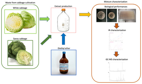
The extraction procedure and mixture characterization are summarized in Figure 1.

Figure 1.
Extraction procedure; biological and chemical characterization of the produced mixture.
White cabbage (cultivar certified by the producer) (1.4 kg) was exfoliated, and the leaves were pressed at the bottom of a 5 L round glass bottle, provided with a closure tip, and then soaked under 1.5 L of fresh diethyl ether. The bottle was firmly tipped, after having ascertained that every single leaf was covered by the organic solvent, and left standing for over 7 days, to allow full chemical extraction of the cabbage lipophilic components. Next, the bottle was opened, and the liquid was transferred into large beakers. During the organic extraction, two distinct phases were observed and separated with a separatory funnel: a pale-yellow upper one, containing the diethyl ether solvent with extracted cabbage lipophilic formulations, and a denser orange lower one. The solution in the upper fraction was dried in a rotavapor (40 °C, under reduced pressure), yielding about 500 mg, corresponding on average to 0.06 weight percent of the total cabbage mass. This was then stored in a freezer (−28 °C) prior to chemical analyses and biological antibacterial and antifungal assays. Upon residual solvent evaporation under a ventilated hood, the denser, lower fraction provided a highly viscous, dark-orange, honey-like formulation, extracted from vegetables after a severe long-term organic solvent extraction. Similarly, this formulation was stored in freezer prior to analyses and biological tests.
Savoy cabbage (cultivar certified by the producer) was processed with a closely related extraction protocol, yielding parallel results. Chemical extractions were maintained by soaking up to 10 days, without relevant differences in the observed extracted results.
2.2. Antifungal and Antibacterial Assays
The agar disc diffusion method was employed to determine the antifungal and antibacterial activity of the cabbage extracts, according to the methods previously published [15,16,17,18]. The extract was provided as powder and suspended in 1,4 dioxane (Sigma-Aldrich, St. Louis, MO, USA) at a concentration of 0.60 mg µL−1 that was considered the starting solution for the following assays.
The antifungal assays were carried out with C. albicans ATCC 14053 and C. glabrata ATCC 15126. The antifungal effects of clotrimazole (10 μg) and extracts were evaluated according to the M44-A method proposed by the Clinical and Laboratory Standards Institute Standard (CLSI). Clotrimazole (Biolife, Italy) (10 μg) discs were used as positive control. 1,4 dioxane (Sigma-Aldrich, St. Louis, MO, USA; 10 μL) and discs were used as negative control. Plates were incubated at 37 °C for 48 h. All experiments were performed in triplicate. The sensitivity test for the extract was considered positive if it resulted in an inhibition halo higher than that induced by clotrimazole (positive control ≥100%).
The antibacterial assays were performed with Staphylococcus aureus NCTC6571, Pseudomonas aeruginosa ATCC27853 and Klebsiella pneumoniae ATCC13883 reference strains. Vancomycin (Biolife, Italy), imipenem (Biolife, Italy) and meropenem (Biolife, Italy) effects were evaluated according to EUCAST disk diffusion method for antimicrobial susceptibility v. 7.0. 1,4 dioxane (10 μL) disks were used as negative controls, while vancomycin, meropenem and imipenem were considered as positive control. Plates were incubated at 37 °C for 24 h. All experiments were performed in triplicate. The halos were measured in mm using calipers. The extract was considered active when it produced a halo equal to or higher than positive control (positive control ≥100%).
Moreover, the minimal inhibitory concentration (MIC) of the extracted mixture against bacterial and yeast reference strains was measured according to the EUCAST antifungal MIC method for yeasts (EUCAST definitive document E.DEF 7.3) and EUCAST reference method for bacteria with some modifications performed by the authors and previously published [15,16,17,18]. All microtiter plates were incubated at 37 °C for 24 h. Each experiment was repeated three times.
2.3. FT-IR Analyses
Fourier-transform infrared (FT-IR) measurements were performed by a Thermo-Fisher Scientific Nicolet iSC50 spectrophotometer (Thermo Fisher Scientific, Waltham, MA, USA).
The analyses were performed in transmission mode on the dryed cabbage extracts by the classical method normally employed for solid samples.
In more detail, 3 mg of the solid sample was pestled with 100 mg of KBr (FT-IR grade) in an agate mortar using a rotatory movements until a homogeneus and impalpable mixture was obtained. Then, the mixture was transferred into a tablet press and pressed at 10 tons cm−2, obtaining a 1 cm-diameter disc which was gently transferred into the sample holder of the FT-IR and analysed.
Spectra were collected in transmission mode averaging 100 scans performing a scan at 2 cm−1 resolution in the wavelength region from 400 to 4000 cm−1.
2.4. GC-MS
The chromatographic characterization was performed using a Finnigan Trace GC-Ultra and Trace DSQ. In particular, the gas chromatographic separation was performed using a capillary column Phenomenex ZB-WAX (30 m length, 0.25 mm I.D., and 0.25 μm film thickness). The inlet temperature was set to 250 °C in splitless mode, and helium was used as the carrier gas with a constant flow of 1.0 mL min−1. The initial oven temperature was set to 45 °C and reached 250 °C with the ramps reported in Table 1.

Table 1.
Oven temperature program.
The mass spectrometer (MS) transfer line temperature was set to 290 °C. The MS signal was acquired through El+ mode with an ionization energy of 70.0 eV and a source temperature of 290 °C. The solvent delay was set to 6.50 min. The detection was carried out in full-scan mode in the range of 35–500 m/z.
The samples were prepared by dissolving the cabbage extracts in dichloromethane (50 mg in 1.0 mL) and filtrating by PTFE membrane filters of 0.20 μm porosity. After a dilution of 1:5 in dichloromethane, the extracts were injected in the GC.
2.5. Statistical Analysis
The disk diffusion results were statistically analyzed using one-way ANOVA followed by Tukey’s HSD multiple comparisons of means using R (v. 3.5.1) (R Core Team, 2020). Data are presented as boxplots. Differences were considered significant for p-values < 0.05.
3. Results and Discussion
The white cabbages and savoy cabbages purchased from the local market were extracted by a solvent extraction and a liquid–liquid purification approach without the aid of temperature and pressure to avoid any kind of degradation of the molecules extracted. The extracts were extensively characterized using FT-IR and GC-MS analyses. Meanwhile, the biological activity was evaluated and quantified by antifungal and antibacterial assays together with the determination of minimal inhibitory concentration (MIC).
3.1. Biological Activity
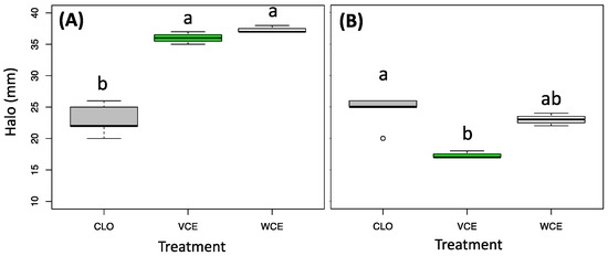
The results from the antifungal assays, obtained by the disk diffusion method, are presented in Figure 2.

Figure 2.
Antifungal assay results, using the disk diffusion method (halo diameter (mm)) from extracts obtained from Savoy cabbage (green box, VCE) and white cabbage (white box, WCE) induced in C. albicans ATCC 14053 (A) and C. glabrata ATCC 15126 (B) compared with Clotrimazole (Grey-CLO). Different letters above the bars indicate significant differences, according to Kruskal-Wallis followed by Nemenyi’s post hoc test (p-value cutoff = 0.05).
In more detail, both the two tested extracts demonstrated an antifungal activity statistically higher than clotrimazole (positive control and reference antifungal drugs for candidiasis) against C. albicans (Figure 2A), while against C. glabrata, only the extract from the white cabbage showed an activity like the antifungal drug (Figure 2B).
The disk diffusion assay results against Gram-positive and Gram-negative bacterial strains are presented in Figure 3. The white cabbage extract showed a statistically higher inhibition effect against S. aureus (Gram-positive bacteria) in respect to vancomycin, while the savoy cabbage extract induced an inhibition effect comparable to the reference drug. P. aeruginosa and K. pneumoniae, both Gram-negative bacteria, showed a similar growth inhibition, lower than the positive control, in the presence of the two cabbage extracts. Nevertheless, the halo size indicated the effectiveness of these two extracts in the reduction of bacterial growth, suggesting their potential use as adjuvants in the antibiotic treatment.

Figure 3.
Antibacterial assay results using the disk diffusion method (halo diameter (mm)) from extracts obtained from Savoy cabbage (green box, VCE) and white cabbage (white box, WCE) induced in S. aureus NCTC6571 (A), P. aeruginosa ATCC27853 (B), and K. pneumoniae ATCC13883 (C) compared with the reference antibiotic drug (grey): vancomycin (VAN) and meropenem (MRP). Different letters above the bars indicate significant differences, according to Kruskal–Wallis followed by Nemenyi’s post-hoc test (p-value cutoff = 0.05).
The MIC results obtained by the standard microdilution method are shown in Table 2. The MIC results confirmed a higher inhibitory effect against both the two Candida species and Gram-positive bacteria (e.g., S. aureus). Moreover, these extracts showed a lower, but effective, inhibition activity against Gram-negative bacteria. These results demonstrated the “universal” or “comprehensive” potential use of these cabbage extracts. The results obtained against P. aeruginosa and K. pneumoniae, even if not statistically significant, are very promising because of the difficulty in finding molecules of natural origin that have minimal efficacy against these microorganisms carrying multiple antibiotic resistances. Therefore, even a minimal effectiveness of different extracts allows us to think of mixtures that can have an efficacy similar to that of effective antibiotics such as meropenem (an antibiotic that is currently very effective).

Table 2.
Minimal inhibitory concentration (MIC) obtained by the microdilution method using extracts from Savoy and white cabbage.
3.2. FT-IR
FT-IR is a useful non-destructive method for the preliminary characterization of the molecular structure of different chemical compounds present in plant extracts [19].
Even if from such complex mixtures, which are the natural extracts that contain many molecules, a punctual identification of all the components is not possible; the measurement is useful to gain a first piece of evidence of having extracted the desired components.
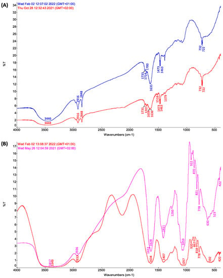
FT-IR spectra of formulations extracted from savoy cabbage (blue pattern) and white cabbage (red pattern) are reported in Figure 4. In particular, in Figure 4A, the results related to the biologically active fraction of the obtained mixture are presented, while in Figure 4B, spectral data are reported for the recovered inactive byproducts. In Figure 4A, the two spectra indeed show a closely similar profile, which suggests a related composition extracted from the savoy and white cabbages. Considering that cabbages of different varieties produced in different years and in different production sites were processed, the spectral profile obtained, almost overlapping, guarantees that the extraction method, although unconventional, yields a biologically active fraction in a highly reproducible way.

Figure 4.
(A) Comparison of the IR profiles of the extracts featured with biological activity obtained using diethyl ether from Savoy cabbage (blue line) and white cabbage (red line). Experimental observed peaks (in cm−1): savoy cabbage, 3450 (vs, vbr), 3013 (vw), 2960 (s), 2920 (vs), 2851 (vs), 1737 (s), 1712 (br), 1641 (vs, br), 1471 (vs, sh), 1465 (vs, sh), 1414 (w, br), 1382 (vs, sh), 1299 (vs, br),1263 (br), 1198, 1175 (s), 1135, 1111 (s), 1078 (vs), 989 (w), 965, 922, 892 (s), 861 (w), 821 (w), 780 (w, br), 746 (w, br), 732 (vs, vsh), 722 (vs, vsh), 560 (br); white cabbage, 3450 (vs, vbr), 3013 (vw), 2960 (s), 2920 (vs), 2851 (vs), 1737 (s), 1712 (br), 1641 (vs, br), 1471 (vs, sh), 1465 (vs, sh), 1414 (w, br), 1380 (s, br), 1299 (vs, br), 1263 (br), 1198, 1175 (s), 1135, 111 (s), 1078 (vs), 989 (w), 965, 922, 892 (s), 861 (w), 821 (w), 780 (w, br), 746 (w, br), 732 (vs, vsh), 722 (vs, vsh), and 560 (br). (B) Comparison of the IR profiles of the sulfured honey-like by-products, extracted with diethyl ether, without biological activity, from Savoy cabbage (red line) and white cabbage (purple line). Experimental observed peaks (in cm−1): Savoy cabbage, 3400 (vs, vbr), 2938 (vs, br), 2885 (s, br), 1670 (s, br), 1639 (vs, br), 1457 (br), 1412 (vs, br), 1348 (br), 1263 (s, br), 1184 (br), 1145 (br), 1100 (br), 1078 (br), 1060 (br), 1032 (br), 987 (br), 922 (vs, br), 898 (br), 870 (vs, br), 821 (vs, br), 782 (vs, br), 703 (br), 635 (vs, br), 594 (vs, br), 562 (vs, br), 525 (vs, br), 422 (s, br); white cabbage, 3400 (vs, vbr), 2938 (vs, br), 2885 (s, br), 1670 (s, br), 1639 (vs, br), 1457 (br), 1410 (vs, br), 1348 (br), 1263 (s, br), 1184 (br), 1145 (br), 1100 (br), 1078 (br), 1060 (br), 1032 (br), 987 (br), 922 (vs, br), 898 (br), 870 (vs, br), 821 (vs, br), 782 (vs, br), 703 (br), 635 (vs, br), 594 (vs, br), 562 (vs, br), 525 (vs, br), 422 (s, br). Legend: s strong, vs very strong, w weak, br broad, vbr very broad, sh sharp, vsh very sharp.
In more detail, in the biologically active fractions under examination (Figure 4A), the expected lipophilic nature of the formulations can be confirmed from the abundant aliphatic moieties (hereafter, identified by GC-MS analyses, referred to as manifold structures, among which are tetratetracontane and octacosane), providing strong C-H stretching peaks in the 3000–2750 cm−1 spectral range, accompanied by different C-H bending modes (normally mixed with other hydrocarbon C-C skeletal modes) in the 1500–1250 cm−1 region which can be observed. Moreover, the intense broad band at 3448 cm−1 contains O-H stretching modes that can be ascribed to molecules with alcoholic functions (as 1-hydroxy-4-methyl-2,6-di-tert-butylbenzene (BHT) identified by GC-MS).
In the 1800–1650 cm−1 spectral range, intense indented IR profiles account for different organic C=O carbonyl groups (ascribed, for example, to myristaldehyde, 2-hexadecanone and methyl tridecyl ketone in both cabbages), whereas broad patterns in the 1200–1000 cm−1 range can be related to different C-O stretching modes of ether moieties (e.g., β-sitosterol in white cabbage and BHT in savoy cabbage). Of note, the two diagnostic sharp, strong peaks in the 800–650 cm−1 spectral range can be related to out-of-plane gamma (C-H) bending modes of aromatic hydrogens (among the most intense IR vibrations in the entire spectral pattern) [20,21].
3.3. Cabbage Extract GC-MS Characterization
Both the white and savoy cabbage extract samples were analyzed by GC-MS in the conditions detailed in the experimental section.
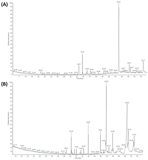
A typical chromatogram obtained by GC-MS is reported in Figure 5. The extracts from the two cabbages contain several volatile (molecules present in the first part of the chromatograms at low retention time values) and semivolatile molecules (molecules with higher retention time). The chromatographic peaks’ identification was performed by a comparison of the spectra recorded with the Wiley spectra library with the criterion of almost 40% matching probability.

Figure 5.
GC-MS chromatograms of diethyl-ether extracts from Savoy (A) and white (B) cabbage leaves. The chromatographic characterization was performed using a Finnigan Trace GC-Ultra and Trace DSQ. The inlet temperature was set to 250 °C in splitless mode, and helium was used as the carrier gas with a constant flow of 1.0 mL/min.
The identified compounds are reported in Table 3, together with semiquantative data of the amount measured in the samples. In fact, the peak areas’ values are reported. The comparison of such values is possible since the weights and the analytical procedure performed on the samples are identical both for white and savoy cabbage.

Table 3.
Compounds identified by GC-MS method.
The results of the peak identification showed a higher complexity of the white cabbage (WCE) compared to the savoy cabbage extract (VCE); this complexity could explain the higher biological performance in growth inhibition. Different compounds, with demonstrated antimicrobial activity, were exclusively present in the WCE: 4-(methylsulfanyl) butanenitrile, 1-tridecanal, 1-isothiocyanato-3-(methylthio)-propane, S-methylmethanethiosulphonate, myristic acid, β-sitosterol, 15-nonacosanone, 17-pentatriacontene and 1,2-hexadecanediol. Eight different compounds were present in both cabbage extracts (highlighted in grey in Table 3): dimethyltrisulfide, 5-methyl-1,3-thiazole, 2-hexadecanone, myristaldehyde, methyl tridecyl ketone, 3-(tert-Butyl)-3,4-dihydro-2H-1,4-benzoxazine, tetratetracontane and octacosane. Four of these are known in the literature for their antimicrobial activity. Dimethyltrisulfide is a sulfur compound reported in different cabbage species. 2-Hexadecanone, present both in white and savoy cabbage, was also identified in the essential oil of Aquilaria crassna, a tropical plant from south-east Asia and New Guinea belonging to the Thymelaeaceae family. The powder from this plant is used as incense, and the essential oil demonstrates antibacterial activity against S. aureus and C. albicans [56]. This compound is also one of the main components of essential oil from two species of Fabaceae, Cassia fistula, growing in Egypt [57], and Senna podocarpa, from different central–north West African countries [58]. In detail, Adebayo et al. demonstrated the antibacterial activity of the essential oil from S. podocarpa against Bacillus subtilis, S. aureus, Escherichia coli, Kiebsiella spp., Proteus spp., Pseudomonas spp., Salmonella spp., Penicillium notatum and Rhizopus stolonifer. Myristaldehyde, a component of Sagittaria trifolia and Cinnamomum loureiroi essential oil, has antimicrobial and antibacterial activities, as reported in Table 3. Moreover, 5-methyl-1,3-thiazole, present in both extracts and other molecular derivates, has also demonstrated antimicrobial activity. Furthermore, from the point of view of the amount of compounds extracted, the results indicated that white cabbage is characterized by a greater number of peaks with a higher quantity present (the peak area is almost always greater than white cabbage).
4. Conclusions
Extracts from savoy and white cabbages were obtained using a solid–liquid extraction procedure by diethyl ether and successfully tested for their antibacterial and antifungal properties. In fact, they perform well against two Candida species and a Pseudomonas aeruginosa, Klebsiella pneumoniae and Staphylococcus aureus reference-strain blend.
The proposed approach is positioned as a feasibility study with respect to exploiting agricultural waste, easily available and grown anywhere in the world.
In light of the properties shown by the extracts, they will be able to find applications as treatment for mucosal infections from Candida strains and for S. aureus skin infections. They were less effective against Gram-negative bacteria, even if the demonstrated inhibitory effect must be further investigated. These results are of interest regardless, considering the difficulty in finding natural mixtures that have minimal efficacy against these microorganisms similar to that of effective antibiotics such as meropenem (an antibiotic that is currently very effective).
Given the preliminary nature of the work, the continuation will go in different directions. On the one hand, the antimicrobial potential of other varieties, not only of cabbage but also of other popular vegetables, will be evaluated. From the point of view of the procedure, we will evaluate the scalability from the laboratory to the industrial scale.
Finally, the authors will not only compare the ethyl ether extraction method used in the present work with more conventional extractions—such as ethyl acetate for flavone extraction and n-hexane for the essential oil—but will also test new green solvents (eutectic, deep eutectic, and ionic liquid) in order to obtain a better extraction yield.
Author Contributions
Conceptualization, A.A. and E.B.; methodology, A.A.; formal analysis, A.A., V.G. and E.B.; investigation, F.T., R.C., M.R. and A.C.; resources, V.G., E.B. and A.A.; data curation, N.M. and M.R.; writing—original draft preparation, A.A., V.G., E.B., V.T. and N.M.; writing—review and editing, A.A., V.G., E.B., V.T. and N.M. All authors have read and agreed to the published version of the manuscript.
Funding
This research received no external funding.
Institutional Review Board Statement
Not applicable.
Informed Consent Statement
Not applicable.
Data Availability Statement
Not applicable.
Conflicts of Interest
The authors declare no conflict of interest.
References
- Tacconelli, E.; Carrara, E.; Savoldi, A.; Harbarth, S.; Mendelson, M.; Monnet, D.L.; Pulcini, C.; Kahlmeter, G.; Kluytmans, J.; Carmeli, Y.; et al. Discovery, Research, and Development of New Antibiotics: The WHO Priority List of Antibiotic-Resistant Bacteria and Tuberculosis. Lancet Infect. Dis. 2018, 18, 318–327. [Google Scholar] [CrossRef]
- Santajit, S.; Indrawattana, N. Mechanisms of Antimicrobial Resistance in ESKAPE Pathogens. BioMed Res. Int. 2016, 2016, 2475067. [Google Scholar] [CrossRef] [PubMed] [Green Version]
- Zhen, X.; Lundborg, C.S.; Sun, X.; Hu, X.; Dong, H. Economic Burden of Antibiotic Resistance in ESKAPE Organisms: A Systematic Review. Antimicrob. Resist. Infect. Control 2019, 8, 137. [Google Scholar] [CrossRef] [PubMed] [Green Version]
- Benedict, K.; Jackson, B.R.; Chiller, T.; Beer, K.D. Estimation of Direct Healthcare Costs of Fungal Diseases in the United States. Clin. Infect. Dis. 2019, 68, 1791–1797. [Google Scholar] [CrossRef] [PubMed] [Green Version]
- Fasciana, T.; Cortegiani, A.; Ippolito, M.; Giarratano, A.; Di Quattro, O.; Lipari, D.; Graceffa, D.; Giammanco, A. Candida auris: An Overview of How to Screen, Detect, Test and Control This Emerging Pathogen. Antibiotics 2020, 9, 778. [Google Scholar] [CrossRef] [PubMed]
- Bhandari, S.R.; Jo, J.; Lee, J. Comparison of Glucosinolate Profiles in Different Tissues of Nine Brassica Crops. Molecules 2015, 20, 15827–15841. [Google Scholar] [CrossRef]
- Bhandari, S.R.; Kwak, J.H. Chemical Composition and Antioxidant Activity in Different Tissues of Brassica Vegetables. Molecules 2015, 20, 1228–1243. [Google Scholar] [CrossRef] [Green Version]
- Singh, J.; Upadhyay, A.K.; Trasad, K.; Bahadur, A.; Rai, M. Variability of Carotenes, Vitamin C, E and Phenolics in Brassica Vegetables. J. Food Compos. Anal. 2007, 20, 106–112. [Google Scholar] [CrossRef]
- Li, X.; Pang, W.; Piao, Z. Omics Meets Phytonutrients in Vegetable Brassicas: For Nutritional Quality Breeding. Hortic. Plant J. 2017, 3, 247–254. [Google Scholar] [CrossRef]
- Le, T.N.; Chiu, C.-H.; Hsieh, P.-C. Bioactive Compounds and Bioactivities of Brassica oleracea L. var. Italica Sprouts and Microgreens: An Updated Overview from a Nutraceutical Perspective. Plants 2020, 9, 946. [Google Scholar] [CrossRef]
- Singh, J.; Upadhyay, A.K.; Bahadur, A.; Singh, B.; Singh, K.P.; Rai, M. Antioxidant Phytochemicals in Cabbage (Brassica oleracea L. Var. Capitata). Sci. Hortic. 2006, 108, 233–237. [Google Scholar] [CrossRef]
- Jaiswal, A.K.; Rajauria, G.; Abu-Ghannam, N.; Gupta, S. Phenolic Composition, Antioxidant Capacity and Antibacterial Activity of Selected Irish Brassica Vegetables. Nat. Prod. Commun. 2011, 6, 1299–1304. [Google Scholar] [CrossRef] [PubMed] [Green Version]
- Pacheco-Cano, R.D.; Salcedo-Hernández, R.; López-Meza, J.E.; Bideshi, D.K.; Barboza-Corona, J.E. Antimicrobial Activity of Broccoli (Brassica oleracea Var. Italica) Cultivar Avenger against Pathogenic Bacteria, Phytopathogenic Filamentous Fungi and Yeast. J. Appl. Microbiol. 2018, 124, 126–135. [Google Scholar] [CrossRef]
- Andini, S. Antimicrobial Isothiocyanates from Brassicaceae Glucosinolates: Analysis, Reactivity, and Quantitative Structure-Activity Relationships; Wageningen University: Wageningen, The Netherlands, 2020; ISBN 978-94-6395-454-9. [Google Scholar]
- Bona, E.; Cantamessa, S.; Pavan, M.; Novello, G.; Massa, N.; Rocchetti, A.; Berta, G.; Gamalero, E. Sensitivity of Candida albicans to Essential Oils: Are They an Alternative to Antifungal Agents? J. Appl. Microbiol. 2016, 121, 1530–1545. [Google Scholar] [CrossRef] [PubMed]
- Massa, N.; Cantamessa, S.; Novello, G.; Ranzato, E.; Martinotti, S.; Pavan, M.; Rocchetti, A.; Berta, G.; Gamalero, E.; Bona, E. Antifungal Activity of Essential Oils against Azole-Resistant and Azole-Susceptible Vaginal Candida glabrata Strains. Can. J. Microbiol. 2018, 64, 647–663. [Google Scholar] [CrossRef]
- Bona, E.; Arrais, A.; Gema, L.; Perotti, V.; Birti, B.; Massa, N.; Novello, G.; Gamalero, E. Chemical Composition and Antimycotic Activity of Six Essential Oils (Cumin, Fennel, Manuka, Sweet Orange, Cedar and Juniper) against Different Candida spp. Nat. Prod. Res. 2019, 35, 4600–4605. [Google Scholar] [CrossRef]
- Bona, E.; Massa, N.; Novello, G.; Pavan, M.; Rocchetti, A.; Berta, G.; Gamalero, E. Essential Oil Antibacterial Activity against Methicillin-Resistant and -Susceptible Staphylococcus aureus Strains. Microbiol. Res. 2019, 10, 8331. [Google Scholar] [CrossRef] [Green Version]
- Patle, T.K.; Shrivas, K.; Kurrey, R.; Upadhyay, S.; Jangde, R.; Chauhan, R. Phytochemical Screening and Determination of Phenolics and Flavonoids in Dillenia pentagyna Using UV–Vis and FTIR Spectroscopy. Spectrochim. Acta Part A Mol. Biomol. Spectrosc. 2020, 242, 118717. [Google Scholar] [CrossRef]
- Arrais, A.; Diana, E.; Gervasio, G.; Gobetto, R.; Marabello, D.; Stanghellini, P.L. Synthesis, Structural and Spectroscopic Characterization of Four [(η6-PAH)Cr(CO)3] Complexes (PAH = Pyrene, Perylene, Chrysene, 1,2-Benzanthracene). Eur. J. Inorg. Chem. 2004, 2004, 1505–1513. [Google Scholar] [CrossRef]
- Arrais, A.; Diana, E.; Marabello, D.; Gervasio, G.; Stanghellini, P.L. Syntheses of Chromium Tricarbonyl Organometals of 1-Methyl-Naphthalene and Different Polycyclic Aromatic Hydrocarbons, Characterisation of the (C11H10)Cr(CO)3 Isomers and the Crystal Structure of the [(η6-5,6,7,8,9,10-C11H10)Cr(CO)3] Complex. J. Organomet. Chem. 2011, 696, 2299–2305. [Google Scholar] [CrossRef]
- Kyung, K.H.; Fleming, H.P. Antimicrobial Activity-of Sulfur Compounds Derived from Cabbage. J. Food Prot. 1997, 60, 67–71. [Google Scholar] [CrossRef] [PubMed]
- Fischer, J. Sulphur- and Nitrogen-Containing Volatile Components of Kohlrabi (Brassica oleracea Var. Gongylodes L.). Z. Lebensm. Unters. Forsch. 1992, 194, 259–262. [Google Scholar] [CrossRef]
- Laribi, B.; Kouki, K.; M’Hamdi, M.; Bettaieb, T. Coriander (Coriandrum sativum L.) and Its Bioactive Constituents. Fitoterapia 2015, 103, 9–26. [Google Scholar] [CrossRef]
- Edziri, H.; Mastouri, M.; Mahjoub, M.A.; Mighri, Z.; Mahjoub, A.; Verschaeve, L. Antibacterial, Antifungal and Cytotoxic Activities of Two Flavonoids from Retama raetam Flowers. Molecules 2012, 17, 7284–7293. [Google Scholar] [CrossRef] [Green Version]
- Ihsan, S.A.; Ali, Z.; Zaki, A.A.; Khan, S.I.; Khan, I.A. Chemical Analysis and Biological Activities of Salvia lavandulifolia Vahl. Essential Oil. J. Biol. 2017, 7, 71–78. [Google Scholar]
- Politowicz, J.; Lech, K.; Lipan, L.; Figiel, A.; Carbonell-Barrachina, Á.A. Volatile Composition and Sensory Profile of Shiitake Mushrooms as Affected by Drying Method: Aroma Profile of Fresh and Dried Lentinula Edodes. J. Sci. Food Agric. 2018, 98, 1511–1521. [Google Scholar] [CrossRef] [PubMed]
- Oh, J.; Cho, I.H. The Aroma Profile and Aroma-Active Compounds of Brassica oleracea (Kale) Tea. Food Sci. Biotechnol. 2021, 30, 1205–1211. [Google Scholar] [CrossRef]
- Wu, H.-Y.; Xu, Y.-H.; Wei, L.-N.; Bi, J.-R.; Hou, H.-M.; Hao, H.-S.; Zhang, G.-L. Inhibitory Effects of 3-(Methylthio) Propyl Isothiocyanate in Comparison with Benzyl Isothiocyanate on Listeria monocytogenes. Food Meas. 2022, 16, 1768–1775. [Google Scholar] [CrossRef]
- Wang, N.; Shen, L.; Qiu, S.; Wang, X.; Wang, K.; Hao, J.; Xu, M. Analysis of the Isothiocyanates Present in Three Chinese Brassica Vegetable Seeds and Their Potential Anticancer Bioactivities. Eur. Food Res. Technol. 2010, 231, 951–958. [Google Scholar] [CrossRef]
- Kyung, K.H.; Han, D.C.; Fleming, H.P. Antibacterial Activity of Heated Cabbage Juice, S-Methyl-L-Cysteine Sulfoxide and Methyl Methanethiosulfonate. J. Food Sci. 1997, 62, 406–409. [Google Scholar] [CrossRef]
- Kouokam, J.C.; Jahns, T.; Becker, H. Antimicrobial Activity of the Essential Oil and Some Isolated Sulphur Rich Compounds from Scorodophloeus zenkeri. Planta Med. 2002, 68, 1082–1087. [Google Scholar] [CrossRef] [PubMed]
- Casiglia, S.; Bruno, M.; Rosselli, S.; Senatore, F. Chemical Composition and Antimicrobial Activity of the Essential Oil from Flowers of Eryngium triquetrum (Apiaceae) Collected Wild in Sicily. Nat. Prod. Commun. 2016, 11, 1019–1024. [Google Scholar] [CrossRef] [PubMed] [Green Version]
- Sadek, B.; Al-Tabakha, M.M.; Fahelelbom, K.M.S. Antimicrobial Prospect of Newly Synthesized 1,3-Thiazole Derivatives. Molecules 2011, 16, 9386–9396. [Google Scholar] [CrossRef] [Green Version]
- Ahmad, M.H.; Jatau, A.I.; Khalid, G.M.; Alshargi, O.Y. Traditional Uses, Phytochemistry, and Pharmacological Activities of Cochlospermum tinctorium A. Rich (Cochlospermaceae): A Review. Futur. J. Pharm. Sci. 2021, 7, 20. [Google Scholar] [CrossRef]
- Abdulaziz, R.; Usman, M.H.; Ibrahim, U.B.; Tambari, B.M.; Nafiu, A.; Jumare, I.F.; Said, M.A.; Ibrahim, A.D. Studies on the Antibacterial Activity and Chemical Composition of Methanol Extract of Cochlospermum tinctorium Root. Asian Plant Res. J. 2019, 2, 1–11. [Google Scholar] [CrossRef]
- Onyenekwe, P.C.; Hashimoto, S. The Composition of the Essential Oil of Dried Nigerian Ginger (Zingiber officinale Roscoe). Eur. Food Res. Technol. 1999, 209, 407–410. [Google Scholar] [CrossRef]
- Ozek, G.; Ozek, T.; Baser, K.H.C.; Hamzaoglu, E.; Duran, A. Composition of Essential Oils from Salvia Anatolica, a New Species Endemic from Turkey. Chem. Nat. Compd. 2007, 43, 667–671. [Google Scholar] [CrossRef]
- Amudha, P.; Jayalakshmi, M.; Pushpabharathi, N.; Vanitha, V. Identification Of Bioactive Components In Enhalus Acoroides Seagrass Extract By Gas Chromatography–Mass Spectrometry. Asian J. Pharm. Clin. Res. 2018, 11, 313. [Google Scholar] [CrossRef]
- Tanapichatsakul, C.; Khruengsai, S.; Monggoot, S.; Pripdeevech, P. Production of Eugenol from Fungal Endophytes Neopestalotiopsis sp. and Diaporthe sp. Isolated from Cinnamomum loureiroi Leaves. PeerJ 2019, 7, e6427. [Google Scholar] [CrossRef] [Green Version]
- Xiangwei, Z.; Xiaodong, W.; Teng, N.; Yang, Z.; Jiakuan, C. Chemical Composition and Antimicrobial Activity of the Essential Oil of Sagittaria trifolia. In Chemistry of Natural Compounds; Springer: New York, NY, USA, 2006; pp. 520–522. [Google Scholar]
- Bajer, T.; Šilha, D.; Ventura, K.; Bajerová, P. Composition and Antimicrobial Activity of the Essential Oil, Distilled Aromatic Water and Herbal Infusion from Epilobium parviflorum Schreb. Ind. Crops Prod. 2017, 100, 95–105. [Google Scholar] [CrossRef]
- Sharma, A.; Rai, P.K.; Prasad, S. GC–MS Detection and Determination of Major Volatile Compounds in Brassica juncea L. Leaves and Seeds. Microchem. J. 2018, 138, 488–493. [Google Scholar] [CrossRef]
- Narasimhan, B.; Dhake, A.S. Antibacterial Principles from Myristica fragrans Seeds. J. Med. Food 2006, 9, 395–399. [Google Scholar] [CrossRef] [PubMed]
- Badar, N.; Arshad, M.; Farooq, U. Characteristics of Anethum graveolens (Umbelliferae) Seed Oil: Extraction, Composition and Antimicrobial Activity. Int. J. Agric. Biol. 2008, 10, 329–332. [Google Scholar]
- Formisano, C.; Mignola, E.; Rigano, D.; Senatore, F.; Nelly Apostolides, A.; Bruno, M.; Rosselli, S. Constituents of Leaves and Flowers Essential Oils of Helichrysum pallasii (Spreng.) Ledeb. Growing Wild in Lebanon. J. Med. Food 2009, 12, 203–207. [Google Scholar] [CrossRef] [PubMed]
- Lomarat, P.; Chancharunee, S.; Anantachoke, N.; Kitphati, W.; Sripha, K.; Bunyapraphatsara, N. Bioactivity-Guided Separation of the Active Compounds in Acacia pennata Responsible for the Prevention of Alzheimer’s Disease. Nat. Prod. Commun. 2015, 10, 1934578X1501000. [Google Scholar] [CrossRef] [Green Version]
- Jayalakshmi, M.; Vanitha, V.; Sangeetha, V. Determination Of Phytocomponents In Ethanol Extract Of Brassica Oleracea—Using Gas Chromatography–Mass Spectroscopy Technique. Asian J. Pharm. Clin. Res. 2018, 11, 133. [Google Scholar] [CrossRef]
- Khatua, S.; Pandey, A.; Biswas, S.J. Phytochemical Evaluation and Antimicrobial Properties of Trichosanthes dioica Root Extract. J. Pharmacogn. Phytochem. 2016, 5, 410. [Google Scholar]
- Akpuaka, A.; Ekwenchi, M.M.; Dashak, D.A.; Dildar, A. Biological Activities of Characterized Isolates of N-Hexane Extract of Azadirachta Indica A.Juss (Neem) Leaves. Nat. Sci. 2013, 11, 141–147. [Google Scholar]
- Krishnaveni, M.; Dhanalakshmi, R.; Nandhini, N. GC-MS Analysis of Phytochemicals, Fatty Acid Profile, Antimicrobial Activity of Gossypium Seeds. Int. J. Pharm. Sci. Rev. Res. 2014, 27, 273–276. [Google Scholar]
- Igwe, O.U.; Okwu, D.E. GC-MS Evaluation of Bioactive Compounds and Antibacterial Activity of the Oil Fraction from the Seeds of Brachystegia Eurycoma (HARMS). Asian J. Plant Sci. Res. 2013, 3, 47–54. [Google Scholar]
- Dinesh Kumar, G.; Karthik, M.; Rajakumar, R. GC-MS Analysis of Bioactive Compounds from Ethanolic Leaves Extract of Eichhornia Crassipes (Mart) Solms. and Their Pharmacological Activities. Pharma Innov. J. 2018, 7, 459–462. [Google Scholar]
- Kekuda, T.R.P.; Mukunda, S.; Sudharshan, S.J.; Murthuza, S.; Rakesh, G.M. Studies on Phytochemical and Antimicrobial Activity of Ethanol Extract of Curcuma Aromatica and Coscinium fenestratum. Nat. Prod. Indian J. 2008, 4, 77–80. [Google Scholar]
- Nair, N.M.; Kanthasamy, R.; Mahesh, R.; Selvam, S.I.K.; Ramalakshmi, S. Production and Characterization of Antimicrobials from Isolate Pantoea agglomerans of Medicago sativa Plant Rhizosphere Soil. J. Appl. Nat. 2019, 11, 267–272. [Google Scholar] [CrossRef]
- Wetwitayaklung, P.; Thavanapong, N.; Charoenteeraboon, J. Chemical Constituents and Antimicrobial Activity of Essential Oil and Extracts of Heartwood of Aquilaria crassna Obtained from Water Distillation and Supercritical Fluid Carbon Dioxide Extraction. Sci. Eng. Health Stud. 2009, 10, 25–33. [Google Scholar]
- Tzakou, O.; Loukis, A.; Said, A. Essential Oil from the Flowers and Leaves of Cassia fistula L. J. Essent. Oil Res. 2007, 19, 360–361. [Google Scholar] [CrossRef]
- Adebayo, M.A.; Lawal, O.A.; Sikiru, A.A.; Ogunwande, I.A.; Avoseh, O.N. Chemical Constituents and Antimicrobial Activity of Essential Oil of Senna Podocarpa (Guill. et Perr.) Lock. Am. J. Plant Sci. 2014, 5, 2448–2453. [Google Scholar] [CrossRef] [Green Version]
Publisher’s Note: MDPI stays neutral with regard to jurisdictional claims in published maps and institutional affiliations. |
© 2022 by the authors. Licensee MDPI, Basel, Switzerland. This article is an open access article distributed under the terms and conditions of the Creative Commons Attribution (CC BY) license (https://creativecommons.org/licenses/by/4.0/).