Article

Isolation of Efficient Xylooligosaccharides-Fermenting Probiotic Lactic Acid Bacteria from Ethnic Pickled Bamboo Shoot Products

by
Apinun Kanpiengjai
1,2,3,*,
Pongsakorn Nuntikaew
1,
Jirat Wongsanittayarak
1,
Nalapat Leangnim
4 and
Chartchai Khanongnuch
3,5
1
Division of Biochemistry and Biochemical Innovation, Department of Chemistry, Faculty of Science, Chiang Mai University, Chiang Mai 50200, Thailand
2
Research Center of Microbial Diversity and Sustainable Utilization, Faculty of Science, Chiang Mai University, Chiang Mai 50200, Thailand
3
Research Center for Multidisciplinary Approaches to Miang, Chiang Mai University, Chiang Mai 50200, Thailand
4
Graduate Program in Biotechnology, The Graduate School, Chiang Mai University, Chiang Mai 50200, Thailand
5
Division of Biotechnology, School of Agro-Industry, Faculty of Agro-Industry, Chiang Mai University, Chiang Mai 50100, Thailand
*
Author to whom correspondence should be addressed.
Biology 2022, 11(5), 638; https://doi.org/10.3390/biology11050638
Submission received: 29 March 2022 / Revised: 9 April 2022 / Accepted: 20 April 2022 / Published: 21 April 2022
(This article belongs to the Special Issue Medical and Industrial Biotechnology)

Abstract

:

Simple Summary

Naw Mai Dong is an ethnic fermented bamboo shoot product that is popular in the upper northern region of Thailand. Xylan is a component of hemicellulose that can be found in bamboo shoots. It is a raw material utilized in xylooligosaccharide production. To produce Naw Mai Dong, bamboo shoots are sliced and pickled in bottles or jars that contain water rinsed from rice crops with or without salt. This would differ from the simple brine comprised of water and salt that is associated with most other pickled products. Naw Mai Dong is sour due to its acidity. Importantly, fermented fruits and vegetables are a potential source of lactic acid bacteria, which have been well-documented to possess a number of probiotic characteristics. This would imply that some of the lactic acid bacteria present during bamboo shoot fermentation could serve as efficient xylooligosaccharides-fermenting probiotic lactic acid bacteria.

Abstract

Xylooligosaccharides (XOSs) are produced from xylan, which is a component of the hemicellulose that can be found in bamboo shoots. Naw Mai Dong, an ethnic pickled bamboo shoot product of northern Thailand, is generally characterized as acidic and has a sour taste. It can be considered a potential source of probiotic lactic acid bacteria (LAB). This study aimed to isolate efficient XOSs-fermenting probiotic LAB from ethnic pickled bamboo shoot products. A total of 51 XOSs-fermenting LAB were recovered from 24 samples of Naw Mai Dong, while 17 strains exhibited luxuriant growth in xylose and XOSs. Among these, seven strains belonging to Levicaseibacillus brevis and Pediococcus acidilactici exhibited similar growth in glucose, xylose, and XOSs, while the rest showed a weaker degree of growth in xylose and XOSs than glucose. Sixteen strains exhibited resistance under gastrointestinal tract conditions and displayed antimicrobial activity against foodborne pathogens. Notably, Lv. brevis FS2.1 possessed the greatest probiotic properties, with the highest %hydrophobicity index and %auto-aggregation. Effective degradation and utilization of XOSs by probiotic strains are dependent upon xylanase and β-xylosidase production, as well as xylose metabolism. It can be concluded that pickled bamboo shoot products can be a beneficial source of XOSs-fermenting probiotic LAB.

1. Introduction

Xylooligosaccharides (XOSs) are oligosaccharides composed of 2–10 xylose residues that are linked through β-(1,4) bonds. They can be produced via the enzymatic hydrolysis of xylan [1]. After being recognized as important dietary fibers, one of the most significant features of XOSs as a food ingredient is their potential to be used as a prebiotic in enhancing the growth of intestinal beneficial flora, particularly Bifidobacterium spp. [2]. In addition, various species of Lactobacillus sp. have been extensively investigated for their XOSs-fermenting ability, such as Lb. acidophilus, Levilactobacillus brevis (new name of Lb. brevis), Lb. casei, Lb. crispatus, Lb. delbrueckii, Limosilactobacillus fermentum (new name of Lb. fermentum), Lb. gasseri, Lb. johnsonii, Lactococcus lactis, Lb. mucosae, Lb. pentosus, Lactiplantibaillus plantarum (new name of Lb. plantarum), Lacticaseibacillus rhamnosus (new name of Lb. rhamnosus), and Lb. sakei. However, there have only been a few reports of probiotic strains with effective XOSs-fermentation capabilities [3]. The microbial metabolism of homologous XOSs depends upon xylanase, β-xylosidase [4], and the ability to metabolize xylose [5], while the dependencies of those same enzymes that are produced by other probiotic LAB have yet to be established. The ability to metabolize XOSs in vitro may be a rare trait among LAB. This determination may limit the application of LAB and XOSs as functional food ingredients.
Naw Mai Dong is an ethnic fermented bamboo shoot product that is popular in the upper northern region of Thailand (Figure 1). The annual mass production period takes place in the early rainy season (May to July), when bamboo shoots are generally abundant. Bamboo shoots are collected from the edges of rice fields, around people’s homes, in the woods, and up on hills. They are sliced thin and pickled in bottles or jars that contain water rinsed from rice crops with or without salt, making it different from the simple brine comprised of water and salt associated with other pickled products. Fermentation is carried out at ambient temperatures for at least three days. The resulting product is sour due to its acidity, with pH values ranging from 4 to 5.5. The chemical composition of bamboo shoots primarily consists of cellulose and hemicellulose, respectively [6]. Since xylan is a major component of hemicellulose, this would imply that the microorganisms that are associated with bamboo shoot fermentation may be able to efficiently degrade and utilize xylan and its oligomers (XOSs) through their xylanolytic enzyme-producing ability. With regard to the acidity of the final pickled bamboo shoot product, it is entirely possible that acid-tolerant microorganisms could be employed as probiotics. In addition, it is expected that pickled bamboo shoot products may selectively provide conditions for the selective enrichment of XOSs-fermenting probiotic LAB. The aims of this research study were to isolate efficient XOSs-fermenting LAB and evaluate probiotic properties of the isolated LAB in vitro for further food applications. The enzymes associated with XOSs degradation were also identified.

2. Materials and Methods

2.1. Chemicals

Analytical-grade xylose was purchased from Loba Chemie (Mumbai, India). Food-grade XOSs for culture medium preparation were obtained from Triple Nine Solution Co. Ltd. (Bangkok, Thailand). Food-grade XOSs were confirmed to be free of xylose contamination before being used. Standard XOSs, including xylobiose (X2), xylotriose (X3), xylotetraose (X4), and xylopentaose (X5), were purchased from Megazyme (Wicklow, Ireland). The nutrient medium, trypticase soy broth, and Columbia blood agar were purchased from HiMedia (Nashik, India). The pepsin, bile salt, and pancreatin used in the preparation of simulated gastrointestinal tract (GIT) conditions were obtained from Sigma-Aldrich (St. Louis, MO, USA).

2.2. Microorganisms and Culture Conditions

Bacterial pathogens, including Bacillus cereus TISTR 747, Escherichia coli TISTR 527, and Salmonella enterica ser. Thyphimurium TISTR 1472, were cultured in a nutrient broth (NB), while Listeria monocytogenes DMST 17303 was cultured in trypticase soy broth (TSB). The first four bacteria were grown aerobically on nutrient agar (NA) at 37 °C. On the other hand, L. monocytogenes was grown anaerobically on trypticase soy agar (TSA) in an anaerobic jar at 37 °C. Isolated XOSs-fermenting LAB were cultured in modified deMan Rogosa and Sharpe broth (mMRSB) (10 g/L peptone, 10 g/L beef extract, 5 g/L yeast extract, 2 g/L di-ammonium hydrogen citrate, 5 g/L sodium acetate, 1 mL/L tween80, 0.2 g/L MgSO4∙7H2O, 0.2 g/L MnSO4∙H2O, and 10 g/L carbon source) containing 10 g/L XOSs (mMRSB-XOSs) as the sole carbon source. When necessary, each bacterium was static-cultured in the mMRSB-XOSs at 37 °C for 24 h and then plated on mMRS agar (mMRSA) containing 10 g/L XOSs as the sole carbon source (mMRSA-XOSs) using the streak plate technique.

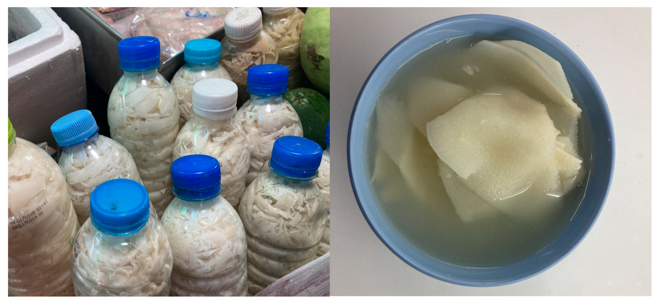
2.3. Sampling Locations and Sample Collection

The sampling sites for pickled bamboo shoot products were located in six provinces of northern Thailand, including Chiang Mai, Nan, Phrae, Chiang Rai, Phayao, and Lampang. All samples were collected during the period from May to July 2020.

2.4. Enrichment and Isolation of Xylose-Fermenting LAB

Approximately 5 g of bamboo shoot products were homogenized in 10 mL of sterile 0.85% NaCl as a diluent using a Masticator® homogenizer blender (Barcelona, Spain) for 5 min. The homogenate was then transferred to a laboratory bottle containing 40 mL mMRSB-XOSs. An enrichment step was carried out at 37 °C for 24–48 h. The culture that exhibited optical density at 600 nm (OD600) of more than 1.00 was selected for the isolation of LAB. The turbid culture was serially diluted from 100 to 105 using a diluent. Accordingly, 0.1 mL of each dilution was then plated on mMRSA-XOSs supplemented with 125 ppm of bromocresol purple and then incubated at 37 °C for 48 h. The distinct colonies that presented clear yellow zones around the colonies, on a purple background, were isolated and streaked on the same medium for the purposes of purification. They were assumed to be XOSs-fermenting LAB. The yellow clear zone produced on mMRSA-XOSs was associated with the hydrolysis of XOS in releasing fermentable sugars for the production of short-chain fatty acids, i.e., lactic acid and acetic acid [7].

2.5. Confirmation and Screening for XOSs-Fermenting LAB

XOSs-fermenting LAB were grown in a screw-cap tube containing 10 mL of mMRSB-XOSs at 37 °C. After 24 h of cultivation, the cells were harvested by centrifugation, washed twice with phosphate buffer saline (PBS) solution, and resuspended in the same solution to a final OD600 of 0.5. The resulting cell suspension was used as an inoculum. A total of 10% (v/v) of inoculum was transferred to a screw-cap tube containing 10 mL of two different media comprised of mMRSB-X (mMRSB containing 10 g/L xylose as the sole carbon source) and mMRSB-XOSs. The mMRSB containing 10 g/L glucose was used as a control. After being incubated at 37 °C for 24 h, the culture was measured for OD600 prior to cell removal by centrifugation. The supernatant was collected for pH measurement and the determination of residual carbohydrates using the phenol–sulfuric acid method [8].

2.6. Bacterial Identification

The genomic DNA of LAB was extracted using a Wizard® Genomic DNA purification kit (Promega Corp., Madison, WI, USA) according to the manufacturer’s instructions. The 16S rRNA gene was amplified with the use of a set of primers, namely 27F (5′-AGAGTTTGATCCTGGCTCAG-3′) and 1492R (5′-GGTACCTTGTTACGACTT-3′), while the genomic DNA was used as a template according to the protocol previously described [7]. The amplicon of the 16S rRNA gene was purified and sequenced using a sequencing service provider (1st BASE Pte Ltd., Singapore). For bacterial identification, pairwise alignment of the 16S rRNA gene sequence was performed using the standard nucleotide blast tool offered by the National Center for Biotechnology Information (NCBI) available at https://blast.ncbi.nlm.nih.gov/Blast.cgi (accessed on 14 December 2021). The neighbor-joining method employing bootstrap resampling from 1000 replicates of the MEGA software version 11.0.10 [9] was used to construct a phylogenetic tree of selected yeast species. The identified sequences were then deposited in Genbank.

2.7. In Vitro Survival Test for Simulated Gastric Conditions

A total of 0.5 mL of the prepared cell suspension (108 cells/mL) was transferred to a screw-cap tube containing 49.5 mL of MRSB-XOSs comprised of 0.3% (w/v) pepsin. It was then adjusted to a pH of 2.0 by the addition of 0.1 N HCl. The incubation period was carried out at 37 °C for 4 h. Samples were periodically collected for the determination of viable cells using the plate-count technique. The initial cell concentration was set to 100%.

2.8. In Vitro Survival Test for Simulated Intestinal Conditions

A total of 0.5 mL of the prepared cell suspension (108 cells/mL) was transferred to a screw-cap tube containing 49.5 mL of MRSB-XOSs that contained 0.3% (w/v) bile salts and 0.3% (w/v) pancreatin. Incubation was carried out at 37 °C for 8 h. Samples were periodically collected for the determination of viable cells using the plate count technique. The initial cell concentration was set to 100%.

2.9. Cell Surface Hydrophobicity Assay

Bacterial adherence was determined by cell surface hydrophobicity [10]. Hydrophobicity is an indirect screening tool that can be used to test and select the adhesion potential of LAB into host intestinal mucosa [11]. A total of 3 mL of the bacterial suspension in PBS (Ainitial) was transferred to a glass tube (12 × 100 mm) containing 1 mL of chloroform, agitated using a vortex mixer for 2 min, and allowed to stand at 37 °C for 30 min. The optical density of the aqueous phase (Afinal) was measured at a wavelength of 600 nm. The hydrophobicity index (HPBI) was calculated using the following equation:
HPBI   ( % ) = ( 1 A final A initial )   ×   100

2.10. Auto-Aggregation Assay

The auto-aggregation of probiotics is believed to be aligned with the adhesion of LAB to the intestinal epithelium [11]. A total of 3 mL of the bacterial suspension in PBS (Ainitial) was transferred to a glass tube (12 × 100 mm), vortexed for 10 s, and incubated at 37 °C for 12 h. The absorbance of the upper part of the mixture (approximately 1 mL) was measured at 600 nm (Afinal) [10]. Auto-aggregation (AA) was calculated using the following equation:
Auto-aggregation   ( % ) = ( 1 A final A initial )   ×   100

2.11. Fermentation of XOSs

A total of 10% (v/v) inoculum was transferred to a screw-cap tube containing 40 mL of mMRSB-XOSs and incubated at 37 °C for 24 h. The culture was centrifuged at 17,350× g for 10 min at 4 °C. The clear supernatant was assigned as a crude extracellular enzyme. It was also used for the determination of the residual carbohydrate concentration, the profile of XOSs, antimicrobial activity, and short-chain fatty acids (SCFAs). Subsequently, cell pellets were washed twice with PBS solution and resuspended with the same volume of 100 mM sodium phosphate buffer at a pH of 6.5. A half-volume of the cell suspension was used as a cell-associated enzyme, while the rest was disrupted for 5 min using a sonicator. The resulting cell-free extract was designated as an intracellular enzyme.

2.11.1. Investigation of Antimicrobial Activity

The antimicrobial activity of the XOSs-fermented broth obtained from the previous preparation, as described in Section 2.11, was assessed against foodborne pathogens using the agar well diffusion method [10]. The foodborne pathogens included B. cereus TISTR 747, E. coli TISTR 527, S. Thyphimurium TISTR 1472, and L. monocytogenes DMST 17303.

2.11.2. Determination of Xylanase and β-Xylosidase Activities

For the xylanase activity assay, 50 μL of the enzyme was reacted with 50 μL of the substrate (0.5% (w/v) xylan in 100 mM of sodium phosphate buffer, pH 6.5. The reaction was carried out at 37 °C for 10 min. Then, 100 μL of DNS solution was added to stop the reaction prior to heating at 100 °C for 10 min. After allowing the reaction to cool, 800 μL of distilled water was added to the reaction. It was then mixed, and the absorbance was measured at 540 nm. One unit (1 U) of xylanase was defined as the amount of the enzyme that released 1 μmole of reducing sugars in 1 min under assay conditions. The xylanase activity was expressed in terms of mU/mL culture.
For the β-xylosidase activity assay, 25 μL of enzyme was reacted with 225 μL of substrate (2 mM p-nitrophenyl xylopyranoside in sodium phosphate buffer at pH 6.5). The reaction was carried out at 37 °C for 10 min. After that, 500 μL of 2% (w/v) sodium carbonate was added to stop the reaction. Finally, the absorbance at 405 nm was measured. One unit (1 U) of β-xylosidase was defined as the amount of the enzyme that released 1 μmole of p-nitrophenol in 1 min under assay conditions. The β-xylosidase activity was expressed in terms of mU/mL culture.

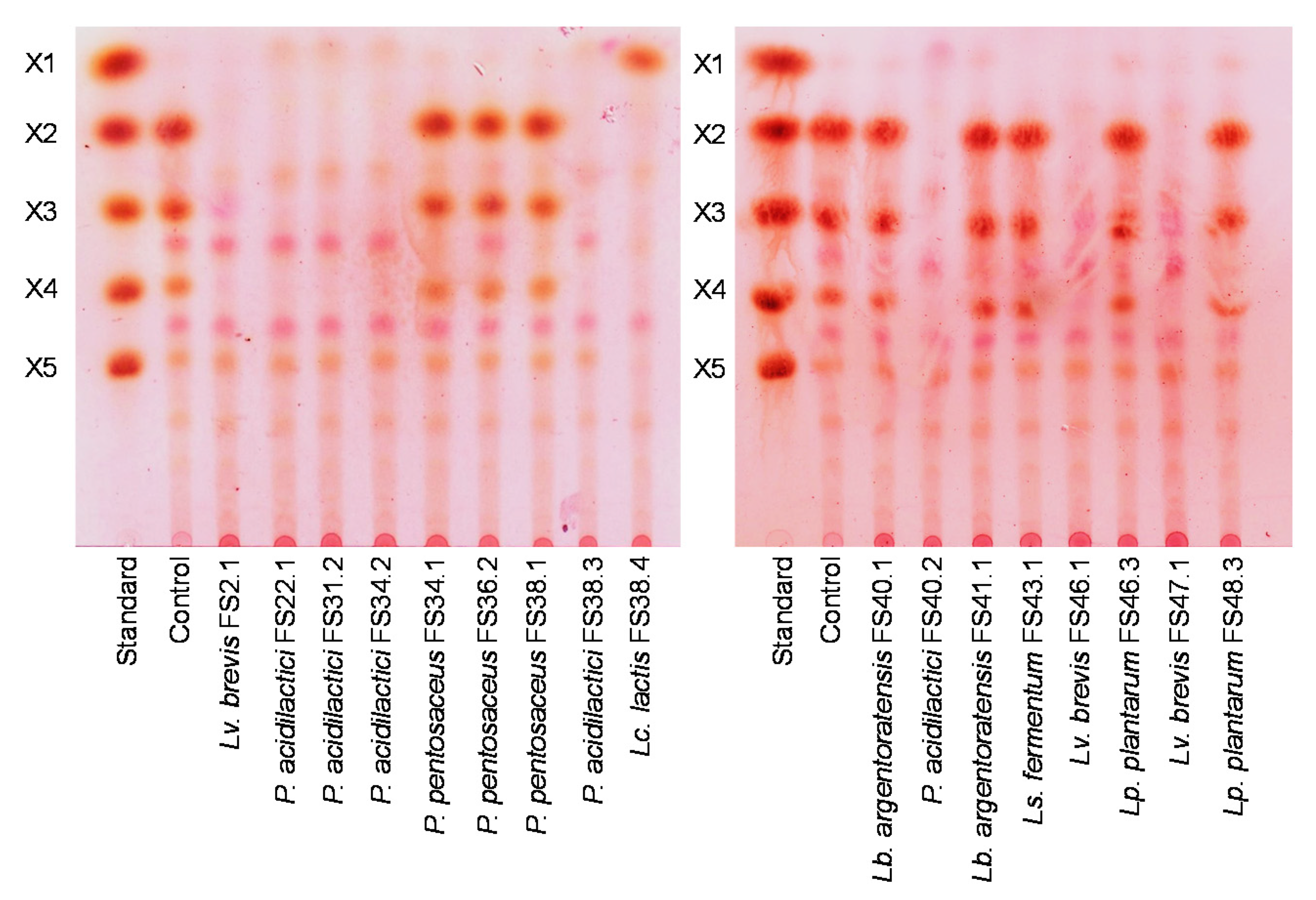
2.11.3. Thin-Layer Chromatography

TLC was performed according to a previously described method [7]. A sample of 1 μL was spotted on an aluminum silica gel plate. After applying a dryer, the plate was developed in a chamber containing n-butanol/ethanol/water (5:3:2 v/v/v). The developing procedure was conducted twice to separate XOSs and xylose better. The TLC plate was then dried and sprayed with 0.5% (w/v) thymol in a mixed solution of 5% (v/v) H2SO4 in ethanol. The spots of the carbohydrates were visualized by heating at 100 °C for 10 min.

2.11.4. Determination of SCFAs

SCFAs, including acetic acid, lactic acid, propionic acid, and butyric acid, were analyzed by HPLC according to the previously described method [7].

2.11.5. Antibiotic resistance

Antibiotic susceptibility was determined using Hicomb Mic test® strips (HiMedia, Nashik, India) of ampicillin (Amp), vancomycin (Van), gentamicin (Gen), kanamycin (Kan), erythromycin (Ery), clindamycin (Cli), tetracycline (Tet), and chloramphenicol (Chl). Briefly, an overnight culture (approximately 106–108 CFU/mL) of the LAB was swabbed onto mMRSA-XOS plates. The antibiotic strip was carefully placed onto the plates. The plates were then incubated at 37 °C for 24 h. The results were expressed as susceptible, S; intermediate, I; or resistant, R, as has been established by the Clinical and Laboratory Standard Institute [12].

2.12. Hemolytic Activity Test

A hemolysis assay was conducted using Columbia blood agar supplemented with 5% (v/v) sheep’s blood. LAB strains were streaked on a blood agar plate and incubated at 37 °C for 48 h. Colonies that formed a clear zone and that were surrounded with a green hue were identified as β-hemolysis and α-hemolysis, respectively, while those that exhibited no clear zone were identified as γ-hemolysis.

2.13. Analytical Methods

All experiments were performed in duplicate. The results are presented as values of the mean ± standard deviation. Data analysis of the mean values was performed based on a full factorial complete randomized design (CRD). Briefly, data were subjected to analysis of variance (ANOVA). Multiple comparison tests were performed based on all pairwise comparisons using Tukey’s HSD test at a 95% confidence level. All analyses were carried out using the Statistix software version 8.0 (Analytical software, Tallahassee, FL, USA). A probability value of p < 0.05 was considered significant.

3. Results

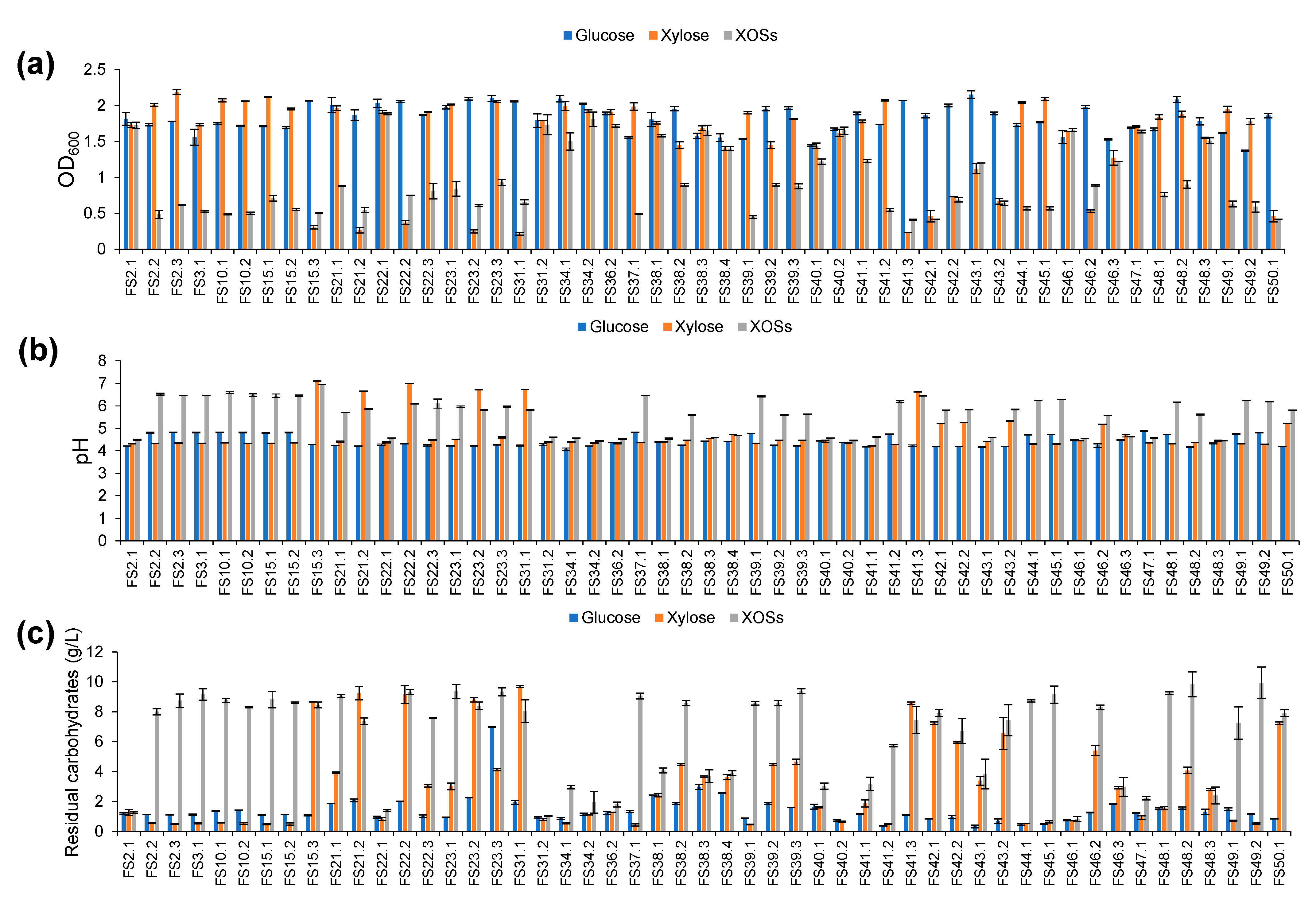
3.1. Isolation of XOSs-Fermenting LAB

A total of 51 XOSs-fermenting LAB were recovered from 24 samples of pickled bamboo shoot products collected from the Chiang Mai, Chiang Rai, Phayao, Phrae, and Nan Provinces of Thailand. All were characterized as Gram-positive. Notably, the assumed XOSs-fermenting LAB were found in all samples, and the number of isolates varied among all samples. Nine out of 24 samples possessed more than two different colonies (Table 1). All assumed XOSs-fermenting LAB were further confirmed by their ability to ferment XOSs.

3.2. Selection of XOSs-Fermenting LAB

All tested LAB were able to grow in the medium containing XOSs as the sole carbon source to different degrees of growth. Glucose, as a representative simple sugar, was used as a control, while xylose was used as the representative pentose sugar. As judged by the OD600 value, LAB strains that exhibited growth in XOSs with OD600 values greater than 1.0 and at least their growth showed equal extents with xylose and glucose, were considered as potential XOSs-fermenting LAB (Figure 2a). Among 51 strains of the assumed XOSs-fermenting LAB, 17 strains were confirmed in terms of their XOSs-fermentability. This information corresponded to a decrease in the pH value (Figure 2b) and the resulting residue carbohydrate (Figure 2c). When the carbon source was fermented by the potential XOSs-fermenting LAB, the pH value decreased from an initial value of 6.5 to a final value within a range of pH 4.0 to 4.5, while less than 4 g/L of the initial carbon source was retained.

3.3. Identification of XOSs-Fermenting LAB

The 16S rRNA gene sequence analysis of 17 LAB strains was performed to assess the level of diversity among potential XOSs-fermenting LAB. An online similarity analysis of all 16S rRNA gene sequences revealed that all strains were closely related to Lv. brevis, P. acidilactici, P. pentosaceus, Lc. lactis, Lb. argentoratensis, Ls. fermentum, and Lp. plantarum. The differentiation of Lb. argentoratensis and Lp. plantarum was performed by phylogenetic analysis according to the previously described protocol [13]. The 16S rRNA gene was deposited in GenBank under an individual accession number (Table 2).

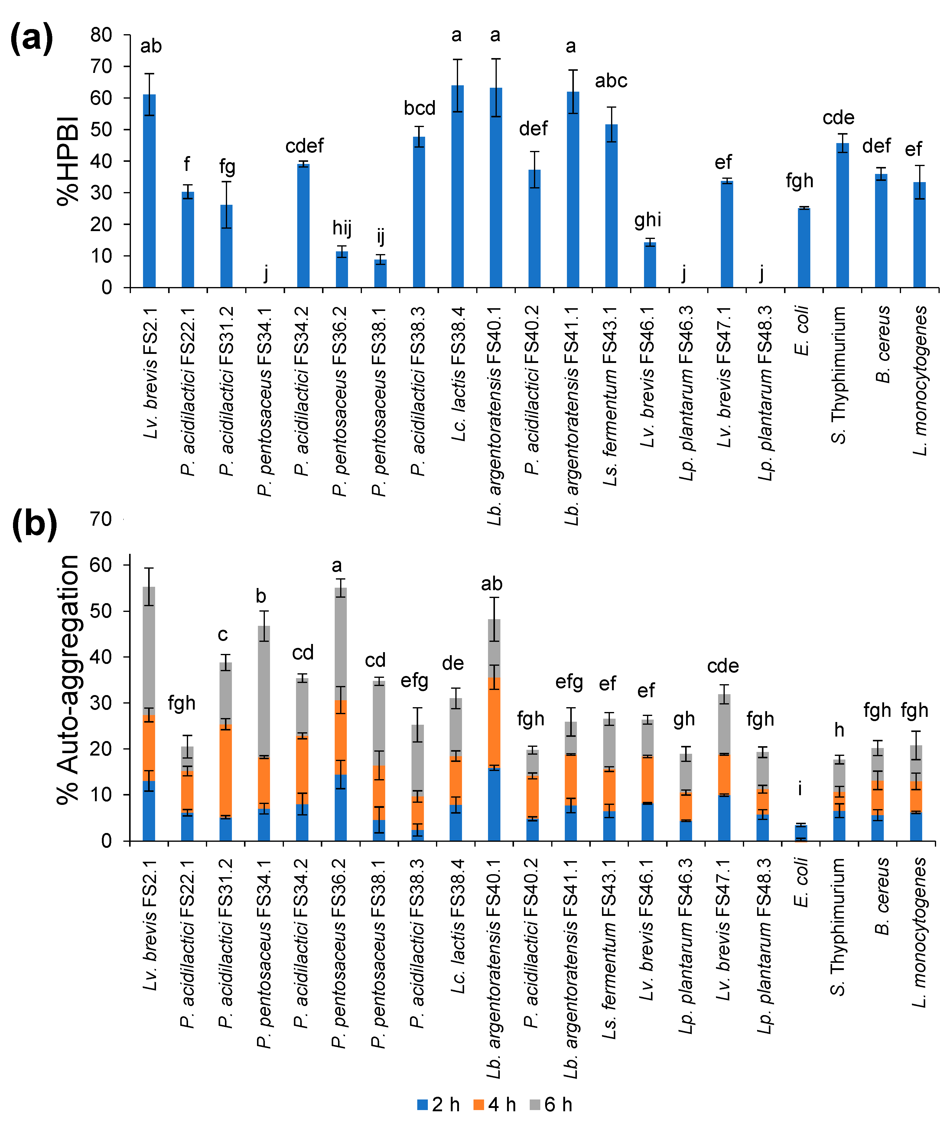
3.4. Tolerance of XOSs-Fermenting LAB under Simulated GIT Conditions

Under simulated gastric conditions, all XOSs-fermenting LAB were able to survive to different degrees, with the exception of Lc. lactis FS38.4. After treatment for 1 h, Lv. brevis FS2.1, P. acidilactici FS38.3, Lb. argentoratensis FS40.1, Ls. fermentum FS43.1, Lv. brevis FS46.1, Lp. plantarum FS46.3, Lv. brevis FS47.1, and Lp. plantarum FS48.3 were partially inhibited, while more than 75% of the initial cell viability was retained after 2 h of incubation. Other strains were inhibited to a higher degree and retained 37% to 50% of the initial cell viability. Notably, bile salt and pancreatin had no effect on the survival of all tested XOSs-fermenting LAB (Table 3).

3.5. Cell Surface Hydrophobicity and Auto-Aggregation

Five XOS-fermenting LAB showed the highest %HPBI within a range of 50–60%. These included Lv. brevis FS2.1, Lc. lactis FS38.4, Lb. argentoratensis strains FS40.1 and FS41.1, and Ls. fermentum FS43.1. Others exhibited no significance or significantly lower %HPBI than those of the tested pathogenic bacteria (Figure 3a).
Various strains of XOSs-fermenting LAB displayed different values of %AA (Figure 3b). Eight out of seventeen strains presented significantly higher %AA than certain representative foodborne pathogens. The first four strains that demonstrated the highest %AA were Lv. brevis FS2.1, P. pentosaceus FS36.2, Lb. argentoratensis FS40.1, and P. pentosaceus FS34.1, respectively. In addition, the %AA obtained from P. acidilactici strains FS31.2 and FS34.2, P. pentosaceus FS38.1, Lc. lactis FS38.4, and Lv. brevis FS47.1 were greater than 20%. These values were higher than those of the pathogens.

3.6. Antimicrobial Activity

The culture supernatant obtained from XOS fermentation by all XOSs-fermenting LAB displayed antimicrobial activity against two different Gram-positive and Gram-negative pathogenic bacteria with regard to the formation of a clear zone on the agar plate. This evidence would indicate that pathogenic bacterial growth was inhibited. The wider clear zone value reflects more potential antimicrobial activity of the LAB against the tested foodborne pathogenic bacteria. The level of antimicrobial activity was indicative of both strain- and species-dependent variations (Table 4).

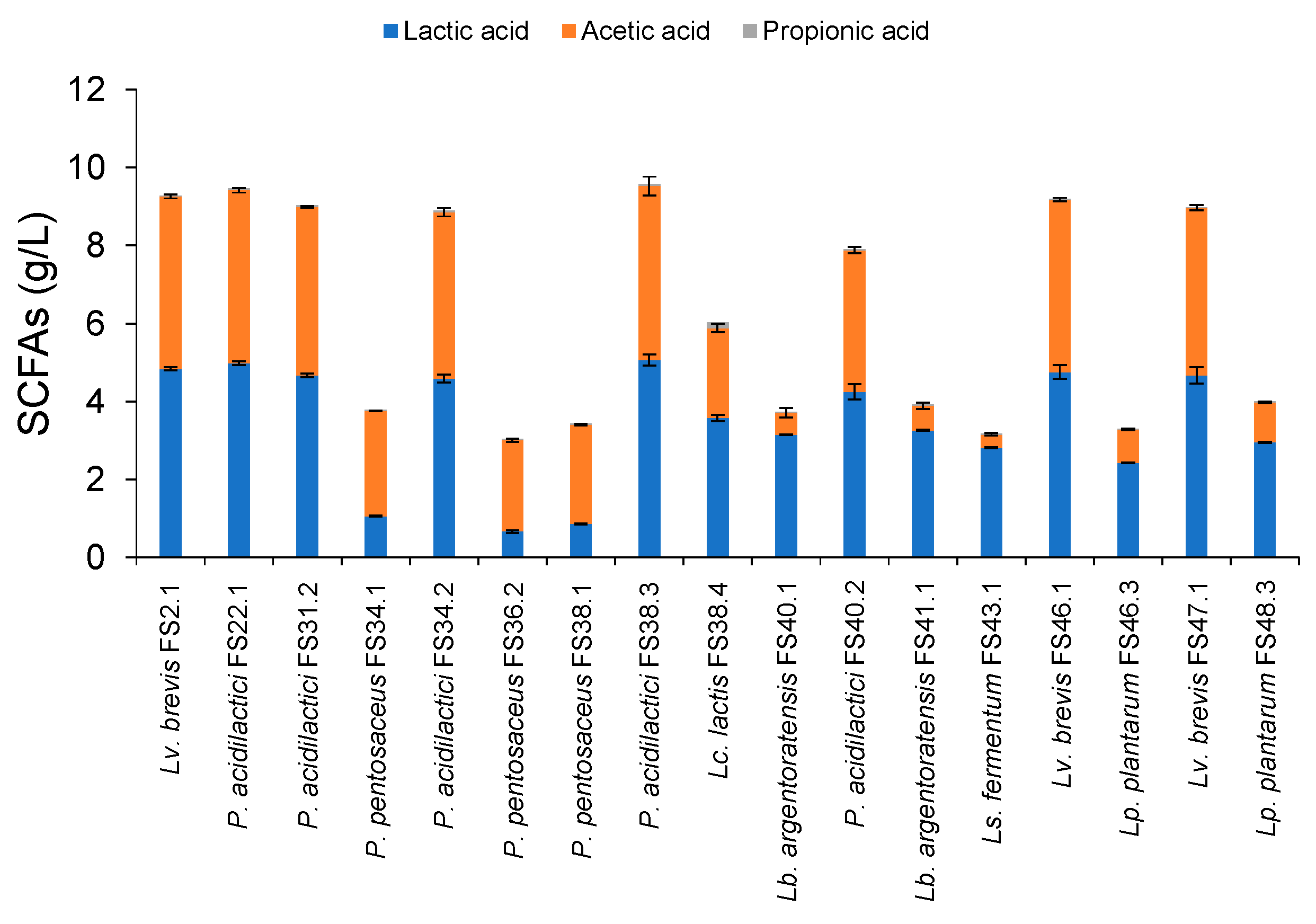
3.7. SCFAs Production

SCFAs production was observed after XOSs fermentation at 37 °C for 24 h. The main SCFAs were lactic acid and acetic acid (Figure 4). Trace amounts of propionic acid were detected from all fermentation cultures. All strains of Lv. lactis and P. acidilactici produced markedly higher amounts of total SCFAs within a range of 8–9 g/L, wherein lactic acid and acetic acid were equally produced at approximately 4–4.5 g/L. The LAB that exhibited weak levels of growth on XOSs produced significantly lower levels of SCFAs than those that exhibited high amounts of growth on XOSs. This may have been associated with the degree of inefficiency in XOSs and xylose fermentation (Figure 2).

3.8. Antibiotic Susceptibility and Hemolytic Activity

All XOSs-fermenting LAB were susceptible to ampicillin, chloramphenicol and erythromycin, tetracycline, and clindamycin, while they were resistant to vancomycin (Table 5). Resistance against gentamycin and kanamycin was associated with species-dependent variations. Intermediately susceptible strains against gentamycin included only P. acidilactici, while other LAB were found to be susceptible. On one hand, all strains of Lv. brevis were susceptible to kanamycin. In addition, all XOSs-fermenting LAB showed no clear zone on blood agar plates, indicating γ-hemolysis.

3.9. Detection of Xylanolytic Enzymes

Xylanase activity was detected from the culture supernatant of all XOSs-fermenting LAB, while it was undetectable for cell suspension and cell extract. This appears to function in contrast with the β-xylosidase activity, which was found to be associated with the cells and partly found in cell extract (Table 6). Considering the residual total carbohydrate values, strains that produced both xylanase and cell-associated β-xylosidase resulted in significantly lower amounts of total carbohydrates than those that produced xylanase alone. Notably, the strain Lb. argentoratensis FS40.1 exhibited the highest degree of xylanase activity at 2187.2 ± 198.1 mU/mL.
A profile of the residual XOSs is shown in Figure 5. Here, three different patterns of the retained carbohydrates were found as follows; pattern I, XOSs were clearly degraded, and xylose was effectively fermented (Lv. brevis strains FS2.1, FS 46.1, and FS 47.1, and P. acidilactici strains FS22.1, FS31.2, FS34.2, FS38.3, and FS40.2); pattern II, XOSs were clearly degraded, and residual xylose was retained (Lc. lactis FS38.4); pattern III, XOSs were partially degraded to shorter-chain XOSs, and trace amounts of residual xylose were retained (P. pentosaceus strains FS34.1, FS36.2, and FS38.1, Lb. argentoratensis strains FS40.1 and FS41.1, Ls. fermentum FS43.1, Lp. plantarum strains FS46.3, and FS48.3).

4. Discussion

Fermentation is a slow decomposition process of organic substrates that may be induced by microorganisms or enzymes in order to convert carbohydrates to alcohols or organic acids [14]. Fermented foods and beverages are associated with different traditions and cultural preferences across the various geographical areas from where they are produced, and this can result in different microbial diversities. As bamboo shoots are one of the various xylan-containing plant materials, Naw Mai Dong would be a beneficial microbial resource in the isolation of microorganisms that are associated with the degradation of xylan, as well as XOSs. The production process of Naw Mai Dong is relatively similar to that of Soidon, a non-salted acidic fermented bamboo shoot product of Manipur, India [15], and other pickled bamboo shoot products [16], but it is produced by a more simplified process than has been previously described. It is further believed that the different aspects of local wisdom that correlate with different geographical regions may be a significant point in the search for the distinct microbial diversity that is associated with each fermented product, as has been found in Phak-Gad-Dong, a pickled mustard green of northern Thailand [17]. Thus, the bacterial diversity of Naw Mai Dong may be different to that of other kinds of pickled bamboo shoots. There has been no record of the isolation of XOSs-fermenting LAB from pickled fruits and vegetables through an enrichment step. The confirmation step was established to ensure the XOSs-fermenting ability of the isolated LAB. Strains that are able to ferment XOSs, at least an equal extent with respect to the reference sugars, xylose, and glucose, could be considered XOSs-fermenting LAB. Consequently, 17 LAB strains were selected for further study. These LAB included seven species, namely Lv. brevis, P. acidilactici, P. pentosaceus, Lc. lactis, L. argentorantensis, Ls. fermentum, and Lp. plantarum. Two LAB species, namely Lv. brevis and P. acidilactici, are recognized as good candidates for the efficient degradation and utilization of XOSs, because their growth in glucose, xylose, and XOSs was luxuriant, while Lc. lactis, Lb. argentoratensis, Lp. plantarum, and Ls. fermentum showed moderate growth in xylose and XOSs. Remarkably, their degree of growth was weaker than that of Lv. brevis and P. acidilactici.
The selected XOSs-fermenting LAB species investigated in this study were similar to the species in the fermented bamboo shoot products of Indian origin, i.e., Lv. brevis, P. pentosaceus, and Lp. plantarum [15,18]. It is noteworthy to mention that Lb. argentoratensis is an elevated species of Lb. plantarum [13], yet the recovery of this species is assumed to be similar to that of Lp. plantarum. Although it has been well-documented that Lc. lactis is generally found in naturally fermented dairy products [19], it can also be found in fresh and fermented vegetables [20]. Its xylan-degrading ability has been well characterized [21]. Previously, P. acidilactici was isolated from various dairy products [22], malt [23], and kimchi [24]. However, the detection of this species in other ethnic pickled bamboo shoot products is still unknown, according to the results of many current studies [15,18,25]. It appears that the sampling location would limit the recovery of LAB species. Most XOSs-fermenting LAB isolated from samples collected from Chiang Mai Province were highly distributed with Pediococcus sp., while those isolated from samples derived from the Chiang Rai, Phayao, Phrae, and Nan Provinces were Lactobacillus sp.
One of the most essential characteristics of probiotic strains is their survival under GIT conditions. This attribute is required in order to confirm that the probiotic strains can pass through the GIT and reach the large intestines, where they exert a range of beneficial effects. Here, 16 strains of XOSs-fermenting LAB resisted gastric and intestinal conditions by retaining more than 40% of their initial cell viability. On the other hand, Lc. lactis FS38.4 lacked probiotic properties due to its inability to survive under gastric conditions. Consequently, this strain was excluded from the cell surface hydrophobicity, auto-aggregation, and antibiotic susceptibility experiments.
Cell surface hydrophobicity is defined as a nonspecific interaction in adhesion between probiotic microorganisms onto GIT epithelial cells, where they may provide prophylactic and therapeutic benefits [26]. Colonization in the epithelial cells of the large intestine and mucosal surfaces can prevent pathogenic bacteria adhesion and inflammatory reactions [27]. Auto-aggregation is defined as aggregation among LAB to form flocs and colonize the intestinal environment of the host when the cells approach harmful conditions [26]. Auto-aggregation and cell surface hydrophobicity are characteristics that provide potential advantages for microorganisms in colonizing the intestinal tract [28]. Factually, probiotics should be associated with higher values of %HPBI and %AA than pathogens. However, these attributes may or may not be correlated with one another [28]. Remarkably, Lv. brevis FS2.1 and Lv. brevis FS40.1 were the only two strains that exhibited high %auto-aggregation and %HPBI.
The production of antimicrobial substances is an important advantage of probiotics. During growth, probiotics produce antimicrobial compounds, i.e., ethanol, fatty acids, hydrogen peroxide, and bacteriocins that can inhibit various bacterial pathogens [29]. In agreement with the findings of this study, all XOSs-fermenting LAB displayed antimicrobial activity against foodborne pathogens. In the case of Lv. brevis and P. acidilactici, it was confirmed that the inhibitory effect might have been caused by the secretion of SCFAs, namely lactic acid and acetic acid, as these species produced approximately 90% of the total SCFAs from XOSs. On the other hand, the remaining XOS-fermenting LAB species produced a low amount of total SCFAs; thus, they may have other inhibitory mechanisms i.e., bacteriocins and bacteriocin-like substances [30].
Antibiotics are the most effective drugs available for the treatment of bacterial infections [31]. When LAB are used as probiotics, they are known to harbor antibiotic-resistant genes that can be transferred to pathogenic bacteria and the commensal bacteria present in the gut [32], which in turn can cause significant suffering to people with pathogen infections [11]. Therefore, probiotics that are sensitive to commonly prescribed antibiotics at low concentrations have a desirable character. In addition, it is recommended that probiotics should be susceptible to at least two clinically relevant antibiotics [33]. In this study, 16 strains of XOSs-fermenting LAB were susceptible to six out of eight different tested antibiotics, and their safety was confirmed with regard to their negative hemolytic activity.
To evaluate the efficiency for XOSs fermentation, the production of a xylanolytic enzyme is of particular relevance. Efficient degradation and utilization of XOSs are affected by the degree of polymerization (DP) of XOSs while also being strain-specific. Xylanolytic enzymes associated with XOSs degradation are mostly inducible enzymes. These include β-xylosidase, α-glucuronidase, α-L-arabinosidase, and acetyl xylan esterase, as well as xylanase [34]. The XOSs used in this study were homologous XOSs with a backbone mainly composed of β-(1,4) linkages between xylose residues; thus, β-xylosidase and xylanase were the primary focus of this study.
As far as our research is concerned, the utilization of XOSs by probiotic LAB has been extensively studied in terms of metabolic activity, enzyme activity, and transcriptomic analysis. The discovery of a coding region for xylanase and β-xylosidase was recorded in P. acitilactici [35], P. pentosaceus [36], Lp. plantarum, and Lv. brevis [34]. Previously, XOSs were proposed to be degraded by intracellular xylanase and β-xylosidase [34,37]. It has been well documented that xylanase is an endo-type enzyme that randomly catalyzes the hydrolysis of xylan to release XOSs and xylose, while β-xylosidase acts as an exo-type enzyme that catalyzes the hydrolysis of non-reducing end xylose residues from XOSs [38]. Strains that were capable of secreting xylanase resulting from random cleavages of XOSs retained shorter-chain XOSs, mainly xylobiose (X2) and xylotriose (X3). It has also been noted that all strains might be able to produce intracellular β-xylosidase, as is commonly known; however, the amount might not be enough for effective detection. On the other hand, effective levels of degradation and utilization of XOSs were found in strains that produced both xylanase and β-xylosidase. The findings of this study were slightly different from those of a number of previously discussed reports, since xylanase was detected as an extracellular enzyme, while β-xylosidase could be found in either intracellular fractions, the whole cell, or both. The profiles of XOSs obtained after XOSs fermentation support the detected xylanolytic activity. Based on these results, it has been proposed that xylanase is a key enzyme that is capable of producing shorter-chain XOSs in promoting the release of xylose with regard to the cell-associated or intracellular β-xylosidase. LAB with cell-associated β-xylosidase resulted in more considerable XOSs degradation than those that produced only intracellular enzymes. The production of extracellular xylanase is not commonly found in LAB, thus providing new insight into alternative sources of xylanase. It is important to note that not only will the ability to produce xylanolytic enzymes, but also the characteristic of xylose fermentability, effectively support proficient XOSs fermentation.
According to its overall attributes, Lv. brevis FS2.1 is considered the most promising strain among all XOSs-fermenting LAB. This has been determined by its resistance to GIT conditions, high values of %auto-aggregation, %cell surface hydrophobicity, antimicrobial production, and efficient degradation and utilization of XOSs. In consideration of their survival under GIT conditions and antimicrobial activity against foodborne pathogens, all 16 strains of XOSs-fermenting LAB could be used as probiotics; however, further in vivo model experiments would be necessary to confirm their health-beneficial properties.

5. Conclusions

This study revealed that the pickled bamboo shoot products of northern Thailand can be considered an alternative beneficial source of XOSs-fermenting LAB. Sixteen strains of the isolated LAB were confirmed to have probiotic properties according to survival under simulated GIT conditions and for their antimicrobial activity. The most promising strain was Lv. brevis FS2.1, as it exhibited the highest percentages of survival under GIT conditions, cell surface hydrophobicity, auto-aggregation, and efficient degradation and utilization of XOSs. Other XOSs-fermenting LAB also exhibited certain beneficial attributes, indicating their potential to be used as probiotics. It is strongly believed that the XOSs-fermenting LAB could be used for the development of synbiotic supplementation containing the approved LAB and XOSs. This would occur in conjunction with the LAB’s fermenting ability of xylose being released after the effective enzymatic degradation of XOSs was recognized as a crucial attribute in promoting the successful fermentation of xylose release after the effective enzymatic degradation of XOSs. Remarkably, xylanase was identified and classified as an extracellular enzyme, while also emerging as a potential source of XOSs-fermenting probiotic LAB. This finding may shed light on Naw Mai Dong as a promising source of xylanolytic enzyme-producing LAB.

Author Contributions

Conceptualization, A.K.; funding acquisition, A.K.; investigation, A.K., P.N., J.W. and N.L.; methodology, A.K., P.N., J.W. and N.L.; project administration, A.K. and C.K.; supervision, A.K., writing–original draft, A.K.; writing-review and edition, A.K. All authors have read and agreed to the published version of the manuscript.

Funding

This research study was partially supported by funding provided by Chiang Mai University.

Institutional Review Board Statement

Not applicable.

Informed Consent Statement

Not applicable.

Data Availability Statement

Not applicable.

Conflicts of Interest

The authors declare that they hold no conflict of interest.

References

  1. de Freitas, C.; Terrone, C.C.; Masarin, F.; Carmona, E.C.; Brienzo, M. In vitro study of the effect of xylooligosaccharides obtained from banana pseudostem xylan by enzymatic hydrolysis on probiotic bacteria. Biocatal. Agric. Biotechnol. 2021, 33, 101973. [Google Scholar] [CrossRef]
  2. Rashid, R.; Sohail, M. Xylanolytic Bacillus species for xylooligosaccharides production: A critical review. Bioresour. Bioprocess 2021, 8, 16. [Google Scholar] [CrossRef]
  3. Li, Z.; Summanen, P.H.; Komoriya, T.; Finegold, S.M. In vitro study of the prebiotic xylooligosaccharide (XOS) on the growth of Bifidobacterium spp. and Lactobacillus spp. Int. J. Food Sci. Nutr. 2015, 66, 919–922. [Google Scholar] [CrossRef] [PubMed]
  4. Petrova, P.; Petrov, K. Chapter 7—Prebiotic–Probiotic relationship: The genetic fundamentals of polysaccharides conversion by Bifidobacterium and Lactobacillus Genera. In Food Bioconversion; Grumezescu, A.M., Holban, A.M., Eds.; Academic Press: Amsterdam, The Netherlands, 2017; pp. 237–278. [Google Scholar]
  5. Michlmayr, H.; Hell, J.; Lorenz, C.; Böhmdorfer, S.; Rosenau, T.; Kneifel, W. Arabinoxylan oligosaccharide hydrolysis by family 43 and 51 glycosidases from Lactobacillus brevis DSM 20054. Appl. Environ. Microbiol. 2013, 79, 6747–6754. [Google Scholar] [CrossRef] [PubMed] [Green Version]
  6. Li, K.; Wang, X.; Wang, J.; Zhang, J. Benefits from additives and xylanase during enzymatic hydrolysis of bamboo shoot and mature bamboo. Bioresour. Technol. 2015, 192, 424–431. [Google Scholar] [CrossRef] [PubMed]
  7. Kanpiengjai, A.; Rieantrakoonchai, W.; Pratanaphon, R.; Pathom-aree, W.; Lumyong, S.; Khanongnuch, C. High efficacy bioconversion of starch to lactic acid using an amylolytic lactic acid bacterium isolated from Thai indigenous fermented rice noodles. Food Sci. Biotechnol. 2014, 23, 1541–1550. [Google Scholar] [CrossRef]
  8. Dubois, M.; Gilles, K.; Hamilton, J.K.; Rebers, P.A.; Smith, F. A colorimetric method for the determination of sugars. Nature 1951, 168, 167. [Google Scholar] [CrossRef] [PubMed]
  9. Tamura, K.; Stecher, G.; Kumar, S. MEGA11: Molecular Evolutionary Genetics Analysis Version 11. Mol. Biol. Evol. 2021, 38, 3022–3027. [Google Scholar] [CrossRef]
  10. Kanpiengjai, A.; Khanongnuch, C.; Lumyong, S.; Kummasook, A.; Kittibunchakul, S. Characterization of Sporidiobolus ruineniae A45.2 cultivated in tannin substrate for use as a potential multifunctional probiotic yeast in aquaculture. J. Fungi 2020, 6, 378. [Google Scholar] [CrossRef]
  11. Unban, K.; Chaichana, W.; Baipong, S.; Abdullahi, A.D.; Kanpiengjai, A.; Shetty, K.; Khanongnuch, C. Probiotic and antioxidant properties of lactic acid bacteria isolated from indigenous fermented tea leaves (Miang) of north Thailand and promising application in synbiotic formulation. Fermentation 2021, 7, 195. [Google Scholar] [CrossRef]
  12. CLSI. CLSI Document M07-A9. Methods for Dilution Antimicrobial Susceptibility Tests for Bacteria That Grow Aerobically: Approved Standard, 19th ed.; CLSI: Wayne, PA, USA, 2012. [Google Scholar]
  13. Liu, D.D.; Gu, C.T. Proposal to reclassify Lactobacillus zhaodongensis, Lactobacillus zeae, Lactobacillus argentoratensis and Lactobacillus buchneri subsp. silagei as Lacticaseibacillus zhaodongensis comb. nov., Lacticaseibacillus zeae comb. nov., Lactiplantibacillus argentoratensis comb. nov. and Lentilactobacillus buchneri subsp. silagei comb. nov., respectively and Apilactobacillus kosoi as a later heterotypic synonym of Apilactobacillus micheneri. Int. J. Syst. Evol. Microbiol. 2020, 70, 6414–6417. [Google Scholar] [PubMed]
  14. Swain, M.R.; Anandharaj, M.; Ray, R.C.; Parveen Rani, R. Fermented fruits and vegetables of Asia: A potential source of probiotics. Biotechnol. Res. Int. 2014, 2014, 250424. [Google Scholar] [CrossRef]
  15. Jeyaram, K.; Romi, W.; Singh, T.A.; Devi, A.R.; Devi, S.S. Bacterial species associated with traditional starter cultures used for fermented bamboo shoot production in Manipur state of India. Int. J. Food Microbiol. 2010, 143, 1–8. [Google Scholar] [CrossRef] [PubMed]
  16. Nongdam, P.; Tikendra, L. The nutritional facts of bamboo shoots and their usage as important traditional foods of northeast India. Int. Sch. Res. Not. 2014, 2014, 679073. [Google Scholar] [CrossRef] [PubMed]
  17. Yongsawas, R.; Inta, A.; Kampuansai, J.; Pandith, H.; Suwannarach, N.; Lamyong, S.; Chantawannakul, P.; Chitov, T.; Disayathanoowat, T. Bacterial communities in Lanna Phak-Gard-Dong (pickled mustard green) from three different ethnolinguistic groups in northern Thailand. Biology 2022, 11, 150. [Google Scholar] [CrossRef] [PubMed]
  18. Tamang, B.; Tamang, J.P.; Schillinger, U.; Franz, C.M.; Gores, M.; Holzapfel, W.H. Phenotypic and genotypic identification of lactic acid bacteria isolated from ethnic fermented bamboo tender shoots of North East India. Int. J. Food Microbiol. 2008, 121, 35–40. [Google Scholar] [CrossRef]
  19. Li, W.; Ren, M.; Duo, L.; Li, J.; Wang, S.; Sun, Y.; Li, M.; Ren, W.; Hou, Q.; Yu, J.; et al. Fermentation characteristics of Lactococcus lactis subsp. lactis isolated from naturally fermented dairy products and screening of potential starter isolates. Front. Microbiol. 2020, 11, 1477–1483. [Google Scholar] [CrossRef]
  20. Kelly, W.J.; Davey, G.P.; Ward, L.J. Characterization of lactococci isolated from minimally processed fresh fruit and vegetables. Int. J. Food Microbiol. 1998, 45, 85–92. [Google Scholar] [CrossRef]
  21. Tanaka, K.; Komiyama, A.; Sonomoto, K.; Ishizaki, A.; Hall, S.J.; Stanbury, P.F. Two different pathways for D-xylose metabolism and the effect of xylose concentration on the yield coefficient of L-lactate in mixed-acid fermentation by the lactic acid bacterium Lactococcus lactis IO-1. Appl. Microbiol. Biotechnol. 2002, 60, 160–167. [Google Scholar]
  22. Abbasiliasi, S.; Tan, J.S.; Ibrahim, T.A.T.; Ramanan, R.N.; Vakhshiteh, F.; Mustafa, S.; Ling, T.C.; Rahim, R.A.; Ariff, A.B. Isolation of Pediococcus acidilactici Kp10 with ability to secrete bacteriocin-like inhibitory substance from milk products for applications in food industry. BMC Microbiol. 2012, 12, 260. [Google Scholar] [CrossRef] [Green Version]
  23. Ahn, H.; Kim, J.; Kim, W.J. Isolation and characterization of bacteriocin-producing Pediococcus acidilactici HW01 from malt and its potential to control beer spoilage lactic acid bacteria. Food Control 2017, 80, 59–66. [Google Scholar] [CrossRef]
  24. Kim, M.; Choi, E.; Kim, J.; Ahn, H.; Han, H.; Kim, W.J. Effect of bacteriocin-producing Pediococcus acidilactici K10 on beer fermentation. J. Inst. Brew. 2016, 122, 422–429. [Google Scholar] [CrossRef] [Green Version]
  25. Xia, X.; Ran, C.; Ye, X.; Li, G.; Kan, J.; Zheng, J. Monitoring of the bacterial communities of bamboo shoots (Dendrocalamus latiflorus) during pickling process. Int. J. Food Sci. Technol. 2017, 52, 1101–1110. [Google Scholar] [CrossRef]
  26. Gut, A.M.; Vasiljevic, T.; Yeager, T.; Donkor, O.N. Characterization of yeasts isolated from traditional kefir grains for potential probiotic properties. J. Funct. Foods 2019, 58, 56–66. [Google Scholar] [CrossRef]
  27. Kavitha, M.; Raja, M.; Perumal, P. Evaluation of probiotic potential of Bacillus spp. isolated from the digestive tract of freshwater fish Labeo calbasu. Aquac. Rep. 2018, 11, 59–69. [Google Scholar] [CrossRef]
  28. Krausova, G.; Hyrslova, I.; Hynstova, I. In vitro evaluation of adhesion capacity, hydrophobicity, and auto-aggregation of newly isolated potential probiotic strains. Fermentation 2019, 5, 100. [Google Scholar] [CrossRef] [Green Version]
  29. Chen, C.C.; Lai, C.C.; Huang, H.L.; Huang, W.Y.; Toh, H.S.; Weng, T.C.; Chuang, Y.C.; Lu, Y.C.; Tang, H.J. Antimicrobial activity of Lactobacillus species against carbapenem-resistant Enterobacteriaceae. Front. Microbiol. 2019, 10, 789. [Google Scholar] [CrossRef]
  30. Jiang, S.; Cai, L.; Lv, L.; Li, L. Pediococcus pentosaceus, a future additive or probiotic candidate. Microb. Cell Fact. 2021, 20, 45. [Google Scholar] [CrossRef]
  31. Neut, C.; Mahieux, S.; Dubreuil, L.J. Antibiotic susceptibility of probiotic strains: Is it reasonable to combine probiotics with antibiotics? Med. Mal. Infect. 2017, 47, 477–483. [Google Scholar] [CrossRef]
  32. Cave, R.; Cole, J.; Mkrtchyan, H.V. Surveillance and prevalence of antimicrobial resistant bacteria from public settings within urban built environments: Challenges and opportunities for hygiene and infection control. Environ. Int. 2021, 157, 106836. [Google Scholar] [CrossRef]
  33. Sanders, M.E.; Akkermans, L.M.; Haller, D.; Hammerman, C.; Heimbach, J.; Hörmannsperger, G.; Huys, G.; Levy, D.D.; Lutgendorff, F.; Mack, D.; et al. Safety assessment of probiotics for human use. Gut Microbes 2010, 1, 164–185. [Google Scholar] [CrossRef] [PubMed]
  34. Iliev, I.; Vasileva, T.; Bivolarski, V.; Momchilova, A.; Ivanova, I. Metabolic profiling of xylooligosaccharides by Lactobacilli. Polymers 2020, 12, 2387. [Google Scholar] [CrossRef] [PubMed]
  35. Lei, Z.; Wu, Y.; Nie, W.; Yin, D.; Yin, X.; Guo, Y.; Aggrey, S.E.; Yuan, J. Transcriptomic analysis of xylan oligosaccharide utilization systems in Pediococcus acidilactici strain BCC-1. J. Agric. Food Chem. 2018, 66, 4725–4733. [Google Scholar] [CrossRef] [PubMed]
  36. Han, D.; Yan, Q.; Liu, J.; Jiang, Z.; Yang, S. Transcriptomic analysis of Pediococcus pentosaceus reveals carbohydrate metabolic dynamics under lactic acid stress. Front. Microbiol. 2021, 12, 736411. [Google Scholar] [CrossRef]
  37. Gilad, O.; Jacobsen, S.; Stuer-Lauridsen, B.; Pedersen, M.B.; Garrigues, C.; Svensson, B. Combined transcriptome and proteome analysis of Bifidobacterium animalis subsp. lactis BB-12 grown on xylo-oligosaccharides and a model of their utilization. Appl. Environ. Microbiol. 2010, 76, 7285–7291. [Google Scholar] [CrossRef] [Green Version]
  38. Jordan, D.B.; Wagschal, K. Properties and applications of microbial beta-D-xylosidases featuring the catalytically efficient enzyme from Selenomonas ruminantium. Appl. Microbiol. Biotechnol. 2010, 86, 1647–1658. [Google Scholar] [CrossRef]
Figure 1. Appearance of Naw Mai Dong commercially available in a local market.
Biology 11 00638 g001
Figure 2. Effect of glucose, xylose, and XOSs on growth (a), change in pH (b), and residual carbohydrate (c) of the assumed XOSs-fermenting LAB. The reported pH is the final value.
Biology 11 00638 g002
Figure 3. Percentages of HPBI (a) and AA (b) of the selected XOSs-fermenting LAB. Lower-case letters indicate statistical differences in values at p < 0.05.
Biology 11 00638 g003
Figure 4. SCFAs production obtained from the selected XOSs-fermenting LAB after XOSs fermentation at 37 °C for 24 h.
Biology 11 00638 g004
Figure 5. TLC chromatogram of the XOSs fermentation broth of the selected XOSs-fermenting LAB after XOSs fermentation at 37 °C for 24 h. X1 = xylose; X2 = xylobiose; X3 = xylotriose; X4 = xylotetraose; X5 = xylopentaose.
Biology 11 00638 g005
Table 1. Sample code, sampling locations, date achieved of pickled bamboo shoot products, number of LAB isolates, and their codes.
No.Sample CodeSubdistrict, District, ProvinceLocationDate AchievedNo. of IsolatesIsolate Code
1FS2Mae Faek Mai, San Sai, Chiang Mai18.9950, 98.97788 June 20203FS2.1, FS2.2, FS2.3
2FS3Suthep, Muang, Chiang Mai18.7904, 98.95959 June 20201FS3.1
3FS10Rim Tai, Mae Rim, Chiang Mai18.9133, 98.944516 June 20202FS10.1, FS10.2
4FS15Pa Daet, Pa Daet, Chiang Rai19.4998, 99.990520 June 20203FS15.1, FS15.2, FS15.3
5FS21Pong Tam, Chai Prakarn, Chiang Mai19.7312, 99.102911 August 20202FS21.1, FS21.2
6FS22Mae Sa, Mae Rim, Chiang Mai18.8924, 98.679812 August 20203FS22.1, FS22.2, FS22.3
7FS23Mae Sa, Mae Rim, Chiang Mai18.8924, 98.679816 August 20203FS23.1, FS23.2, FS23.3
8FS31Ki Lek, Mae Taeng, Chiang Mai19.0781, 98.940516 August 20202FS31.1, FS31.2
9FS34San Phi Suea, Mueang, Chiang Mai18.8371, 98.987223 August 20202FS34.1, FS34.2
10FS36Mae Taeng, Mae Taeng, Chiang Mai19.0945, 98.901224 August 20201FS36.2
11FS37San Phak Wan, Hang Dong, Chiang Mai18.7227, 98.957525 August 20201FS37.1
12FS38Nong Kwai, Hang Dong, Chiang Mai18.7381, 98.931231 August 20204FS38.1, FS38.2, FS38.3, FS38.4
13FS39Han Kaeo, Hang Dong, Chiang Mai18.6535, 98.903531 August 20203FS39.1, FS39.2, FS39.3
14FS40Hang Dong, Hang Dong, Chiang Mai18.6894, 98.90365 September 20202FS40.1, FS40.2
15FS41Rong Chang, Pa Daet, Chiang Rai19.4948, 99.94145 September 20203FS41.1, FS41.2, FS41.3
16FS42Pa Ngae, Pa Daet, Chiang Rai19.5545, 99.94635 September 20202FS42.1, FS42.2
17FS43Pa Daet, Pa Daet, Chiang Rai19.5116, 99.99255 September 20202FS43.1, FS43.2
18FS44Tha Wang Thong, Muang, Phayao19.1768, 99.89325 September 20201FS44.1
19FS45Wang Nuer, Wang Nuer, Lampang19.1355, 99.63235 September 20201FS45.1
20FS46Wang Nuer, Wang Nuer, Lampang19.1355, 99.63235 September 20203FS46.1, FS46.2, FS46.3
21FS47Rong Kwang, Rong Kwang, Phrae18.3371, 100.316520 September 20201FS47.1
22FS48Rong Kwang, Rong Kwang, Phrae18.3371, 100.316520 September 20203FS48.1, FS48.2, FS48.3
23FS49Klang Wiang, Wiang Sa, Nan18.5626, 100.746620 September 20202FS49.1, FS49.2
24FS50Klang Wiang, Wiang Sa, Nan18.5626, 100.746620 September 20201FS50.1
Table 2. Molecular identification of 16S rRNA gene sequences of XOSs-fermenting LAB.
IsolatesNucleotides% SimilarityIdentification ResultsAccession Number
FS2.1143099.86Levilactobacillus brevisOM899733
FS22.1145499.79Pediococcus acidilacticiOM899734
FS31.2144799.79Pediococcus acidilacticiOM899735
FS34.11450100.00Pediococcus pentosaceusOM899736
FS34.2146099.79Pediococcus acidilacticiOM899737
FS36.21440100.00Pediococcus pentosaceusOM899738
FS38.11440100.00Pediococcus pentosaceusOM899739
FS38.3144099.79Pediococcus acidilacticiOM899740
FS38.4142099.93Lactococcus lactisOM899741
FS40.1142899.93Lactobacillus argentoratensisOM899742
FS40.2144099.79Pediococcus acidilacticiOM899743
FS41.1144099.86Lactobacillus argentoratensisOM899744
FS43.1144099.93Limosilactobacillus fermentumOM899745
FS46.1143099.79Levilactobacillus brevisOM899746
FS46.3142499.93Lactiplantibacillus plantarumOM899747
FS47.1143099.86Levilactobacillus brevisOM899748
FS48.3142899.93Lactiplantibacillus plantarumOM899749
Table 3. Survival of XOSs-fermenting LAB under gastric and intestinal conditions.
StrainsGastric ConditionsIntestinal Conditions
0 h1 h2 h0 h4 h8 h
Lv. brevis FS2.1100.0 ± 0.1593.0 ± 1.7074.5 ± 0.94100.0 ± 0.98103.1 ± 0.43109.1 ± 0.38
P. acidilactici FS22.1100.0 ± 0.1985.5 ± 1.2347.2 ± 2.16100.0 ± 0.41101.3 ± 0.07104.7 ± 0.18
P. acidilactici FS31.2100.0 ± 0.1376.8 ± 0.5146.8 ± 2.14100.0 ± 0.37102.5 ± 0.23108.0 ± 0.66
P. pentosaceus FS34.1100.0 ± 0.1185.3 ± 1.7843.0 ± 2.41100.0 ± 0.22101.6 ± 0.22101.9 ± 0.23
P. acidilactici FS34.2100.0 ± 0.2084.6 ± 0.3737.9 ± 4.12100.0 ± 0.09101.2 ± 0.08106.1 ± 0.67
P. pentosaceus FS36.2100.0 ± 0.3087.3 ± 0.6543.9 ± 2.46100.0 ± 0.47100.6 ± 0.19101.4 ± 0.20
P. pentosaceus FS38.1100.0 ± 0.5382.3 ± 2.2153.0 ± 2.21100.0 ± 0.42102.3 ± 0.50103.0 ± 0.35
P. acidilactici FS38.3100.0 ± 0.1691.8 ± 0.1078.6 ± 0.48100.0 ± 0.29103.0 ± 0.51108.6 ± 0.33
Lc. lactis FS38.4100.0 ± 1.340.0 ± 0.000.0 ± 0.00100.0 ± 0.93102.9 ± 0.11103.7 ± 0.08
L. argentoratensis FS40.1100.0 ± 1.1577.4 ± 2.0873.6 ± 0.27100.0 ± 0.47102.7 ± 0.79108.4 ± 0.27
P. acidilactici FS40.2100.0 ± 0.1579.5 ± 1.5540.8 ± 1.23100.0 ± 0.48102.7 ± 0.28106.8 ± 0.28
L. argentoratensis FS41.1100.0 ± 0.1753.4 ± 2.3639.0 ± 0.00100.0 ± 0.05104.4 ± 0.20105.6 ± 0.38
Ls. fermentum FS43.1100.0 ± 2.2993.8 ± 0.5091.6 ± 0.92100.0 ± 0.9597.9 ± 0.7997.0 ± 1.54
Lv. brevis FS46.1100.0 ± 0.4894.2 ± 0.7790.0 ± 0.45100.0 ± 0.91101.4 ± 0.37106.3 ± 0.11
Lp. plantarum FS46.3100.0 ± 0.8995.6 ± 0.1398.3 ± 0.90100.0 ± 0.77105.3 ± 0.14105.4 ± 0.18
Lv. brevis FS47.1100.0 ± 1.1797.5 ± 0.0584.3 ± 0.77100.0 ± 0.08101.0 ± 0.80102.4 ± 0.19
Lp. plantarum FS48.3100.0 ± 0.7994.3 ± 0.1694.0 ± 0.42100.0 ± 0.41108.6 ± 0.55112.5 ± 0.68
Table 4. Range of clear zone (centimeter) representing antimicrobial activity against some foodborne pathogens of culture broth obtained from XOSs fermentation by XOSs-fermenting LAB.
StrainsE. coliB. cereusS.
Thyphimurium
L. monocytogenes
Lv. brevis FS2.10.2 ± 0.00.4 ± 0.00.4 ± 0.01.0 ± 0.1
P. acidilactici FS22.10.7 ± 0.00.8 ± 0.00.3 ± 0.00.3 ± 0.0
P. acidilactici FS31.20.6 ± 0.10.6 ± 0.30.5 ± 0.10.5 ± 0.1
P. pentosaceus FS34.10.8 ± 0.11.0 ± 0.11.0 ± 0.31.0 ± 0.1
P. acidilactici FS34.20.6 ± 0.00.7 ± 0.00.4 ± 0.10.4 ± 0.0
P. pentosaceus FS36.20.4 ± 0.11.1 ± 0.00.7 ± 0.40.7 ± 0.1
P. pentosaceus FS38.10.4 ± 0.10.9 ± 0.01.5 ± 0.20.5 ± 0.1
P. acidilactici FS38.30.2 ± 0.11.0 ± 0.11.3 ± 0.40.3 ± 0.0
Lc. lactis FS38.40.5 ± 0.11.0 ± 0.10.8 ± 0.10.4 ± 0.1
L. argentorantensis FS40.10.6 ± 0.11.0 ± 0.10.6 ± 0.10.5 ± 0.1
P. acidilactici FS40.20.6 ± 0.10.7 ± 0.10.9 ± 0.10.8 ± 0.1
L. argentorantensis FS41.10.8 ± 0.10.3 ± 0.10.5 ± 0.10.8 ± 0.1
Ls. fermentum FS43.10.7 ± 0.00.5 ± 0.10.6 ± 0.10.7 ± 0.1
Lv. brevis FS46.10.9 ± 0.00.7 ± 0.10.9 ± 0.00.9 ± 0.0
Lp. plantarum FS46.30.6 ± 0.10.6 ± 0.10.9 ± 0.00.8 ± 0.1
Lv. brevis FS47.10.6 ± 0.00.7 ± 0.10.9 ± 0.00.9 ± 0.0
Lp. plantarum FS48.31.0 ± 0.11.0 ± 0.21.0 ± 0.11.1 ± 0.1
Table 5. Antibiotic susceptibility test of selected XOSs-fermenting LAB.
StrainsAmpChlEryGenVanKanTetCli
Lv. brevis FS2.1SSSSRSSS
P. acidilactici FS22.1SSSIRRSS
P. acidilactici FS31.2SSSIRRSS
P. pentosaceus FS34.1SSSIRRSS
P. acidilactici FS34.2SSSSRRSS
P. pentosaceus FS36.2SSSSRRSS
P. pentosaceus FS38.1SSSSRRSS
P. acidilactici FS38.3SSSIRRSS
L. argentoratensis FS40.1SSSSRRSS
P. acidilactici FS40.2SSSIRRSS
L. argentoratensis FS41.1SSSSRRSS
Ls. fermentum FS43.1SSSSRRSS
Lc. brevis FS46.1SSSSRSSS
Lp. plantarum FS46.3SSSSRRSS
Lv. brevis FS47.1SSSSRSSI
Lp. plantarum FS48.3SSSSRRSS
S = susceptible; I = intermediate; R = Resistant.
Table 6. β-xylosidase and xylanase activities of XOSs-fermenting LAB after being grown in MRSB-XOSs at 37 °C for 24 h.
Strainsβ-Xylosidase (mU/mL)Xylanase (mU/mL)
ExtracellularCell-AssociatedIntracellularExtracellularCell-AssociatedIntracellular
Lv. brevis FS2.1ND29.4 ± 3.1ND320.8 ± 17.3NDND
P. acidilactici FS22.1ND79.2 ± 1.89.3 ± 0.1280.9 ± 76.6NDND
P. acidilactici FS31.2ND110.2 ± 0.512.5 ± 0.7450.5 ± 7.2NDND
P. pentosaceus FS34.1NDNDND871.4 ± 10.1NDND
P. acidilactici FS34.2ND122 ± 7.29.8 ± 0.4278.9 ± 67.9NDND
P. pentosaceus FS36.2NDNDND545.5 ± 52.0NDND
P. pentosaceus FS38.1NDNDND476.1± 89.6NDND
P. acidilactici FS38.3ND106.5 ± 3.55.8 ± 0.3256.4 ± 7.2NDND
Lc. lactis FS38.4ND103.6 ± 3.831.2 ± 0.1524.1 ± 70.8NDND
Lb. argentoratensis FS40.1NDNDND2187.2 ± 198.1NDND
P. acidilactici FS40.2ND151.5 ± 28.46.6 ± 0.6422.9 ± 23.1NDND
Lb. argentoratensis FS41.1NDNDND673.2 ± 79.5NDND
Ls. fermentum FS43.1NDND5.1 ± 0.1602.7 ± 23.1NDND
Lv. brevis FS46.1ND54.0 ± 3.0ND279.9 ± 28.9NDND
Lp. plantarum FS46.3NDND20.3 ± 0.9561.9 ± 46.2NDND
Lv. brevis FS47.1ND67.9 ± 7.424.5 ± 1.1446.4 ± 85.2NDND
Lp. plantarum FS48.3NDNDND578.2 ± 60.7NDND
Note: The term “extracellular” refers to extracellular enzymes that were obtained from the cell-free supernatant of the XOSs fermentation culture; “cell-associated” refers to cell-associated enzymes that were obtained from cell pellets after collecting cell-free supernatants; “intracellular” refers to intracellular enzymes that were obtained from the cell extracts of the cell pellet. ND refers to not detected.
Publisher’s Note: MDPI stays neutral with regard to jurisdictional claims in published maps and institutional affiliations.

