You are currently viewing a new version of our website. To view the old version click .
Biology
  • Article
  • Open Access

1 February 2022

miRNA-Profiling in Ejaculated and Epididymal Pig Spermatozoa and Their Relation to Fertility after Artificial Insemination

,
,
and
1
Department of Biomedical & Clinical Sciences (BKV), BKH/Obstetrics & Gynaecology, Faculty of Medicine and Health Sciences, Linköping University, SE-58185 Linköping, Sweden
2
Department of Medicine and Animal Surgery, Faculty of Veterinary Medicine, University of Murcia, 30100 Murcia, Spain
*
Author to whom correspondence should be addressed.

Simple Summary

The present study searched for the presence and abundance of porcine spermatozoa small RNA sequences (microRNAs) that have the potential to alter gene expression patterns. Four different sperm sources were compared: spermatozoa from three different sections of the ejaculate and from the caudal epididymis, also classed as spermatozoa from higher (HF) or lower (LF) fertility boars. Sperm miRNAs were compared using high-output small RNA sequencing. We identified five sperm miRNAs not previously reported in pigs. Differences in abundance of four miRNAs known to affect the expression of genes with key roles in fertility were related to boar fertility. These miRNAs could be used as fertility markers in artificial insemination programs.

Abstract

MicroRNAs (miRNAs) are short non-coding RNAs (20–25 nucleotides in length) capable of regulating gene expression by binding -fully or partially- to the 3’-UTR of target messenger RNA (mRNA). To date, several studies have investigated the role of sperm miRNAs in spermatogenesis and their remaining presence toward fertilization and early embryo development. However, little is known about the miRNA cargo in the different sperm sources and their possible implications in boar fertility. Here, we characterized the differential abundance of miRNAs in spermatozoa from the terminal segment of the epididymis and three different fractions of the pig ejaculate (sperm-peak, sperm-rich, and post-sperm rich) comparing breeding boars with higher (HF) and lower (LF) fertility after artificial insemination (AI) using high-output small RNA sequencing. We identified five sperm miRNAs that, to our knowledge, have not been previously reported in pigs (mir-10386, mir-10390, mir-6516, mir-9788-1, and mir-9788-2). Additionally, four miRNAs (mir-1285, mir-92a, mir-34c, mir-30), were differentially expressed among spermatozoa sourced from ejaculate fractions and the cauda epididymis, and also different abundance was found between HF and LF groups in mir-182, mir-1285, mir-191, and mir-96. These miRNAs target genes with key roles in fertility, sperm survival, immune tolerance, or cell cycle regulation, among others. Linking the current findings with the expression of specific sperm proteins would help predict fertility in future AI-sires.

1. Introduction

The expression of more than 60% of all genes is estimated to be under the regulation of non-coding small RNAs (ncsRNAs) [1]. Currently, several families of ncsRNAs: microRNAs (miRNAs), short interfering RNAs (siRNAs), piwi-interacting RNAs (piRNAs), and transfer RNA (tRNA) have been identified within somatic cells [2,3,4], with the miRNA family being the best described among the species studied so far. The miRNAs are endogenous ncsRNA molecules of 20–25 nucleotides (nt) in length, capable of altering the translation of their messenger RNA (mRNA) targets [5], to influence protein production [6,7,8] by regulating protein translation during the cell cycle [9]. The abundance of several miRNAs has been described in mammalian spermatozoa (both in epidydimal and ejaculated spermatozoa [10]). Specifically, studies of miRNAs in spermatozoa searched for their role in spermatogenesis and their remaining presence toward fertilization and early embryo development, attempting to disclose if their presence or absence in mature spermatozoa might be related to abnormal development and functioning [11,12] and/or fertility modulation, as found in the murine species where the transfer of sperm miRNAs into the oocyte after fertilization was associated with differential early embryo development [3,13,14,15,16]. Moreover, Dicer1 (an enzyme required for miRNA synthesis) in deficient mice developed morphologically abnormal spermatids, low sperm motility, and low fertility [17]. In the porcine genome, up to 457 mature miRNAs have been identified and annotated to date (miRbase 22, www.mirbase.org (accessed 2 March 2018)), among which, several of these miRNAs have been previously studied for profile and functionality [18,19]. However, much remains to be unveiled regarding the identification and biological functions of miRNAs in pig spermatozoa, [12,20], and of differences in the expression of specific miRNAs among semen samples, with different sperm morphology and/or motility. Furthermore, pig spermatozoa recovered from the epididymis terminal section (EpiTS, e.g., mature spermatozoa in the cauda and the convoluted first portion of the vas deferens) [21] or from the ejaculate, depicted miRNAs with the ability to influence the expression of target genes partially responsible for spermatogenesis, sperm maturation, and zygote development [22,23]. The boar ejaculate consists of three different major fractions: the pre-sperm fraction (pre-SRF, with absence of spermatozoa), the sperm–rich fraction (SRF), and the post sperm-rich fraction (post-SRF). The SRF can be further divided into a sperm-peak fraction (SPF), consisting of the first 10–15 mL of the SRF holding approximately 25% of the total ejaculate sperm numbers, and being rich in epididymal caudal fluid [24], and the remaining SRF, which maintains an elevated sperm concentration and protein secretions from the vesicular and prostate glands. The post-SRF not only presents relatively fewer spermatozoa but also increasing secretions of vesicular, prostate, and bulbourethral glands [25]. In the present study, we report deep sequencing of miRNAs from spermatozoa collected from three fractions of the ejaculate (ejaculated spermatozoa exposed to different secretions of the accessory sexual glands, namely SPF, rest of SRF and post-SRF) of mature breeding boars. Furthermore, since miRNAs have been suggested as potential biomarkers in breeding programs [26], the boars used in this study were classified as having higher (HF) or lesser (LF) fertility subsequent to commercial artificial insemination (AI) as well as spermatozoa retrieved from their EpiTS. Our aim was the genome-wide identification and profiling of miRNAs in the different sperm sources to disclose which miRNAs were present after sperm maturation and whether they changed after ejaculation. Moreover, since miRNAs are crucial for fertilization and preimplantation embryo development [27,28], we further postulate that although many factors can influence male fertility, the identification of specific miRNAs could help its prognosis. Therefore, we additionally analyzed whether a differential abundance of specific miRNAs between boars with higher- or lower-fertility could have a putative role as fertility biomarkers, by their relation to potential gene targets. Selected preliminary data had been reported elsewhere [29].

2. Materials and Methods

2.1. Ethics Statement

All experiments were accomplished in accordance with the European Directive 2010/63/EU EEC for animal experiments and accepted by the University of Murcia’s Research Ethics Commission (research code: 639/2012) and the “Regional Committee for Ethical Approval of Animal Experiments” (Linköpings Djurförsöksetiska nämnd) in Linköping, Sweden (permits Dnr 75-12, ID1400 and 03416-2020).

2.2. Experimental Design

All boars used in this study (n = 6) presented normal semen quality. The miRNA expression profile was studied in in ejaculated spermatozoa manually collected from each of three ejaculate fractions (SPF, rest of SRF, and post-SRF) as well as in epididymal spermatozoa collected from the EpiTS from the same boars, slaughtered due to genetic renewal at the enterprise. The ejaculates (n = 18) were monthly collected (one collection/boar/month) in within 3 months. In this same 3-month period, other ejaculates were weekly collected as routine in the AI center and used to prepare commercial insemination doses for farms. In total, 923 sows were inseminated, and after farrowing, both farrowing rate (FR, proportion of inseminated sows that farrowed) and litter size (LS, number of piglets born per farrowing) were recorded. The raw AI-fertility data, provided by Topics Norsvin España (Madrid, Spain), were subjected to statistically correction for factors associated to farm and sow to separate the individual contribution of each boar to fertility; accounted as the deviation of FR (in percentage) and LS (in number) of each boar [29,30]. The boars were then, according to their FR (high-fertility boars: FR > 0.94; low-fertility boars: FR < −0.42) or LS (high-fertility boars: LS > 0.11; low-fertility boars: LS < −0.14), classified as having higher (HF; n = 3, 540 AIs) or lower (LF; n = 3, 315 AIs) fertility. The miRNAs from HF and LF boar spermatozoa (ejaculated and epididymal) were compared.

2.3. Boars Handling and Sample Collection

Spermatozoa from ejaculates and the epididymis terminal section (EpiTS) used in the experiment were collected from six healthy, mature cross-bred boars (Landrace × Large-White breeds; 2–3 years old) of known fertility, housed in a commercial AI-station (Topigs Norsvin España, Calasparra, Murcia, Spain). To ensure good semen production over time, the boars were housed in a building with constant lighting 16 h per day and equipped with evaporative coolers to maintain temperature and air humidity within comfortable ranges.
Using the gloved-hand method, three separate fractions of the ejaculate (SPF, SRF and post-SRF) were collected from each boar over a period of three months (one ejaculation per boar and month). All ejaculates used in the experiment fulfilled the requirements of sperm quantity and quality to produce commercial AI-doses (> 200 × 106 spermatozoa/mL, >70% progressive motile and >75% with normal morphology). Considering the ejaculates were fractioned, the concentration of spermatozoa was measured (SP-100 NucleoCounter; ChemoMetec A/S, Allerød, Denmark) in a recomposed sample (mixing specific aliquots of the fractions) while sperm motility was assessed using CASA system (ISASV1® CASA; Proiser R + D, Valencia, Spain) and sperm morphology assessed with phase contrast microscopy. The results were compared to data of previous and posterior ejaculates, and they were found to be non-deviating. Each of the three ejaculate fractions was double centrifuged (1500× g at room temperature (RT) for 10 min (Rotofix 32A; Hettich Centrifuge UK, Newport Pagnell, Buckinghamshire, England, UK), to harvest the spermatozoa. The boars were slaughtered after being culled for genetic improvement decisions while they were still healthy and fertile, to collect the contents of their EpiTS via cannulation [30]. All sperm pellets obtained were kept in 15 or 2 mL-tubes at −80 °C until further analyses.

2.4. RNA Extraction

Total RNA was isolated from spermatozoa using a miRNeasy kit (Qiagen, Hilden, Germany) designed to isolate RNA for low quantity samples following the manufacturer’s instructions. Briefly, each sample (200 µL) was thawed on ice and incubated with 200 µL of QIAzol Lysis Reagent (Qiagen, Hilden, Germany) at room temperature (RT) for 2 h. Samples were mixed by pipetting and vortexing, treated with 100 µL of chloroform, hand-shaken (RT, 15 s) and incubated at RT for 3 min. Then, samples were centrifugated (12,000× g, 4 °C, 15 min) and the aqueous phase (200 µL) was mixed with 300 µL of 100% ethanol. Samples were placed into a RNeasy MinElute spin column (Qiagen, Hilden, Germany) in a 2 mL collection tube and centrifuged (8000× g, RT, 15 s). Several washes were performed to the column (500 µL of RWT buffer (8000× g, RT,15 s), 500 µL of RPE buffer (8000× g, RT, 15 s), and 500 µL of 80% ethanol (8000× g, RT, 2 min), discarding the flow-through after each washing. Subsequently, the column was centrifuged (15,000× g, RT, 5 min) and the flow-through was discarded. The RNA was eluted by centrifuging the spin column membrane containing 10 µL nuclease-free water (15,000× g, RT, 1 min). Total RNA content and its quality was determined by NanoDrop® 1000 (Thermo Fisher Scientific, Waltham, MA, USA). RNA concentration in the samples ranged from 50 to 100 ng/μL and an A260/A280 ratio of ~1.8 was achieved in all samples, indicating good RNA purity. Samples were kept at −80 °C until further analysis.

2.5. Small RNA Library Preparation

Small RNA libraries were built using the Illumina TruSeq Small RNA Sample kit (RS-200-0012 and RS-200-0024: Indexes 1-24, Illumina, San Diego, CA, USA) following the manufacturer’s instructions. The aim of this procedure was to ligate adapters to each end of the RNA molecule, and then reverse-transcribe and -amplify to generate a cDNA library. A gel purification step prepared the library for clustering and sequencing. Briefly, 1 μg total RNA in 5 μL of molecular grade water was used for each sample. The 3’ and 5’ adapters were ligated to the samples by incubation at 28 °C for 1 h in a thermal cycler. Then, samples were subjected to reverse transcription (50 °C for 1 h) followed by amplification by PCR to create cDNA constructs based on the small RNA ligated with 3’ and 5’ adapters. This step selectively enriched RNA fragments with adapter molecules on both ends. Amplification was performed with 2 primers that annealed to the adapter ends. The PCR settings for the amplification step were as follows: 98 °C for 30 s, 14 cycles of 98 °C for 10 s, 60 °C for 30 s, 72 °C for 15 s, and then 72 °C for 10 min and held at 4 °C. Each library was then run on a High Sensitivity DNA chip (Agilent Technologies, Santa Clara, CA, USA). A total of 24 libraries were then pooled in equal molar amounts and run in a 6% TBE gel (Life Technologies, Carlsbad, CA, USA) at 140 V for 55 min. Bands between 140 and 160 bp containing miRNAs were excised from the gels. These gel pieces were centrifuged in a gel breaker tube (IST Engineering, Milpitas, CA, USA) at 20,000× g for 2 min to move the gel through the holes of the gel braker tube into the 2 mL tube and incubated on a rotating chamber at RT for 2 h in 200 µL molecular grade water. The cDNA construct was then checked for quantity and quality with the Agilent High Sensitivity DNA Kit (Agilent Technologies, Santa Clara, CA, USA). For clustering, the total of all molarities from the peaks observed in the Bioanalyzer were used and the libraries were normalized to 2 nM using Tris-HCl 10 mM, pH 8.5. The cDNA construct was then denatured and clustered on a single read Illumina V2 flow cell (Illumina, San Diego, CA, USA) and ran on the Illumina NextSeq sequencing platform (NextSeq 500/550; Illumina, San Diego, CA, USA) with a Mid Output kit v2.5, with at least 10 million reads per sample for 150 cycles for with a 6-cycle indexing read.

2.6. Overview of Sequencing Performance

A total of 24 small-RNA libraries were performed (Boars n = 6 × Sperm sources n = 4) (six boars and four sperm sources per boar), which were internally quality filtered using the procedure chastity pass filter (PF, i.e., the ratio of the brightest base intensity divided by the sum of the brightest and second brightest base intensities) (Illumina System Guide (15050091 v03)) that Illumina’s NextSeq sequencer performs to calculate the percent of passing on patterned and non-patterned Illumina flow cells. The NextSeq flow cell contained four physical lanes and the pooled library was loaded in all lanes. The outcomes of the sequencing process are depicted in Table 1 and Supplementary File S1.
Table 1. Sequencing outcomes for RNA-Seq procedure.

2.7. Bioinformatic Analyses

Prior to alignment, reads were trimmed for 3’ and 5’ adapters using Trimmomatic (version 0.36) [31]. The ENCODE miRNA-Seq data were processed using STAR aligner v. 2.4.2a [32]. Clean reads were aligned on porcine genome (ssc10.2), and miRNA-Seq data were studied using Partek Genomics Suite 7.0 (Partek). A Principal Component Analysis (PCA) was performed on all samples (Supplementary File S1). Data were first normalized using the total count normalization method [33,34,35,36]. Differential expression of miRNAs among sperm sources was established by using a one-way ANOVA, setting parameters as a fold change (FC) > 1 or < −1 with p-value < 0.05. The raw datasets were deposited at Sequence Read Archive (SRA) with the BioProject accession number: PRJNA762225 (https://www.ncbi.nlm.nih.gov/sra/PRJNA762225 accessed on 12 March 2018).

2.8. Target Gene Prediction and Functional Analysis

In the present study, miRDB (http://mirdb.org (accessed on 12 March 2018)) was used to predict which potential mRNA targets could the differentially expressed sperm miRNAs relate to. For target prediction analyses, the miRNAs were aligned with their human homologous considering that the miRNA-target database has been established exclusively for human and some model organisms. Only target genes with a score >90 (miRbase) were selected.
The network of biological functions and pathways based on the GO and KEGG databases was investigated using Cytoscape Software v3.0.0 (http://www.cytoscape.org/ (accessed on 12 March 2018)) application ClueGO v2.0.3.

3. Results

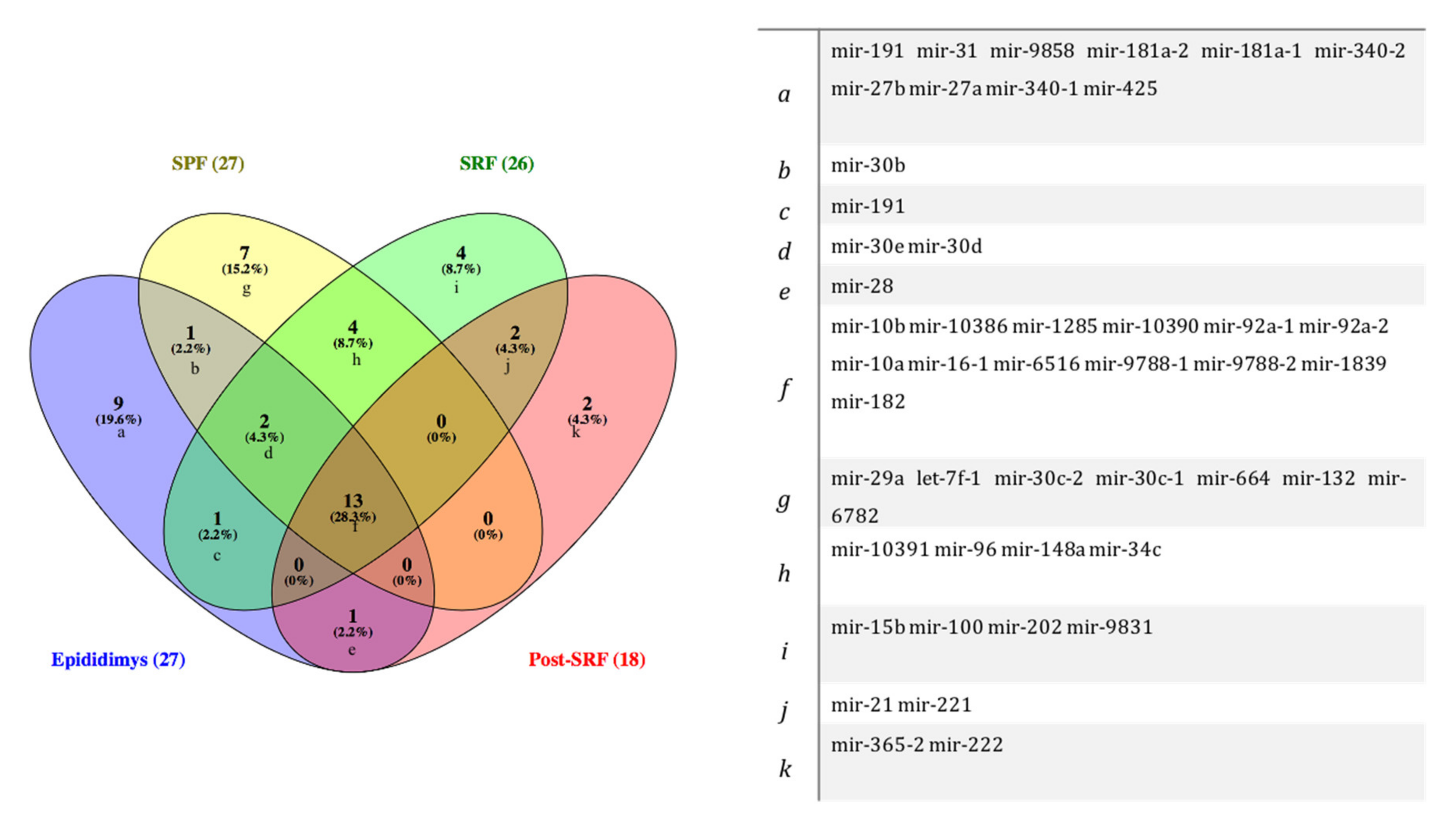
3.1. Identification of miRNAs in Spermatozoa from Cauda Epididymis and Ejaculate Fractions

Table 2 lists the miRNAs identified in boar spermatozoa that showed differences in abundance among sperm sources, highlighting six miRNAs that have not been described before in pig spermatozoa and five miRNAs that were significantly dysregulated (p < 0.005).
Table 2. List of identified miRNAs with different abundance among spermatozoa from three ejaculate fractions: sperm peak fraction (SPF), sperm rich fraction (SRF), and post-SRF of mature boars (n = 6), and the epididymal terminal segment (EpiTS).
Figure 1 depicts the miRNAs commonly expressed among spermatozoa recovered from the three ejaculate fractions studied: SPF (n = 27), SRF (n = 26), and the post-SRF (n = 18), and from the EpiTS (n = 27), and where all sources shared the expression of 13 miRNAs.
Figure 1. Venn diagram showing microRNAs (miRNAs) commonly identified among spermatozoa recovered from three ejaculate fractions: sperm peak fraction (SPF), sperm rich fraction (SRF), and post-SRF, and from the functional epididymal terminal segment (EpiTS).

3.2. miRNAs Were Differentially Expressed among Spermatozoa Ejaculate Fractions and from Cauda Epididymis, as Well as between Boars with Higher (HF) or Lower (LF) Fertility: Assessment of Chromosome Location and Structure

Some miRNA abundance differed significantly (p < 0.05) among sperm sources (Figure 2). The mir-1285 was upregulated in spermatozoa from the EpiTS compared to spermatozoa derived from any of the ejaculate fractions studied, as well as when comparing the SPF and the post-SRF fractions. The SPF-spermatozoa showed a higher expression of mir-92a-1, mir-92a-2, and mir-34c than spermatozoa from the SRF. The mir-30e was upregulated in SRF-spermatozoa compared with post-SRF spermatozoa (Table 3). Additionally, there was an overexpression of mir-191 and mir-96 (in spermatozoa from EpiTS and SRF, respectively), while a repression of mir-182 and mir-1285 was observed in spermatozoa from the SPF and the SRF, respectively, when comparing spermatozoa from boars of higher- or lesser fertility (Table 3). Figure 2 depicts chromosome location and precursor structure of those miRNAs that were differentially expressed. Figure 3 depicts a hierarchical clustering of the pattern followed by all miRNAs in spermatozoa from different sources within all boars (n = 6) and in boars with higher- or lower-fertility (n = 3) within the same sperm source.
Figure 2. Chromosome location (left image) and miRNA precursor structure (right image) of all differentially expressed microRNAs (miRNAs) (p-value < 0.05 and ≥ 1.0-Fold Change (FC) or ≤ −1.0) presented in this study: mir-182 (A,a), mir-191 (B,b), mir-34c-1 (C,c), mir-96 (D,d), mir-92a (E,e), mir-30e (F,f), mir-1285 (G,g). The images were created in Rfam version 14.5 [37]. The black color in the miRNA structure represents a template model, while differences between the template and the sequences are highlighted in color, depending on whether it is a modification (green), an insertion (pink), or a reposition (blue). Structures were generated by R2DT using the d.5.e.P.waltl template provided by CRW [38], copyright © 2021 Rfam Team.
Table 3. Differentially abundant microRNAs (miRNAs, p-value < 0.05) in spermatozoa collected from the three ejaculate fractions studied (SPF: sperm-peak fraction; SRF: sperm-rich fraction; and post-SRF) and from the functional epididymal terminal segment (EpiTS) among all boars or comparing high-and low fertility boars.
Figure 3. Hierarchical clustering of the expression levels of miRNAs identified in spermatozoa collected from the three ejaculate fractions studied (SPF: sperm-peak fraction; SRF: sperm-rich fraction; and post-SRF) and from the functional epididymal terminal segment (EpiTS) from all boars used in this study (n = 6, upper- and mid- figures), and also, expression levels of miRNAs when selecting boars with either higher- (HF) or lower- fertility (LF) (n = 3, lower figures). The color scale indicates the relative expression of miRNAs: the red color shows a higher expression and the green color depicts a lower expression. Each row represents one biological sample, and each column represents one miRNA.

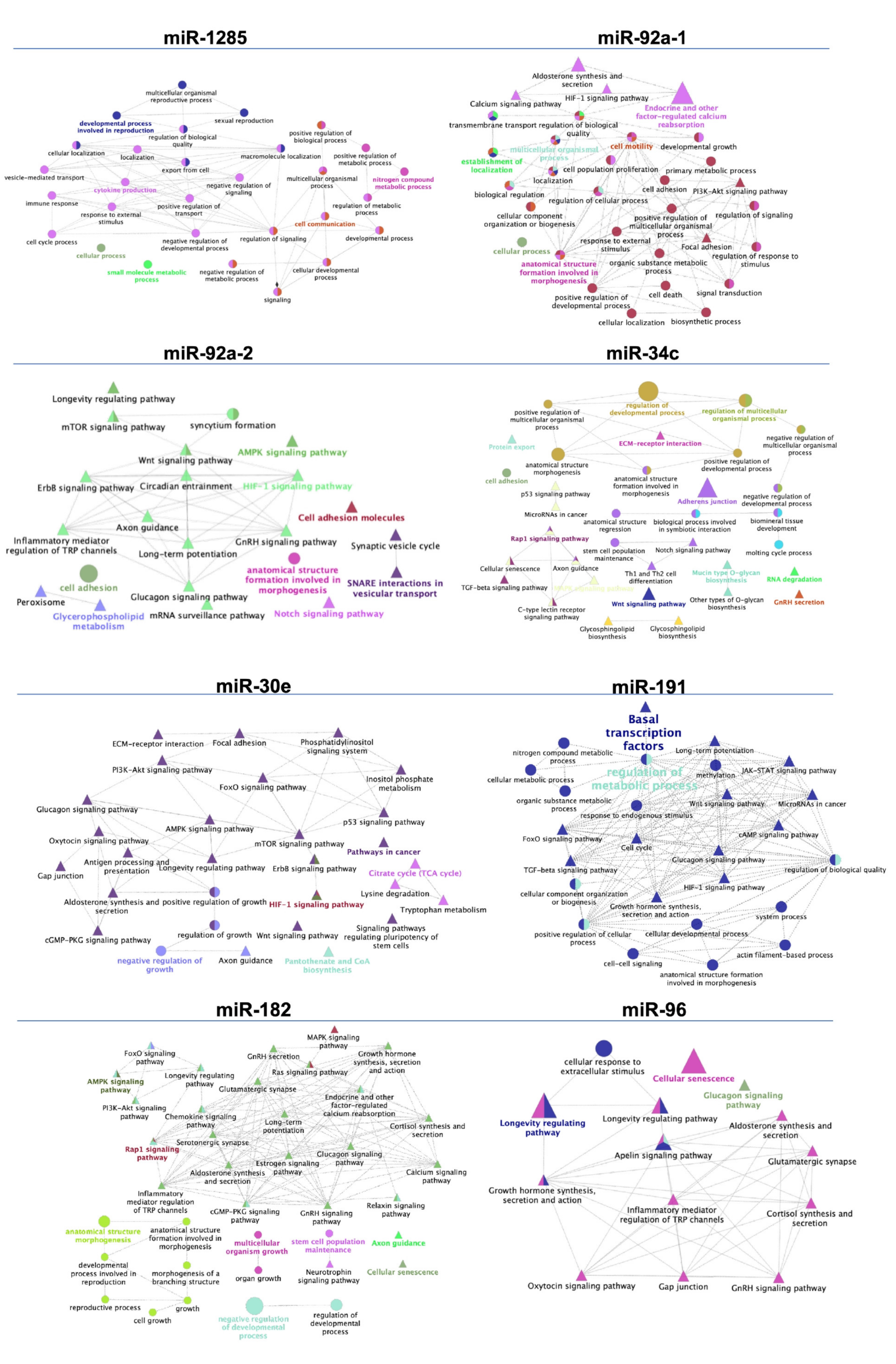
3.3. Target Prediction and Functional Annotations of Differentially Expressed Sperm miRNAs

To gain insight into the biological function of the differentially expressed sperm miRNAs found in this study, we identified which target genes whose expressions could be potentially and/or partially regulated by these sperm miRNAs. We focused on those target genes with a score ≥90 of homology (Table 4 and Table 5) in the miRNA-target human database (miRDB), against which the miRNAs were aligned owing to the highly conserved degree of homology among species. A total number of 246 potential target genes likely influenced by the expression of the five miRNAs (mir-1285, mir-92a-1, mir-92a-2, mir-34c, and mir-30e) were found after comparing epididymal spermatozoa with those from ejaculate fractions (Table 4). Additionally, a total of 208 target genes were listed as putatively regulated by four miRNAs (mir-191, mir-182, mir-96, and mir-1285) found comparing spermatozoa from higher- with lower-fertile boars (Table 5). The networks representing interactions between those GO terms and biological pathways are shown in Figure 4. ClueGO software (Cytoscape) revealed the considered miRNA-targeted genes (target genes scoring > 90 in miRbase) in several immune-related pathways and cellular processes.
Table 4. List of predicted target genes of differentially expressed miRNAs (p value < 0.05 and > 1.0-Fold Change (FC) or< −1.0-FC) in spermatozoa from three different ejaculate fractions: sperm peak fraction (SPF), sperm rich fraction (SRF), or post-SRF, or from the epididymal terminal segment (EpiTS).
Table 5. List of predicted target genes of differentially expressed miRNAs (p value < 0.05 and > 1.0-fold change (FC) or < −1.0-FC) in spermatozoa from ejaculate fractions (sperm-peak fraction (SPF), sperm-rich fraction (SRF), and post-SRF), and the functional epididymal terminal segment (EpiTS), and of higher fertility (HF) compared to lower fertility boars (LF).
Figure 4. Schematic representation of biological processes and pathways (KEGG) associated with potential target genes for differentially expressed microRNAs (miRNAs, p-value < 0.05) found in spermatozoa collected either from the three ejaculate fractions (SPF: sperm-peak fraction; SRF: sperm-rich fraction; and post-SRF), or from the functional epididymal terminal segment (EpiTS) among boars (mir-1285, mir-92a-1, mir-92a-2, mir-34c, and miR-30e) or comparing boars with higher- or lower fertility after AI (mir-191, mir-182, mir-96, and mir-1285). Cytoscape v3.0.0 application ClueGO v2.0.3 was used to build the networks for overrepresented biological processes and pathways. Terms are functionally grouped based on shared genes (kappa score) and are shown in different colors. The following ClueGO parameters were used: GO tree levels, 1–3 (first level = 0), minimum number of genes, 2, minimum percentage of genes, 1, GO term fusion, GO term connection restriction (kappa score), 0.4. Some redundant or unnecessary terms were discarded, and the network was manually rearranged.

4. Discussion

Multiple lines of evidence indicate that sperm miRNAs have crucial gene-regulatory roles in many fundamental biological processes, including spermatogenesis, prompting for sperm motility, fertilization, and early embryo development [14,15,19]. Although pig spermatozoa have been shown to express miRNAs ruling sperm motility, structural integrity and metabolism before [12,39], the present study aimed to perform comprehensive miRNA expression profiling of pig spermatozoa retrieved from different sources using a genome-wide deep sequencing approach. The results have provided novel findings regarding the miRNA content and its differential abundance in the spermatozoa from the EpiTS and from three ejaculate fractions (SPF, SRF, and post-SRF), for their plausible implication in sperm-related biological processes post-fertilization, ultimately influencing fertility by controlling the expression of specific genes.
After sperm production in the testis, spermatozoa acquire motility and fertilizing capacity during their journey along the epididymis [29], interacting with different ncsRNAs (including miRNAs), transcripts, and proteins that are released from the epithelium of the epididymis mostly via epididymosomes [40,41,42]. We have hereby identified a total of 27 miRNAs in spermatozoa from the EpiTS and the SPF, 26 in the SRF, and only 18 in the post-SRF. Interestingly, the differences between the numbers of commonly expressed miRNAs was not large between the SPF and the SRF (n = 19) or between EpiTS and SRF (n = 16), which seems reasonable considering the SRF (including its SPF) presents higher contents of epidydimal fluid [43].
Some of the miRNAs found in this study have not been described in pig spermatozoa before (mir-10386, mir-10390, mir-10391, mir-9788-1, mir-9788-2, and mir-6516, present in all sperm sources). mir-10386, mir-10390 and mir-10391 have only been described in porcine liver [44]. mir-9788-1 and mir-9788-2 were identified in sow milk exosomes [45], while mir-6516 has been reported in other tissues in several species (porcine liver [44], human platelets [46], chicken embryos, [47] or mouse brain [48]. Further experimental evidence is needed to validate the presence and to identify the roles ascribed to these miRNAs in pig spermatozoa.
Few of the miRNAs identified in the present study were differentially expressed among sperm sources. Mir-1285 was found upregulated in spermatozoa from the epididymis terminal segment compared to spermatozoa from all of the different ejaculate fractions.
This miRNA was previously identified by our group in spermatozoa retrieved from the SRF [49], but, to the best of our knowledge, the present study is the first to report an overexpression of this miRNA in spermatozoa from the EpiTS compared to spermatozoa from the ejaculate fractions. The fact that mir-1285 abundance differed when compared to the other sperm sources could be explained by the fact that, during spermatogenesis, post-transcriptional control of gene expression is highly active [50] and mir-1285 appears to be involved in spermiogenesis by inhibiting boar Sertoli cell proliferation through the regulation of AMPK [51]. Phosphorylated AMPK is implicated in the regulation of 17β-estradiol-mediated inhibition of Sertoli cell viability through increasing p53 and p27 expression and inhibiting mTOR and Skp2 expression [51]. Moreover, mir-1285 regulates the expression of many genes associated with reproductive processes, for instance, the potential target gene DAZAP1 seems essential for normal development of spermatozoa in mice [52]. This finding supports the theory that miRNAs may play key roles in spermatogenesis. Additionally, we observed an overexpression of three miRNAs (mir-92a-1, mir-92a-2, and mir-34c) in SPF compared with the SRF spermatozoa. The differences in miRNAs abundance between sperm from different parts of the ejaculate suggests that miRNAs regulate mRNA expression in the final processing of spermatozoa during ejaculation [22]. MiR-92 has been shown to play a role in early chicken gonadogenesis by regulating the expression of the ATRX and DDX3X genes. The mir-92a target genes are implicated in the regulation of the NOTCH signaling pathway, which regulates many cellular events: inhibition of cell differentiation, cell proliferation, and preservation of stem cell population [53]. In addition, NOTCH genes are considered potential targets for mir-34 [54]. The mir-34c belongs to a family of evolutionarily conserved miRNAs (mir-34a, mir-34b, and mir-34c) with active influence on genes that control the cell cycle [55,56], including cell cycle arrest, cellular senescence [57], and apoptosis [58]. Furthermore, the abundance of mir-34c is regulated by the p53 signaling pathway and could constitute a central inhibitor of p53 functions, which are responsible of a variety of intrinsic and extrinsic stress signals that impact upon cellular homeostatic mechanisms regulating DNA replication, chromosome segregation, and cell division [58]. mir-34c has been suggested as fundamental for the development of both male and female bovine gametes and therefore suggested as a potential biomarker of male bovine fertility [59]. Moreover, decreased levels of mir-34c have been observed in samples of infertile individuals [60]. Among the mir-34c target genes, three genes downregulated by mir-34c (TGIF2, E2F5, and BMP3), all implicated in the regulation of Transforming Growth Factor-beta (TGF-β) signaling pathway drew our attention. A recent study pointed the TGF-β signaling as a crucial mechanism for immune tolerance to spermatozoa, a very relevant capacity present in the epididymis to tolerate spermatozoa in the lumen, despite they present xenoantigens, which could cause a response from the male immune system [61]. After sperm deposition in the female reproductive tract, similar concepts would apply for the TGF-β signaling pathway, considering its role in preventing spermatozoa from autoimmune responses [49], while simultaneously, the female immune system provides effective protection against ascending pathogens [62]. The genes mentioned above are transcriptional repressors and inhibitors of the TGF-β signaling pathway; its down-regulation triggers the signal elicited by this factor [63,64,65]. The fact that spermatozoa from the SPF may carry epididymosomes containing miRNAs in the cauda epididymal fluid [24,66] could explain these findings. These epididymosomes can provide relevant miRNAs to the spermatozoa besides the endogenous ones above listed, potentially enhancing sperm tolerance and female immune tolerance for a successful fertilization and embryo development, influencing fertility. Overall, the current findings contribute to an increased information regarding sperm miRNAs abundance and possible roles of miRNAs via mRNA regulation on the physiological functions and regulatory mechanisms in pig spermatozoa.
Since the breeding boars used in our study were highly selected for sperm quality, an important requisite for incorporation in AI centers, their fertility was far from being a binary variable, and thus it was defined as of higher- or lower- nature, considering farrowing rate (FR) and litter size (LS) as end-points. Consequently, we additionally investigated whether sperm miRNA expression could be somehow related to boar fertility. Increased abundance of mir-182 (SPF) and mir-1285 (post-SRF) and decreased levels of mir-191 (EpiTS) and mir-96 (SRF) were observed in spermatozoa from higher-fertility boars compared with those retrieved from boars with lower fertility. Moreover, since sperm miRNAs could regulate the expression of genes playing important roles in fertilization and embryo developmental ability, as it has been reported before [14], we further investigated the biological implications of the sperm miRNAs we localized and their target genes.
Mir-191 is improperly expressed in some diseases, including cancer, type 2 diabetes, Crohn’s disease, pulmonary hypertension, and Alzheimer’s disease. Nevertheless, little information has been found regarding its role in reproductive processes. mir-191 was proposed as a key factor involved in embryo development. Additionally, higher concentrations of mir-191 in IVF/ICSI medium were observed in non-successful procedures [67]. Interestingly, mir-191 was found within the epididymosomal miRNA content in the bull [40], suggesting to modulate intercellular communication within the epididymis in low-fertility boars, disrupting epididymal function.
The SPF spermatozoa from boars ranked with higher fertility revealed increased levels of mir-182. This finding adds to the findings of Curry et al., 2011, where high expression of mir-182 was observed in boars depicting high sperm motility and intact structure [39]. Many of the identified miRNA target genes are involved in several cellular processes, including cell structure and growth, cellular senescence and calcium signaling, among others. Of particular interest was the influence of mir-182 on the regulation of BCL2L12, IGF1R, MED1, and RARG genes, since these genes are implicated in the promotion of cell progression and the negative regulation of apoptotic processes, mainly by direct neutralization of caspase-7 (CASP7) and indirect neutralization of caspase-3 (CASP3), which are known to play essential roles in the execution phase of apoptosis, through the phosphoinositide-3 kinase (PI-3K)-Akt and Ras-Raf-MAPK pathways [68,69]. From our specific analysis of differentially expressed genes, we found an overexpression of three target genes for mir-182 (ABHD13, MFAP3, and PCNX) in HF compared to LF in the SPF. Of special interest was the PCNX gene, which has been speculated to play an important role in the testis, related to spermatogenesis [70]. Overall, these results suggest that mir-182 regulates the survival of spermatozoa towards an adequate progression of spermatozoa within the oviduct. mir-96 has been widely involved in the diagnosis of human cancer progression and development because of its involvement in cellular apoptosis and death [71]. Our results indicate that mir-96 was downregulated in the SRF fraction of the boars depicting higher fertility, but the connection with sperm apoptosis in mature pig spermatozoa is still a debated issue, compared to other species (humans for instance, where the degree of chromatin compaction is lower). In consequence, this issue ought to be followed. CACNA2D2, SPEN, and EBF3 are mir-96 target genes found upregulated in spermatozoa from HF compared to LF boars in the SRF. The calcium channel, voltage-dependent, alpha 2/delta subunit 2 (CACNA2D2) regulates calcium current density and is highly expressed in the testis and has been found under expressed in varicocele patients [72]. Mutations of CACNA2D2 have been associated with reduced male fertility in transgenic mice [73]. SPEN (spen family transcription repressor) is a gene that encodes a nucleic acid-binding protein putatively involved in repression of gene expression. SPEN is involved in general downregulation of the transcription during the heat shock response in mouse spermatogenic cells through its interactions with chromatin [74], and methylation changes in EBF3 (Early B cell Factor 3) have been associated with loss of fertility in human [75]. Although the present study revealed some interesting information regarding the abundance of miRNAs in the different sperm sources, it is important to note that other ncsRNAs, such as iRNAs, tsRNAs, or piRNAs are present in spermatozoa, and might be playing an important role in reproductive functions. Further studies are needed to explore potential involvement of other ncsRNAs of interest in sperm function.

5. Conclusions

In conclusion, the results presented in this study revealed novel miRNAs in pig spermatozoa whose relative degrees of abundance vary among spermatozoa, from the functional terminal segment of the epididymis and different fractions of the ejaculate. Some of these specific miRNAs could be linked, for homology to predicted target genes with relevant functions related to sperm survival, immune tolerance, or cell cycle regulation, among others. Such regulation could influence embryo development and, ultimately, fertility of the sires, a matter hereby explored. If these genes are essential, then exploration of sperm miRNAs would be beneficial towards the identification of fertility biomarkers, benefiting the efficiency of artificial insemination techniques through safer selections of the most fertile breeders. However, further research is needed to shed evidence on the mechanistic and physiological roles of such miRNAs, and whether they are intrinsic in spermatozoa or derived from epididymosomes and/or seminal plasma.

Supplementary Materials

The following supporting information can be downloaded at: https://www.mdpi.com/article/10.3390/biology11020236/s1, File S1. Sample grouping represented in a PCA plot. Information regarding library quality and sequencing outcomes.

Author Contributions

Conceptualization, H.R.-M. and J.R.; methodology, C.A.M. and J.R.; software, C.A.M. and M.A.-R.; validation, M.A.-R. and H.R.-M.; writing—original draft preparation, C.A.M.; writing—review and editing, J.R., M.A.-R. and H.R.-M.; supervision, J.R. and H.R.-M.; project administration, H.R.-M.; funding acquisition, H.R.-M. All authors have read and agreed to the published version of the manuscript.

Funding

This research was supported by grant PID2020-113493RB-I00 funded by MCIN/AEI /10.13039/501100011033 (Spain), Seneca Foundation Murcia, Spain (19892/GERM-15), and the Swedish Research Council FORMAS (2017-00946 and 2019-00288), Stockholm, Sweden. CAM was financially supported by the European Union’s Horizon 2020 research and innovation program under the MSCA (grant agreement no. 891663).

Institutional Review Board Statement

The study was conducted according to the European Directive 2010/63/EU EEC for animal experiments and approved by the Bioethics Committee of Murcia University (research code: 639/2012) and the “Regional Committee for Ethical Approval of Animal Experiments” (Linköpings Djurförsöksetiska nämnd) in Linköping, Sweden (permits Dnr 75-12, ID1400, and 03416-2020).

Data Availability Statement

The raw datasets generated during and/or analyzed during the current study are available at Sequence Read Archive (SRA) with BioProject accession number: PRJNA762225 (https://www.ncbi.nlm.nih.gov/sra/PRJNA762225 (accessed on 12 March 2018)).

Acknowledgments

The authors of this manuscript thank Topigs Norsvin España for supplying the boar ejaculates and the boar fertility records; Annette Molbaek and Åsa Schippert, from the Genomics Core Facility at LiU for expert assistance with NGS; and Jonathan J.M. Landry from EMBL, Genomics Core Facilities, Services & Technology Unit (Germany), for his help with the bioinformatic analyses.

Conflicts of Interest

The authors declare no conflict of interest. The funders had no role in the design of the study; in the collection, analyses, or interpretation of data; in the writing of the manuscript, or in the decision to publish the results.

References

  1. Visconti, P.E.; Stewart-Savage, J.; Blasco, A.; Battaglia, L.; Miranda, P.; Kopf, G.S.; Tezon, J.G. Roles of bicarbonate, cAMP, and protein tyrosine phosphorylation on capacitation and the spontaneous acrosome reaction of hamster sperm. Biol. Reprod. 1999, 61, 76–84. [Google Scholar] [CrossRef] [PubMed]
  2. Donlic, A.; Hargrove, A.E. Targeting RNA in mammalian systems with small molecules. Wiley Interdiscip. Rev. RNA 2018, 9, e1477. [Google Scholar] [CrossRef] [PubMed]
  3. Fu, Y.; Fan, P.; Wang, L.; Shu, Z.; Zhu, S.; Feng, S.; Li, X.; Qiu, X.; Zhao, S.; Liu, X. Improvement, identification, and target prediction for miRNAs in the porcine genome by using massive, public high-throughput sequencing data. J. Anim. Sci. 2021, 99, skab018. [Google Scholar] [CrossRef] [PubMed]
  4. Hua, R.; Wang, Y.; Lian, W.; Li, W.; Xi, Y.; Xue, S.; Kang, T.; Lei, M. Small RNA-seq analysis of extracellular vesicles from porcine uterine flushing fluids during peri-implantation. Gene 2021, 766, 145117. [Google Scholar] [CrossRef] [PubMed]
  5. Hausser, J.; Syed, A.P.; Bilen, B.; Zavolan, M. Analysis of CDS-located miRNA target sites suggests that they can effectively inhibit translation. Genome Res. 2013, 23, 604–615. [Google Scholar] [CrossRef]
  6. Bartel, D.P. MicroRNAs: Genomics, biogenesis, mechanism, and function. Cell 2004, 116, 281–297. [Google Scholar] [CrossRef]
  7. Bagga, S.; Bracht, J.; Hunter, S.; Massirer, K.; Holtz, J.; Eachus, R.; Pasquinelli, A.E. Regulation by let-7 and lin-4 miRNAs results in target mRNA degradation. Cell 2005, 122, 553–563. [Google Scholar] [CrossRef]
  8. Wu, L.; Fan, J.; Belasco, J.G. MicroRNAs direct rapid deadenylation of mRNA. Proc. Natl. Acad. Sci. USA 2006, 103, 4034–4039. [Google Scholar] [CrossRef]
  9. Vasudevan, S.; Tong, Y.; Steitz, J.A. Switching from repression to activation: microRNAs can up-regulate translation. Science 2007, 318, 1931–1934. [Google Scholar] [CrossRef]
  10. Miller, D. Analysis and significance of messenger RNA in human ejaculated spermatozoa. Mol. Reprod. Dev. 2000, 56, 259–264. [Google Scholar] [CrossRef]
  11. Yan, W.; Morozumi, K.; Zhang, J.; Ro, S.; Park, C.; Yanagimachi, R. Birth of mice after intracytoplasmic injection of single purified sperm nuclei and detection of messenger RNAs and MicroRNAs in the sperm nuclei. Biol. Reprod. 2008, 78, 896–902. [Google Scholar] [CrossRef] [PubMed]
  12. Curry, E.; Ellis, S.E.; Pratt, S.L. Detection of porcine sperm microRNAs using a heterologous microRNA microarray and reverse transcriptase polymerase chain reaction. Mol. Reprod. Dev. 2009, 76, 218–219. [Google Scholar] [CrossRef] [PubMed]
  13. Amanai, M.; Brahmajosyula, M.; Perry, A.C.F. A restricted role for sperm-borne microRNAs in mammalian fertilization. Biol. Reprod. 2006, 75, 877–884. [Google Scholar] [CrossRef] [PubMed]
  14. Liu, W.-M.; Pang, R.T.K.; Chiu, P.C.n.; Wong, B.P.C.; Lao, K.; Lee, K.-F.; Yeung, W.S.B. Sperm-borne microRNA-34c is required for the first cleavage division in mouse. Proc. Natl. Acad. Sci. USA 2012, 109, 490–494. [Google Scholar] [CrossRef]
  15. Wu, C.; Blondin, P.; Vigneault, C.; Labrecque, R.; Sirard, M.-A. Sperm miRNAs—Potential mediators of bull age and early embryo development. BMC Genom. 2020, 21, 798. [Google Scholar] [CrossRef]
  16. Alves, M.B.R.; de Arruda, R.P.; De Bem, T.H.C.; Florez-Rodriguez, S.A.; de Sá Filho, M.F.; Belleannée, C.; Meirelles, F.V.; da Silveira, J.C.; Perecin, F.; Celeghini, E.C.C. Sperm-borne miR-216b modulates cell proliferation during early embryo development via K-RAS. Sci. Rep. 2019, 9, 10358. [Google Scholar] [CrossRef]
  17. Maatouk, D.M.; Loveland, K.L.; McManus, M.T.; Moore, K.; Harfe, B.D. Dicer1 is required for differentiation of the mouse male germline. Biol. Reprod. 2008, 79, 696–703. [Google Scholar] [CrossRef]
  18. Chen, X.; Zheng, Y.; Li, X.; Gao, Q.; Feng, T.; Zhang, P.; Liao, M.; Tian, X.; Lu, H.; Zeng, W. Profiling of miRNAs in porcine Sertoli cells. J. Anim. Sci. Biotechnol. 2020, 11, 85. [Google Scholar] [CrossRef]
  19. Reza, A.M.M.T.; Choi, Y.-J.; Han, S.G.; Song, H.; Park, C.; Hong, K.; Kim, J.-H. Roles of microRNAs in mammalian reproduction: From the commitment of germ cells to peri-implantation embryos. Biol. Rev. 2019, 94, 415–438. [Google Scholar] [CrossRef]
  20. Kasimanickam, V.; Buhr, M.; Kasimanickam, R. Patterns of expression of sperm and seminal plasma microRNAs in boar semen. Theriogenology 2019, 125, 87–92. [Google Scholar] [CrossRef]
  21. Rodriguez-Martinez, H. Aspects of the Electrolytic Composition of Boar Epididymal Fluid with Reference to Sperm Maturation and Storage, Boar Semen Preservation II. In Reproduction in Domestic Animals; John Wiley and Sons: Hoboken, NJ, USA, 1991; Volume 57, pp. 13–27. [Google Scholar]
  22. Chang, Y.; Dai, D.-H.; Li, Y.; Zhang, Y.; Zhang, M.; Zhou, G.-B.; Zeng, C.-J. Differences in the expression of microRNAs and their predicted gene targets between cauda epididymal and ejaculated boar sperm. Theriogenology 2016, 86, 2162–2171. [Google Scholar] [CrossRef] [PubMed]
  23. Chen, C.; Wu, H.; Shen, D.; Wang, S.; Zhang, L.; Wang, X.; Gao, B.; Wu, T.; Li, B.; Li, K.; et al. Comparative profiling of small RNAs of pig seminal plasma and ejaculated and epididymal sperm. Reproduction 2017, 153, 785–796. [Google Scholar] [CrossRef] [PubMed]
  24. Rodríguez-Martínez, H.; Kvist, U.; Saravia, F.; Wallgren, M.; Johannisson, A.; Sanz, L.; Peña, F.J.; Martínez, E.A.; Roca, J.; Vázquez, J.M.; et al. The physiological roles of the boar ejaculate. Soc. Reprod. Fertil. Suppl. 2009, 66, 1–21. [Google Scholar] [CrossRef] [PubMed]
  25. Rodríguez-Martínez, H.; Saravia, F.; Wallgren, M.; Tienthai, P.; Johannisson, A.; Vázquez, J.M.; Martínez, E.; Roca, J.; Sanz, L.; Calvete, J.J. Boar spermatozoa in the oviduct. Theriogenology 2005, 63, 514–535. [Google Scholar] [CrossRef] [PubMed]
  26. Raza, S.H.A.; Kaster, N.; Khan, R.; Abdelnour, S.A.; El-Hack, M.E.A.; Khafaga, A.F.; Taha, A.; Ohran, H.; Swelum, A.A.; Schreurs, N.M.; et al. The Role of MicroRNAs in Muscle Tissue Development in Beef Cattle. Genes 2020, 11, 295. [Google Scholar] [CrossRef] [PubMed]
  27. Cheong, A.W.Y.; Pang, R.T.K.; Liu, W.-M.; Kottawatta, K.S.A.; Lee, K.-F.; Yeung, W.S.B. MicroRNA Let-7a and dicer are important in the activation and implantation of delayed implanting mouse embryos. Hum. Reprod. 2014, 29, 750–762. [Google Scholar] [CrossRef]
  28. Yang, P.; Wu, Z.; Ma, C.; Pan, N.; Wang, Y.; Yan, L. Endometrial miR-543 Is Downregulated During the Implantation Window in Women with Endometriosis-Related Infertility. Reprod. Sci. 2019, 26, 900–908. [Google Scholar] [CrossRef]
  29. Rodriguez-Martinez, H.; Roca, J.; Alvarez-Rodriguez, M.; Martinez-Serrano, C.A. How does the boar epididymis regulate the emission of fertile spermatozoa? Anim. Reprod. Sci. 2021, 106829. [Google Scholar] [CrossRef]
  30. Barranco, I.; Perez-Patiño, C.; Tvarijonaviciute, A.; Parrilla, I.; Vicente-Carrillo, A.; Alvarez-Rodriguez, M.; Ceron, J.J.; Martinez, E.A.; Rodriguez-Martinez, H.; Roca, J. Active paraoxonase 1 is synthesised throughout the internal boar genital organs. Reproduction 2017, 154, 237–243. [Google Scholar] [CrossRef]
  31. Bolger, A.M.; Lohse, M.; Usadel, B. Trimmomatic: A flexible trimmer for Illumina sequence data. Bioinformatics 2014, 30, 2114–2120. [Google Scholar] [CrossRef]
  32. Dobin, A.; Davis, C.A.; Schlesinger, F.; Drenkow, J.; Zaleski, C.; Jha, S.; Batut, P.; Chaisson, M.; Gingeras, T.R. STAR: Ultrafast universal RNA-seq aligner. Bioinformatics 2013, 29, 15–21. [Google Scholar] [CrossRef] [PubMed]
  33. Evans, C.; Hardin, J.; Stoebel, D.M. Selecting between-sample RNA-Seq normalization methods from the perspective of their assumptions. Brief. Bioinform. 2018, 19, 776–792. [Google Scholar] [CrossRef] [PubMed]
  34. Wang, Y.; Chen, Z.-P.; Hu, H.; Lei, J.; Zhou, Z.; Yao, B.; Chen, L.; Liang, G.; Zhan, S.; Zhu, X.; et al. Sperm microRNAs confer depression susceptibility to offspring. Sci. Adv. 2021, 7, eabd7605. [Google Scholar] [CrossRef] [PubMed]
  35. Wu, Q.; Song, R.; Ortogero, N.; Zheng, H.; Evanoff, R.; Small, C.L.; Griswold, M.D.; Namekawa, S.H.; Royo, H.; Turner, J.M.; et al. The RNase III enzyme DROSHA is essential for microRNA production and spermatogenesis. J. Biol. Chem. 2012, 287, 25173–25190. [Google Scholar] [CrossRef]
  36. Bolstad, B.M.; Irizarry, R.A.; Astrand, M.; Speed, T.P. A comparison of normalization methods for high density oligonucleotide array data based on variance and bias. Bioinformatics 2003, 19, 185–193. [Google Scholar] [CrossRef]
  37. Kalvari, I.; Nawrocki, E.P.; Ontiveros-Palacios, N.; Argasinska, J.; Lamkiewicz, K.; Marz, M.; Griffiths-Jones, S.; Toffano-Nioche, C.; Gautheret, D.; Weinberg, Z.; et al. Rfam 14: Expanded coverage of metagenomic, viral and microRNA families. Nucleic Acids Res. 2021, 49, D192–D200. [Google Scholar] [CrossRef]
  38. Sweeney, B.A.; Hoksza, D.; Nawrocki, E.P.; Ribas, C.E.; Madeira, F.; Cannone, J.J.; Gutell, R.; Maddala, A.; Meade, C.D.; Williams, L.D.; et al. R2DT is a framework for predicting and visualising RNA secondary structure using templates. Nat. Commun. 2021, 12, 3494. [Google Scholar] [CrossRef]
  39. Curry, E.; Safranski, T.J.; Pratt, S.L. Differential expression of porcine sperm microRNAs and their association with sperm morphology and motility. Theriogenology 2011, 76, 1532–1539. [Google Scholar] [CrossRef]
  40. Belleannée, C.; Calvo, É.; Caballero, J.; Sullivan, R. Epididymosomes convey different repertoires of microRNAs throughout the bovine epididymis. Biol. Reprod. 2013, 89, 30. [Google Scholar] [CrossRef]
  41. Reilly, J.N.; McLaughlin, E.A.; Stanger, S.J.; Anderson, A.L.; Hutcheon, K.; Church, K.; Mihalas, B.P.; Tyagi, S.; Holt, J.E.; Eamens, A.L.; et al. Characterisation of mouse epididymosomes reveals a complex profile of microRNAs and a potential mechanism for modification of the sperm epigenome. Sci. Rep. 2016, 6, 31794. [Google Scholar] [CrossRef]
  42. Sharma, U.; Sun, F.; Conine, C.C.; Reichholf, B.; Kukreja, S.; Herzog, V.A.; Ameres, S.L.; Rando, O.J. Small RNAs Are Trafficked from the Epididymis to Developing Mammalian Sperm. Dev. Cell 2018, 46, 481–494.e6. [Google Scholar] [CrossRef] [PubMed]
  43. Hossain, M.S.; Johannisson, A.; Siqueira, A.P.; Wallgren, M.; Rodriguez-Martinez, H. Spermatozoa in the sperm-peak-fraction of the boar ejaculate show a lower flow of Ca(2+) under capacitation conditions post-thaw which might account for their higher membrane stability after cryopreservation. Anim. Reprod. Sci. 2011, 128, 37–44. [Google Scholar] [CrossRef] [PubMed][Green Version]
  44. Pawlina, K.; Gurgul, A.; Oczkowicz, M.; Bugno-Poniewierska, M. The characteristics of the porcine (Sus scrofa) liver miRNAome with the use of next generation sequencing. J. Appl. Genet. 2015, 56, 239–252. [Google Scholar] [CrossRef] [PubMed]
  45. Chen, T.; Xi, Q.-Y.; Ye, R.-S.; Cheng, X.; Qi, Q.-E.; Wang, S.-B.; Shu, G.; Wang, L.-N.; Zhu, X.-T.; Jiang, Q.-Y.; et al. Exploration of microRNAs in porcine milk exosomes. BMC Genom. 2014, 15, 100. [Google Scholar] [CrossRef] [PubMed]
  46. Plé, H.; Landry, P.; Benham, A.; Coarfa, C.; Gunaratne, P.H.; Provost, P. The repertoire and features of human platelet microRNAs. PLoS ONE 2012, 7, e50746. [Google Scholar] [CrossRef]
  47. Shao, P.; Liao, J.-Y.; Guan, D.-G.; Yang, J.-H.; Zheng, L.-L.; Jing, Q.; Zhou, H.; Qu, L.-H. Drastic expression change of transposon-derived piRNA-like RNAs and microRNAs in early stages of chicken embryos implies a role in gastrulation. RNA Biol. 2012, 9, 212–227. [Google Scholar] [CrossRef]
  48. Inukai, S.; de Lencastre, A.; Turner, M.; Slack, F. Novel microRNAs differentially expressed during aging in the mouse brain. PLoS ONE 2012, 7, e40028. [Google Scholar] [CrossRef]
  49. Alvarez-Rodriguez, M.; Martinez, C.; Wright, D.; Barranco, I.; Roca, J.; Rodriguez-Martinez, H. The Transcriptome of Pig Spermatozoa, and Its Role in Fertility. Int. J. Mol. Sci. 2020, 21, 1572. [Google Scholar] [CrossRef]
  50. Papaioannou, M.D.; Nef, S. microRNAs in the testis: Building up male fertility. J. Androl. 2010, 31, 26–33. [Google Scholar] [CrossRef]
  51. Jiao, Z.J.; Yi, W.; Rong, Y.W.; Kee, J.D.; Zhong, W.X. MicroRNA-1285 Regulates 17β-Estradiol-Inhibited Immature Boar Sertoli Cell Proliferation via Adenosine Monophosphate-Activated Protein Kinase Activation. Endocrinology 2015, 156, 4059–4070. [Google Scholar] [CrossRef]
  52. Hsu, L.C.-L.; Chen, H.-Y.; Lin, Y.-W.; Chu, W.-C.; Lin, M.-J.; Yan, Y.-T.; Yen, P.H. DAZAP1, an hnRNP protein, is required for normal growth and spermatogenesis in mice. RNA 2008, 14, 1814–1822. [Google Scholar] [CrossRef] [PubMed]
  53. Zhu, S.; Zhang, L.; Zhao, Z.; Fu, W.; Fu, K.; Liu, G.; Jia, W. MicroRNA-92a-3p inhibits the cell proliferation, migration and invasion of Wilms tumor by targeting NOTCH1. Oncol. Rep. 2018, 40, 571–578. [Google Scholar] [CrossRef] [PubMed]
  54. De Antonellis, P.; Medaglia, C.; Cusanelli, E.; Andolfo, I.; Liguori, L.; De Vita, G.; Carotenuto, M.; Bello, A.; Formiggini, F.; Galeone, A.; et al. MiR-34a targeting of Notch ligand delta-like 1 impairs CD15+/CD133+ tumor-propagating cells and supports neural differentiation in medulloblastoma. PLoS ONE 2011, 6, e24584. [Google Scholar] [CrossRef] [PubMed]
  55. Corney, D.C.; Flesken-Nikitin, A.; Godwin, A.K.; Wang, W.; Nikitin, A.Y. MicroRNA-34b and MicroRNA-34c are targets of p53 and cooperate in control of cell proliferation and adhesion-independent growth. Cancer Res. 2007, 67, 8433–8438. [Google Scholar] [CrossRef] [PubMed]
  56. Lujambio, A.; Calin, G.A.; Villanueva, A.; Ropero, S.; Sánchez-Céspedes, M.; Blanco, D.; Montuenga, L.M.; Rossi, S.; Nicoloso, M.S.; Faller, W.J.; et al. A microRNA DNA methylation signature for human cancer metastasis. Proc. Natl. Acad. Sci. USA 2008, 105, 13556–13561. [Google Scholar] [CrossRef]
  57. Kumamoto, K.; Spillare, E.A.; Fujita, K.; Horikawa, I.; Yamashita, T.; Appella, E.; Nagashima, M.; Takenoshita, S.; Yokota, J.; Harris, C.C. Nutlin-3a activates p53 to both down-regulate inhibitor of growth 2 and up-regulate mir-34a, mir-34b, and mir-34c expression, and induce senescence. Cancer Res. 2008, 68, 3193–3203. [Google Scholar] [CrossRef]
  58. Bommer, G.T.; Gerin, I.; Feng, Y.; Kaczorowski, A.J.; Kuick, R.; Love, R.E.; Zhai, Y.; Giordano, T.J.; Qin, Z.S.; Moore, B.B.; et al. p53-mediated activation of miRNA34 candidate tumor-suppressor genes. Curr. Biol. 2007, 17, 1298–1307. [Google Scholar] [CrossRef]
  59. Tscherner, A.; Gilchrist, G.; Smith, N.; Blondin, P.; Gillis, D.; LaMarre, J. MicroRNA-34 family expression in bovine gametes and preimplantation embryos. Reprod. Biol. Endocrinol. 2014, 12, 85. [Google Scholar] [CrossRef]
  60. Pantos, K.; Grigoriadis, S.; Tomara, P.; Louka, I.; Maziotis, E.; Pantou, A.; Nitsos, N.; Vaxevanoglou, T.; Kokkali, G.; Agarwal, A.; et al. Investigating the Role of the microRNA-34/449 Family in Male Infertility: A Critical Analysis and Review of the Literature. Front. Endocrinol. 2021, 12, 806. [Google Scholar] [CrossRef]
  61. Voisin, A.; Damon-Soubeyrand, C.; Bravard, S.; Saez, F.; Drevet, J.R.; Guiton, R. Differential expression and localisation of TGF-β isoforms and receptors in the murine epididymis. Sci. Rep. 2020, 10, 995. [Google Scholar] [CrossRef]
  62. Martinez, C.A.; Rubér, M.; Rodriguez-Martinez, H.; Alvarez-Rodriguez, M. Pig Pregnancies after Transfer of Allogeneic Embryos Show a Dysregulated Endometrial/Placental Cytokine Balance: A Novel Clue for Embryo Death? Biomolecules 2020, 10, 554. [Google Scholar] [CrossRef]
  63. Melhuish, T.A.; Gallo, C.M.; Wotton, D. TGIF2 interacts with histone deacetylase 1 and represses transcription. J. Biol. Chem. 2001, 276, 32109–32114. [Google Scholar] [CrossRef] [PubMed]
  64. Majumder, S.; Bhowal, A.; Basu, S.; Mukherjee, P.; Chatterji, U.; Sengupta, S. Deregulated E2F5/p38/SMAD3 Circuitry Reinforces the Pro-Tumorigenic Switch of TGFβ Signaling in Prostate Cancer. J. Cell. Physiol. 2016, 231, 2482–2492. [Google Scholar] [CrossRef]
  65. Bahamonde, M.E.; Lyons, K.M. BMP3: To be or not to be a BMP. J. Bone Joint Surg. Am. 2001, 83, S56–S62. [Google Scholar] [CrossRef] [PubMed]
  66. Rodriguez-Martinez, H.; Kvist, U.; Ernerudh, J.; Sanz, L.; Calvete, J.J. Seminal plasma proteins: What role do they play? Am. J. Reprod. Immunol. 2011, 66 (Suppl S1), 11–22. [Google Scholar] [CrossRef] [PubMed]
  67. Rosenbluth, E.M.; Shelton, D.N.; Wells, L.M.; Sparks, A.E.T.; Van Voorhis, B.J. Human embryos secrete microRNAs into culture media-a potential biomarker for implantation. Fertil. Steril. 2014, 101, 1493–1500. [Google Scholar] [CrossRef]
  68. Stegh, A.H.; Kim, H.; Bachoo, R.M.; Forloney, K.L.; Zhang, J.; Schulze, H.; Park, K.; Hannon, G.J.; Yuan, J.; Louis, D.N.; et al. Bcl2L12 inhibits post-mitochondrial apoptosis signaling in glioblastoma. Genes Dev. 2007, 21, 98–111. [Google Scholar] [CrossRef]
  69. Riedemann, J.; Macaulay, V.M. IGF1R signalling and its inhibition. Endocr. Relat. Cancer 2006, 13 (Suppl. S1), S33–S43. [Google Scholar] [CrossRef]
  70. Geisinger, A.; Alsheimer, M.; Baier, A.; Benavente, R.; Wettstein, R. The mammalian gene pecanex 1 is differentially expressed during spermatogenesis. Biochim. Biophys. Acta Gene Struct. Expr. 2005, 1728, 34–43. [Google Scholar] [CrossRef]
  71. Cai, T.; Long, J.; Wang, H.; Liu, W.; Zhang, Y. Identification and characterization of miR-96, a potential biomarker of NSCLC, through bioinformatic analysis. Oncol. Rep. 2017, 38, 1213–1223. [Google Scholar] [CrossRef]
  72. Agarwal, A.; Sharma, R.; Samanta, L.; Durairajanayagam, D.; Sabanegh, E. Proteomic signatures of infertile men with clinical varicocele and their validation studies reveal mitochondrial dysfunction leading to infertility. Asian J. Androl. 2016, 18, 282–291. [Google Scholar] [CrossRef] [PubMed]
  73. Eppig, J.T.; Blake, J.A.; Bult, C.J.; Kadin, J.A.; Richardson, J.E. The Mouse Genome Database (MGD): Facilitating mouse as a model for human biology and disease. Nucleic Acids Res. 2015, 43, D726–D736. [Google Scholar] [CrossRef] [PubMed]
  74. Korfanty, J.; Stokowy, T.; Chadalski, M.; Toma-Jonik, A.; Vydra, N.; Widłak, P.; Wojtaś, B.; Gielniewski, B.; Widlak, W. SPEN protein expression and interactions with chromatin in mouse testicular cells. Reproduction 2018, 156, 195–206. [Google Scholar] [CrossRef] [PubMed]
  75. Sujit, K.M.; Sarkar, S.; Singh, V.; Pandey, R.; Agrawal, N.K.; Trivedi, S.; Singh, K.; Gupta, G.; Rajender, S. Genome-wide differential methylation analyses identifies methylation signatures of male infertility. Hum. Reprod. 2018, 33, 2256–2267. [Google Scholar] [CrossRef]
Publisher’s Note: MDPI stays neutral with regard to jurisdictional claims in published maps and institutional affiliations.

Article Metrics

Citations

Article Access Statistics

Multiple requests from the same IP address are counted as one view.