Article

Enhanced Stability and Bioactivity of Curcuma comosa Roxb. Extract in Electrospun Gelatin Nanofibers

by
Chi-Ming Chiu
1,2,
Jukkrit Nootem
1,
Thanapich Santiwat
1,
Choladda Srisuwannaket
1,
Kornkanya Pratumyot
1,
Wei-Chao Lin
2,
Withawat Mingvanish
1 and
Nakorn Niamnont
1,*
1
Organic Synthesis, Electrochemistry & Natural Product Research Unit, Department of Chemistry, Faculty of Science, King Mongkut’s University of Technology Thonburi, Bangkok 10140, Thailand
2
Department of Cosmetic Science, College of Pharmacy and Science, Chia Nan University of Pharmacy and Science, Tainan 71710, Taiwan
*
Author to whom correspondence should be addressed.
Fibers 2019, 7(9), 76; https://doi.org/10.3390/fib7090076
Submission received: 3 July 2019 / Revised: 24 July 2019 / Accepted: 10 August 2019 / Published: 27 August 2019
(This article belongs to the Special Issue Electrospun Polymer Nanofibers for Food and Health Applications)

Abstract

:
Electrospun fiber can be used as a carrier for releasing active ingredients at the target site to achieve the effects of drug treatment. The objectives of this research work were to study suitable conditions for producing electrospun gelatin fiber loaded with crude Curcuma comosa Roxb. extract (CE) and to study antioxidant, anti-tyrosinase and anti-bacterial activities and its freeze–thaw stability as well. To achieve optimal conditions for producing electrospun gelatin fiber, the concentration of gelatin was adjusted to 30% w/v in a co-solvent system of acetic acid/water (9:1 v/v) with a feed rate of 3 mL/h and an applied voltage of 15 kV. The lowest percent loading of 5% (w/v) CE in gelatin nanofiber exhibited the highest DPPH radical scavenging activity of 94% and the highest inhibition of tyrosinase enzyme of 35%. Moreover, the inhibition zones for antibacterial activities against S. aureus and S. epidermidis were 7.77 ± 0.21 and 7.73 ± 0.12 mm, respectively. The freeze–thaw stability of CE in electrospun gelatin nanofiber was significantly different (p < 0.05) after the 4th cycle as compared to CE. Electrospun gelatin nanofiber containing CE also showed the capacity of the release of bioactive ingredients possessing anti-oxidant properties and, therefore, it could potentially be used for face masks.

1. Introduction

Curcuma comosa Roxb. is a species in the genus Curcuma of the family Zingiberaceae. It has been used in traditional folk medicine for the treatment of a wide range of diseases in many Asian countries [1,2]. The individual compounds isolated from C. comosa have displayed various biological properties such as estrogenic [3], choleretic [4], nematocidal [5], anti-inflammatory [6], antioxidant [7], and antibacterial activities [8]. The active constituents of C. comosa extract have been identified as acetophenones, curcuminoids, sesquiterpenes, and diarylheptanoids [9]. Additionally, the major bioactive compounds were diarylheptanoid derivatives at a level of 80–90% in crude extract [10]. Amongst non-phenolic diarylheptanoids, (3R)-1,7-diphenyl-(4E,6E)-4,6-heptadien-3-ol exhibited the highest anti-inflammatory [11], anti-oxidant [12,13] antibacterial activities [14] and anti-tyrosinase. However, its backbone consisting of double bonds is easily oxidized and transformed upon exposure to oxygen, light, and temperature, which decreases its bioactivities [15].
Various encapsulation techniques have been employed to improve the stability of diarylheptanoids using lipid nanoemulsions, beta-cyclodextrin, and calcium alginate [16,17,18]. These have demonstrated an increase in the stability and bioactivity of diarylheptanoids under various environmental conditions. However, encapsulation is associated with several limitations such as handling, drug-loading, and upscaling. Therefore, an alternative encapsulation method, known as electrospinning, has been developed to produce continuous fibers in levels of submicron diameters down to nanometer diameters by transforming polymer solutions or polymer melts to nonwoven-fibrous materials using electricity. Furthermore, an electrospun nanofibrous mat exhibited superior properties such as effective drug encapsulation, high drug loading efficacy, wide range of polymers to accommodate compatibility, and the ability to adjust drug releasing level through its high surface to volume ratio [19,20,21].
Several polymeric materials such as cellulose [22,23,24], polycaprolactone [25], poly (lactic acid) [26], polyvinyl alcohol [27,28] and gelatin [29,30] have been currently used to encapsulate many drugs or bioactive compounds by electrospinning. Gelatin, a hydrophilic polymer, has commonly been electrospun together with other polymers to carry bioactive compounds due to its biocompatible, nontoxic and biodegradable characteristics [31]. Many research studies have confirmed that bioactive compounds loaded into electrospun gelatin fibers, including silver nanoparticles [32], antibiotic drugs [33], and vitamins A and E [34] showed significant releasing property, increased bioactivity, and highly enhanced stability. Herein, we reported for the first time on the optimal conditions for the fabrication of the electrospun fibers of gelatin loaded with crude Curcuma comosa Roxb extract (CE). The bioactivities of the electrospun fibers containing bioactive compounds were also investigated using 2,2-diphenyl-1-picrylhydrazyl (DPPH) radical scavenging activity assay, anti-tyrosinase activity assay and antibacterial activity assay. Moreover, its freeze–thaw stability testing was carried out.

2. Experiments

2.1. Reagents and Apparatus

Gelatin powder from porcine skin with the gel strength of ~300 g Bloom (Type A), 2,2-diphenyl-1-picrylhydrazyl 3,4-dihydroxy-l-phenylalanine (DPPH; ≥98%), phosphate-buffered saline (PBS), 3-(3,4-Dihydroxyphenyl)-l-alanine (l-DOPA), and tyrosinase were purchased from Sigma–Aldrich (St. Louis, MO, USA). All analytical grade reagents of methanol, ethanol, acetone, acetic acid, and ethyl acetate (EA) with 99% purification were obtained from RCI Labscan (Thailand). The other chemicals used were of analytical grade. Powder of Curcuma comosa Roxb. rhizome was purchased from a local Thai Herb Trading market in Bangkok, Thailand in May 2016. Muller-Hinton Agar (MHA), Gentamicin, dimethyl sulfoxide (DMSO), Staphylococcus aureus, and Staphylococcus epidermidis were all supplied by the laboratory at Rangsit University, in Pathum Thani, Thailand.

2.2. Preparation of Crude Ethanol Extract of Curcuma comosa Roxb. Rhizome Powder

A total of 500 g of powder of Curcuma comosa Roxb. rhizome was macerated with 2 × 500 mL of 95% ethanol at room temperature for 3 days. The ethanol extract solution was then concentrated by using a rotary evaporator to obtain the crude ethanol extract of Curcuma comosa Roxb. rhizome (CE). CE was kept at 4 °C with light protection. The amounts of diarylheptanoid derivatives in CE were measured using high performance liquid chromatography (HPLC, Agilent Technologies, CA, USA) at detector wavelength of 259 nm [35]. The purity of diarylheptanoid derivatives was found to be 89.3% by HPLC technique.

2.3. Identification of Bioactive Compounds of the Crude Ethanol Extract of Curcuma comosa Roxb. Rhizome Powder

Preliminary phytochemical testing was used to detect the presence of plant secondary metabolites, i.e., alkaloids, flavonoids, saponins, steroids, tannins, phenols according to Prashanth et al.’s method [36] with little modifications. Furthermore, the crude ethanol extract of Curcuma comosa Roxb. rhizome (CE) powder was characterized by Fourier-transform infrared spectrometer (FTIR, Thermo scientific, CA, USA) in the region of 4000 to 600 cm−1 and 400 MHz 1H, and 100 MHz 13C-NMR spectrometer (Avance III HD 400 MHz of NMR, Bruker, Zurich, Switzerland) for determining the presence of diarylheptanoid derivatives in CE. The deuterated solvent for NMR analysis was chloroform-d (CDCl3).

2.4. Electrospinning and Characterization of Gelatin Nanofibrous Mat

The gelatin solutions of known concentrations (20, 25 and 30% (w/v)) were prepared in a mixed solvent system containing acetic acid and H2O (9:1 (v/v)). Each concentration of the gelatin solution was then loaded into a 5-mL syringe connected with a metal needle of inner diameter of 0.22 mm. The needle was connected to the positive electrode (Gamma high voltage research FL 32174, United States) and the negative electrode was connected to the rotary spinning collector. The distance of needle tip away from the collector was located about 15 cm. Each gelatin solution was injected under the applied voltage of 15 kV at a flow rate of 3 mL/h and nanofibers were collected on the rotary spinning collector at 1200 rpm [24]. In order to study morphology of electrospun nanofibers fabricated under various experimental electrospinning conditions, all nanofiber samples were dried in a vacuum oven at 50 °C for 3 h. After that, each sample was cut into a small piece with a size of 1 × 1 cm, which was coated with gold before examined using a scanning electron microscope (JSM-6610 LV; SEM, JEOL, Tokyo, Japan). The diameters of each electrospun fiber were then determined using ImageJ 1.51 K. software (n ≥ 100) [29,30].

2.5. Studies of Characteristics of Electrospun Gelatin Nanofibers Loaded with the Crude Ethanol Extract of Curcuma comosa Roxb. Rhizome

Percent loading. To determine the suitable content of the crude ethanol extract of Curcuma comosa Roxb. rhizome (CE) loaded into electrospun gelatin nanofibrous mat, a predetermined weight of CE (1, 5, 10, 15% (w/v)) was dissolved in 2 g of gelatin solution (30% and the solution mixture was electrospun under optimum conditions. Crude diarylheptanoid derivatives extract (1, 5, 10, 15% (w/v)) of a predetermined weight was dissolved in 2 g gelatin solution to generate electrospun fiber under optimum conditions. 10 mg of a gelatin nanofibrous mat loaded with CE was then re-dissolved in 2 mL of methanol. The content of diarylheptanoids in the solution mixture was determined using a UV-Vis spectrometer at 254 nm and 285 nm based on the method of Yingngam et al. [10] and reported as mean value ± SD (n = 3).
Anti-oxidant activity. 50 mg of a electrospun gelatin nanofibrous mat containing various concentrations of the crude ethanol extract of Curcuma comosa Roxb. rhizome (CE) (1, 5, 10 and 15% w/v) was re-dissolved in 5 mL of methanol by aid of sonication machine for 5 min and was subjected to examine its DPPH radical scavenging activity in accordance to the method of Devi et al. [37] with some modifications. 100 µL of each sample was mixed with 100 µL of 0.5 mM DPPH solution in methanol. The solution mixture was incubated in the dark at room temperature for 15 min. The absorbance of the solution mixture was measured at 517 nm using Perkin-Elmer multimode plate reader. All tests were done in triplicate.
The percentage of DPPH radical scavenging activity was calculated as follows:
DPPH   scavenging   activity   ( % ) = [ A 0 ( A 1 A 2 ) A 0 ] × 100
where A0 was the absorbance of the blank solution; A1 was the absorbance of the nanofiber solution with DPPH; A2 was the absorbance of the nanofiber solution without DPPH.
Anti-tyrosinase activity. The anti-tyrosinase activity of electrospun gelatin nanofibrous mat loaded with various concentrations of the crude ethanol extract of Curcuma comosa Roxb. rhizome (CE) was carried out based on inhibition of mushroom tyrosinase-catalyzed oxidation of L-DOPA. According to Masamoto et al.’s [38] method with some modifications, 30 µg of each electrospun gelatin nanofibrous mat loaded with various concentrations of CE was dissolved in 3 mL of sodium phosphate buffer pH 6.8 and l.0 mL of a 1.5 mM L-DOPA, 0.1 mL of DMSO with or without a test sample and 1.8 mL of a phosphoric acid (pH 6.8) were mixed well in a test tube. The reaction mixture was preincubated at 25 °C for 10 min, before 0.1 mL of an aqueous tyrosinase solution (100 U/mL, Sigma Chemical Co. (St. Louis, MO, USA)) was added into the reaction mixture and the reaction was monitored at 475 nm. The control reaction was conducted with DMSO. Each measurement was performed at least in triplicate.
The inhibitory percentage of tyrosinase activity was calculated using the following formula:
Tyrosinase   inhibitory   activity   ( % ) = [ ( A 1 A 2 ) ( A 3 A 4 ) ( A 1 A 2 ) ] × 100
where A1 was the absorbance of the blank solution with enzyme; A2 was the absorbance of the blank solution without enzyme; A3 was the absorbance of the nanofiber solution with enzyme; A4 was the absorbance of the nanofiber solution without enzyme.
Anti-bacterial activity. The initial evaluation of anti-bacterial activity of electrospun nanofibrous mat loaded with various concentrations of the crude ethanol extract of Curcuma comosa Roxb. rhizome (CE) was carried out using agar diffusion method (Kirby-Bauer test). The anti-bacterial activity testing was done by following the Clinical and Laboratory Standard Institute (CLSI) Guidelines 2017 with some modifications. Two Gram-positive bacteria; Staphylococcus aureus and Staphylococcus epidermidis, were used for this study. Briefly, electrospun gelatin nanofiber sheets loaded and unloaded 500–1000 µg/mL CE and 0.25 µg/mL gentamicin (positive control) were dissolved in DMSO. Paper discs (6 mm in diameter) were soaked with 5 μL of test samples for 15 min and let them dry in a laminar air flow cabinet for 2 h. After that, they were placed on Mueller-Hinton agar (MHA) plates adjusted to pH 7.2–7.4, which have been inoculated with test bacteria at 37 °C for 18 h. The diameters of inhibition zones (mm) were measured after 18 h. Filter papers containing only DMSO were used a control and no inhibition zones were not observed. Testing was performed in triplicate and repeated three times. The minimum inhibitory concentration (MIC) of electrospun nanofibrous mat loaded with various concentrations of CE was carried out using agar dilution method, which also followed the Clinical & Laboratory Standard Institute (CLSI) Guidelines 2017 with some modifications. The MIC value of the test sample was determined as the lowest concentration of the test sample that completely prevent any turbidity or growth of the test bacteria. All test samples were performed in triplicate.

2.6. Stability of Electrospun Gelatin Nanofibrous Mat Loaded with Crude Ethanol Extract of Curcuma comosa Roxb. Rhizome Powder

The stability of electrospun gelatin nanofibrous mat loaded by crude ethanol extract of Curcuma comosa Roxb. rhizome powder (CE) have been evaluated by using freeze–thaw cycle test via their remaining content of bioactive compounds in CE and remaining DPPH radical scavenging activity [34]. 30 mg of nanofibrous mat mixed with CE dissolved in 3 mL of methanol and an only CE solution in methanol at the same concentration were prepared. All test solutions from gelatin-CE nanofibrous mats and CE solution were freezed in freezer at −5 °C for 16 h and then thawed in a conventional oven at 45 °C for 8 h. After that, all test solutions were determined for the remaining contents of bioactive compounds in CE by comparison of calibration curves of CE using an UV-Vis spectrophotometer at 254 and 285 nm. The remaining DPPH scavenging activity of each test sample at any freeze–thaw cycle were assessed as mentioned before. The freeze/thaw cycle testing were repeated up to 20 times (a duration of around 3 weeks).

2.7. Statistical Analysis

All experiments were carried out in triplicate. The experimental results are reported as mean ± standard deviation (SD). One-way analysis of variance (ANOVA) was performed for the triplicate (n = 3) testing data using SigmaPlot 10.0.

3. Results and Discussion

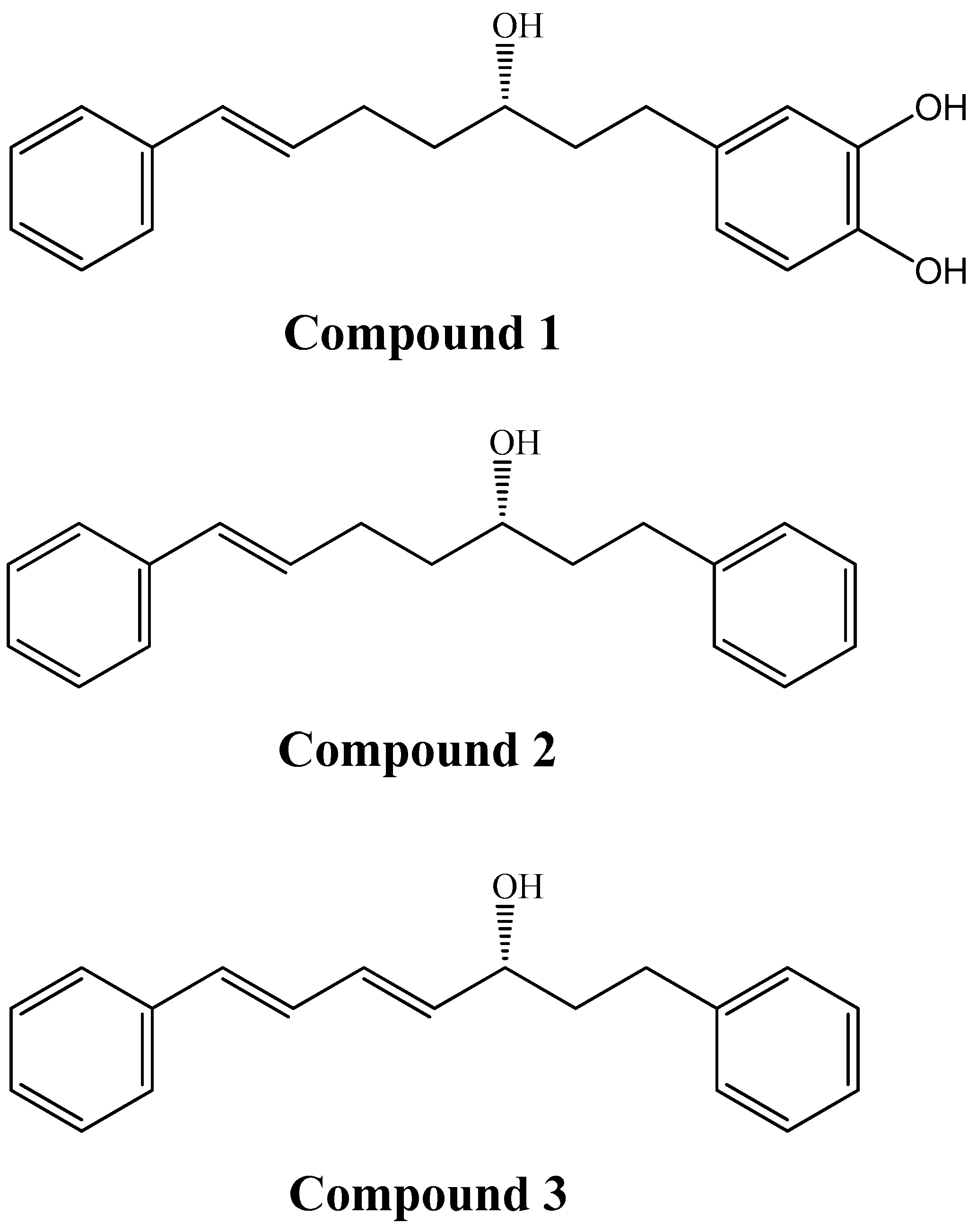
3.1. Extraction and Characterisation of Diarylheptanoid Derivatives in Crude Ethanol Extract of Curcuma comosa Roxb. Rhizome Powder

Crude ethanol extract of Curcuma comosa Roxb. rhizome powder (CE) was obtained as viscous and dark brown solid with 5.37% yield (w/w) of dried plant material. The presence of diarylheptanoid derivatives (Figure 1) and bioactive compounds in CE was studied using spectroscopic techniques (Figures S1–S3 in Supplementary Data). Furthermore, the quantity of diarylheptanoid derivatives in CE was estimated to be 89.3% by using HPLC. The 1H-NMR spectrum of CE (Figure S1) showed important peaks at 6.5–7.5 ppm (Ar–H; aromatic protons), 3.3–5.0 ppm (H–Csp3–OH; protons attached to carbon atoms singly bonded to oxygen), and 2.0–3.0 ppm (HCsp3-C; aliphatic protons of sp3 hybridized carbon atoms of diarylheptanoids). In addition, the results of preliminary phytochemical screening testing presented in Table S1 in the Supplementary Data revealed the existence of other secondary metabolites in CE, which may include cardiac glycosides, phenols, flavonoids, tannins, and terpenoids. These results confirm the phytochemical evidences reported previously for CE [35,36].

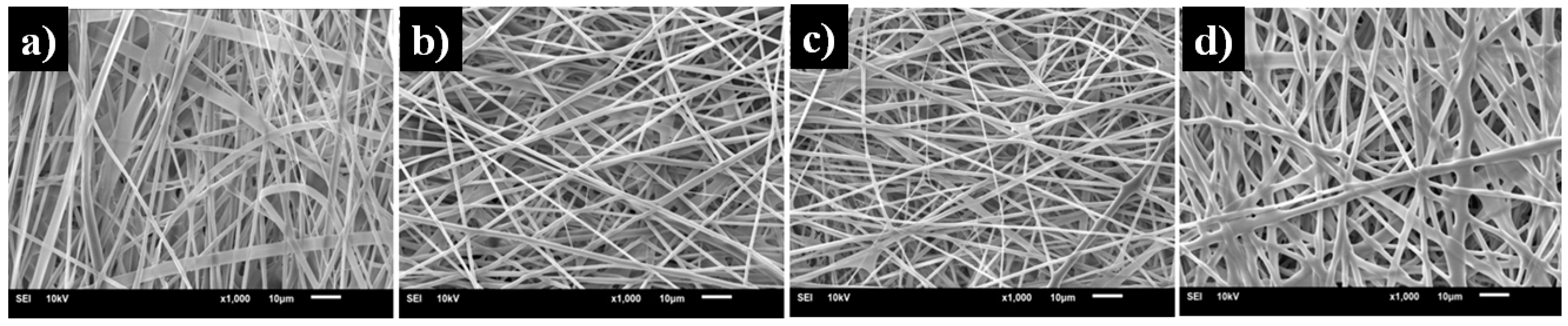
3.2. Effect of Gelatin Concentrations on the Morphology of Electrospun Gelatin Nanofibers

The acetic acid/water co-solvent system at the volumetric ratio of 9:1 was chosen for fabricating gelatin nanofibers by using electrospinning technique [29,30]. The morphology of electrospun gelatin nanofibers fabricated from gelatin solutions in acetic acid/water (9:1 v/v) at various gelatin concentrations was examined using scanning electron microscopy (SEM) From SEM image (Figure 2), the average diameters of electrospun nanofibers obtained from 20, 25, and 30% (w/v) gelatin solutions were 183 ± 29, 367 ± 64 and 547 ± 94 nm, respectively. This may be because polymer molecules and solvents affect the surface tension and the proper evaporation rate of the solution to produce smooth and uniform fibers. Although 20% (w/v) gelatin solution gave electrospun nanofibers the finest average diameter (mean = 183.4 nm), its nanofibers has relatively low toughness. From SEM micrographs of electrospun nanofibers, gelatin concentrations lower than 25% (w/v) could easily cause the cracking of nanofibers. However, cracking is less likely to be seen in electrospun nanofibers obtained from 30% (w/v) gelatin solution, and its average diameter is relatively in range of 500–600 nm. Therefore, a 30% (w/v) gelatin concentration in acetic acid/water (9:1 v/v) was selected as the optimal condition in this study.

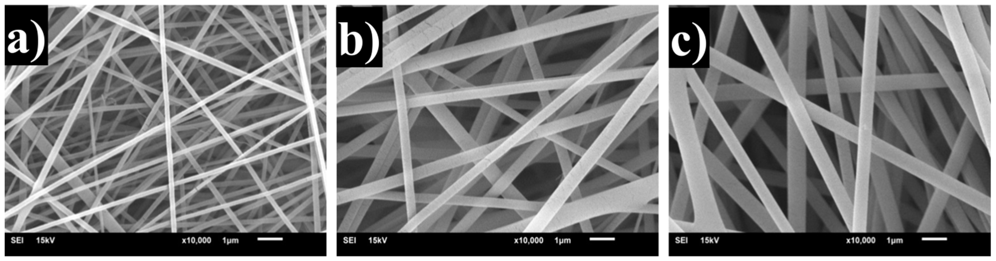
3.3. Effect of Various Concentrations of Crude Ethanol Extract of Curcuma comosa Roxb. Rhizome Powderloaded on the Morphology of Electrospun Gelatin Nanofibers

The optimal conditions for electrospinning were chosen to fabricate gelatin nanofibrous mat containing crude ethanol extract of Curcuma comosa Roxb. rhizome powder (CE) with various concentrations (1, 5, 10 and 15% (w/v)). Figure 3 showed that the gelatin nanofibers loaded with 5% (w/v) of CE were relatively with an average diameter of 1089 ± 611.38 nm. However, 10 and 15% (w/v) of CE made gelatin solutions more viscous and resulted in an increase in the average diameters of gelatin nanofibers, which might cause to the effect of the high CE extract concentration with the increase viscosity, and reduced conductivity to main affect in an increase the average diameters of the electrospun gelatin nanofibrous mat. Thus, the addition of 5% (w/v) of CE into 30% w/v of gelatin solution is the most suitable condition in this study.

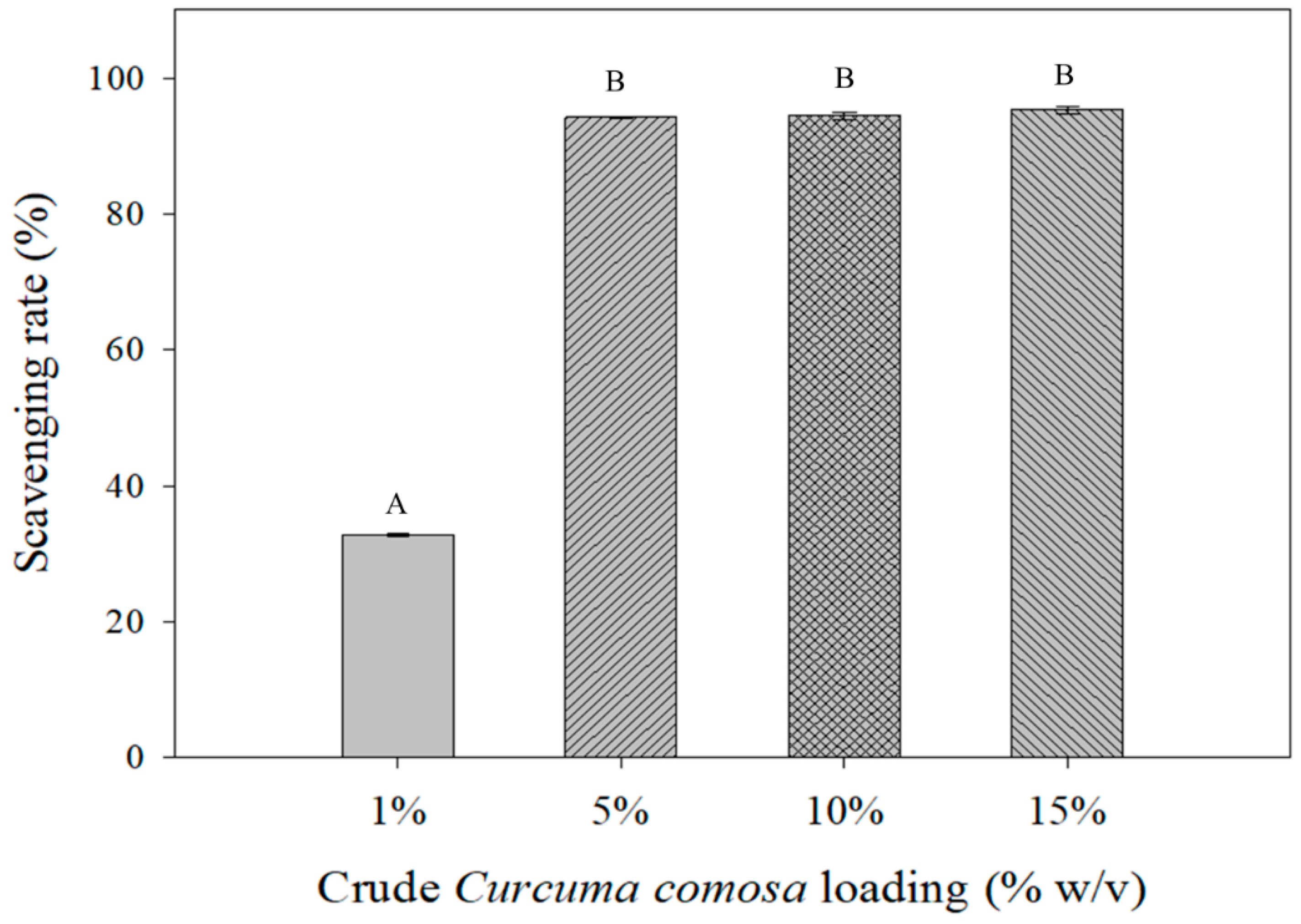
3.4. Effect of Percent Loading on Anti-Oxidant Activity of Electrospun Gelatin Nanofibrous Mat Loaded with Crude Ethanol Extract of Curcuma comosa Roxb. Rhizome Powder

The effect of percent loading of crude ethanol extract of Curcuma comosa Roxb. rhizome powder (CE) on anti-oxidant activity of electrospun gelatin nanofibers, which were fabricated from 30% (w/v) gelatin solution in acetic acid/H2O (9:1 (v/v)) was explored by DPPH radical scavenging assay. Figure 4 presents the plot of percent loading of CE against DPPH radical scavenging rate. The percent loadings at 1, 5, 10, and 15% w/v yielded DPPH radical scavenging rates of 35%, 94%, 94%, and 94.5%, respectively. However, the DPPH radical scavenging rates at the percent loadings of 5, 10 and 15% w/w, they were not statistically significant (p < 0.05). Therefore, to achieve significant free radical scavenging activity, the lowest percent loading with the highest DPPH radical scavenging rate of 5% w/v was preferred. This showed that CE was a good source of natural antioxidants.

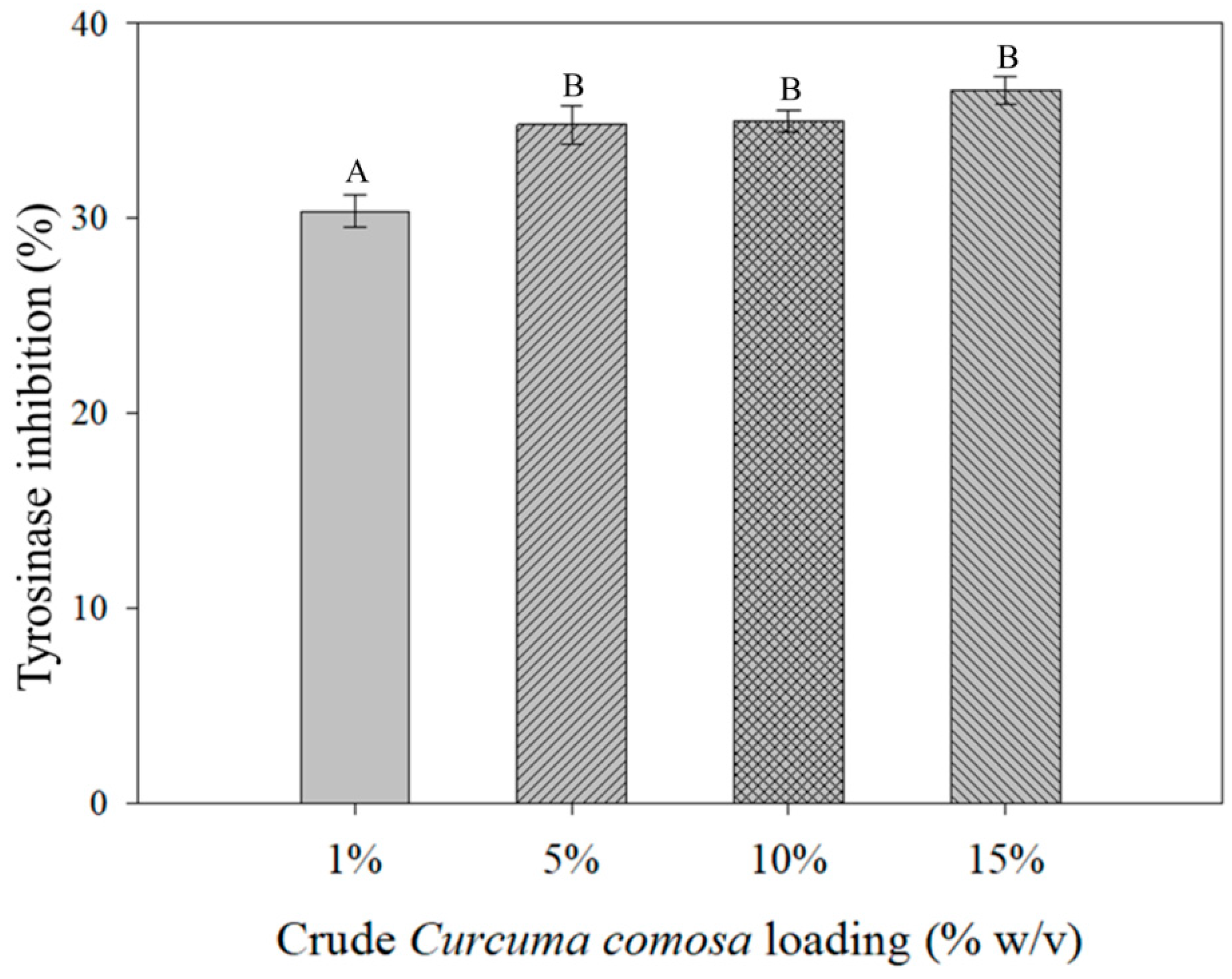
3.5. Effect of Percent Loading on Anti-Tyrosinase Activity of Electrospun Gelatin Nanofibrous Mat Loaded with Crude Ethanol Extract of Curcuma comosa Roxb. Rhizome Powder

Figure 5 is the plot of anti-tyrosinase activity of electrospun gelatin nanofibrous mat against percent loading of crude ethanol extract of Curcuma comosa Roxb. rhizome powder (CE). The results showed that, at the percent loading of 1% (w/v) of CE, the inhibition rate of tyrosinase enzyme was 30%. As the percent loading of CE increased from 5% to 15% (w/v), the inhibition rate of tyrosinase enzyme slightly increased from 35% to 37%. The lowest percent CE loading with the highest anti-tyrosinase activity of 5% w/v was therefore selected due to its good performance in inhibiting melanin production.

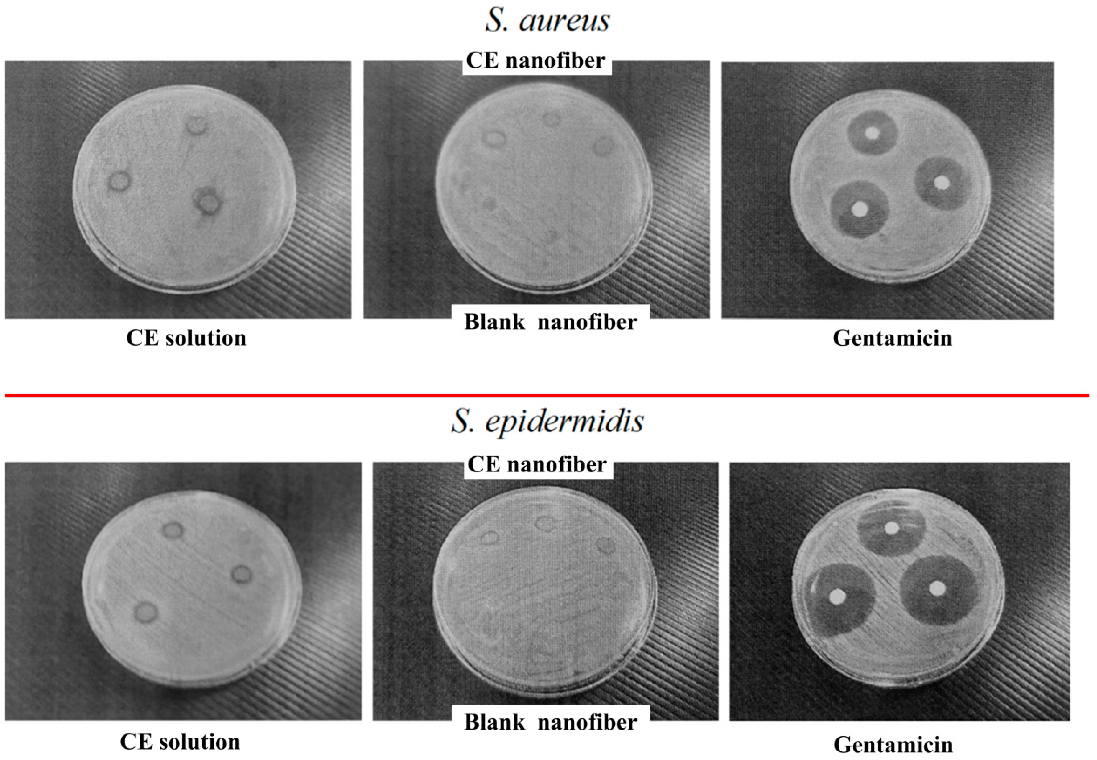
3.6. Effect of Percent Loading on Antibacterial Activity of Electrospun Gelatin Nanofibrous Mat Loaded with Crude Ethanol Extract of Curcuma comosa Roxb. Rhizome Powder

Electrospun gelatin nanofiber sheets loaded with various concentrations of crude ethanol extract of Curcuma comosa Roxb. rhizome powder (CE) were tested for their antibacterial activity by the disc diffusion method and by the broth dilution method. Gentamicin was used for this study as a positive control. It gave inhibitory activity against S. aureus with the inhibition zone of 24.30 ± 0.00 mm and MIC of 0.25 µg/mL and against S. epidermidis with the inhibition zone of 30.50 ± 0.17 mm and MIC of 0.25 µg/mL. The results of the antibacterial studies with regard to the zone of inhibition are shown in Table 1. The electrospun gelatin nanofibrous mat without CE loading (blank nanofiber) showed no inhibitory activity against S. aureus and S. epidermidis. CE solution showed inhibitory activity against S. aureus with the inhibition zone of 9.80 ± 0.17 mm and MIC of 1000 µg/mL and against S. epidermidis with the inhibition zone of 9.93 ± 0.06 mm and MIC of 500.0 µg/mL (Figure 6). The electrospun gelatin nanofibrous mat with CE loading of 5% (w/v) (CE nanofiber) showed inhibitory activity against S. aureus with the inhibition zone of 7.77 ± 0.21 mm and against S. epidermidis with the inhibition zone of 7.73 ± 0.12 mm (Table 1). This means that CE nanofiber could inhibit S. epidermidis and S. aureus in median level of inhibition. This may be due to the good adhesion of the bioactive compounds in electrospun gelatin nanofibrous mat. Although, the limit of maximum loading of CE concentration in nanofibrous mat of the work was low concentration level of bioactive compounds. Additionally, CE nanofiber could be the efficacy of the antimicrobial treatment for atopic dermatitis (AD, eczema) as a common inflammatory skin.

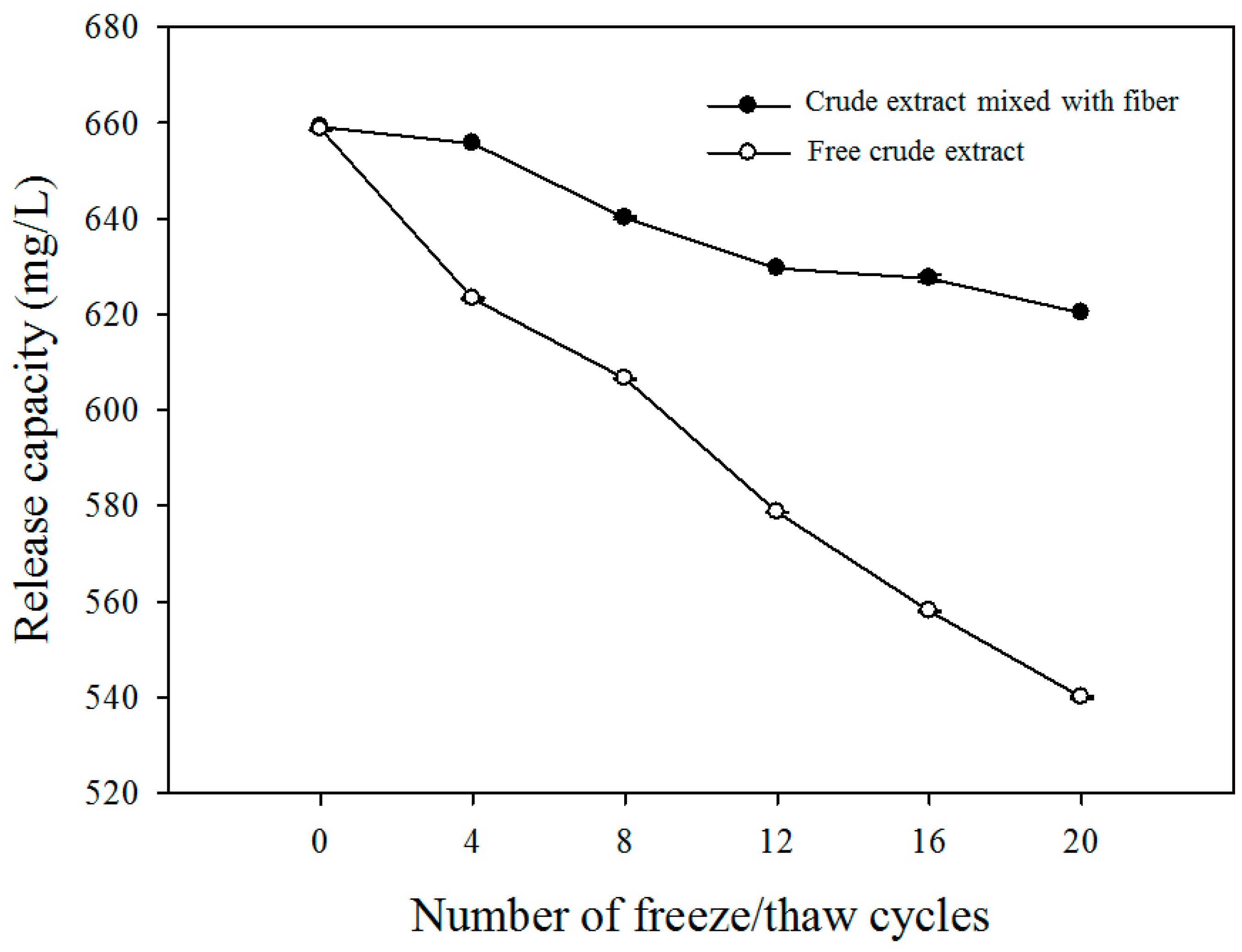
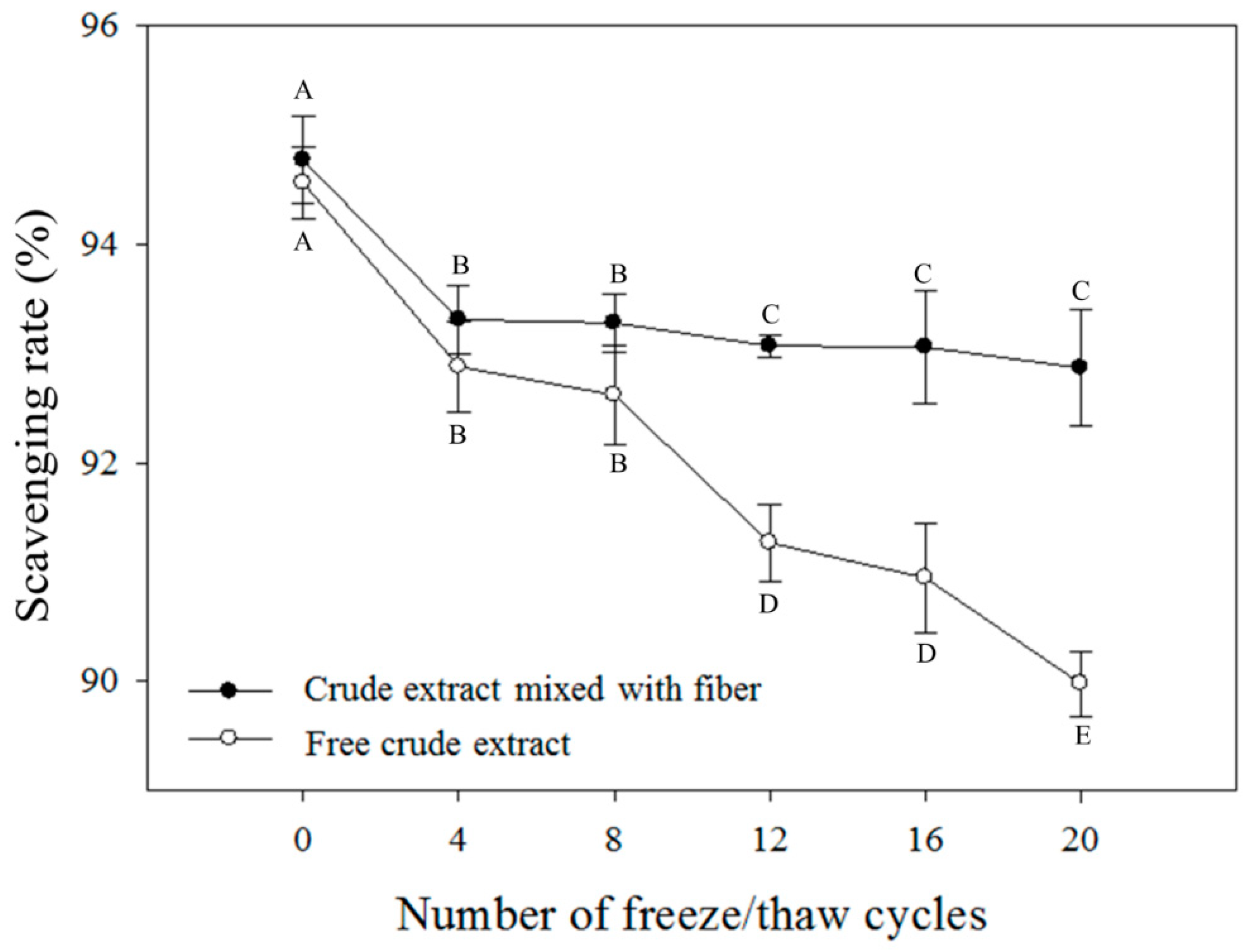
3.7. Stability of Crude Extract-Loaded Gelatin Nanofibrous Mat

The stability of crude ethanol extract of Curcuma comosa Roxb. rhizome powder (CE) loaded into electrospun gelatin nanofibrous mat was investigated using a freeze–thaw cycle test. The concentrations of bioactive compounds released from electrospun gelatin nanofibrous mats were determined by following the method’s Li [33]. At the 20th cycle, the released concentration of bioactive compounds from tested electrospun gelatin nanofibrous mat was 620 mg/L, which was significantly higher than the remaining concentration of CE in the tested solution (540 mg/L) (Figure 7). This confirmed that the bioactive compounds loaded into the gelatin nanofiber showed increasing stability. Moreover, Figure 8 showed that the DPPH radical scavenging activity of both free CE and CE loaded into electrospun gelatin nanofibrous mat decreased rapidly with progressing freeze–thaw cycles. However, free CE exhibited decreasing activity faster than CE loaded into electrospun gelatin nanofibrous mats. At the 20th cycle, the electrospun gelatin nanofibrous mats loaded with CE had the DPPH radical scavenging rate of 93% whereas the free CE had the DPPH radical scavenging rate of 89%. The results clearly indicate that electrospun gelatin nanofibrous mat can be used as a drug carrier for CE.

4. Conclusions

The suitable conditions for fabricating electrospun gelatin nanofibrous mat loaded with crude ethanol extract of Curcuma comosa Roxb. rhizome powder (CE) were 30% (w/v) gelatin solution in acetic acid/H2O (9:1 (v/v)) mixed with 5% (w/v) of CE. The electrospun gelatin nanofibrous mat loaded with 5% (w/v) of CE showed significant antioxidant activity, and anti-tyrosinase activity. In addition, it exhibited antibacterial activity against S. aureus and S. epidermidis as compared to the positive control gentamicin. Notably, the electrospun gelatin nanofibrous mat was proved to stabilize the bioactive compunds in CE under an accelerated storage condition. The results showed that drugs loaded into electrospun gelatin nanofibers were appropriate for facial mask applications.

Supplementary Materials

The following are available online at https://www.mdpi.com/2079-6439/7/9/76/s1, Figure S1: 400 MHz 1H-NMR spectrum of crude Curcuma comosa Roxb. extract., Table S1: Results of premilinary phytochemical screening of the crude ethanol extract of Curcuma comosa Roxb.

Author Contributions

Conceptualization, N.N. and W.M.; data curation, C.-M.C. and N.N.; formal analysis, C.-M.C. and W.M.; funding acquisition, N.N.; investigation, C.-M.C., J.N. and T.S.; methodology, N.N., C.-M.C., J.N. and T.S.; project administration, N.N.; supervision, N.N. and W.M.; writing—original draft, C.-M.C.; and writing—review and editing, N.N., W.M., C.S., W.-L.L. and K.P.

Funding

This research was funded by the Thailand research fund [MRG6180070], the research fund for DPST graduate with first placement, Thai government [No.11/2557] and the APC was funded by the KMUTT 55th Anniversary Commemorative Fund. C. C gratefully acknowledges the scholarship of Faculty of Science, King Mongkut’s University of Technology Thonburi.

Acknowledgments

The authors would like to thanks Wei-Jing Hung and Wei-Chao Lin, Department of Cosmetic Science, Chia Nan University of Pharmacy and Science for support and encouragement.

Conflicts of Interest

The authors declare no conflicts of interest.

References

  1. Saensouk, S.; Saensouk, P.; Pasorn Chantaranothai, P. Diversity and uses of zingiberaceae in nam nao national park, chaiyaphum and phetchabun provinces, Thailand, with a new record for Thailand. Agric. Nat. Resour. 2016, 50, 445–453. [Google Scholar] [CrossRef]
  2. Su, J.; Sripanidkulchai, K.; Suksamrarn, A.; Hu, Y.; Piyachuturawat, P.; Sripanidkulchai, B. Pharmacokinetics and organ distribution of diarylheptanoid phytoestrogens from Curcuma comosa in rats. J. Nat. Med. 2012, 66, 468–475. [Google Scholar] [CrossRef] [PubMed]
  3. Winuthayanon, W.; Suksen, K.; Boonchird, C.; Chuncharunee, A.; Ponglikitmongkol, M.; Suksamrarn, A.; Piyachaturawat, P. Estrogenic Activity of Diarylheptanoids from Curcuma comosa Roxb. Requires Metabolic Activation. J. Agric. Food Chem. 2009, 57, 840–845. [Google Scholar] [CrossRef] [PubMed]
  4. Suksamrarn, A.; Eiamong, S.; Piyachaturawat, P.; Byrne, L.T. A Phloracetophenone Glucoside with Choleretic Activity from Curcuma comosa. Phytochemistry 1997, 45, 103–105. [Google Scholar] [CrossRef]
  5. Jurgens, T.M.; Frazier, E.G.; Schaeffer, J.M.; Jones, T.E.; Zink, D.L.; Borris, R.P.; Nanakorn, W.; Beck, H.T.; Balick, M.J. Novel nematocidal agents from Curcuma comosa. J. Nat. Prod. 1994, 57, 230–235. [Google Scholar] [CrossRef] [PubMed]
  6. Matsumoto, T.; Nakamura, S.; Fujimoto, K.; Ohta, T.; Ogawa, K.; Yoshikawa, M.; Onishi, E.; Fukaya, M.; Matsuda, H. Structure of diarylheptanoids with antiallergic activity from the rhizomes of Curcuma comosa. J. Nat. Med. 2015, 69, 142–147. [Google Scholar] [CrossRef] [PubMed]
  7. Wang, J.; Windbergs, M. Functional electrospun fibers for the treatment of human skin wounds. Eur. J. Pharm. Biopharm. 2017, 119, 283–299. [Google Scholar] [CrossRef] [PubMed]
  8. Li, T.; Zhang, D.; Oo, T.N.; San, M.M.; Mon, A.M.; Hein, P.P.; Wang, Y.; Lu, C.; Yang, X. Investigation on the Antibacterial and Anti-T3SS Activity of Traditional Myanmar Medicinal Plants. Evid. Based Complement. Altern. Med. 2018, 2018, 2812908. [Google Scholar] [CrossRef] [PubMed]
  9. Suksamrarn, A.; Ponglikitmongkol, M.; Wongkrajang, K.; Chindaduang, A.; Kittidanairak, S.; Jankam, A.; Yingyongnarongkul, A.B.E.; Kittipanumat, N.; Chokchaisiri, R.; Khetkam, P.; et al. Diarylheptanoids, new phytoestrogens from the rhizomes of Curcuma comosa: Isolation, chemical modification and estrogenic activity evaluation. Bioorganic Med. Chem. 2008, 16, 6891–6902. [Google Scholar] [CrossRef]
  10. Yingngam, B.; Brantner, A.; Jinarat, D.; Kaewamatawong, R.; Rungseevijitprapa, W.; Suksamrarn, A.; Piyachaturawat, P.; Chokchaisiri, R. Determination of the marker diarylheptanoid phytoestrogens in Curcuma comosa rhizomes and selected herbal medicinal products by HPLC-DAD. Chem. Pharm. Bull. 2018, 66, 65–70. [Google Scholar] [CrossRef]
  11. Intapad, S.; Saengsirisuwan, V.; Prasannarong, M.; Chuncharunee, A.; Suvitayawat, W.; Chokchaisiri, R.; Suksamrarn, A.; Piyachaturawat, P. Long-term effect of phytoestrogens from Curcuma comosa Roxb on vascular relaxation in ovariectomized rats. J. Agric. Food Chem. 2012, 60, 758–764. [Google Scholar] [CrossRef]
  12. Sodsai, A.; Piyachaturawat, P.; Sophasan, S.; Suksamrarn, A.; Vongsakul, M. Suppression by Curcuma comosa Roxb. of proinflammatory cytokine secretion in phorbol-12-myristate-13-acetate stimulated human mononuclear cells. Int. Immunopharmacol. 2007, 7, 524–531. [Google Scholar] [CrossRef] [PubMed]
  13. Wiseman, H. The therapeutic potential of phytoestrogens. Expert Opin. Investig. Drugs 2000, 9, 1829–1840. [Google Scholar] [CrossRef] [PubMed]
  14. Boonmee, A.; Srisomsap, C.; Chokchaichamnankit, D.; Karnchanatat, A.; Sangvanich, P. A proteomic analysis of Curcuma comosa Roxb. Rhizomes. Proteome Sci. 2011, 9, 43. [Google Scholar] [CrossRef] [PubMed]
  15. Xu, F.; Nakamura, S.; Qu, Y.; Matsuda, H.; Pongpiriyadacha, Y.; Wu, L.; Yoshikawa, M. Structures of New Sesquiterpenes from Curcuma comosa. Chem. Pharm. Bull. 2008, 56, 1710–1716. [Google Scholar] [CrossRef]
  16. Su, J.; Sripanidkulchai, K.; Hu, Y.; Chaiittianan, R.; Sripanidkulchai, B. Increased in situ intestinal absorption of phytoestrogenic diarylheptanoids from Curcuma comosa in nanoemulsions. Off. J. Am. Assoc. Pharm. Sci. 2013, 14, 1055–1062. [Google Scholar] [CrossRef] [PubMed]
  17. Ahn, B.K.; Lee, S.G.; Kim, S.R.; Lee, D.H.; Oh, M.H.; Lee, M.W.; Choi, Y.W. Inclusion compound formulation of hirsutenone with beta-cyclodextrin. J. Pharm. Investig. 2013, 43, 453–459. [Google Scholar] [CrossRef]
  18. Wani, T.A.; Shah, A.G.; Wani, S.M.; Wani, I.A.; Masoodi, F.A.; Nissar, N.; Shagoo, M.A. Suitability of different food grade materials for the encapsulation of some functional foods well reported for their advantages and susceptibility. Crit. Rev. Food Sci. Nutr. 2016, 56, 2431–2454. [Google Scholar] [CrossRef]
  19. Radhakrishnan, S.; Lakshminarayanan, R.; Madhaiyan, K.; Barathi, V.A.; Lim, K.H.C.; Ramakrishna, S. Electrosprayed nanoparticles and electrospun nanofibers based on natural materials: Applications in tissue regeneration, drug delivery and pharmaceuticals. Chem. Soc. Rev. 2015, 44, 790–814. [Google Scholar]
  20. STorres-Giner, R.; Pérez-Masiá, R.; Lagaron, J.M. A review on electrospun polymer nanostructures as advanced bioactive platforms. Polym. Eng. Sci. 2016, 56, 500–527. [Google Scholar] [CrossRef]
  21. Bhattarai, R.S.; Bachu, R.D.; Boddu, S.H.S.; Bhaduri, S. Biomedical Applications of Electrospun Nanofibers: Drug and Nanoparticle Delivery. Pharmaceutics 2019, 11, 5. [Google Scholar] [CrossRef] [PubMed]
  22. Papa, A.; Guarino, V.; Cirillo, V.; Oliviero, O.; Ambrosio, L. Optimization of Bicomponent Electrospun Fibers for Therapeutic Use: Post-Treatments to Improve Chemical and Biological Stability. J. Funct. Biomater. 2017, 8, 47. [Google Scholar] [CrossRef] [PubMed]
  23. Yang, F.; Miao, Y.; Wang, Y.; Zhang, L.M.; Lin, X. Electrospun Zein/Gelatin Scaffold-Enhanced Cell Attachment and Growth of Human Periodontal Ligament Stem Cells. Materials 2017, 10, 1168. [Google Scholar] [CrossRef] [PubMed]
  24. Nootem, J.; Chalorak, P.; Meemon, K.; Mingvanish, W.; Pratumyot, K.; Ruckthong, L.; Srisuwannaket, C.; Niamnont, N. Electrospun cellulose acetate doped with astaxanthin derivatives from Haematococcus pluvialis for in vivo anti-aging activity. RSC Adv. 2018, 8, 37151–37158. [Google Scholar] [CrossRef]
  25. Zupacnic, S.; Baumgartner, S.; Lavric, Z.; Petelin, M.; Kristl, J. Local delivery of resveratrol using polycaprolactone nanofibers for treatment of periodontal disease. J. Drug. Deliv. Sci. Technol. 2015, 30, 408–416. [Google Scholar]
  26. El-Refaie, K.; Bowlin, G.L.; Mansfield, K.; Layman, J.; Simpson, D.G.; Sanders, E.H.; Wnek, G.E. Release of tetracycline hydrochloride from electrospun poly (ethylene-co-vinylacetate), poly (lactic acid), and a blend. J. Control. Release 2002, 81, 57–64. [Google Scholar]
  27. Chao, S.; Li, Y.; Zhao, R.; Zhang, L.; Li, Y.; Wang, C.; Li, X. Synthesis and characterization of tigecycline-loaded sericin/poly (vinyl alcohol) composite fibers via electrospinning as antibacterial wound dressings. J. Drug. Deliv Sci. Technol. 2018, 44, 440–447. [Google Scholar] [CrossRef]
  28. Zhu, L.F.; Chen, X.; Ahmad, Z.; Li, J.S.; Chang, M.W. Engineering of Ganoderma lucidum polysaccharide loaded polyvinyl alcohol nanofibers for biopharmaceutical delivery. J. Drug. Deliv Sci. Technol. 2019, 50, 208–216. [Google Scholar] [CrossRef]
  29. Ghorani, B.; Tucker, N. Fundamentals of electrospinning as a novel delivery vehicle for bioactive compounds in food nanotechnology. Food Hydrocoll. 2015, 51, 227–240. [Google Scholar] [CrossRef]
  30. Figueroa-Lopez, K.J.; Castro-Mayorga, J.L.; Andrade-Mahecha, M.M.; Cabedo, L.; Lagaron, J.M. Antibacterial and barrier properties of gelatin coated by electrospun polycaprolactone ultrathin fibers containing black pepper oleoresin of interest in active food biopackaging applications. Nanomaterials 2018, 8, 199. [Google Scholar] [CrossRef]
  31. Mogosanu, G.D.; Grumezescu, A.M. Natural and synthetic polymers for wounds and burns dressing. Int. J. Pharm. 2014, 463, 127–136. [Google Scholar] [CrossRef] [PubMed]
  32. Maytal, F.; Zilberman, M. Drug delivery from gelatin-based systems. Expert Opin. Drug Deliv. 2015, 12, 1547–1563. [Google Scholar]
  33. Nagarajan, S.; Soussan, L.; Bechelany, M.; Teyssier, C.; Cavailles, V.; Pochat-Bohatier, C.; Miele, P.; Kalkura, N.; Janot, J.M.; Balme, S. Novel biocompatible electrospun gelatin fiber mats with antibiotic drug delivery properties. J. Mater. Chem. B 2016, 4, 1134–1141. [Google Scholar] [CrossRef]
  34. Li, H.; Wang, M.; Williams, G.R.; Wu, J.; Sun, X.; Lv, Y.; Zhu, L.M. Electrospun gelatin nanofibers loaded with vitamins A and E as antibacterial wound dressing materials. RSC Adv. 2016, 6, 50267–50277. [Google Scholar] [CrossRef]
  35. Sutjarit, N.; Sueajai, J.; Boonmuen, N.; Sornkaew, N.; Suksamrarn, A.; Tuchinda, P.; Zhu, W.; Weerachayaphorn, J.; Piyachaturawat, P. Curcuma comosa reduces visceral adipose tissue and improves dyslipidemia in ovariectomized rats. J. Ethnopharmacol. 2018, 215, 167–175. [Google Scholar] [CrossRef]
  36. Prashanth, N.; Bhavani, N.L. Phytochemical analysis of two high yielding Curcuma longa varieties from Andhra Pradesh. Int. J. Life Sci. Biotechnol. Pharm. Res. 2013, 2, 103–108. [Google Scholar]
  37. Devi, K.P.; Suganthy, N.; Kesika, P.; Pandian, S.K. Bioprotective properties of seaweeds: In vitro evaluation of antioxidant activity and antimicrobial activity against food borne bacteria in relation to polyphenolic content. BMC Complement. Altern. Med. 2008, 8, 1–11. [Google Scholar] [CrossRef]
  38. Masamoto, Y.; Ando, H.; Murata, Y.; Shimoishi, Y.; Tada, M.; Takahata, K. Mushroom tyrosinase inhibitory activity of esculetin isolated from seeds of Euphorbia lathyris L. Biosci. Biotechnol. Biochem. 2003, 67, 631–634. [Google Scholar] [CrossRef]
Figure 1. Chemical structures of diarylheptanoids (compound 1: (3S)-1-(3,4-Dihydroxyphenyl)-7 -phenyl-(6E)-6-hepten-3-ol; compound 2: (3R)-1,7-Diphenyl-(4E,6E)- 4,6-heptadiene-3-ol; compound 3: (3S)-1,7-Diphenyl-(6E)-6-hepten-3-ol) in crude ethanol extract of Curcuma comosa Roxb. rhizome powder.
Fibers 07 00076 g001
Figure 2. Electrospun gelatin nanofibers fabricated from gelatin solutions in acetic acid/water (9:1 (v/v)) at various gelatin concentrations: (a) 20%, (b) 25%, and (c) 30% (w/v).
Fibers 07 00076 g002
Figure 3. Electrospun gelatin nanofibers with various concentrations of crude ethanol extract of Curcuma comosa Roxb.rhizome powder: (a) 1%, (b) 5%, (c) 10%, and (d) 15% (w/v) in acetic acid/H2O (9:1 (v/v)).
Fibers 07 00076 g003
Figure 4. Effect of percent loading of crude ethanol extract of Curcuma comosa Roxb. powder in electrospun gelatin nanofibers on DPPH radical-scavenging activity. A,B Means with different capital letter superscripts are significantly different (p < 0.05).
Fibers 07 00076 g004
Figure 5. Effect of percent loading of crude ethanol extract of Curcuma comosa Roxb. powder in electrospun gelatin nanofibers on anti-tyrosinase activity. A,B Means with different capital letter superscripts are significantly different (p < 0.05).
Fibers 07 00076 g005
Figure 6. Effect of 5% w/v crude extract of electrospun fiber on antibacterial activity inhibition. Antibacterial activity of crude ethanol extract of Curcuma comosa Roxb. rhizome powder (CE) in electrospun gelatin nanofibrous mat; 5% (w/v) CE solution, blank nanofiber, 5% (w/v) CE nanofiber, and gentamicin positive control (5% (w/v)). Test disc diameter is 6 mm.
Fibers 07 00076 g006
Figure 7. Change in concentrations of bioactive compounds in crude ethanol extract of Curcuma comosa Roxb. rhizome powder (CE) released from electrospun gelatin nanofibrous mats during freeze–thaw cycle testing; (•): electrospun gelatin nanofibrous mats loaded with 5% (w/v) of CE and (ο): 5% (w/v) of only CE solution.
Fibers 07 00076 g007
Figure 8. Change in percent of DPPH radical scavenging activity of bioactive compounds in crude ethanol extract of Curcuma comosa Roxb. rhizome powder (CE) released from electrospun gelatin nanofibrous mat during freeze–thaw cycle testing; (•): electrospun gelatin nanofiber sheets loaded with 5% (w/v) of CE and (ο): 5% (w/v) of CE solution. A,B,C,D,E Means with different capital letter superscripts are significantly different (p < 0.05).
Fibers 07 00076 g008
Table 1. Inhibition zone and MIC value of 5% (w/v) crude ethanol extract of Curcuma comosa Roxb. rhizome powder crude extract on electrospun gelatin nanofiber sheets.
SampleS. aureusS. epidermidis
Inhibition Zone a (mm)MIC (µg/mL)Inhibition Zone a (mm)MIC (µg/mL)
CE solution9.80 ± 0.1710009.93 ± 0.06500
Blank nanofiberNA *-NA *-
CE nanofiber7.77 ± 0.21-7.73 ± 0.12-
Gentamicin24.30 ± 0.000.2530.50 ± 0.170.25
* NA = no activity, a include disc diameter (6.0 mm).

