Article

Commercial, Non-Commercial and Experimental Wound Dressings Based on Bacterial Cellulose: An In-Depth Comparative Study of Physicochemical Properties

by
Sarah Brandão Palácio
1,2,*,
Simone Oliveira Penello
3,4,
Katharine Valéria Saraiva Hodel
2,5,
Willams Teles Barbosa
2,
Gisele Assunção Reis
2,
Bruna Aparecida Souza Machado
2,
Ana Leonor Pardo Campos Godoy
5,6,
Maria Inês Bruno Tavares
4,
Layla Carvalho Mahnke
7,
Josiane Dantas Viana Barbosa
2,* and
José Lamartine de Andrade Aguiar
7,*
1
Salvador Health Group (GSAU-SV), Air Force Health Directorate (DIRSA), Brazilian Air Force, Rio de Janeiro 41650-010, RJ, Brazil
2
SENAI Institute for Innovation in Advanced Health Systems (ISI SAS), SENAI CIMATEC University Center, Salvador 41650-010, BA, Brazil
3
Aeronautics Chemical-Pharmaceutical Laboratory (LAQFA), Brazilian Air Force, Rio de Janeiro 21941-353, RJ, Brazil
4
Institute of Macromolecules Professor Eloisa Mano-IMA-UFRJ, Federal University of Rio de Janeiro (UFRJ), Rio de Janeiro 21941-598, RJ, Brazil
5
Pharmacy Graduate Program, Federal University of Bahia (UFBA), Salvador 40110-100, BA, Brazil
6
Department of Clinical and Toxicological Analysis, Faculty of Pharmacy, Federal University of Bahia (UFBA), Salvador 40110-100, BA, Brazil
7
Post-Graduation Program in Surgery, Department of Surgery, Health Sciences Center, Federal University of Pernambuco (UFPE), Recife 50670-420, PE, Brazil
*
Authors to whom correspondence should be addressed.
Fibers 2025, 13(9), 127; https://doi.org/10.3390/fib13090127
Submission received: 31 July 2025 / Revised: 29 August 2025 / Accepted: 9 September 2025 / Published: 15 September 2025

Abstract

Wound management remains a significant global healthcare challenge, particularly due to chronic wounds that resist healing and impose economic and social burdens. Bacterial cellulose (BC), owing to its biocompatibility, high purity and moisture-handling capabilities, has gained attention as a wound dressing material. This study provides a comparative evaluation of a commercial BC film (Membracel®), a non-commercial BC from POLISA® (BCP) and an experimental BC from SENAI CIMATEC (BCC), all produced via static fermentation using distinct culture conditions. Comprehensive characterization included scanning electron microscopy, X-ray diffraction, Fourier transform infrared spectroscopy, thermogravimetric analysis, solid-state 13C NMR, water interaction assessments, porosity and vapor permeability measurements, optical and mechanical testing and in vitro stability in simulated wound fluid. The three BC films exhibited markedly different structural and functional profiles. BCC displayed the highest crystallinity (78.7%), thermal stability and vapor permeability, indicating suitability for wounds with high exudate. BCP showed the greatest tensile strength (46.2 MPa) and flexibility, suggesting utility where mechanical robustness is required. Membracel® exhibited lower crystallinity and vapor permeability, appropriate for low-exudate wounds. All samples remained dimensionally stable in simulated wound fluid. These findings highlight clear correlations between the physicochemical properties of BC-based dressings and their potential clinical applications, supporting the development of tailored wound care solutions based on wound type and moisture management requirements.

1. Introduction

Wound management remains one of the greatest global healthcare challenges, driven by factors ranging from impaired healing processes to insufficiently targeted treatment strategies. It is estimated that over 10.5 million individuals worldwide suffer from chronic wounds, lesions that fail to heal within three months, imposing substantial economic and social burdens [1,2]. Chronic wounds, including diabetic foot ulcers, pressure injuries and burns, are characterized by prolonged healing times, elevated infection risk and the need for frequent dressing changes, all of which exacerbate patient morbidity and healthcare costs [3,4].
In this manner, the choice of wound dressing is a critical determinant of therapeutic success. Wound dressings serve primarily to protect the wound bed from external contaminants, maintain a favorable moist environment and support tissue repair [5]. Broadly, they are classified into traditional and advanced categories. Traditional wound dressings, though often less expensive, typically lack optimal moisture regulation, exhibit limited biocompatibility and require frequent replacement, thereby increasing both patient discomfort and overall treatment expense [6]. To address these limitations, advanced biomaterial-based wound dressings have been engineered to provide enhanced fluid management, improved cellular support and controlled release of antimicrobial or regenerative agents [7,8].
Within this landscape, biopolymers, which are polymeric biomolecules composed of long chains of covalently linked monomeric units, have garnered special attention. Bacterial cellulose (BC), produced predominantly by Gluconacetobacter species, stands out as a highly promising material for wound care [9]. Its unique nanofibrillar network, significative water-holding capacity and mechanical robustness closely mimic the extracellular matrix [10]. High porosity facilitates gas exchange, while the absence of endotoxins and immunogenic components ensures excellent biocompatibility [11]. Furthermore, the intrinsic hydrogel nature of BC allows the incorporation of antimicrobial compounds, growth factors or other therapeutic agents, thereby extending its functional versatility [12,13].
Several commercial BC dressings, such as Membracel® (Vuelo Pharma, Rio de Janeiro, Brazil), have demonstrated favorable outcomes in preclinical and clinical studies [14,15,16,17]. Simultaneously, different research groups have developed experimental BC formulations by varying culture media, microbial strains and post-treatment protocols, producing materials with tailored porosity, mechanical strength and bioactive loading capacity [18,19,20,21]. However, these studies are often reported in isolation, using heterogeneous characterization methods and in vitro assays that impede direct comparison between commercial products and laboratory-scale prototypes.
Because BC structure and performance are intrinsically tied to production parameters, such as carbon source, strain selection, culture conditions and purification strategy, a direct physicochemical comparison of commercial and experimental BC dressings is essential. Variations in fiber diameter or network density, for example, will alter water-holding capacity and vapor permeability, thereby dictating which formulations best balance exudate management versus moisture retention [22,23]. By systematically evaluating tensile properties, fluid uptake kinetics, porosity metrics and surface characteristics under identical protocols, clear structure–function correlations can be established to inform dressing recommendations tailored to specific wound types, whether heavily exuding ulcers, low-exuding abrasions or infected lesions requiring localized antimicrobial delivery.
Therefore, the aim of this study was to address this gap by conducting a comprehensive comparative analysis of three bacterial cellulose dressings, one commercial and two experimental, evaluating their key physicochemical properties and establishing correlations with their suitability for managing different wound types.

2. Materials and Methods

2.1. Obtaining Samples of Bacterial Cellulose Wound Dressings

The bacterial cellulose wound dressings were obtained via three distinct routes. The commercial product Membracel® was used as received, having been generously donated by the Hospital de Força Aérea do Galeão in Rio de Janeiro, Brazil. The BC produced by POLISA® (BCP) is manufactured using a standardized production process, which operates under a quality management control system registered with the Brazilian Health Regulatory Agency (ANVISA). Production consists of sterilizing a culture medium of sugarcane molasses (10% v/v; 36.57% sucrose, 57.39% reducing sugars) in an autoclave, inoculating it with native Gluconacetobacter hansenii and incubating at 27 °C and 55% relative humidity with HEPA-filtered air [18]. After 15 days of static fermentation, the thick membrane formed at the air–medium interface was harvested, washed repeatedly with purified water, clarified in 0.3% (v/v) sodium hypochlorite and rinsed to neutral pH to yield a purified BC matrix [18]. This hydrated matrix was homogenized for 3 min at 1500 rpm to produce a hydrogel, of which 1.5 L was cast onto a microporous felt (120 threads/cm) under vacuum to remove water and produce the films [18]. The BC produced at University SENAI CIMATEC (BCC) was obtained using G. hansenii ATCC 23769 in modified Hestrin–Schramm medium (25 or 50 g·L−1 glucose, 5 g·L−1 yeast extract, 3 g·L−1 peptone, 2 g·L−1 KH2PO4), sterilized at 121 °C for 15 min and incubated statically at 30 °C for 14 days, with OD600 monitoring to follow production kinetics [19]. Resulting membranes were washed in distilled water at 80 °C for 1 h, treated with 0.3 mol·L−1 K2CO3 at 80 °C for 1 h, rinsed to neutral pH and then dried at 50 °C for 24 h for yield determination and dry film formation [19].

2.2. Characterization of Samples of Bacterial Cellulose Wound Dressings

2.2.1. Scanning Electron Microscopy (SEM)

Scanning electron microscopy (SEM) was conducted to evaluate the surface morphology and structural interconnectivity of BC films. Prior to analysis, the samples were sputter-coated with a thin layer of carbon using a DENTON VACUUM sputter coater (DII-29010SCTR Smart Coater, JEOL Inc., Peabody, MA, USA). SEM imaging was performed using a JSM-6510LV scanning electron microscope (JEOL Inc., Peabody, MA, USA) operated at an accelerating voltage of 20 kV.
Elemental analysis was conducted using a Tescan Mira 4 high-resolution SEM at 15 kV. Energy-dispersive X-ray spectroscopy (EDS) was employed to characterize the BC films. Samples were gold-coated with a Denton Vacuum Desk II sputter coater (Denton Vacuum, Moorestown, NJ, USA) for 60 s at 3 nA. Images were acquired at magnifications of 200×–10,000×, and data were analyzed using Essence™ 1.0 software to identify features and patterns.

2.2.2. X-Ray Diffraction (XRD) and Fourier Transformed InfraRed Spectroscopy (FTIR)

The structural characterization of the samples was performed by X-ray diffraction (XRD) using a model UltimaIV X-ray diffractometer (Rigaku Holdings Corp., Matsubara, Tokyo, Japan), operating with Cu-Kα radiation (λ = 1.5406 Å), under a voltage of 40 kV and a current of 20 mA. The scans were performed in the 2θ range of 2–60°, with a step of 0.02°. The crystallinity index (CrI) for each sample was calculated according to the method proposed by Segal et al. [24] (Equation (1)).
C r I % = ( I 002 I a m ) I 002 × 100
I002 represents the maximum intensity of the (002) diffraction peak at 2θ = 22.8°, and Iam corresponds to the intensity of the amorphous background at 2θ = 18°.
The FTIR spectra of the samples were measured on a Spectrometer model FrontierTM (PerkinElmer, Waltham, MA, USA). Scans (60) were taken at a resolution of 4 cm−1 in the frequency range 4000–600 cm−1 with software Spectrum Version 10.4.2.

2.2.3. Thermogravimetric Analysis

The thermal stability of the samples was assessed by thermogravimetric analysis (TGA) and derivative thermogravimetry (DTG) using a TGA Q500 (TA Instruments, New Castle, DE, USA). The analyses were conducted under an inert nitrogen atmosphere with a continuous gas flow throughout the experiment. The samples were heated from 25 °C to 700 °C at a heating ramp of 10 °C/min.

2.2.4. Solid-State 13C Nuclear Magnetic Resonance (NMR) Analysis

13C CP-MAS NMR analysis was performed with a Bruker BioSpin 500 MHz spectrometer operating at 125 MHz for 13C. The technique employed was cross polarization magic angle spinning (CP/MAS). The delay was 2 s and contact time was 1000 (p15) with 30 k scans.

2.2.5. Water Interaction: Swelling Index, Moisture Content Index, Water Activity (aw) and Contact Angle

The capacity of the BC wound dressing swell in water and their moisture content were quantified gravimetrically, adapting the approach of Du et al. [25]. Rectangular specimens (16 cm2) were first dried to constant weight and their dry mass recorded (mdry). Each piece was then submerged in 30 mL of deionized water at 25 ± 2 °C for 24 h. After equilibration, samples were removed, gently blotted with filter paper to remove surface water and weighed again to obtain the swollen mass (mswollen). Water swelling ratio (WSI, %) (Equation (2)) and moisture content index (MC, %) (Equation (3)) were calculated as follows:
W S I % = ( m swollen m dry ) m dry × 100
M C % = ( m swollen m dry ) m swollen × 100
Water activity (aw) of the perforated BC wound dressing was determined at 25 ± 2 °C using a Novasina Lab Master aw instrument fitted with a CM-2 electrolytic sensor. Square samples (2 × 2 cm) were placed in the chamber and allowed to equilibrate until stable readings were achieved [26].
The contact angle test was conducted using a contact goniometer model ESr-N and OCA 15EC module (DataPhysics Instruments, Filderstadt, Baden-Wüettemberg, Germany), in conjunction with the SCA20 software (version 4.5.14 Build 1064), which captured droplet images and calculated the contact angle. Five µL of distilled water was applied to the samples using a syringe, maintaining a constant plunger speed of 5 µL/s. Measurements were taken at different sample points and in triplicate. The final contact angle value (°), as well as the corresponding droplet image, were automatically determined by the equipment.
All measurements were performed in triplicate, and results are reported as mean ± standard deviation.

2.2.6. Porosity and Water Vapor Transmission Rate (WVTR) Analysis

Porosity of the BC wound dressings was quantified via the liquid-displacement method, adapted by Ao et al. [27]. Square samples (2 × 2 cm) were first dried and weighed to obtain their initial mass (W1) and measured to calculate their volume (Vs). Samples were then immersed in absolute ethanol (PA grade) at 25 ± 2 °C for 24 h to ensure complete infiltration. After removal, excess surface water was blotted away with filter paper, and the wet mass (W2) was recorded. Porosity (%) was calculated for each of three replicates as follows (Equation (4)):
P o r o s i t y % = ( w 2 w 1 ) ρ ( E t O H ) × V × 100
ρ(EtOH) is the density of ethanol;
V is the volume of solvent.
WVTR was determined gravimetrically following the cup method adapted from ASTM E96 [28], using circular samples (≈60 mm diameter). Samples were cut and mounted to fully cover the mouth of permeation cups containing 30 mL of distilled water; cups were closed and sealed to prevent edge leakage and then placed inside a desiccator containing silica gel (nominal 0% RH) at 25 ± 2 °C to establish a constant vapor pressure gradient. Cup assemblies were weighed on an analytical balance (±0.1 mg) at 24 h intervals for five consecutive days. Sample permeation area (A) was calculated from the measured sample diameter (A = πr2, expressed in m2). The WVTR for each sample was calculated by Equation (5), and reported in g·m−2·24 h−1.
W V T R = Δ m / Δ t A
where Δm is the change in mass of the system (g); Δt is the time interval corresponding to the mass change (h); and A is the permeation area of the film (m2).
All measurements were performed in triplicate, and results are reported as mean ± standard deviation.

2.2.7. Optical Properties

Rectangular specimens (1.5 × 4 cm) of each BC wound dressing were analyzed in triplicate for their optical properties. Samples were mounted in the holder of a 700 PLUS UV–Vis spectrophotometer (FEMTO, Bosque da Saúde, SP, Brazil). Opacity was calculated by dividing the absorbance at 500 nm by the film thickness (mm), yielding units of Abs500·mm−1 (adapted from Almeida et al. [29]). Transparency was determined as the transmittance at 600 nm normalized to thickness (T600·mm−1), following the approach of Cazón et al. [30].

2.2.8. Tensile Mechanical Properties

Mechanical testing was conducted in accordance with ASTM D-882 (ASTM, 2018), using a texture analyzer (TA3/100, model CT310k, Brookfield, WI, USA). Five specimens from each sample, measuring 80 mm × 20 mm, were evaluated. The specimens were mounted between the clamps of the equipment with an initial gauge length of 50 mm and subjected to uniaxial tension at a constant crosshead speed of 0.8 mm/s. Film thickness and width are expressed in millimeters (mm). The mechanical parameters determined included the maximum tensile strength at break (MPa) and elongation at break (%), which were calculated using Equations (6) and (7).
T S   ( M P a ) = M a x   f o r c e   ( N ) [ t h i c k n e s s   o f   f i l m   ( m m ) × w i d t h   o f   f i l m   ( m m ) ]
TS is tensile strength, expressed in units as mPa (N/mm2);
Max force (N) means the maximum tensile stress that a material can withstand.
E l o n g a t i o n   a t   b r e a k % E = L L 0 × 100
L is extension of film strip length at rupture;
L0 is initial length of film.

2.2.9. In Vitro Stability and Fluid Uptake in Simulated Wound Fluid

The dry mass of each perforated BC wound dressing (n = 3) was recorded (W0) using an analytical balance. Samples were then submerged in 10 mL of simulated wound fluid (SWF; pH 7.4) in individual tubes and incubated at 37 ± 1 °C [31]. Every 72 h, two-thirds of the SWF was gently replaced with fresh medium to maintain ion balance. At predetermined intervals (4, 7, 10 and 14 days), specimens were retrieved, rinsed briefly in deionized water to remove adherent salts and patted dry with filter paper. Each sample was then dried to constant weight in a vacuum oven at 60 °C and reweighed (Wf). The percentage mass change was calculated as follows (Equation (8)):
Mass   Change   ( % ) = W f W 0 W 0

2.3. Data Analysis

The results obtained related to the characterization of BC wound dressings were analyzed for variance (ANOVA) at 95% significance, and the results that present significant differences between treatments were differentiated by Tukey’s test. All statistical analyses were performed in GraphPad Prism software (version 9.2; San Diego, CA, USA).

3. Results and Discussion

3.1. Morphological Properties

SEM was used to examine morphological properties, as illustrated in Figure 1, which displays micrographs in two different magnifications (1000× and 2000×) of the BCC, BCP and Membracel® samples, which revealed the typical BC morphology related to their three-dimensional fibrous structure [32]. This structure is composed of disordered fibrils and has a rough, wrinkled surface. In contrast, Membracel® displayed a smoother surface with a lower fiber density and a non-undulated appearance. The morphological differences between BC films can affect several physicochemical properties, such as porosity and wettability [33], as well as the incorporation of active compounds and the ability to support cell migration [34]. The presence of compact folded regions and a highly porous nanofibrillar structure in BC films has been shown to enhance their ability to absorb liquids, such as wound exudate, as well as facilitate the incorporation and retention of therapeutic agents. This configuration facilitates a regulated release of active compounds, as the material’s swelling diminishes pore size, thereby reducing the rate of drug diffusion [35].
The BCC and BCP films exhibit folded and compact regions in the SEM micrographs, which contrast with the smoother surface observed in the Membracel® wound dressing. This difference can be attributed to the distinct methodology employed during film preparation, where BCC film was obtained by the casting method and BCP by vacuum water extraction onto a microporous felt. Furthermore, the varying concentrations of precursor cellulose hydrogel used in the fabrication of the BC wound dressing film can also influence the morphological characteristics of the material, thereby determining if the surface texture of the BC film will be smoother or rougher, with a low or high fiber density. In addition, it is imperative to acknowledge the uncharted nature of the industrial process employed in the production of commercial BC Membracel®. Consequently, the incorporation of chemical adjuvants, which have the potential to disrupt the morphological arrangement of cellulose nanofibers, must be taken into consideration. Finally, the morphological variations among the BC samples are also influenced by several fermentation parameters, including the type of culture medium, the microbial strain used, and the purification procedures applied [36].
For detection and determination of the element composition for BCC, BCP and Membracel® films, the Energy Dispersive X-ray Spectroscopy (EDS-SEM) device was used. The results showed that all bacterial cellulose films were composed of the elements carbon (59.57, 62.04, 61.94%) and oxygen (35.31, 37.96, 38.06%) for BCC, BCP and Membracel®, respectively, as observed in (Figure 2A1–C1). The EDS spectra of BCP and Membracel® confirm the presence of key elements only, suggesting the purity of the products. This finding is consistent with the results reported by Ahmed et al. [37], who demonstrated that nanocellulose obtained from the same genus fermentation agent used on BCP and BCC is predominantly composed of carbon and oxygen, with no significant contamination by other elements. Similarly, Dawwam et al. [38] highlighted that the EDS profile of pristine bacterial cellulose is mainly characterized by these two elements, which are intrinsic to the polysaccharide backbone of cellulose. Therefore, the predominance of carbon and oxygen in the spectra of BCP and Membracel® supports the high purity and structural integrity of the materials, and is corroborated with the literature. On the other hand, in the spectrum for BCC (Figure 2A1), an experimental film showed the characteristic peaks corresponding to the elements nitrogen (3.54%), sodium (0.88%), silicon (0.36%), chlorine (0.23%) and magnesium (0.12%), which could be attributed to the components of the fermentation wort.

3.2. Physicochemical Characterization by FTIR and XRD

The physicochemical properties of the BCs were assessed through FTIR spectroscopy, as depicted in Figure 3, which illustrates the spectra for BCC, BCP and Membracel®. The spectra confirmed the presence of functional groups characteristic of bacterial cellulose, presenting typical bands of molecular vibrations associated with the structure of type I cellulose. The bands centered at 3348 cm−1 and 2890 cm−1 are attributed, respectively, to the stretching vibrations of the hydroxyl groups (O–H) and to the asymmetric stretching of the methyl and methylene groups (C–H). These absorptions indicate the presence of intramolecular hydrogen bonds and the aliphatic nature of bacterial cellulose [39,40]. The band observed at 1620 cm−1 is attributed to the bending vibration of the adsorbed water molecule (H–O–H), which reinforces the hydrophilic nature of the material and its absorption capacity [41]. This favors moisture retention for future applications as a dressing. In the region between 1440 and 1275 cm−1, bands associated with the deformation vibration of the O–H groups were identified, while the bands present between 1176 and 933 cm−1 corresponded to the stretching vibrations of the C–O group. These vibrations are directly linked to the ring structure of the glucose unit present in cellulose [42]. The presence of hydroxyl and carboxylic functional groups contributes to surface reactivity and may favor interactions with drugs or bioactive agents [43].
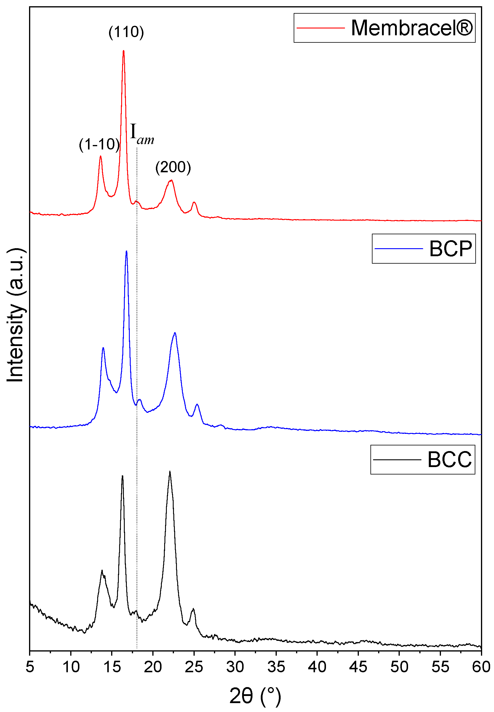
Using the XRD technique, it was possible to investigate the crystalline structure of BCs and the results can be observed in Figure 4, including the XRD patterns of BCC, BCP and Membracel®. The diffractograms of BCC, BCP and Membracel® showed predominant peaks characteristic of cellulose I, observed at 2θ values of 14.0° (plane 1-10), 16.8° (plane 110) and 22.4° (plane 200) [44]. BCC exhibits more intense peaks, particularly in the 200 plane, indicating a higher degree of crystallinity compared to BCP and Membracel®. This behavior is reinforced by the more evident presence of the amorphous region (Iam) for BCP and Membracel®, whose Iam peak intensity is more pronounced in the Membracel®, demonstrating less structural ordering.
The crystallinity index (CrI) values confirm the characteristics observed in the X-ray diffraction patterns. The BCC sample presented the highest CrI (78.74%), consistent with its intense and well-defined diffraction peaks, indicating a highly ordered structure. The BCP sample presented an intermediate CrI of 66.09%. Also, Membracel presented the lowest CrI (53.82%), due to the higher proportion of amorphous regions and less structural organization. Several studies have reported that the crystallinity of bacterial cellulose (BC) varies widely, ranging from 46.7% to 91.62%, with this variation largely influenced by the culture medium, especially the types of carbon and nitrogen sources used [45].
The greater crystallinity observed in the BCC sample may be directly related to the more controlled cultivation and purification conditions used in its production, compared to the process used to obtain BCP. This is because BCC was produced in a laboratory using a commercial strain and culture medium that were well-standardized and controlled. Furthermore, its purification process effectively removed the majority of impurities and amorphous regions, enhancing structural order, although minor elements from the fermentation medium remained detectable. This led to a higher CrI. BCP, obtained with sugarcane molasses, had a lower CrI compared to BCC, possibly due to the variable composition of the substrate, which can affect the organization of the cellulose structure. Membracel®, with the lowest CrI (53.82%), being a commercial product, reflects the effects of large-scale processing, which may involve less selective or more aggressive purification methods.
Therefore, the XRD profiles are not only indicative of crystalline ordering but also align with the fibril orientation seen in SEM and with the differences in tensile performance. For instance, the higher CrI of BCC corresponds to its more compact network and greater rigidity, while Membracel® with lower CrI presents a less ordered structure that translates into reduced mechanical strength. These structure–function correlations highlight the central role of crystallinity in dictating the physicochemical and functional properties of BC dressings.

3.3. Thermal Characterization by TGA/DTG

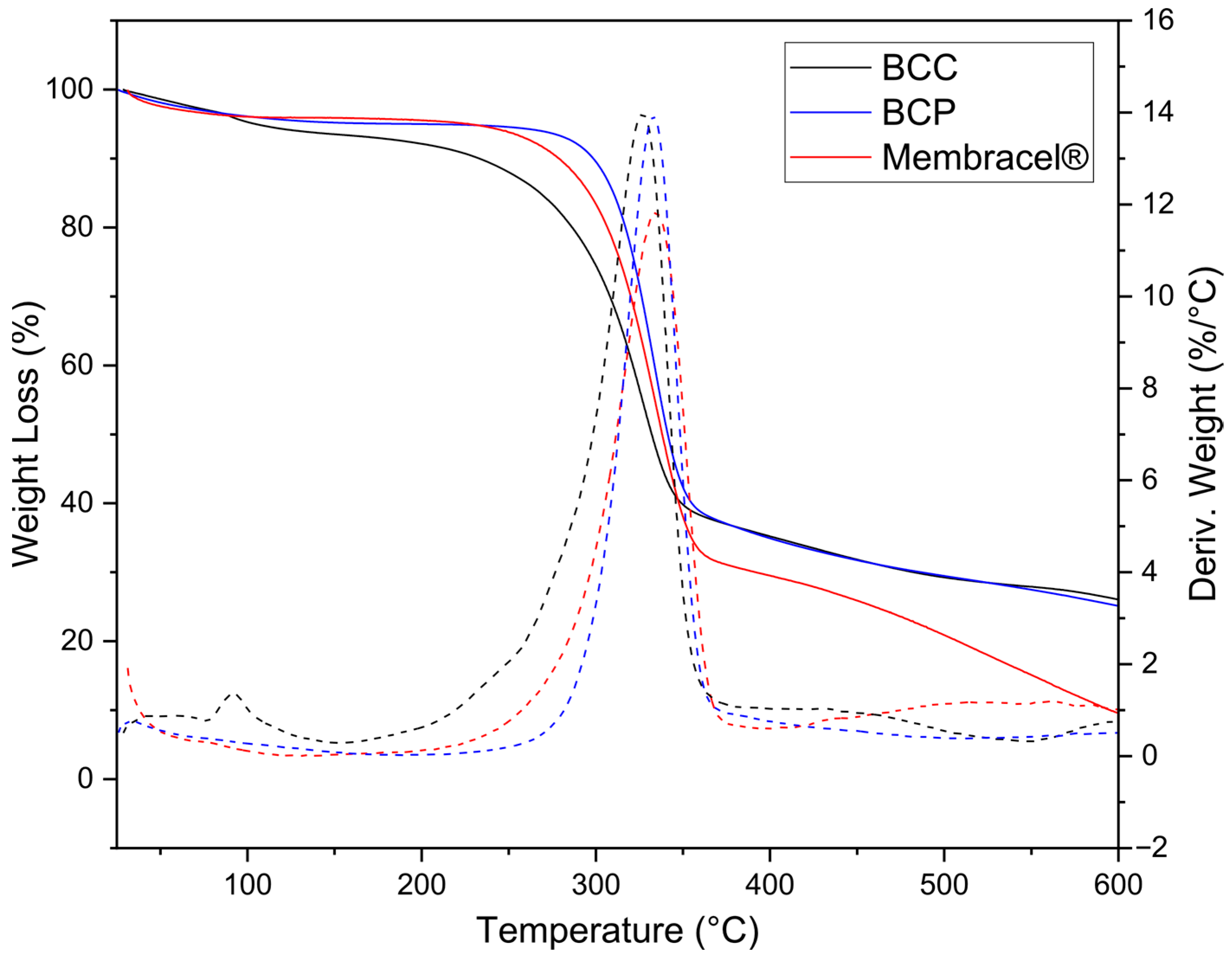
The thermal properties of the BCs were evaluated by TG and DTG analysis, with Figure 5 showing the curves for BCC, BCP and Membracel®. All samples showed two main mass loss events. The first event occurred at approximately 93 °C, associated with the evaporation of free water physically bound to the cellulose structure [46]. It is noted that the more pronounced loss was observed for BCC, indicating greater water retention. The second event occurs between 200 and 400 °C, corresponding to the thermal degradation of cellulose, involving the breaking of glycosidic bonds and the decomposition of the hydroxyl groups of the polymer chain [47]. At the end of the analysis, the total weight loss for BCP, Membracel® and BCC was 100%, 91.37% and 80.25%, respectively. BCC showed lower total weight loss; this behavior may be related to its higher CrI (78.74%), as determined by XRD. In general, although the relationship is not always direct or linear, materials with greater crystallinity tend to present greater thermal stability and less degradation, as the crystalline regions are more organized and less susceptible to thermal breakdown [48]. In addition to this relationship, the chemical composition, processing method, production process residues and industrial additions also affect thermal behavior. Therefore, while BCC’s lower mass loss is consistent with its higher crystallinity, this effect cannot be attributed exclusively to CrI. It more likely results from a synergistic contribution of ordered fibrillar packing, reduced amorphous impurities, and efficient purification, which together improve thermal resistance.

3.4. Solid-State 13C NMR Analysis

The solid-state 13C NMR analysis was used to examine the properties of crystalline BCs, as illustrated in the Figure 6 spectra for BCC, BCP and Membracel®. Resonance signals characteristic of crystalline cellulose, observed between 110 and 50 ppm, have been extensively reported in the literature [48,49]. The singlet peak at 106 ppm is attributed to the C1 carbon (C-O-C) in all samples, while two signals around 84 and 90 ppm correspond to the C4 carbon (CH-O), representing the crystalline regions. Signals observed between 71 and 76 ppm arise from the overlapping resonances of the C2, C3 and C5 carbons (CH-O) derived from the amorphous region. Finally, the region between 60 and 65 ppm is assigned to the C6 carbons (CH2–O). The line broadening observed for BCP and BCC indicates a behavior typical of more organized materials, whereas the line shape of Membracel® is less ordered compared to the other two samples, which is consistent with the X-ray diffraction data [49,50].

3.5. Water Interaction Properties

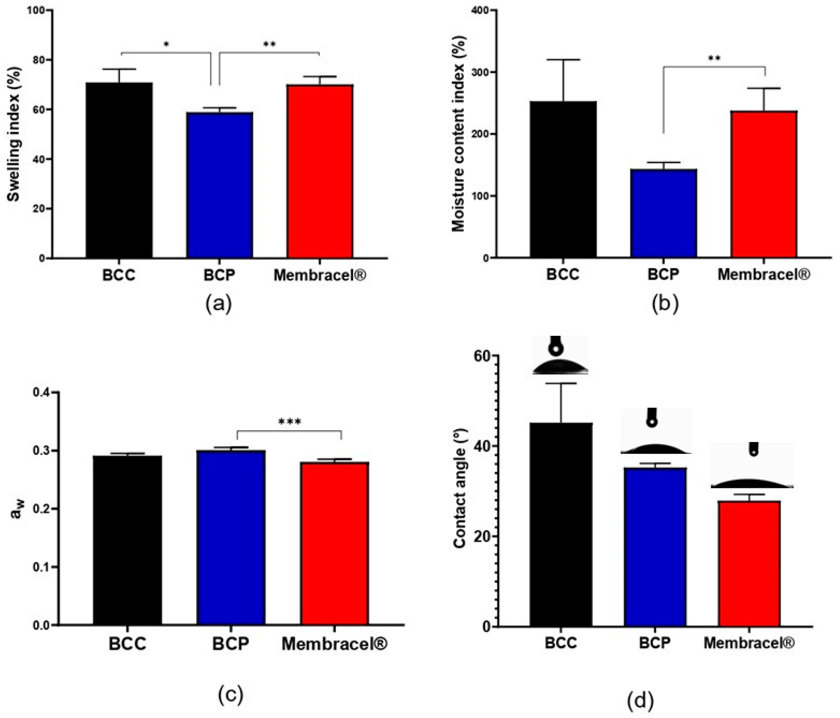
In this work, the water interaction metrics for the three BC wound dressings were also analyzed, as shown in Figure 7, which presents the hydration characteristics associated with their microstructural organization. BCP exhibited the lowest swelling index (58.9 ± 1.8%) and moisture content (143.5 ± 10.5%), significantly lower than both BCC (70.9 ± 5.4% swelling; p = 0.0366) and Membracel® (70.1 ± 3.1% swelling; p = 0.0023). Although Deng et al. [51] and Mo et al. [52] have reported BC-based dressings swelling by more than 3000%, those materials incorporated polyvinyl alcohol and silver-loaded zeolitic imidazolate frameworks, which substantially alter matrix porosity and hydration. By contrast, Teshima et al. [53] suggest that a swelling index below ~200% may help prevent temporary wound expansion, a threshold that was not exceeded by any of the samples evaluated.
Water activity (aw) further discriminates these dressings: Membracel® displayed the lowest aw (0.281 ± 0.004), significantly below BCP (0.301 ± 0.005; p = 0.0001), indicating that its retained water is more tightly bound and less available for microbial metabolism. BCC showed no significant difference to any of the other samples (0.292 ± 0.005; p > 0.05). As values above 0.85 typically support bacterial growth, the low aw in all samples (0.281–0.301) suggests inherently low susceptibility to infection—a critical advantage in wound management, where aw modulates both microbial control and local hydration [54,55,56].
Contact angle measurements (Figure 7d) mirrored these trends in surface topography and crystallinity. Membracel®—with the lowest crystallinity index (CrI = 53.8%)—exhibited the smallest contact angle (27.9°), reflecting maximal surface hydrophilicity. BCP, of intermediate CrI (66.1%), showed a moderate angle (35.2°), while BCC—most crystalline (CrI = 78.7%)—had the largest angle (45.1°), indicating lower wettability. This decoupling of surface wetting from bulk water retention (as seen in TGA) underscores that a highly organized fibrillar network can internalize large water volumes while resisting initial fluid spreading, whereas an amorphous-rich network promotes rapid surface hydration.
Furthermore, it is important to emphasize that parameters such as the swelling index and moisture content encapsulate a dressing’s capacity to sequester, retain and modulate fluid availability within the wound microenvironment. When considered together with physicochemical properties such as aw and the water vapor transmission rate (WVTR)—presented in the next section—these metrics allow prediction of clinical performance and potential risks (e.g., wound desiccation, infection, and dressing-change frequency) [57,58,59]. Appropriate moisture control also supports key biological processes that promote healing: it facilitates autolytic debridement, reduces pain and scar formation, stimulates collagen synthesis, promotes keratinocyte migration across the wound bed and preserves the activity and availability of nutrients, growth factors and other soluble mediators within the wound milieu [60,61,62,63].
Understanding each dressing’s hydrophilicity profile is essential for matching BC materials to wound exudate. Exudation varies by wound type—burns and venous leg ulcers being highly exudative, superficial abrasions less so—and excessive moisture can impede healing, particularly in chronic wounds [64]. Thus, BCC’s high swelling and moisture retention may suit heavily exuding wounds, while BCP and Membracel®, with their moderated swelling and superior microbial control, may be better directed to low- to moderate-exudate lesions. These structure–function insights provide a rational basis for selecting the optimal BC dressing tailored to specific wound environments.

3.6. Porosity and Water Vapor Permeability Characterization

The porosity and water vapor permeability of the BC samples were evaluated. Figure 8 presents the results, with Figure 8a showing porosity and Figure 8b displaying the water vapor permeability rate (WVPR) for the three BC dressings. All samples exhibited similar porosity—38.6 ± 3.2% for BCC, 35.1 ± 9.0% for BCP and 32.8 ± 6.5% for Membracel®—with no significant differences. Measured WVTR values ranged from 0.0327 ± 0.009 g·m−2·24 h−1 (BCC) to 0.797 ± 0.063 g·m−2·24 h−1 (BCP), with a statistically significant difference between BCC and BCP (p = 0.0102).
Despite exhibiting similar porosity (≈33–39%; p > 0.05), the three BC wound dressings diverge markedly in their vapor transmission and overall fluid-handling behavior. In general, an excessively high WVTR accelerates wound desiccation and crust formation, whereas an excessively low WVTR promotes exudate accumulation, delays healing and increases the risk of bacterial contamination [65]. Thus, WVTR may be considered an important property for performance. Zhang et al. [66] demonstrated that coating BC with silver nanoparticles and granulocyte-macrophage colony-stimulating factor (GM-CSF) progressively reduces the native WVTR of approximately 1.088 g·m−2·24 h−1 exhibited by unmodified BC, implying that the incorporation of exogenous components occupies intrafibrillar pores and increases network density. Conversely, Ciecholewska-Juśko et al. [67] showed that BC cross-linked with citric acid retains bound water for substantially longer durations than untreated BC, a phenomenon functionally equivalent to a reduction in water vapor transmission rate.
In this study we did not apply structural modifications to the BC; all analyses were conducted on unmodified, “native” BC samples. Nevertheless, statistically significant differences were observed among the samples, which appear to be related to their water interaction profiles—specifically swelling index and moisture content. BCC and Membracel® exhibited the highest swelling indices and moisture content, which likely contributed to their distinct vapor-handling behavior. These observations are consistent with previous reports, where a BC-based dressing showing greater swelling and moisture retention also presented higher vapor transmission characteristics, reinforcing the notion that the matrix–water interaction strongly influences WVTR [56].
From an application perspective, the differences measured may imply distinct clinical roles. BCC, which displayed the highest WVTR, will favor rapid moisture removal and may be best suited to moderately or heavily exuding wounds where prevention of fluid pooling is critical [60]. By contrast, BCP is more occlusive and therefore better matched to low-exudate or desiccated wounds in which moisture conservation is desirable. Membracel® occupies an intermediate position, providing a compromise between moisture retention and vapor transmission. These findings emphasize that selecting an appropriate BC dressing should be guided by the wound’s exudate profile and the dressing’s water interaction properties.

3.7. Optical Properties

The optical properties of the BC samples were assessed. Figure 9 provides an overview of these properties for the bacterial cellulose dressings, where Figure 9a illustrates opacity and Figure 9b presents transparency. BCC exhibited the highest opacity (32.1 Abs500 nm·mm−1), which was significantly greater than both BCP (16.8 Abs500 nm·mm−1) and Membracel® (11.5 Abs500 nm·mm−1; p < 0.001 for both). BCP was also significantly opaquer than Membracel® (p < 0.001). Conversely, Membracel® demonstrated the highest transparency (665 T600 nm%·mm−1), significantly exceeding that of BCC (130 T600 nm%·mm−1) and BCP (142 T600 nm%·mm−1; p < 0.001), while BCC and BCP did not differ from one another in transparency. Amorim et al. [68] have shown that BC films produced in a minimal black tea–sugar medium under oxygen-limited conditions attain markedly enhanced optical transparency without synthetic additives or post-formation treatments. Our data suggests that BCC can attenuate visible light, BCP exhibits intermediate optical behavior and the commercial Membracel® is essentially transparent.
The transparency of wound dressings represents an important feature for clinical practice, allowing noninvasive inspection of the wound bed during treatment, without the need for frequent dressing removal. This property allows clinicians to monitor key aspects of the healing process, such as wound color, exudate production and early signs of infection. Consequently, transparent dressings reduce the risk of infection by decreasing the frequency of dressing changes. Moreover, the ability to visually assess the wound in situ supports timely clinical decision-making, enabling early intervention in cases of complications such as infection or necrosis [69,70]. However, highly transparent wound dressings, such as Membracel®, have lower water absorbency, as was previously reported and confirmed in this study, which suggests that these materials may not be suitable for heavily exudative wounds.

3.8. Mechanical Properties

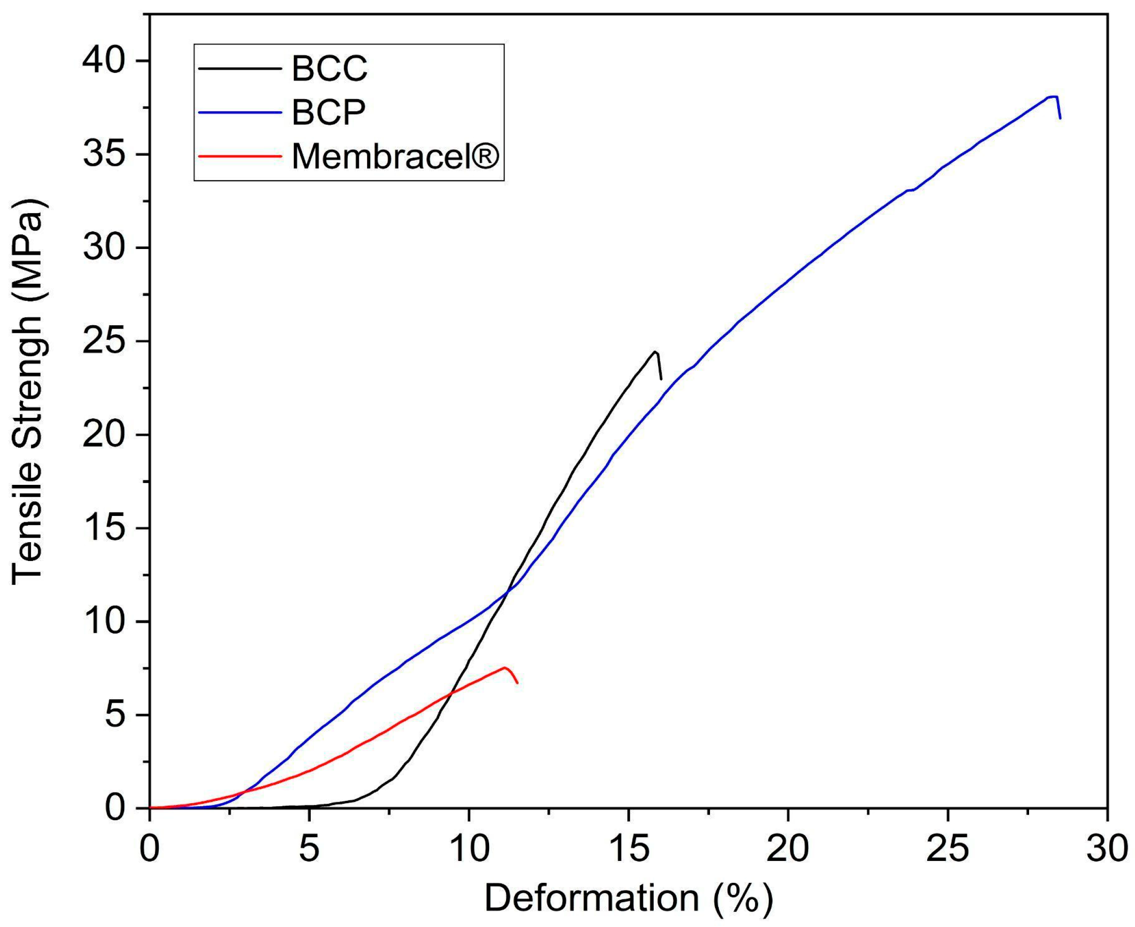
Mechanical analysis showed that the BCP film had the highest tensile strength and elongation at rupture (38.06 ± 7.96 MPa/28.42 ± 17.71%), followed by BCC (24.37 ± 2.49 MPa/Deformation (%) = 15.84 ± 4.83%), while Membracel® showed the lowest values (7.55 ± 3.48 MPa/11.16 ± 7.85%) (Figure 10). The obtained values of tensile strength and elongation of BC film agreed with other studies with this material [18]. These results suggest that the differences in mechanical performance among the samples are strongly influenced by the production methodologies employed, such as the must composition, inoculum quantity, treatment and drying method. The enhanced mechanical properties of the BCP film can be attributed to the culture conditions and film formation techniques, with the use of a concentrated hydrogel to obtain the BC film, which can promote increased nanofibril entanglement and structural integrity, augmenting both tensile strength and elasticity. In this way, Chen et al. [71] tested six different strains of Komagataeibacter genus for BC production and compared their mechanical strength. The authors demonstrated a significant difference between the mechanical properties of BC hydrogels, including tensile strength, stiffness, viscoelasticity, porosity and permeability. These properties were found to be dependent on both the concentration of cellulose and the structure of the fibril network.
In contrast, the BCC film, fabricated through casting and employing a mild-temperature drying process (50 °C), exhibited a reduction in tensile strength and elongation at break. This result can be explained by the hypothesis that the methodology applied to obtain the BC films, such as the cellulose nanofibers concentration used in this process, as well as the differences in fermentation parameters, are associated with variations in tensile strength and deformation properties. Such variations can impact the distribution and the package of nanofibers. A study by Dayal and Catchmark [72] indicated that variations in microbial strain, medium supplement and fermentation conditions significantly impact the degree of polymerization and crystallinity of bacterial cellulose, both of which are key factors in determining mechanical behavior. The authors showed that BC films with higher crystallinity exhibit enhanced tensile strength and modulus of elasticity, attributable to the more compact arrangement of nanofibrils, whereas the deformation capacity (elongation) exhibits a tendency to decrease as crystalline increases, resulting in a more rigid material.
Nevertheless, in this study, the BCC had the highest crystallinity among the other BCs and this result is corroborated by NRM and TGA analysis. However, its tensile strength and deformation were the lowest compared to the BCP. This seems to be a counterintuitive result. As discussed above, it is important to note that crystallinity is not the only parameter that affects the mechanical behavior of the membranes. Moreover, other properties, such as the arrangement of nanofibrils that differs between BCP and BCC due to differences in fermentation conditions, film production methodology and cellulose fibril concentration in the BC films’ hydrogel precursor, also impact the thickness of BC films. The BCP films were thicker (0.085 ± 0.006 mm) than BCC (0.043 ± 0.007 mm) and Membracel® (0.049 ± 0.012 mm), which may explain BCP’s higher tensile strength. Although the water activities of BCP and BCC are similar, BCC exhibits higher swelling and moisture retention levels than BCP, which can also influence the lower tensile strength observed in BCC, since a dressing with greater water retention capacity tends to have lower tensile strength due to the greater molecular mobility.
Conversely, the commercial Membracel® film exhibited the lowest tensile strength and deformation values. This result is consistent with the SEM micrographs of BC films, which showed that, compared to the other BCs, Membracel® is less fibrous and rough. This outcome can be attributed to differences in the industrial production process of BC dressings, which often involve chemical or physical modifications, such as plasticization, blending, perforation of the film and surface treatments. These modifications improve flexibility and facilitate handling, but they may compromise mechanical strength. Additionally, variations in drying techniques, including hot air drying, vacuum drying and vacuum freezing drying, have been shown to affect the pore structure, the strength of hydrogen bonds between cellulose macromolecules and the degree of crystallinity of BC films, thereby influencing the overall tensile properties of the final product [73].
Furthermore, it is imperative to emphasize that the mechanical properties of BC films are highly tunable. As highlighted by Wang et al. [74], several strategies can be employed to improve the mechanical properties of BC, including microscale fibril orientation, chemical modifications and the incorporation of polymers or nanoparticles. These approaches aim to alter the BC microstructure by promoting stronger fibril-to-fibril interactions and enhancing the alignment and organization of the nanofibrils, thereby increasing the material’s structural integrity and mechanical performance.

3.9. In Vitro Stability and Fluid Uptake in Simulated Wound Fluid

The in vitro stability and fluid absorption characteristics of the three bacterial cellulose wound dressings were evaluated over a 14-day immersion period in simulated wound fluid. The corresponding results are presented in Figure 11. Notably, none of the samples exhibited net mass loss indicative of polymer breakdown; instead, all materials absorbed fluid and gained mass, underscoring their structural stability in a proteolytic environment. BCC showed a gradual, modest swelling, with mean mass changes of −2.6% by day 4, −8.7% by day 7, −10.5% by day 10 and −8.4% by day 14. BCP swelled more rapidly, reaching −9.8% at day 4 and −13.7% at day 7, before partially contracting to −8.2% on day 10 and −11.3% on day 14. Membracel® followed a similar overall trajectory but displayed a pronounced mid-term uptake, with −8.6% at day 4, −10.7% at day 7, peaking at −26.5% on day 10 and then returning toward −10.3% by day 14.
These findings align with the literature, which consistently reports that unmodified bacterial cellulose is resistant to enzymatic degradation in wound environments—owing to the absence of endogenous human cellulases—and therefore remains dimensionally stable over time [75,76]. In studies such as that of Atlia et al. [31], similarly unmodified cellulose dressings showed negligible mass loss in simulated wound fluid, further reinforcing the inherent stability of our BC samples under wound conditions.

4. Conclusions

This comparative study demonstrated that bacterial cellulose (BC) wound dressings, including the commercial Membracel®, non-commercial BCP and the experimental BCC, exhibit distinct structural and functional profiles directly linked to their production methods. BCC, characterized by the highest crystallinity index and superior thermal stability, coupled with its high water vapor permeability and moisture retention capacity, appears particularly well suited for managing heavily exuding wounds, where efficient moisture regulation is critical to promote healing. In contrast, BCP showed superior mechanical properties, with the highest tensile strength and elongation, indicating greater robustness and flexibility, important attributes for dressings intended for wounds requiring enhanced durability and adaptability to movement. Membracel® presented lower crystallinity and water vapor permeability, potentially favoring its application in low-exudate wounds, where maintaining a moist yet controlled environment is essential.
Furthermore, in vitro stability and fluid uptake assays in simulated wound fluid over 14 days revealed no significant polymer breakdown in any of the BC dressings, confirming their structural stability under proteolytic conditions typical of chronic wounds. All materials absorbed fluid and gained mass, underscoring their dimensional stability during prolonged use. Swelling behavior varied among samples: BCC showed a gradual and moderate mass increase, BCP demonstrated rapid swelling followed by partial contraction and Membracel® exhibited pronounced mid-term fluid uptake before stabilization. These findings are consistent with literature reports that unmodified bacterial cellulose is inherently resistant to enzymatic degradation due to the lack of endogenous human cellulases.
Elemental analysis confirmed the high purity of BCP and Membracel®, while minor elements in BCC reflect its fermentation origin, supporting the structural integrity underlying their distinct functional profiles. Together, these results elucidate clear structure–function relationships that provide a scientific basis for the targeted selection of BC wound dressings according to wound type and moisture management requirements. This enhanced understanding supports the optimized clinical application of bacterial cellulose dressings, maximizing their therapeutic efficacy across diverse wound care scenarios.

Author Contributions

Conceptualization, S.B.P., S.O.P., K.V.S.H., W.T.B. and G.A.R.; methodology, A.L.P.C.G., G.A.R. and B.A.S.M.; validation, S.B.P., S.O.P., K.V.S.H. and W.T.B.; formal analysis, J.D.V.B., M.I.B.T., L.C.M. and J.L.d.A.A.; investigation, S.B.P., S.O.P., K.V.S.H., W.T.B., A.L.P.C.G., G.A.R., B.A.S.M. and M.I.B.T.; resources, J.D.V.B., L.C.M., J.L.d.A.A.; data curation, S.B.P., S.O.P., K.V.S.H. and W.T.B.; writing—original draft preparation, S.B.P., S.O.P., K.V.S.H. and W.T.B.; writing—review and editing, J.D.V.B., M.I.B.T., L.C.M. and J.L.d.A.A.; supervision, J.D.V.B., J.L.d.A.A.; project administration, S.B.P., J.D.V.B. and J.L.d.A.A.; funding acquisition, J.D.V.B., L.C.M., J.L.d.A.A. All authors have read and agreed to the published version of the manuscript.

Funding

This study was partially funded by the National Council for Scientific and Technological Development (CNPq): J.L.d.A.A. [CNPq/MCTI 424123/2021-5] and the Pernambuco State Science and Technology Support Foundation (FACEPE): J.L.d.A.A. [SIN-0129-4.01/25] and L.C.M. [BFP-0093-4.01/24].

Data Availability Statement

Data are contained within the article and available upon request.

Acknowledgments

The authors are thankful to SENAI-CIMATEC, POLISA® Biopolymers for Health Ltd., Federal University of Pernambuco (UFPE), CNPq and FACEPE.

Conflicts of Interest

The authors declare no conflicts of interest.

Abbreviations

BCBacterial cellulose
BCPBacterial cellulose from POLISA®
BCCBacterial cellulose from SENAI CIMATEC
SEMScanning electron microscopy
XRDX-ray Diffraction
FTIRFourier Transformed Infrared Spectroscopy
TGAThermogravimetric analysis
DTGDerivative Thermogravimetry
NMRNuclear Magnetic Resonance
SWFSimulated wound fluid
ANOVAAnalysis of variance
CrICrystallinity index
WVTRWater vapor transmission rate

References

  1. Falanga, V.; Isseroff, R.R.; Soulika, A.M.; Romanelli, M.; Margolis, D.; Kapp, S.; Granick, M.; Harding, K. Chronic Wounds. Nat. Rev. Dis. Primers 2022, 8, 50. [Google Scholar] [CrossRef]
  2. Sen, C.K. Human Wound and Its Burden: Updated 2022 Compendium of Estimates. Adv. Wound Care 2023, 12, 657–670. [Google Scholar] [CrossRef]
  3. Díaz-Herrera, M.Á.; González-Durán, M.; Rodríguez-Martínez, F.J.; Tujillo-Flores, G.; Tuset-Mateu, N.; Verdú-Soriano, J.; Gea-Caballero, V.; Sanllorente-Melenchón, A.; Almeda-Ortega, J.; Cunillera-Puértolas, O.; et al. The Financial Burden of Chronic Wounds in Primary Care: A Real-World Data Analysis on Cost and Prevalence. Int. J. Nurs. Stud. Adv. 2025, 8, 100313. [Google Scholar] [CrossRef]
  4. Sharma, A.; Shankar, R.; Yadav, A.K.; Pratap, A.; Ansari, M.A.; Srivastava, V. Burden of Chronic Nonhealing Wounds: An Overview of the Worldwide Humanistic and Economic Burden to the Healthcare System. Int. J. Low Extrem. Wounds 2024, 15347346241246339. [Google Scholar] [CrossRef]
  5. Galvão Duarte, J.; Piedade, A.P.; Sarmento, B.; Mascarenhas-Melo, F. The Printed Path to Healing: Advancing Wound Dressings through Additive Manufacturing. Adv. Healthc. Mater. 2025, 14, 2402711. [Google Scholar] [CrossRef]
  6. Hodge, J.G.; Zamierowski, D.S.; Robinson, J.L.; Mellott, A.J. Evaluating Polymeric Biomaterials to Improve next Generation Wound Dressing Design. Biomater. Res. 2022, 26, 50. [Google Scholar] [CrossRef]
  7. Monami, M.; Ragghianti, B.; Scatena, A.; Miranda, C.; Monge, L.; Uccioli, L.; Stefanon, L.; Cappella, C.; Silverii, A.; Vermigli, C. Effectiveness of Different Advanced Wound Dressings versus Standard of Care for the Management of Diabetic Foot Ulcers: A Meta-Analysis of Randomized Controlled Trials for the Development of the Italian Guidelines for the Treatment of Diabetic Foot Syndrome. Acta Diabetol. 2024, 61, 1517–1526. [Google Scholar] [CrossRef]
  8. Pang, Q.; Yang, F.; Jiang, Z.; Wu, K.; Hou, R.; Zhu, Y. Smart Wound Dressing for Advanced Wound Management: Real-Time Monitoring and on-Demand Treatment. Mater. Des. 2023, 229, 111917. [Google Scholar] [CrossRef]
  9. Horue, M.; Silva, J.M.; Berti, I.R.; Brandão, L.R.; Barud, H.d.S.; Castro, G.R. Bacterial Cellulose-Based Materials as Dressings for Wound Healing. Pharmaceutics 2023, 15, 424. [Google Scholar] [CrossRef]
  10. Utoiu, E.; Manoiu, V.S.; Oprita, E.I.; Craciunescu, O. Bacterial Cellulose: A Sustainable Source for Hydrogels and 3D-Printed Scaffolds for Tissue Engineering. Gels 2024, 10, 387. [Google Scholar] [CrossRef]
  11. Rumon, M.M.H. Advances in Cellulose-Based Hydrogels: Tunable Swelling Dynamics and Their Versatile Real-Time Applications. RSC Adv. 2025, 15, 11688–11729. [Google Scholar] [CrossRef]
  12. Hou, S.; Xia, Z.; Pan, J.; Wang, N.; Gao, H.; Ren, J.; Xia, X. Bacterial Cellulose Applied in Wound Dressing Materials: Production and Functional Modification—A Review. Macromol. Biosci. 2024, 24, 2300333. [Google Scholar] [CrossRef]
  13. Aditya, T.; Allain, J.P.; Jaramillo, C.; Restrepo, A.M. Surface Modification of Bacterial Cellulose for Biomedical Applications. Int. J. Mol. Sci. 2022, 23, 610. [Google Scholar] [CrossRef]
  14. Castilhos, C.; Rubira, L.T.; Nogeuira, M.L.S.; Santos, T.S. Estudo de Caso: Tratamento de Amputação de Antepé Com Membrana Regeneradora Membracel. Feridas 2020, 8, 1652–1657. [Google Scholar] [CrossRef]
  15. Barbosa Da Silva, S.; Salani, R.; De Cássia Ferreira, R.; Gazote Eloy Geraldo, Y.; Pavani, C.; Setúbal Destro Rodrigues, M.F.; Motta, L.J.; Fátima Teixeira Silva, D. Can Photons Pass through Primary Coatings Used to Treat Cutaneous Wounds? Adv. Ski. Wound Care 2021, 34, 97–102. [Google Scholar] [CrossRef] [PubMed]
  16. Coelho, G.A.; Magalhães, M.A.B.; Matioski, A.; Ribas-Filho, J.M.; Magalhães, W.L.E.; Claro, F.C.; Ramos, R.K.; de Camargo, T.M.S.; Malafaia, O. Pine Nanocellulose and Bacterial Nanocellulose Dressings are Similar in the Treatment of Second-Degree Burn? Experimental Study in Rats. Arq. Bras. Cir. Dig. 2020, 33, e1533. [Google Scholar] [CrossRef]
  17. Nunes, C.A.d.B.; Melo, P.G.; Malaquias, S.G.; Amaral, K.V.Á.; Alves, G.R.; Meira, A.A.; Cardoso, A.L.; Pereira, L.V.; Bachion, M.M. Effectiveness of Two Bundles in Venous Leg Ulcer Healing: A Randomized Controlled Trial. J. Vasc. Nurs. 2019, 37, 232–245. [Google Scholar] [CrossRef]
  18. de Carvalho, R.S.F.; Mahnke, L.C.; Palácio, S.B.; Barbosa, W.T.; Hodel, K.V.S.; Barbosa, J.D.V.; Melo, F.d.A.D.; Chorilli, M.; Meneguin, A.B.; Pinto, F.C.M.; et al. Bacterial Cellulose Hydrogel Produced by Gluconacetobacter Hansenii Using Sugarcane Molasses as Medium: Physicochemical Characterization for Wound Healing Applications. Carbohydr. Polym. Technol. Appl. 2025, 9, 100632. [Google Scholar] [CrossRef]
  19. Hodel, K.V.S.; Fonseca, L.M.d.S.; Santos, I.M.d.S.; Cerqueira, J.C.; Santos-Júnior, R.E.d.; Nunes, S.B.; Barbosa, J.D.V.; Machado, B.A.S. Evaluation of Different Methods for Cultivating Gluconacetobacter Hansenii for Bacterial Cellulose and Montmorillonite Biocomposite Production: Wound-Dressing Applications. Polymers 2020, 12, 267. [Google Scholar] [CrossRef] [PubMed]
  20. Cacicedo, M.L.; Pacheco, G.; Islan, G.A.; Alvarez, V.A.; Barud, H.S.; Castro, G.R. Chitosan-Bacterial Cellulose Patch of Ciprofloxacin for Wound Dressing: Preparation and Characterization Studies. Int. J. Biol. Macromol. 2020, 147, 1136–1145. [Google Scholar] [CrossRef]
  21. Maria, L.C.S.; Santos, A.L.C.; Oliveira, P.C.; Valle, A.S.S.; Barud, H.S.; Messaddeq, Y.; Ribeiro, S.J.L. Preparation and Antibacterial Activity of Silver Nanoparticles Impregnated in Bacterial Cellulose. Polímeros 2010, 20, 72–77. [Google Scholar] [CrossRef]
  22. Kaczmarek, M.; Jędrzejczak-Krzepkowska, M.; Ludwicka, K. Comparative Analysis of Bacterial Cellulose Membranes Synthesized by Chosen Komagataeibacter Strains and Their Application Potential. Int. J. Mol. Sci. 2022, 23, 3391. [Google Scholar] [CrossRef]
  23. Swingler, S.; Gupta, A.; Gibson, H.; Kowalczuk, M.; Heaselgrave, W.; Radecka, I. Recent Advances and Applications of Bacterial Cellulose in Biomedicine. Polymers 2021, 13, 412. [Google Scholar] [CrossRef]
  24. Segal, L.; Creely, J.J.; Martin, A.E., Jr.; Conrad, C.M. An Empirical Method for Estimating the Degree of Crystallinity of Native Cellulose Using the X-Ray Diffractometer. Text. Res. J. 1959, 29, 786–794. [Google Scholar] [CrossRef]
  25. Du, R.; Zhao, F.; Peng, Q.; Zhou, Z.; Han, Y. Production and Characterization of Bacterial Cellulose Produced by Gluconacetobacter Xylinus Isolated from Chinese Persimmon Vinegar. Carbohydr. Polym. 2018, 194, 200–207. [Google Scholar] [CrossRef] [PubMed]
  26. Leal, I.L.; Rosa, Y.C.d.S.; Penha, J.d.S.; Correia, P.R.C.; Melo, P.d.S.; Guimarães, D.H.; Barbosa, J.D.V.; Druzian, J.I.; Machado, B.A.S. Development and Application Starch Films:PBAT with Additives for Evaluating the Shelf Life of Tommy Atkins Mango in the Fresh-Cut State. J. Appl. Polym. Sci. 2019, 136, 48150–48169. [Google Scholar] [CrossRef]
  27. Ao, H.; Jiang, W.; Nie, Y.; Zhou, C.; Zong, J.; Liu, M.; Liu, X.; Wan, Y. Engineering Quaternized Chitosan in the 3D Bacterial Cellulose Structure for Antibacterial Wound Dressings. Polym. Test. 2020, 86, 106490. [Google Scholar] [CrossRef]
  28. ASTM E96-00; Standard Test Methods for Water Vapor Transmission of Materials. American Society for Testing and Materials (ASTM): West Conshohocken, PA, USA, 2000. [CrossRef]
  29. Almeida, D.M.; Prestes, R.A.; Pinheiro, L.A.; Woicechowski, A.L.; Wosiacki, G. Propriedades Físicas, Químicas e de Barreira Em Filme Formados Por Blenda de Celulose Bacteriana e Fécula de Batata. Polímeros Ciência Tecnol. 2013, 23, 538–546. [Google Scholar] [CrossRef]
  30. Cazón, P.; Vázquez, M.; Velazquez, G. Environmentally Friendly Films Combining Bacterial Cellulose, Chitosan, and Polyvinyl Alcohol: Effect of Water Activity on Barrier, Mechanical, and Optical Properties. Biomacromolecules 2020, 21, 753–760. [Google Scholar] [CrossRef]
  31. Atila, D.; Karataş, A.; Keskin, D.; Tezcaner, A. Pullulan Hydrogel-Immobilized Bacterial Cellulose Membranes with Dual-Release of Vitamin C and E for Wound Dressing Applications. Int. J. Biol. Macromol. 2022, 218, 760–774. [Google Scholar] [CrossRef]
  32. Hasanin, M.S.; Abdelraof, M.; Hashem, A.H.; El Saied, H. Sustainable Bacterial Cellulose Production by Achromobacter Using Mango Peel Waste. Microb. Cell Factories 2023, 22, 24. [Google Scholar] [CrossRef]
  33. Santos, S.M.; Carbajo, J.M.; Gómez, N.; Quintana, E.; Ladero, M.; Sánchez, A.; Chinga-Carrasco, G.; Villar, J.C. Use of Bacterial Cellulose in Degraded Paper Restoration. Part II: Application on Real Samples. J. Mater. Sci. 2016, 51, 1553–1561. [Google Scholar] [CrossRef]
  34. Das, M.; Zandraa, O.; Mudenur, C.; Saha, N.; Sáha, P.; Mandal, B.; Katiyar, V. Composite Scaffolds Based on Bacterial Cellulose for Wound Dressing Application. ACS Appl. Bio Mater. 2022, 5, 3722–3733. [Google Scholar] [CrossRef]
  35. Volova, T.G.; Prudnikova, S.V.; Kiselev, E.G.; Nemtsev, I.V.; Vasiliev, A.D.; Kuzmin, A.P.; Shishatskaya, E.I. Bacterial Cellulose (BC) and BC Composites: Production and Properties. Nanomaterials 2022, 12, 192. [Google Scholar] [CrossRef]
  36. Wang, X.; Guo, C.; Hao, W.; Ullah, N.; Chen, L.; Li, Z.; Feng, X. Development and Characterization of Agar-Based Edible Films Reinforced with Nano-Bacterial Cellulose. Int. J. Biol. Macromol. 2018, 118, 722–730. [Google Scholar] [CrossRef]
  37. Ahmed, E.F.; Shawkat Ali, W.; Hasan Heider, N. Description and determination of the nanocellulose components produced from acetic acid bacteria. Rev. Bionatura 2023, 8, 112. [Google Scholar] [CrossRef]
  38. Dawwan, G.E.; El-Sayed, N.S.; Al-Shemy, M.T. Bacterial cellulose doped with ZnO as a multifunctional bioactive plataform for Curcumin and propolis Immobilization: Synthesis, characterization, and wound healing potential. Microb Cell Fact. 2025, 24, 196. [Google Scholar] [CrossRef] [PubMed]
  39. Don, T.-M.; Lee, K.-T.; Chen, B.-Y.; Tang, S.; Huang, Y.-C.; Chuang, A.E.-Y. Physicochemical Properties of Bacterial Cellulose/Phototherapeutic Polypyrrole/Antibacterial Chitosan Composite Membranes and Their Evaluation as Chronic Wound Dressings. Int. J. Biol. Macromol. 2025, 308, 142183. [Google Scholar] [CrossRef]
  40. Fatima, A.; Yasir, S.; Ul-Islam, M.; Kamal, T.; Ahmad, M.d.W.; Abbas, Y.; Manan, S.; Ullah, M.W.; Yang, G. Ex Situ Development and Characterization of Green Antibacterial Bacterial Cellulose-Based Composites for Potential Biomedical Applications. Adv. Compos. Hybrid Mater. 2022, 5, 307–321. [Google Scholar] [CrossRef]
  41. Pasaribu, K.M.; Ilyas, S.; Tamrin, T.; Radecka, I.; Swingler, S.; Gupta, A.; Stamboulis, A.G.; Gea, S. Bioactive Bacterial Cellulose Wound Dressings for Burns with Collagen In-Situ and Chitosan Ex-Situ Impregnation. Int. J. Biol. Macromol. 2023, 230, 123118. [Google Scholar] [CrossRef]
  42. Zulkarnain, N.N.; Abd Rahman, N.; Othman, A.R.; Md Saleh, N.; Mohd Ali, J.; Shukor, H.; Mohd Said, M.; Wan Zaki, W.R. Characterization and Antibacterial Activity of Bacterial Cellulose Impregnated with Moringa Oleifera Leaf Extract and Silver Nanoparticles. Cellulose 2024, 31, 5213–5227. [Google Scholar] [CrossRef]
  43. Mensah, A.; Chen, Y.; Christopher, N.; Wei, Q. Membrane Technological Pathways and Inherent Structure of Bacterial Cellulose Composites for Drug Delivery. Bioengineering 2022, 9, 3. [Google Scholar] [CrossRef]
  44. Claro, A.M.; Dias, I.K.R.; Fontes, M.d.L.; Colturato, V.M.M.; Lima, L.R.; Sávio, L.B.; Berto, G.L.; Arantes, V.; Barud, H.d.S. Bacterial Cellulose Nanocrystals Obtained through Enzymatic and Acidic Routes: A Comparative Study of Their Main Properties and in Vitro Biological Responses. Carbohydr. Res. 2024, 539, 109104. [Google Scholar] [CrossRef]
  45. Uğurel, C.; Öğüt, H. Optimization of Bacterial Cellulose Production by Komagataeibacter Rhaeticus K23. Fibers 2024, 12, 29. [Google Scholar] [CrossRef]
  46. Mamouri, K.S.; Rahaiee, S.; Zare, M.; Kenari, M.N.; Mirzakhani, N. Physicochemical and Thermal Characterization, and Evaluation of a Bacterial Cellulose/Barhang Gum-Based Dressing for Wound Healing. Int. J. Biol. Macromol. 2023, 242, 124660. [Google Scholar] [CrossRef] [PubMed]
  47. Suneetha, M.; Won, S.-Y.; Zo, S.M.; Han, S.S. Fungal Carboxymethyl Chitosan-Impregnated Bacterial Cellulose Hydrogel as Wound-Dressing Agent. Gels 2023, 9, 184. [Google Scholar] [CrossRef]
  48. Pasaribu, K.M.; Gea, S.; Ilyas, S.; Tamrin, T.; Radecka, I. Characterization of Bacterial Cellulose-Based Wound Dressing in Different Order Impregnation of Chitosan and Collagen. Biomolecules 2020, 10, 1511. [Google Scholar] [CrossRef] [PubMed]
  49. Inoue, B.S.; Streit, S.; dos Santos Schneider, A.L.; Meier, M.M. Bioactive Bacterial Cellulose Membrane with Prolonged Release of Chlorhexidine for Dental Medical Application. Int. J. Biol. Macromol. 2020, 148, 1098–1108. [Google Scholar] [CrossRef]
  50. Mohite, B.V.; Patil, S.V. Physical, Structural, Mechanical and Thermal Characterization of Bacterial Cellulose by G. Hansenii NCIM 2529. Carbohydr. Polym. 2014, 106, 132–141. [Google Scholar] [CrossRef]
  51. Deng, L.; Huang, Y.; Chen, S.; Han, Z.; Han, Z.; Jin, M.; Qu, X.; Wang, B.; Wang, H.; Gu, S. Bacterial Cellulose-Based Hydrogel with Antibacterial Activity and Vascularization for Wound Healing. Carbohydr. Polym. 2023, 308, 120647. [Google Scholar] [CrossRef] [PubMed]
  52. Mo, M.; Wu, C.; Chen, Y. Bacterial Cellulose-Based Superabsorbent Hydrogel for Wet Wound Dressing. Molecules 2025, 30, 737. [Google Scholar] [CrossRef]
  53. Teshima, R.; Osawa, S.; Yoshikawa, M.; Kawano, Y.; Otsuka, H.; Hanawa, T. Low-Adhesion and Low-Swelling Hydrogel Based on Alginate and Carbonated Water to Prevent Temporary Dilation of Wound Sites. Int. J. Biol. Macromol. 2024, 254, 127928. [Google Scholar] [CrossRef]
  54. Su, J.; Li, J.; Liang, J.; Zhang, K.; Li, J. Hydrogel Preparation Methods and Biomaterials for Wound Dressing. Life 2021, 11, 1016. [Google Scholar] [CrossRef]
  55. Wong, C.C.Q.; Tomura, K.; Yamamoto, O. Characterization of Sucrose-Impregnated Crystalline Glucose/Mannose Films as Moisturizing Wound Dressings and Their Significant Healing Effect on Deep Wounds in a Rat Model. Bioengineering 2025, 12, 327. [Google Scholar] [CrossRef]
  56. Hodel, K.V.S.; Machado, B.A.S.; Sacramento, G.d.C.; Maciel, C.A.d.O.; Oliveira-Junior, G.S.; Matos, B.N.; Gelfuso, G.M.; Nunes, S.B.; Barbosa, J.D.V.; Godoy, A.L.P.C. Active Potential of Bacterial Cellulose-Based Wound Dressing: Analysis of Its Potential for Dermal Lesion Treatment. Pharmaceutics 2022, 14, 1222. [Google Scholar] [CrossRef]
  57. Milne, S.D.; Seoudi, I.; Al Hamad, H.; Talal, T.K.; Anoop, A.A.; Allahverdi, N.; Zakaria, Z.; Menzies, R.; Connolly, P. A Wearable Wound Moisture Sensor as an Indicator for Wound Dressing Change: An Observational Study of Wound Moisture and Status. Int. Wound J. 2015, 13, 1309. [Google Scholar] [CrossRef] [PubMed]
  58. Cazón, P.; Velázquez, G.; Vázquez, M. Bacterial Cellulose Films: Evaluation of the Water Interaction. Food Packag. Shelf Life 2020, 25, 100526. [Google Scholar] [CrossRef]
  59. Norahan, M.H.; Pedroza-González, S.C.; Sánchez-Salazar, M.G.; Álvarez, M.M.; Trujillo de Santiago, G. Structural and Biological Engineering of 3D Hydrogels for Wound Healing. Bioact. Mater. 2023, 24, 197–235. [Google Scholar] [CrossRef] [PubMed]
  60. Nuutila, K.; Eriksson, E. Moist Wound Healing with Commonly Available Dressings. Adv. Wound Care 2021, 10, 685. [Google Scholar] [CrossRef]
  61. Schmitz, M.; Eberlein, T.; Andriessen, A. Wound Treatment Costs Comparing a Bio-Cellulose Dressing with Moist Wound Healing Dressings and Conventional Dressings. Wound Med. 2014, 6, 11–14. [Google Scholar] [CrossRef]
  62. Rauscher, M.; Rauscher, A.; Hu, L.Y.; Schlitt, H.J.; Krauß, S.; Illg, C.; Wolfertstetter, P.R.; Hofmann, A.; Knorr, C.; Denzinger, M. Influence of Accumulation of Humidity under Wound Dressings and Effects on Transepidermal Water Loss (TEWL) and Skin Hydration. Appl. Sci. 2024, 14, 7739. [Google Scholar] [CrossRef]
  63. Yue, M.; Lei, M.; Liu, Y.; Gui, N. The Application of Moist Dressings in Wound Care for Tracheostomy Patients: A Meta-analysis. J. Clin. Nurs. 2019, 28, 2724–2731. [Google Scholar] [CrossRef] [PubMed]
  64. Feng, F.; Zhao, Z.; Li, J.; Huang, Y.; Chen, W. Multifunctional Dressings for Wound Exudate Management. Prog Mater Sci 2024, 146, 101328. [Google Scholar] [CrossRef]
  65. Portela, R.; Leal, C.R.; Almeida, P.L.; Sobral, R.G. Bacterial Cellulose: A Versatile Biopolymer for Wound Dressing Applications. Microb. Biotechnol. 2019, 12, 586–610. [Google Scholar] [CrossRef]
  66. Zhang, B.; Huang, Z.; Guo, T.; Jing, J.; Dang, Y. Accelerating Infectious Wound Healing through Bacterial Cellulose/Ag Composite Film Enriched with GM-CSF. Sci. Rep. 2025, 15, 22142. [Google Scholar] [CrossRef]
  67. Ciecholewska-Juśko, D.; Żywicka, A.; Junka, A.; Drozd, R.; Sobolewski, P.; Migdał, P.; Kowalska, U.; Toporkiewicz, M.; Fijałkowski, K. Superabsorbent Crosslinked Bacterial Cellulose Biomaterials for Chronic Wound Dressings. Carbohydr. Polym. 2021, 253, 117247. [Google Scholar] [CrossRef]
  68. de Amorim, J.D.P.; Cavalcanti, Y.d.F.; de Medeiros, A.D.M.; Silva Junior, C.J.G.d.; Durval, I.J.B.; Costa, A.F.d.S.; Sarubbo, L.A. Synthesis of Transparent Bacterial Cellulose Films as a Platform for Targeted Drug Delivery in Wound Care. Processes 2024, 12, 1282. [Google Scholar] [CrossRef]
  69. Kuddushi, M.; Shah, A.A.; Ayranci, C.; Zhang, X. Recent Advances in Novel Materials and Techniques for Developing Transparent Wound Dressings. J. Mater. Chem. B 2023, 11, 6201–6224. [Google Scholar] [CrossRef] [PubMed]
  70. Tan, S.T.; Winarto, N.; Dosan, R.; Aisyah, P.B. The Benefits Of Occlusive Dressings In Wound Healing. Open Dermatol. J. 2019, 13, 27–33. [Google Scholar] [CrossRef]
  71. Chen, S.Q.; Lopez-Sanchez, P.; Wang, D.; Mikkelsen, D.; Gidley, M.J. Mechanical Properties of Bacterial Cellulose Synthesised by Diverse Strains of the Genus Komagataeibacter. Food Hydrocoll. 2018, 81, 87–95. [Google Scholar] [CrossRef]
  72. Dayal, M.S.; Catchmark, J.M. Mechanical and Structural Property Analysis of Bacterial Cellulose Composites. Carbohydr. Polym. 2016, 144, 447–453. [Google Scholar] [CrossRef] [PubMed]
  73. Zhang, C.J.; Wang, L.; Zhao, J.C.; Zhu, P. Effect of Drying Methods on Structure and Mechanical Properties of Bacterial Cellulose Films. Adv. Mater. Res. 2011, 239, 2667–2670. [Google Scholar] [CrossRef]
  74. Wang, Y.Y.; Zhao, X.Q.; Li, D.M.; Wu, Y.M.; Wahid, F.; Xie, Y.Y.; Zhong, C. Review on the Strategies for Enhancing Mechanical Properties of Bacterial Cellulose. J. Mater. Sci. 2023, 58, 15265–15293. [Google Scholar] [CrossRef]
  75. Torgbo, S.; Sukyai, P. Biodegradation and Thermal Stability of Bacterial Cellulose as Biomaterial: The Relevance in Biomedical Applications. Polym. Degrad. Stab. 2020, 179, 109232. [Google Scholar] [CrossRef]
  76. Liu, G.; Zou, F.; He, W.; Li, J.; Xie, Y.; Ma, M.; Zheng, Y. The Controlled Degradation of Bacterial Cellulose in Simulated Physiological Environment by Immobilization and Release of Cellulase. Carbohydr. Polym. 2023, 314, 120906. [Google Scholar] [CrossRef] [PubMed]
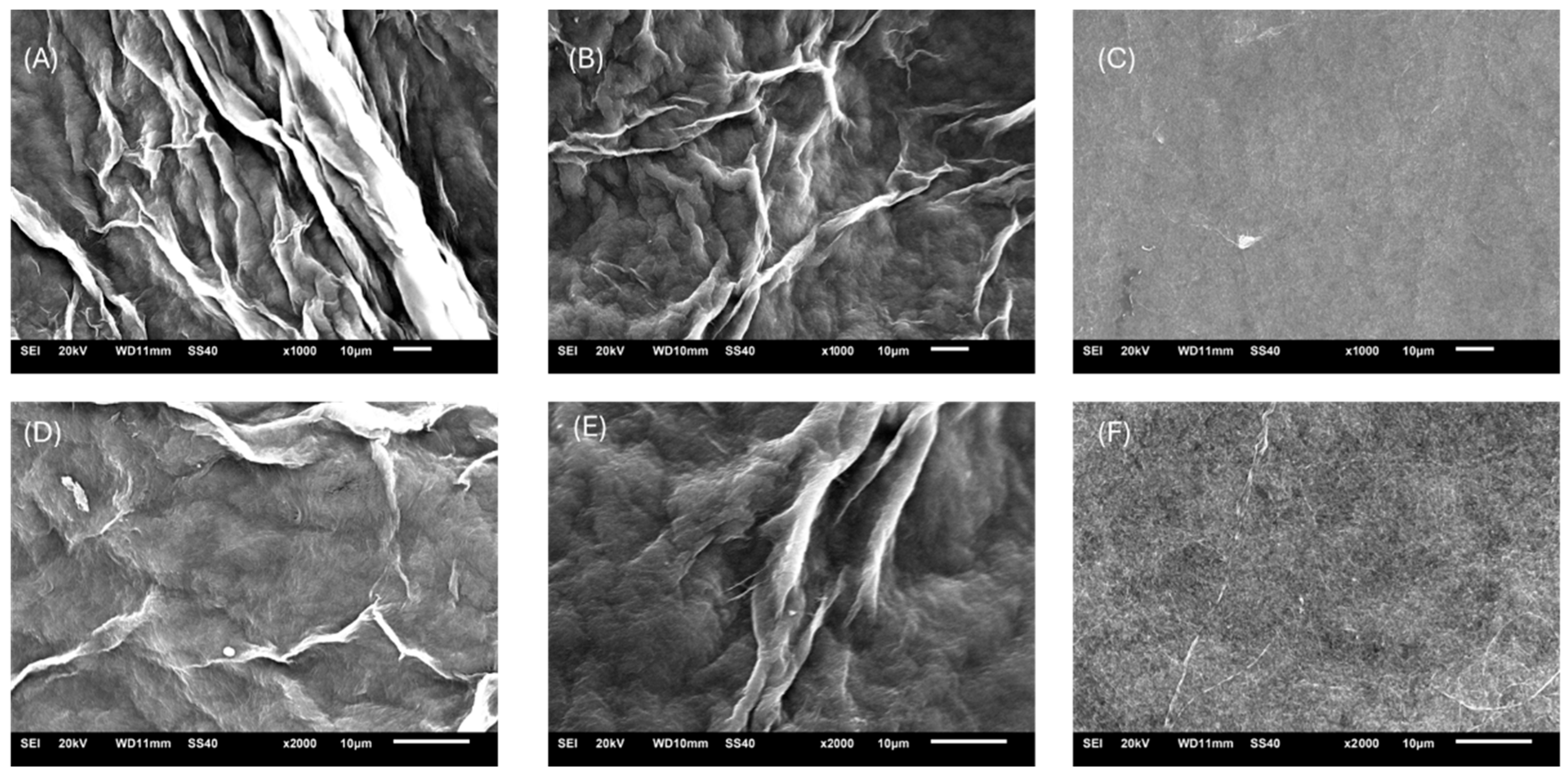
Figure 1. Scanning electron microscopy (SEM) images of three different bacterial cellulose (BC) films: BCC (A,D), BCP (B,E) and Membracel® (C,F). Images (AC) were acquired at a magnification of 1000×, while images (DF) were acquired at 2000×.
Fibers 13 00127 g001
Figure 2. Energy dispersive X-ray spectroscopy (EDX-SEM) elemental mapping image of BCC (A1,A2), BCP (B1,B2) and Membracel® (C1,C2).
Fibers 13 00127 g002
Figure 3. FTIR spectra of BCC, BCP and Membracel®.
Fibers 13 00127 g003
Figure 4. XRD diffractogram of BCC, BCP and Membracel®.
Fibers 13 00127 g004
Figure 5. TG/DTG curves of BCC, BCP and Membracel®.
Fibers 13 00127 g005
Figure 6. 13C solid-state NMR spectra of BCC, BCP and Membracel®.
Fibers 13 00127 g006
Figure 7. Water interaction properties of bacterial cellulose dressing: (a) Swelling index (%); (b) moisture content index (%); (c) water activity (aw) and (d) water contact angle (°). Error bars in all panels represent mean ± SD (n = 3). Statistical analysis by one-way ANOVA with Tukey’s post hoc test: * p < 0.05, ** p < 0.01, *** p < 0.001.
Fibers 13 00127 g007
Figure 8. (a) Porosity and (b) water vapor permeability rate of bacterial cellulose dressings. Error bars in all panels represent mean ± SD (n = 3). Statistical analysis by one-way ANOVA with Tukey’s post hoc test: * p < 0.05, ns, not significant.
Fibers 13 00127 g008
Figure 9. Optical characterization of BC wound dressings: (a) opacity at 500 nm (Abs500 nm·mm−1) and (b) transparency at 600 nm (T600 nm %·mm−1). Error bars in all panels represent mean ± SD (n = 3). Statistical analysis by one-way ANOVA with Tukey’s post hoc test: *** p < 0.001.
Fibers 13 00127 g009
Figure 10. Mechanical properties of three different bacterial cellulose (BC) films. The chart compares tensile strength and elongation at break, highlighting the distinct performance profiles of each BC type.
Fibers 13 00127 g010
Figure 11. Mass change profiles of bacterial cellulose dressings over 14 days in simulated wound fluid. Circles (BCC), squares (BCP) and triangles (Membracel®) indicate mean mass loss (%) at each time point; negative values denote net mass gain from fluid uptake. Error bars represent ± SD (n = 3).
Fibers 13 00127 g011
Disclaimer/Publisher’s Note: The statements, opinions and data contained in all publications are solely those of the individual author(s) and contributor(s) and not of MDPI and/or the editor(s). MDPI and/or the editor(s) disclaim responsibility for any injury to people or property resulting from any ideas, methods, instructions or products referred to in the content.