Article

Fibrillated Nanocellulose Obtained by Mechanochemical Processes from Coconut Fiber Residue

by
Sarah Inglid dos Santos Silva
1,*,
Cassiano Pires
2,
Egon Petersohn Junior
3,
Angela Maria Tribuzy de Magalhães Cordeiro
4,
Rilton Alves de Freitas
2 and
Nataly Albuquerque dos Santos
1,4,*
1
Graduate Program in Chemistry, Center for Exact and Natural Sciences, Department of Chemistry, Federal University of Paraíba, João Pessoa 58025-650, PB, Brazil
2
Graduate Program in Pharmaceutical Sciences, Department of Pharmacy, Federal University of Paraná, Curitiba 81530-900, PR, Brazil
3
Polytechnic Center, Integrated Program in Engineering & Materials Science, Federal University of Paraná, Curitiba 81531-990, PR, Brazil
4
Department of Food Technology, Center for Technology and Regional Development, Federal University of Paraíba, João Pessoa 58025-650, PB, Brazil
*
Authors to whom correspondence should be addressed.
Fibers 2025, 13(9), 123; https://doi.org/10.3390/fib13090123
Submission received: 25 July 2025 / Revised: 29 August 2025 / Accepted: 3 September 2025 / Published: 9 September 2025

Abstract

Rich in cellulose, the agro-industrial residue of “Cocos nucifera L.” stands out due to its high global production. In view of this, this research into the development of cellulose nanofibrils from green coconut fiber residue evaluated the fiber produced from an alkaline pre-treatment associated with a grinding process using a colloidal mill, which produced pure and renewable cellulose with characteristics similar to those of commercial celluloses. FTIR and XRD spectroscopy analyses showed that the methodologies established for coconut fiber are efficient in removing amorphous groups. The XRD corroborated the spectrogram and revealed a peak at 2θ = 22°, corresponding to the crystalline region of cellulose I. Both analyses were preceded by thermal analysis showing a reduction in lignin and an increase in the cellulose fraction. The AFM and SEM morphological micrographic analyses confirm the efficiency of the mechanochemical treatment in producing nanometric fibers, which, when submitted to rheology analyses, presented the desired gel profile.

1. Introduction

In recent decades, growing environmental concerns about products derived from fossil sources—characterized by slow renewability, pollution and price instability—have intensified the search for truly sustainable alternatives. In this context, cellulose stands out as an abundant and renewable biomass, with nanocellulose gaining increasing attention for its versatility and potential to replace synthetic materials [1]. Cellulose nanomaterials, such as nanofibrils (CNFs) and nanocrystals (CNCs), represent a pillar of the bioeconomy, combining economic viability with environmental benefits [2].
One promising approach is to use agro-industrial waste to obtain nanocellulose—an abundant, low-cost raw material that is often disposed of inappropriately. Recent studies emphasize how this type of recovery contributes to the circular economy, highlighting its positive environmental and economic impact [3]. Specifically, green coconut waste (Cocos nucifera L.) stands out, considering its global production of over 62 million tons in 2022. Composed of approximately 35% cellulose, 35% hemicellulose and 25% lignin, this waste has high potential for the production of lignocellulosic materials with added value [3,4].
The industrial interest in nanocellulose obtained from green coconuts is directly associated with the search for extraction routes that preserve its crystalline structure and maximize its physicochemical properties [5]. In this sense, hybrid strategies have been widely adopted, combining alkaline hydrolysis with mechanical processes such as defibrillation or high-pressure homogenization. This methodological arrangement favors the efficient removal of hemicelluloses and lignin, while reducing dependence on aggressive reagents and the energy consumption characteristic of purely mechanical processes. Recent studies confirm that this integration not only increases the yield and structural quality of the nanocellulose, but it is also important to note that this route was not found in literature searches for NFC production from coconut biomass [3,6].
Furthermore, the study [4] illustrates the wide applicability of coconut-derived nanocellulose: the functionalized nanopaper exhibited electromagnetic interference attenuation efficiency (21.9 dB at 10 GHz) and relevant antibacterial activity [4]. Other fronts have shown how coconut nanocellulose can reinforce thermo-responsive hydrogels, improving rheological properties and enabling controlled drug release systems [7]. In addition, research into biodegradable films incorporating coconut nanocellulose has revealed high crystallinity and reinforced performance, with strong relevance for replacing synthetic plastics [8].
Considered a latent matrix for numerous applications, including drug delivery, energy storage, optoelectronic conversion and filtration, nanocelluloses also possess a higher number of OH groups on their surface, facilitating the incorporation of various positively and negatively charged species. New applications can be designed and implemented through the intermolecular electrostatic properties of CNC surfaces [9].
In this study, the hypothesis refers to the fact that, in the context of sustainable development, when working with renewable materials, there is growing interest in clean processes and treatments that do not use oil-derived inputs and generate high-added-value chemical products. For this reason, we are considering obtaining a nanomaterial from the processing of green coconut agro-industrial waste by a cleaner route, using a colloidal mill, to produce a cellulose gel nanofibrillated with a wide range of industrial applications due to its physicochemical properties.
In short, the development of nanocellulose is taking place from green coconut waste using processes that reduce the use of acids and represent a promising solution for the reuse of agro-industrial waste. In view of the above, this study seeks to validate the hypothesis that minimized mechanical and chemical routes can generate a nanomaterial with optimized properties, offering a sustainable and efficient alternative for industry. Investigating the critical parameters and characteristics resulting from this approach will enable us to answer the problem of how to produce nanocellulose in an environmentally responsible and economically viable way.

2. Materials and Methods

2.1. Materials

The green coconut was collected in the city of João Pessoa, PB, and processed according to the methodology used by [10,11] in which the water was removed from the coconut and the shell residue was previously crushed and sieved through sieves with a diameter of 40 mesh. Next, 200 g of fibers was washed with 500 mL of purified water in the ratio (1:5 m/v) for 2 h at 50 °C and under constant mechanical agitation at 200 rpm. The sample was then filtered and the was filtrate discarded [12].

2.2. Pre-Treatment to Obtain Cellulose

The fibers were subjected to an alkaline pre-treatment using a 2% (w/v) NaOH solution under mechanical stirring (200 rpm) for 2 h at 80 °C. The treated fibers were then filtered and washed twice with deionized water, followed by oven drying at 50 °C for 24 h. Depolymerization conditions were evaluated in triplicate using NaOH concentrations of 2% and 5%. Since no significant differences in yield were observed, the minimum concentration of 2% NaOH was selected. Likewise, the experiments confirmed that 2 h was an adequate reaction time, in agreement with previously reported findings [12,13].
For the bleaching step, 100 g of alkali-treated fibers was dispersed in 300 mL of a solution containing 30 g of NaClO2 and 8–10 drops of glacial acetic acid in deionized water. The suspension was stirred for 1 h at 70 °C, cooled in an ice bath and subsequently washed with cold water and acetone, following an adapted procedure described in previous studies [11]

2.3. Obtaining Nanocellulose by the Grinding Method

Cellulose pulp was obtained from the residual biomass of green coconuts at a concentration of 2% m/v, corresponding to 60 g of cellulose in 3 L of water. Processing was carried out using a Supermasscolloider colloid mill (Masuko Sangyo Co, Honcho, Kawaguchi-city, Saitama Prefecture, Japan). The pulp was subjected to shearing forces, promoting the degradation of the fiber’s cell wall and exposing its microfibrils. The process was carried out at a flow rate of 1 L/min and an average speed of 1500 rpm. To obtain a viscous gel, 15 passes were made through the system. It was observed that, from the tenth pass onwards, the suspension showed characteristic gel behavior due to the exposure of the cellulose fibrils [13].

2.4. Characterization Techniques

2.4.1. Fourier Transform Infrared Spectroscopy—FTIR

The infrared spectrophotometer analysis of the raw, delignified and nanofibrillated cellulose (CNF) samples was carried out on a FT-IR spectrophotometer (model IR Prestige-2, SHIMADZU, Nakagyo-ku, Kyoto, Japan) SHIMADZU Fourier Transform Spectrometer. The samples were dried and crushed and mixed in a quantity of potassium bromide (KBr) at a sample/KBr ratio of 1:100 to prepare the pellet used in the analysis. The scanning patterns were within the wavenumber range of 400–4000 cm−1 with a resolution of 4 cm−1 and 20 scans for each sample.

2.4.2. Elemental Analysis (CHNS)

The elemental composition of nanofibrillated cellulose (NFC) was determined by CHNS analysis using the UNICUBE elemental analyzer (Elementar Analysensysteme GmbH, Langenselbold, Germany). An amount of 1.0 g of CNF sample freeze-dried for 24 h was analyzed.

2.4.3. X-Ray Diffraction (XRD) Analysis

X-ray diffraction (XRD) analysis was carried out using a Shimadzu XRD-6000 diffractometer (Shimatzu, Kyoto, Japan) equipped with Cu Kα radiation (λ = 1.5418 Å). Samples were mounted on glass sample holders. The measurements were performed at 40 kV and 30 mA, with a scan rate of 0.5° min−1. The crystallinity index (C.I.) was calculated using the peak fitting method (Equation (1)).
C . I = ( A r e a   o f   c r y s t a l l i n e   p e a k s ) ( A r e a   o f   c r y s t a l l i n e   a n d   a m o r p h o u s   p e a k s ) × 100 %

2.4.4. Thermogravimetric Analysis and Differential Thermal Analysis

The Thermogravimetric/Differential Thermal Analysis (TG/DT) curves of the samples raw fiber, delignified fiber and NFC were obtained under non-isothermal (dynamic) conditions on a TA Instruments SDT 2960 Thermal Analyzer (Lukens Drive, New Castle, DE, USA). The samples were analyzed in an inert atmosphere (nitrogen) at a gas flow rate of 50 mL/min and a heating rate of 10 °C min−1, with a temperature range of 25–900 °C and approximately 10 mg of mass in an alumina crucible.

2.4.5. Morphological Surfaces (SEM, AFM)

  • Scanning Electron Spectroscopy (SEM)
The morphological characterization of the samples was performed using a field-emission scanning electron microscope (FE-SEM), model MIRA3 LMH (TESCAN, Brno, Czech Republic), operating at an accelerating voltage of 20 kV. The equipment was equipped with an energy-dispersive X-ray spectroscopy (EDS) detector and MEV TM4000PLUS II under ambient conditions. Prior to analysis, the fibers were freeze-dried for 24 h, mounted on carbon adhesive tape and sputter-coated with a thin gold layer using a Quorum Q150R sputter coater for 35 s at 20 mA under an argon atmosphere.
  • Atomic Force Microscopy (AFM)
For AFM analysis, the samples were diluted in water at a concentration of 1:100 then deposited on mica and dried overnight in a vacuum oven at 60 °C. The topography images were obtained in tapping mode using a Mikromasch NSC-35 cantilever (Watsonville, CA, USA), with a force constant of 8.9 N m−1 and a resonance frequency of 200 kHz.

2.4.6. Rheology

The viscoelastic properties of the samples were evaluated using a Discovery Hybrid Rheometer HR-10 (TA Instruments, New Castle, DE, USA) equipped with a cone-and-plate geometry (40 mm diameter, 2° cone angle and 0.100 mm gap) and a Peltier temperature control system set at 25 °C. Stress sweep tests were conducted at a constant frequency of 10 Hz over a stress range of 0.001 to 500 Pa to determine the linear viscoelastic region (LVR). Subsequently, frequency sweep measurements were performed within the LVR in the range of 0.01 to 10 Hz, where the storage modulus (G’), loss modulus (G”) and loss tangent (tan δ) were recorded as a function of frequency. Flow behavior was assessed through a controlled shear rate ramp, starting with an upward ramp from 1 to 100 s−1, with each ramp lasting 300 s. All tests were conducted at 25 °C, with a 60 s sample rest time and a resolution of five points per decade. This methodology was adapted from Petersohn Junior et al. [14].

3. Results

We estimated the yield of the initial coconut mass and the resulting mass after bleaching. For an initial mass of 200 g, we obtained an average of 60 g of bleached fiber, which corresponds to a yield of approximately 30%. This figure is in line with the literature, which reports yields ranging from 20% to 40% for lignocellulosic fibers after delignification and bleaching treatments [14,15]. Furthermore, it is superior to the traditional method of obtaining coconut nanocellulose, which uses rigorous acid hydrolysis and has yields of between 8% and 20% [11].

3.1. Fourier Transform Infrared Spectroscopy—FTIR

The three samples exhibited similar patterns with stretching waves for the first bands found in the range of 3400 cm−1 in the raw fiber, 3355 cm−1 in the delignified fiber and 3350 cm−1 in the NFC, as seen in Figure 1.
Behavior similar to that observed in this study for the in natura, delignified and nanofibrillated fiber is reported for other lignocellulosic materials. According to [16], this behavior is attributed to the stretching vibration of OH groups, which is closely related to hydrogen bonds in cellulose fibers and nanofibrils.
Cellulose nanofibrils underwent a more severe process, and it is evident that the NFC obtained has a spectrum similar to that of amorphous cellulose, with some bands disappearing. From the 2922 cm−1 bands of the raw sample and the 2899 cm−1 bands of the delignified sample, the -CH functional groups can be seen, whereas in the nanofibrillated sample these groups were absent.
The absence of the -CH stretch in the nanofibrillated sample is due to the reduction in the aliphatic wax fraction during the successive dewaxing treatment, since these groups are predominantly present in materials with a lignocellulosic structure [17]. Another characteristic of this vibration, which extends between the interval of 3400 cm−1 and 2900 cm −1 [18], points out that this stretching is due to the presence of moisture retained in the samples.
In the absorption range of 1734 cm−1, there is a prominent band for fibers as lignocellulosic waste, corresponding to the carbonyl group -C=O, present in the ester bonds of the carboxylic groups of both lignin and hemicellulose [19,20]. In the case of cellulose nanofibrils produced using two isolation techniques, significant changes were observed in which the intensities of the absorption band at 1639 cm−1 were increased. This may be related to the increase in the mass proportion of cellulose content in the treated fiber, which is correlated with the OH curvature of water absorption in cellulose [20].
Extending the analysis to 1058 cm−1, it is possible to find the main bands referring to the aromatic rings typical of lignin. In the 1514 cm−1 range of the raw fiber we will still have the carbonyl bond; however, in the delignified fiber, as well as in the cellulose nanofibril, there is no band in this absorption range. In the range of 1369 cm−1 and 1371 cm−1, we have the -C-O and -C-H bonds present in the phenolic bonds only for the in natura and delignified samples. In the 1323 cm−1 range, there is a band in the less processed fibers, which represents the absorption region of the -C-O-C bonds. In the band referring to the -C-O bond present in the in natura fiber in the 1249 cm−1 range, although only one band is observed, the decrease in relation to the in natura fiber in this absorption range is clearly visible.
Another bond found in both samples is -C-O-C, in the 1058 cm−1 band in the raw fiber and 1060 cm−1 in the delignified fiber. A visible point is the considerable decrease in phenolic bonds in the delignified sample, which is completely absent in the cellulose nanofibril that underwent the treatment precisely to remove these compounds (Figure 2). Results also found in studies by [11,21] proved that the delignification process applied to coconut fiber was effective in removing lignin and hemicellulose, as well as promoting a higher concentration of cellulose.

3.2. Elemental Analysis (CHNS)

The elemental composition expressed carbon~(40.18%), hydrogen~(5.24%), sulfur (0.00%) and nitrogen (0.00%), i.e., sulfur and nitrogen were not detectable. The oxygen content was determined by the difference between the compounds present, resulting in~(54.58%) oxygen. The results obtained corroborate the FTIR sample presented earlier, through the -C-O, -C-H, -C-O-C and -C=O bonds of the bands expressed in the spectroscopy.
The data obtained Table 1 is in agreement with the literature on nanocellulose purified by mild alkaline treatment, which promotes the effective extraction of lignin and hemicellulose from lignocellulosic fibers. Studies such as those by [6,22] show that the absence of nitrogen and sulfur in the elemental composition is indicative of the complete removal of nitrogenous components (such as proteins) and sulphate functional groups, respectively, suggesting a material of high cellulose purity.
We supplemented our CHNS discussion by comparing our elemental results with recent studies on coconut-based and other lignocellulosic feedstocks [23,24] (Soriano et al., 2021; Qureshi et al., 2024). The elemental composition of the NFC, reinforced by the literature, shows that the alkaline pre-treatment was satisfactory in depolymerizing the lignin and degrading the hemicellulose fraction, thus obtaining a nanocellulose with a high degree of purification [25,26].
Table 1. Results of the CHNS percentages of the NFC sample.
SampleN [%]C [%]H [%]S [%]
NFC040.185.240
Wood [24]0.1849.346.240.2
Sugarcane bagasse [25]044.85.40

3.3. Analysis of the Crystallinity of Green Coconut Fibers and Nanocellulose (XRD)

The diffractograms obtained for the three samples in this study showed the expected pattern due to the treatments they underwent to remove the amorphous region, a characteristic of lignocellulosic materials. The first sample, coconut fiber in natura, shows a slight stretching in the intensity axis at an angle of approximately 21.3°; however, the main characteristic for this sample is the high amorphous fraction, demonstrated by the width of the peak in Figure 2a, fiber in natura.
The amorphous lignocellulosic fiber underwent a chemical treatment to promote the depolymerization and solubilization of lignin and the hydrolysis of the highly branched hemicellulose content to reveal the cellulose and increase its accessibility to subsequent processes. The degradation of lignin and hemicellulose alters the degree of polymerization of all the components of the fiber, which leads to physical alterations in the surface, porosity and crystallinity, the latter being one of the most relevant due to the removal of amorphous regions, and the degree of crystallinity increases significantly [27,28]. The C.I. increased from 30% in the in natura sample to 49% in the NFC (Table 2). The lower C.I. value suggests that there was a large amorphous region initially and that these regions were removed after the chemical stages; subsequently, after mechanical treatment, there was a considerable increase. So, after the delignification process, the sample showed the presence of two peaks of greater intensity and definition, in contrast to the first sample in natura, the first being in the region of 16.2° and the second suffering a slight shift to 22° (see Figure 2b, delignified fiber).
In the diffractogram for NFC in Figure 2c, we can see the result of the bleaching treatments that removed the lignin and hemicellulose fraction from the fibers and how the mechanical treatment can organize a periodicity over long distances in an orderly fashion, which reflects in a greater incidence the narrower and more intense crystalline peaks.
The fourth diffractogram in Figure 2d shows the difference in the order of organization of the samples before and after treatment, resulting in different degrees of crystallinity. Significant increases in the relative intensity of the peaks at 2θ = 21.3° of the raw sample to 2θ = 22.4° of the NFC were observed, giving rise to a pattern typical of cellulose I patterns, results similar to those obtained by [29,30].

3.4. Thermogravimetric Analysis and Differential Thermal Analysis (TG/DTG)

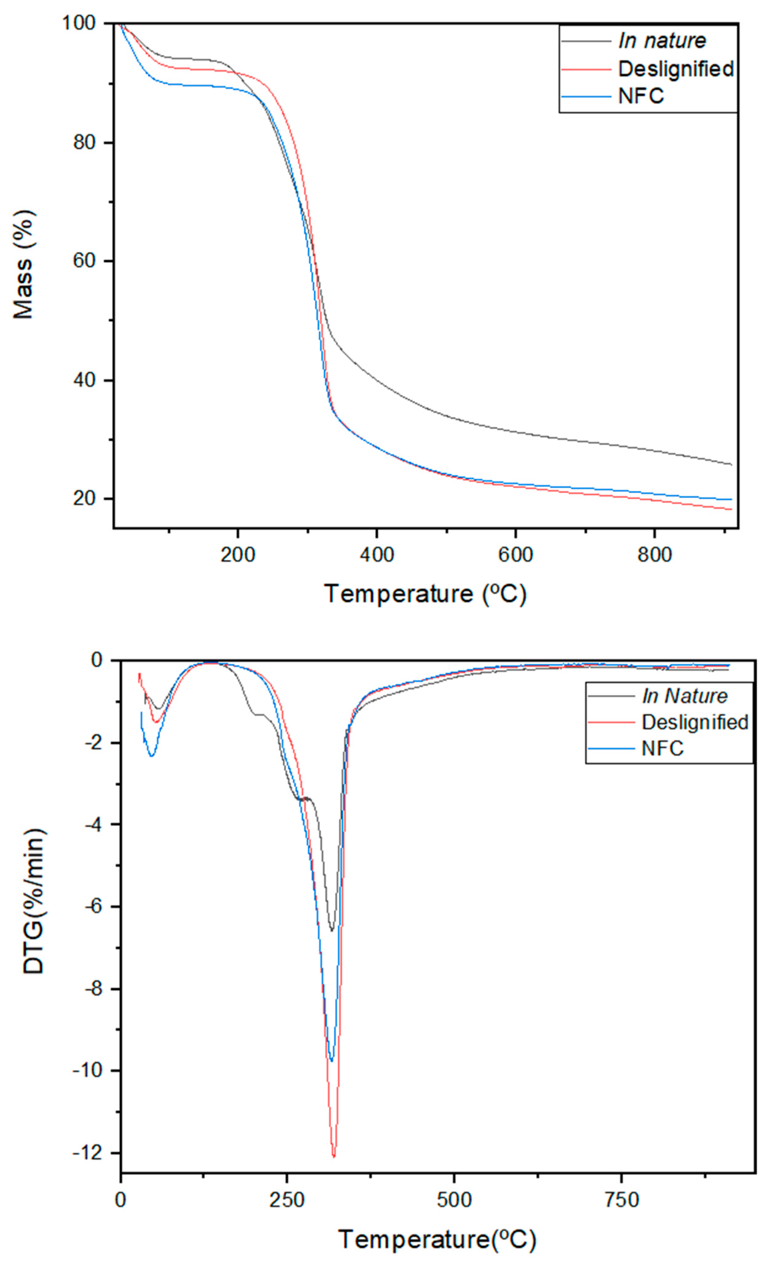
The Thermogravimetric curves (TGs) and their respective derivatives (DTGs) of the samples of raw fiber, delignified fiber and cellulose nanofibrils (NFCs) are shown in Figure 3, showing the thermal behavior of the materials in the face of decomposition. Thermogravimetric analysis makes it possible to indirectly assess the occurrence of mercerization as well as the efficiency of the pre-treatments applied to remove lignin and hemicellulose. In addition, it is possible to observe the thermal degradation profile of NFCs obtained through mechanical treatment after delignification. The data obtained provides evidence for understanding the thermal performance of the samples based on the variation in mass as a function of temperature and the heating rate applied.
The three samples showed similar thermal behavior to each other and were in line with the literature for coconut fiber and cellulose nanofibril samples. The initial loss of mass observed in the 30 °C to 130 °C range is related to the elimination of the moisture present in the samples. In this interval, the raw fiber showed a loss of 5%, the delignified sample 7% and the NFC 10%. In the second stage, the raw fiber showed two more degradation peaks, the first of which occurred between 150 and 224 °C, with a degradation percentage of 7% of mass, mainly due to the depolymerization of hemicellulose (Figure 3).
According to [31], this loss is due to the relatively lower thermal stability of hemicellulose compared to cellulose and lignin. The second peak, which appears at a temperature of 265 °C in the DTG curve of the raw sample and does not appear in the purified samples, demonstrates the efficiency of the treatment in removing xylan and lignin [32,33].
After the treatment steps to remove lignin and hemicellulose with the aim of preserving cellulose, Figure 3 shows that the cellulose degradation peaks in the three samples occurred in the 200–360 °C range. The efficiency of fiber purification was preserved between 50–60% in the samples that underwent alkaline and mechanical treatments due to the increase in thermal stability for these samples [34,35].
The raw fiber, with its lower thermal stability, exhibited additional degradation peaks within the same temperature range, with a mass decomposition of 27% of the cellulose present in the sample. After 400 °C, irrelevant mass loss events were observed, which are attributed to the efficiency of the lignin removal treatment. Generally, a wider decomposition range is required for the removal of lignin due to the thermal stability of the aromatic groups, which begins at approximately 200 °C and continues until the formation of the carbonized residue [34,36].

3.5. Morphological Surfaces (SEM and AFM)

The micrographs obtained by SEM in Figure 4 and AFM in Figure 5, followed by their measurements, show the efficiency of the treatment in obtaining nanocellulose. The micrographs show a morphological analysis of the surface of the nanofibrils, showing elongated and interwoven structures. The AFM shows the nanometer-scale topography of the fibrils.
The micrographs follow a sequence of magnifications. In the first image (A), obtained using an SEM TM4000PLUS II (DPUNION), no metallization was required, and a large number of well-defined fibers can be observed. Image (B), as well as the subsequent ones, underwent metallization and were acquired with a MIRA3 LMH (TESCAN) microscope. In this case, the fibrils appear as an entangled web, and their visualization is less clear due to the metallization layer that tends to mask their individuality.
Image (C), at a magnification of 5 kx, reveals several dispersed fibrils, while image (D) shows the dimensional characterization of isolated fibrils by field-emission microscopy at different magnifications, with individual nanometric widths ranging from 72 nm to 82 nm. Finally, image (E) isolates a single fibril, measuring approximately 82.25 nm in diameter. At higher FEG magnifications, due to the presence of small amorphous regions within the crystalline cellulose structure, the nanocellulose fibrils (NCFs) appear at the nanoscale with an irregular surface.
The AFM shows the morphology of the fibers after the mechanochemical treatment, as seen in Figure 5. Due to the convolution effect of the tip, the measurement of the width of the fibers may present values greater than the real ones for a more assertive value, so the height was used. The auxiliary graph in Figure 5 illustrates the measurement of the cut of the fibers, where it can be seen that their height is 8 ± 2 nm, confirming their nanometric dimension; it can also be seen that the fibers have a micrometric length. The bars and scale are meant to show the magnification for the cropped section of the whole fibrillated nanocellulose.

3.6. Rheology

Rheological analysis provides information on the viscoelastic properties and mechanical behavior of aqueous suspensions, so only the NFC sample was subjected to rheology to obtain the information of interest in the technique. The stress scan (Figure 6a) provides information on the elastic limit of the gel formed, while the frequency scan (Figure 6b) allows us to understand the relationship between the storage (G’) and loss (G’’) moduli.
The results of the graph in Figure 6a, referring to the stress scan aimed at determining the linear viscoelastic region (LVR), show that the storage modulus (G’) remained constant, while the loss modulus (G”) showed little variation. The fact that G’ is significantly higher than G” over the entire stress range indicates that the sample exhibits a mostly elastic behavior, as observed in concentrated nanocellulose suspensions [37,38]. This behavior is associated with nanofibrillated suspensions, with strong hydrogen bonds and interlacing between fibrils, which gives a very resistant network formation [15,39].
The results observed for the frequency scan in Figure 6b suggest gel-like behavior [40]. The formation of more compact internal cross-linked network structures justifies the behavior of the G’ and G” curves, which show an upward trend as the frequency increases. As reported in the literature, the behaviors follow a pattern throughout the frequency range G’, which remained higher than G”, indicating the presence of a network structure formed by cellulose entanglements within the emulsion system [41,42]. Both results suggest that the material remained in a linear viscoelastic regime, demonstrating that, within the range tested, nanofibrillated coconut cellulose shows structural robustness and predictable behavior.

4. Conclusions

This study demonstrated the feasibility of using green coconut fibers through mechanochemical processes to produce nanocellulose. The results indicate that the chemical and mechanical treatments applied were effective in removing lignin and hemicellulose, yielding fibers with a high degree of crystallinity, which increased from 30% in the raw fiber to 49% in the nanocellulose and enhanced thermal stability, making them suitable for various industrial applications.
Morphological analysis by SEM and AFM confirmed the formation of nanofibrils with widths ranging from 72 to 82 nm and heights of approximately 8 ± 2 nm, while rheological characterization showed predominantly elastic behavior, with the storage modulus (G’) higher than the loss modulus (G”) across the entire tested stress range, indicating the formation of a robust structural network.
These findings highlight the potential of coconut-derived nanocellulose as a sustainable, high-value-added material for industrial applications, emphasizing the reduction of aggressive chemical usage and promoting circular economy principles. The study also demonstrates that incorporating agro-industrial residues into nanomaterial synthesis is a viable strategy for developing functional and environmentally friendly materials.
In summary, this work not only provides solid quantitative data on the structural, morphological and rheological properties of the obtained nanocellulose but also suggests promising avenues for applying agricultural residues in cleaner and more sustainable industrial practices.

Author Contributions

Conceptualization, S.I.d.S.S., R.A.d.F. and N.A.d.S.; methodology, S.I.d.S.S., C.P., E.P.J. and A.M.T.d.M.C.; formal analysis, S.I.d.S.S. and C.P.; investigation, S.I.d.S.S., R.A.d.F. and N.A.d.S.; writing—original draft preparation, S.I.d.S.S.; writing—review and editing, S.I.d.S.S., C.P., R.A.d.F. and N.A.d.S.; supervision, R.A.d.F., N.A.d.S. and A.M.T.d.M.C.; project administration, S.I.d.S.S., R.A.d.F. and N.A.d.S. All authors have read and agreed to the published version of the manuscript.

Funding

This study was financed in part by the Coordenação de Aperfeiçoamento de Pessoal de Nível Superior—Brasil (CAPES)—Finance Code 001, Doctoral Scholarship No. 88887.508791/2020-00. This work was funded by the Public Call No. 03/2020 Research Productivity PROPESQ/PRPG/UFPB proposal code PVM13616-2020. Grant 3080/2021, Paraiba State Research Foundation (FAPESQ). This work was supported by the CNPq Productivity Scholarship Program (PQ-2).

Data Availability Statement

Data related to this study are available in the manuscript.

Acknowledgments

The authors would like to acknowledge the Biopolymers and Colloids Laboratory (BIOPOL/UFPR) and Embrapa Florestas for their support with equipment. During the preparation of this manuscript, the authors used the DeepL tool (AI translation) for the purpose of translating this article from Portuguese to English. The authors have reviewed and edited the output and take full responsibility for the content of this publication.

Conflicts of Interest

The authors declare no conflicts of interest.

Abbreviations

The following abbreviations are used in this manuscript:
CNFCellulose Nanofibril
CNCCellulose Nanocrystal
FTIRFourier transform infrared spectroscopy
TGThermogravimetric Analysis
DTGThermogravimetric Derivative Analysis
DRXX-ray diffraction
SEMScanning Electron Spectroscopy
AFMAtomic Force Microscopy

References

  1. Nagarajan, K.J. A Comprehensive Review on Cellulose Nanocrystals and Cellulose Nanofibers: Pretreatment, Preparation, and Characterization; Wiley: Hoboken, NJ, USA, 2021; ISBN 4860771060964. [Google Scholar]
  2. TAPPI International Nanotecnology Division. Roadmap for the Development of International Standards for Nanocellulose; TAPPI: Peachtree Corners, GA, USA, 2011; Volume 33. [Google Scholar]
  3. Nargotra, P.; Sharma, V.; Tsai, M.L.; Hsieh, S.L.; Dong, C.D.; Wang, H.M.D.; Kuo, C.H. Recent Advancements in the Valorization of Agro-Industrial Food Waste for the Production of Nanocellulose. Appl. Sci. 2023, 13, 6159. [Google Scholar] [CrossRef]
  4. Jose, J.; Vinod, T.P. Nanocellulose from Coconut Midrib Used for Antibacterial and Electromagnetic Interference Shielding Applications. New J. Chem. 2025, 49, 9475–9483. [Google Scholar] [CrossRef]
  5. Vincent, S.; Kandasubramanian, B. Cellulose Nanocrystals from Agricultural Resources: Extraction and Functionalisation. Eur Polym. J. 2021, 160, 110789. [Google Scholar] [CrossRef]
  6. Klemm, D.; Kramer, F.; Moritz, S.; Lindström, T.; Ankerfors, M.; Gray, D.; Dorris, A. Nanocelluloses: A New Family of Nature-Based Materials. Angew. Chem. Int. Ed. 2011, 50, 5438–5466. [Google Scholar] [CrossRef]
  7. Leow, Y.; Boo, Y.J.; Lin, M.; Tan, Y.C.; Goh, R.Z.R.; Zhu, Q.; Loh, X.J.; Xue, K.; Kai, D. Coconut Husk-Derived Nanocellulose as Reinforcing Additives in Thermal-Responsive Hydrogels. Carbohydr. Polym. 2024, 323, 121453. [Google Scholar] [CrossRef]
  8. Arun, R.; Shruthy, R.; Preetha, R.; Sreejit, V. Biodegradable Nano Composite Reinforced with Cellulose Nano Fiber from Coconut Industry Waste for Replacing Synthetic Plastic Food Packaging. Chemosphere 2022, 291, 132786. [Google Scholar] [CrossRef]
  9. Marakana, P.G.; Dey, A.; Saini, B. Isolation of Nanocellulose from Lignocellulosic Biomass: Synthesis, Characterization, Modification, and Potential Applications. J. Environ. Chem. Eng. 2021, 9, 106606. [Google Scholar] [CrossRef]
  10. Rosa, M.d.F.; Abreu, F.A.P.d.; Furtado, Â.A.L.; Brígido, A.K.L.; Norões, E.R.d.V. Processo Agroindustrial: Obtenção de Pó de Casca de Coco Verde; Embrapa Comunicado Técnico: Brasília, DF, Brazil, 2001; Volume 61, pp. 1–4. [Google Scholar]
  11. Rosa, M.F.; Medeiros, E.S.; Malmonge, J.A.; Gregorski, K.S.; Wood, D.F.; Mattoso, L.H.C.; Glenn, G.; Orts, W.J.; Imam, S.H. Cellulose Nanowhiskers from Coconut Husk Fibers: Effect of Preparation Conditions on Their Thermal and Morphological Behavior. Carbohydr. Polym. 2010, 81, 83–92. [Google Scholar] [CrossRef]
  12. Mahrouz, M.; Malainine, M.E.; Dufresne, A.; Vuong, R.; Vignon, M.R. Structure and Morphology of Cladodes and Spines of Opuntia Ficus-Indica. Cellul. Extr. Characterisation 2003, 51, 77–83. [Google Scholar]
  13. Zenni, M.R.; Santos, C.P.d.; Bianchi, R.M.d.C. Celulose Nanofibrilada: Estudo Da Obtenção E Aplicação Na Indústria Papeleira. Eng. Mod. Soluções Probl. Soc. Indústria 2021, 2, 107–119. [Google Scholar] [CrossRef]
  14. Petersohn Junior, E.; Pires, C.; de Freitas, R.A.; Magalhães, W.L.E. Effect of the Addition of Hydroxypropyl Methylcellulose and Hydroxyethyl Cellulose on the Rheological Properties and Thermogravimetric Kinetics of Dried and Redispersed Microfibrillated Cellulose. Fibers Polym. 2025, 26, 1465–1478. [Google Scholar] [CrossRef]
  15. Jonoobi, M.; Oladi, R.; Davoudpour, Y.; Oksman, K.; Dufresne, A.; Hamzeh, Y.; Davoodi, R. Different Preparation Methods and Properties of Nanostructured Cellulose from Various Natural Resources and Residues: A Review. Cellulose 2015, 22, 935–969. [Google Scholar] [CrossRef]
  16. Nechyporchuk, O.; Belgacem, M.N.; Bras, J. Production of Cellulose Nanofibrils: A Review of Recent Advances. Ind. Crops Prod. 2016, 93, 2–25. [Google Scholar] [CrossRef]
  17. Fonseca, A.S.; Panthapulakkal, S.; Konar, S.K.; Sain, M.; Bufalino, L.; Raabe, J.; Miranda, I.P.A.; Martins, M.A.; Tonoli, G.H.D. Improving Cellulose Nanofibrillation of Non-Wood Fiber Using Alkaline and Bleaching Pre-Treatments. Ind. Crops Prod. 2019, 131, 203–212. [Google Scholar] [CrossRef]
  18. Chen, Y.W.; Lee, H.V.; Abd Hamid, S.B. Facile Production of Nanostructured Cellulose from Elaeis Guineensis Empty Fruit Bunch via One Pot Oxidative-Hydrolysis Isolation Approach. Carbohydr. Polym. 2017, 157, 1511–1524. [Google Scholar] [CrossRef]
  19. Clecius, A.; Lima, A.D.; Nascimento, R.F.; Sousa, F.F.D.; Filho, J.M.; Oliveira, A.C. Modified Coconut Shell Fibers: A Green and Economical Sorbent for the Removal of Anions from Aqueous Solutions. Chem. Eng. J. 2012, 185–186, 274–284. [Google Scholar] [CrossRef]
  20. Ferreira, H.S.; Pires, C.A.D.M. Ag/TiO2 Photocatalyst Immobilized onto Modified Natural Fibers for Photodegradation of Anthracene. Chem. Eng. Sci. 2020, 227, 115939. [Google Scholar] [CrossRef]
  21. Johar, N.; Ahmad, I.; Dufresne, A. Extraction, Preparation and Characterization of Cellulose Fibres and Nanocrystals from Rice Husk. Ind. Crops Prod. 2012, 37, 93–99. [Google Scholar] [CrossRef]
  22. Ruiz, A.; Silvino, E.; Avelino, F. Bioethanol Production by Saccharomyces Cerevisiae, Pichia Stipitis and Zymomonas Mobilis from Deligni Fi Ed Coconut Fi Bre Mature and Lignin Extraction According to Biore Fi Nery Concept. Renew. Energy 2016, 94, 353–365. [Google Scholar] [CrossRef]
  23. Soriano, J.A.; García-Contreras, R.; Carpio de Los Pinos, A.J. Study of the Thermochemical Properties of Lignocellulosic Biomass from Energy Crops. Energies 2021, 14, 3780. [Google Scholar] [CrossRef]
  24. Qureshi, S.S.; Nizamuddin, S.; Xu, J.; Vancov, T.; Chen, C. Cellulose Nanocrystals from Agriculture and Forestry Biomass: Synthesis Methods, Characterization and Industrial Applications. Environ. Sci. Pollut. Res. 2024, 31, 58745–58778. [Google Scholar] [CrossRef]
  25. Trache, D.; Tarchoun, A.F.; Derradji, M.; Hamidon, T.S.; Masruchin, N.; Brosse, N.; Hussin, M.H. Nanocellulose: From Fundamentals to Advanced Applications. Front. Chem. 2020, 8, 392. [Google Scholar] [CrossRef] [PubMed]
  26. Kargarzadeh, H.; Ioelovich, M.; Ahmad, I.; Thomas, S.; Dufresne, A. Methods for Extraction of Nanocellulose from Various Sources. In Handbook of Nanocellulose and Cellulose Nanocomposites; Wiley: Hoboken, NJ, USA, 2017; pp. 1–49. [Google Scholar] [CrossRef]
  27. Dhali, K.; Ghasemlou, M.; Daver, F.; Cass, P.; Adhikari, B. A Review of Nanocellulose as a New Material towards Environmental Sustainability. Sci. Total Environ. 2021, 775, 145871. [Google Scholar] [CrossRef]
  28. Kim, J.S.; Lee, Y.Y.; Kim, T.H. A Review on Alkaline Pretreatment Technology for Bioconversion of Lignocellulosic Biomass. Bioresour. Technol. 2016, 199, 42–48. [Google Scholar] [CrossRef]
  29. Hernandez, C.C.; Ferreira, F.F.; Rosa, D.S. X-Ray Powder Diffraction and Other Analyses of Cellulose Nanocrystals Obtained from Corn Straw by Chemical Treatments. Carbohydr. Polym. 2018, 193, 39–44. [Google Scholar] [CrossRef]
  30. Wu, J.; Du, X.; Yin, Z.; Xu, S.; Xu, S.; Zhang, Y. Preparation and Characterization of Cellulose Nanofibrils from Coconut Coir Fibers and Their Reinforcements in Biodegradable Composite Films. Carbohydr. Polym. 2019, 211, 49–56. [Google Scholar] [CrossRef] [PubMed]
  31. Chirayil, C.J.; Joy, J.; Mathew, L.; Mozetic, M.; Koetz, J.; Thomas, S. Isolation and Characterization of Cellulose Nanofibrils from Helicteres Isora Plant. Ind. Crops Prod. 2014, 59, 27–34. [Google Scholar] [CrossRef]
  32. Reddy, K.O.; Maheswari, C.U.; Dhlamini, M.S.; Mothudi, B.M.; Kommula, V.P.; Zhang, J.; Zhang, J.; Rajulu, A.V. Extraction and Characterization of Cellulose Single Fibers from Native African Napier Grass. Carbohydr. Polym. 2018, 188, 85–91. [Google Scholar] [CrossRef]
  33. Rosa, S.M.L.; Rehman, N.; De Miranda, M.I.G.; Nachtigall, S.M.B.; Bica, C.I.D. Chlorine-Free Extraction of Cellulose from Rice Husk and Whisker Isolation. Carbohydr. Polym. 2012, 87, 1131–1138. [Google Scholar] [CrossRef]
  34. Mishra, L.; Basu, G. Coconut Fibre: Its Structure, Properties and Applications; Elsevier Ltd.: Amsterdam, The Netherlands, 2020; Volume 1, ISBN 9780128206669. [Google Scholar]
  35. Nagarajan, K.J.; Balaji, A.N.; Ramanujam, N.R. Extraction of Cellulose Nano Fi Bers from Cocos Nucifera Var Aurantiaca Peduncle by Ball Milling Combined with Chemical Treatment. Carbohydr. Polym. 2019, 212, 312–322. [Google Scholar] [CrossRef]
  36. Moura, H.O.M.A.; Câmara, A.B.F.; Silva, B.R.; Albuquerque, I.M.; Oliveira, K.G.; Campos, L.M.A.; Carvalho, L.S. Nova Metodologia De Caracterização Composicional Da Lignocelulose Aplicando a Deconvolução De Dados Termogravimétricos. Open Sci. Res. IV 2022, 4, 604–619. [Google Scholar] [CrossRef]
  37. Iotti, M.; Gregersen, Ø.W.; Moe, S.; Lenes, M. Rheological Studies of Microfibrillar Cellulose Water Dispersions. J. Polym. Environ. 2011, 19, 137–145. [Google Scholar] [CrossRef]
  38. Moberg, T.; Sahlin, K.; Yao, K.; Geng, S.; Westman, G.; Zhou, Q.; Oksman, K.; Rigdahl, M. Rheological Properties of Nanocellulose Suspensions: Effects of Fibril/Particle Dimensions and Surface Characteristics. Cellulose 2017, 24, 2499–2510. [Google Scholar] [CrossRef]
  39. Pääkko, M.; Ankerfors, M.; Kosonen, H.; Nykänen, A.; Ahola, S.; Österberg, M.; Ruokolainen, J.; Laine, J.; Larsson, P.T.; Ikkala, O.; et al. Enzymatic Hydrolysis Combined with Mechanical Shearing and High-Pressure Homogenization for Nanoscale Cellulose Fibrils and Strong Gels. Biomacromolecules 2007, 8, 1934–1941. [Google Scholar] [CrossRef] [PubMed]
  40. Schramm, G. A Practical Approach to Rheology and Rheometry, 2nd ed.; Rheology; Gebrueder HAAKE GmbH: Karlsruhe, Germany, 1994. [Google Scholar]
  41. Zhang, H.; Wang, H.; Dai, H.; Chen, H.; Ma, L.; Zhang, Y. Regulating Emulsion Properties through Tannin Acid-Mediated Gelatin/Cellulose Nanocrystal Complexes: From Low-Oil Emulsion to High Internal Phase Emulsion Gel for 3D Printing. Food Hydrocoll. 2025, 159, 110647. [Google Scholar] [CrossRef]
  42. Tian, Y.; Huang, R.; Chen, Y.; Wang, T.; Wu, J.; Wang, S. The Preparation and Characterization of Pineapple Peel Cellulose Nanofibers and Its Application in Oil-Water Emulsions. Carbohydr. Polym. 2025, 353, 123245. [Google Scholar] [CrossRef] [PubMed]
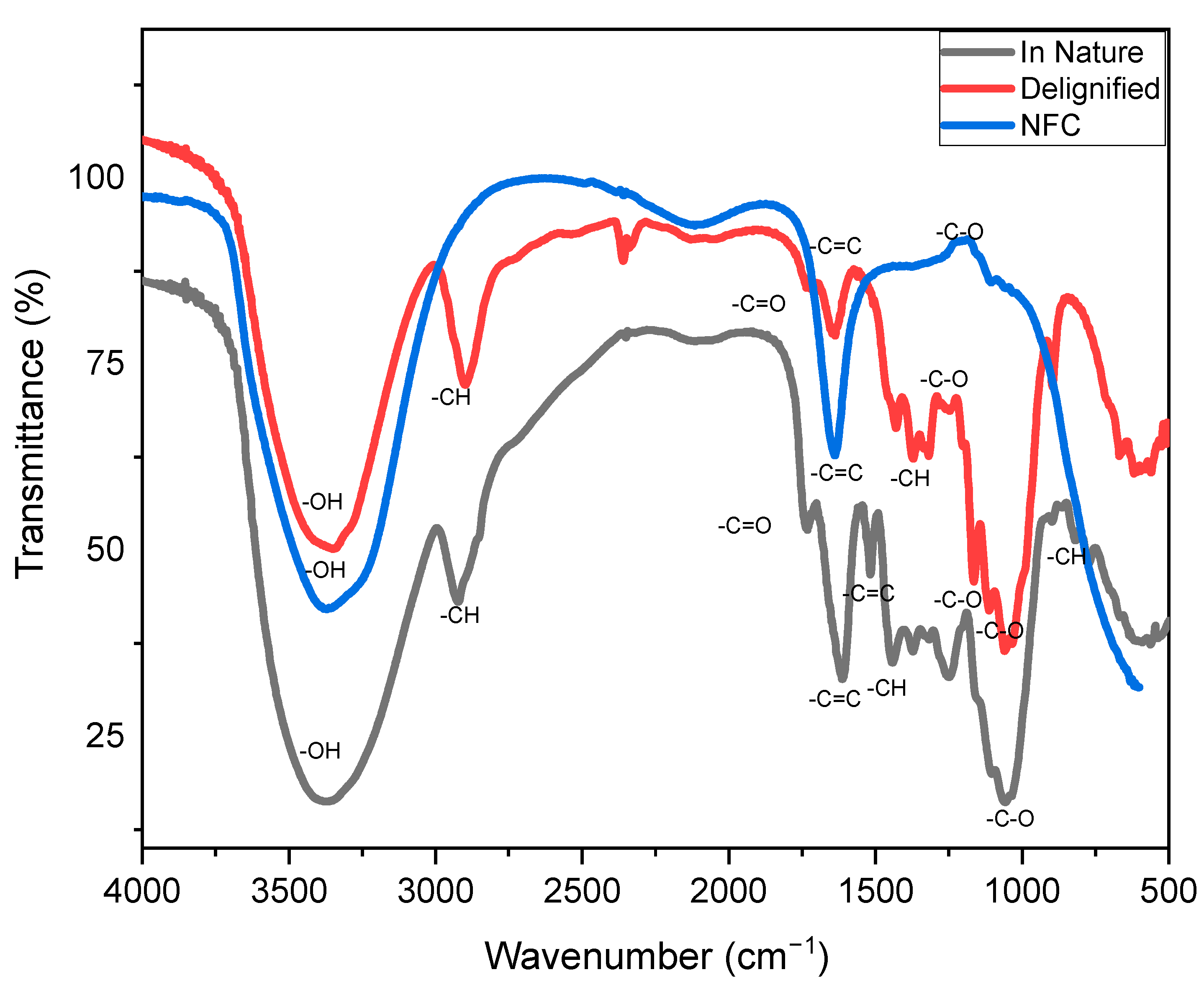
Figure 1. FTIR spectrum of the in natura, delignified and nanofibrillated fiber.
Fibers 13 00123 g001
Figure 2. Diffractograms obtained for the samples of in nature fibers (a), delignified fibers (b) and NFC (c) and diffractogram of the analysis of the three superimposed samples (d).
Fibers 13 00123 g002aFibers 13 00123 g002b
Figure 3. Thermogravimetry and Thermogravimetric Derivative of raw, delignified and nanofibrillated fiber.
Fibers 13 00123 g003
Figure 4. SEM micrographs of nanofibrillated cellulose (NCF) at different magnifications, showing well-defined, entangled, and isolated fibrils. Diameters ranged from 72–82 nm, with a single fibril measured at 82.25 nm.
Fibers 13 00123 g004aFibers 13 00123 g004b
Figure 5. Morphological analysis of cellulose nanofibrils by atomic force microscopy.
Fibers 13 00123 g005
Figure 6. Graph of the voltage sweep (a) and the frequency modulus (b).
Fibers 13 00123 g006aFibers 13 00123 g006b
Table 2. Crystallinity indices (C.I.) of in natura fibers; delignified fibers; NFC.
SampleCrystallinity Index (C.I.), %
In natura30
Delignified
NFC
40
49
Disclaimer/Publisher’s Note: The statements, opinions and data contained in all publications are solely those of the individual author(s) and contributor(s) and not of MDPI and/or the editor(s). MDPI and/or the editor(s) disclaim responsibility for any injury to people or property resulting from any ideas, methods, instructions or products referred to in the content.