Article

Coating of Nanolipid Structures by a Novel Simil-Microfluidic Technique: Experimental and Theoretical Approaches

by
Anna Angela Barba
1,2,
Sabrina Bochicchio
1,
Paolo Bertoncin
3,
Gaetano Lamberti
1,4 and
Annalisa Dalmoro
1,2,*
1
Eng4Life Srl, Spin-off Accademico, Via Fiorentino, 32, 83100 Avellino, Italy
2
Dipartimento di Farmacia, Università degli Studi di Salerno, via Giovanni Paolo II, 132, 84084 Fisciano, Italy
3
Dipartimento di Scienze della Vita - Centro Microscopia Elettronica, Università degli Studi di Trieste, Via Fleming 31, A/B, 34127 Trieste, Italy
4
Dipartimento di Ingegneria Industriale, Università degli Studi di Salerno, via Giovanni Paolo II, 132, 84084 Fisciano, Italy
*
Author to whom correspondence should be addressed.
Coatings 2019, 9(8), 491; https://doi.org/10.3390/coatings9080491
Submission received: 25 June 2019 / Revised: 26 July 2019 / Accepted: 30 July 2019 / Published: 2 August 2019

Abstract

:
Nanolipid vesicular structures are ideal candidates for the controlled release of various ingredients, from vitamins for nutraceutical purposes to chemoterapic drugs. To improve their stability, permeability, and some specific surface properties, such as mucoadhesiveness, these structures can require a process of surface engineering. The interaction of lipid vesicles with oppositely charged polyelectrolytes seems to be an interesting solution, especially when the negatively charged liposomes are complexed with the cationic chitosan. In this work, a novel simil-microfluidic technique was used to produce both chitosan-coated vesicles and a vegan alternative composed of cholesterol-free liposomes coated by Guar Hydroxypropyltrimonium Chloride (Guar-HC). The combination between the experimental approach, based on experimental observations in terms of Z-potential, and size evolutions, and the theoretical approach, based on concepts of saturation, was the methodology applied to define the best polycation concentration to fairly cover (vegan or not) liposomes without aggregation. The smart production of coated nanolipid structures was confirmed by characterizations of morphology, mucoadhesiveness, and stability.

Graphical Abstract

1. Introduction

Liposomes are lipid vesicular structures formed by one or more phospholipid bilayers surrounding an aqueous core. Due to their low intrinsic toxicity and immunogenicity, and their ability to incorporate hydrophilic and hydrophobic molecules, liposomes are ideal candidates in the controlled release of many kinds of active ingredients. Nanolipid structures are able to overcome the typical drawbacks of bioactive compounds, such as low stability, limited membrane permeability, a short half-life, and low bioavailability [1]. However, liposomes present poor stability during storage (with a high tendency to aggregate) and in biological fluids [2]. In particular, they can release an active molecule before reaching the target, or they can be cleared from the blood stream by the reticuloendothelial system, reducing the half life time of the circulating liposomes [3]. Therefore, the engineering of liposome surfaces is required to control their stability and permeability, as well as to impart some specific surface properties, such as mucoadhesiveness [4]. The interaction of lipid vesicles with oppositely charged polyelectrolytes seems to be an interesting way to modify liposomes’ surfaces [2]. Different kinds of polymers, especially polycations, have been used for the surface modification of negatively charged liposomes, such as poly(N-ethyl-4-vinylpyridinium bromide) [5], polylysine [6], and polyquaternium-1 [7]. The coating of lipid structures by polycations has the ability to minimize drug release in undesired sites and increase the cellular uptake of vesicles by cells due to the polycations’ coatings’ positive charge. However the most used polycation for coating liposomes is chitosan, thanks to its ability to increase the vesicles’ stability, encapsulation efficiency, and mucoadhesive properties [8,9]. The coating process of vesicles by chitosan is largely studied and applied, but it is essentially based on drop-wise bulk methods, leading to small product output volumes, which are not useful for industrial purposes. Therefore, an innovative continuous method based on microfluidic principles was previously developed to produce chitosan covered nanoliposomes [2] that also encapsulating nutraceuticals [10]. Moreover, the trial and error approach is usually used to detect the best concentration for coating vesicles by chitosan. McClements et al. conducted a mathematical analysis of the main factors influencing the formation and stability of colloidal dispersions containing spherical particles surrounded by oppositely charged polymeric membranes for a system composed of negative liposomal vesicles and cationic chitosan [11,12,13]. In this work, a novel simil-microfluidic technique was used to produce both the already experienced chitosan-coated vesicles and a vegan alternative, i.e., an alternative composed exclusively of vegetal compounds, made by coating cholesterol-free liposomes with the vegan accepted polycation, Guar Hydroxypropyltrimonium Chloride (Guar HC). The idea of this work was to perform a parallel study on two possible marketable products, one of which is vegan accepted (when cholesterol and chitosan are not desired ingredients). No information is found in the literature about Guar HC as a coating for liposomes. It was chosen because it is personalized guar gum in which hydroxyl groups are replaced with trimethyl ammonium groups, giving a cationic charged structure, which allows easy cross-linking with anionic systems. Moreover, it acts as a viscosity, volume, and foam enhancer [14]. Moreover, the theoretical approach in the literature for choosing the best polycation concentration for vesicle coating was revised to apply it to both chitosan-coated liposomes and Guar HC-coated vegan liposomes. It is important to highlight that the simil-microfluidic method allows one to easily apply a theoretical approach to liposome covering thanks to the fact that in this method the same volumes of polycation solution and liposomes are continuously in contact with each other, unlike the drop-wise method, where the volume of polycation solution increases with time. By combining the experimental approach in the experienced novel simil-microfluidic technique and the revised theoretical approach, a useful production of nanolipid structures coated with a polycation was possible.

2. Materials and Methods
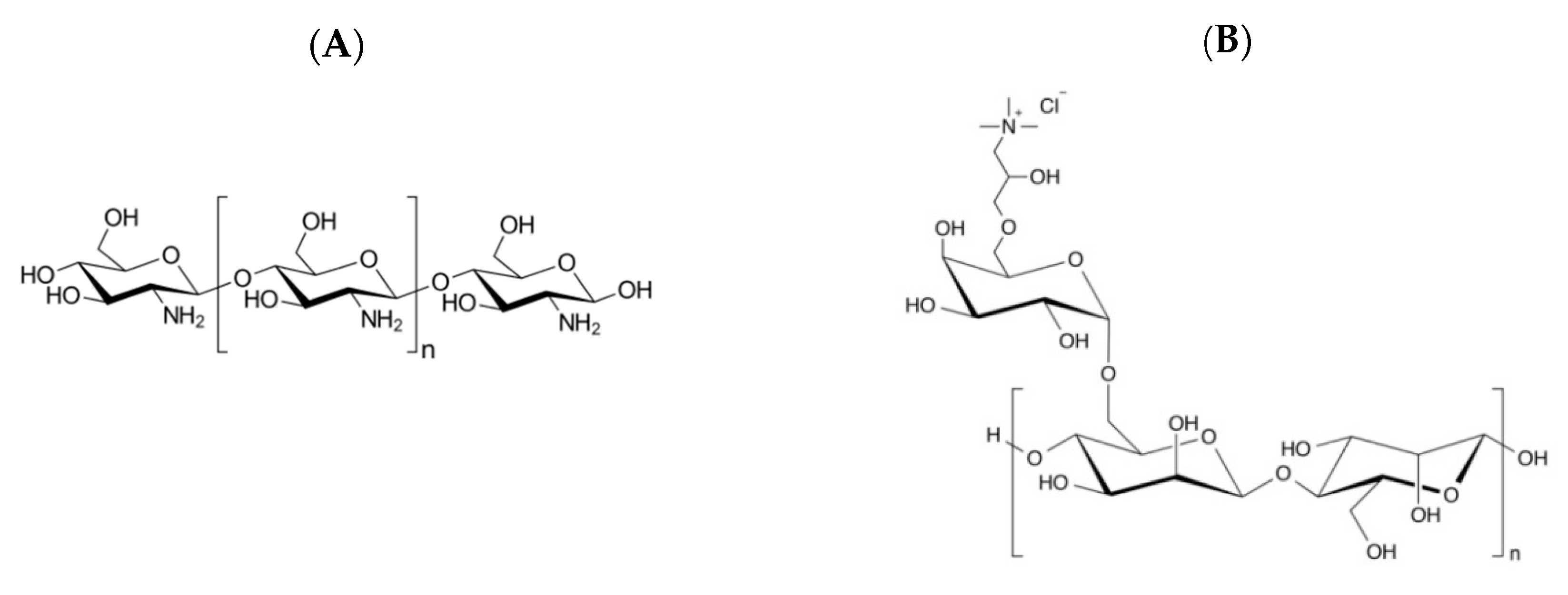
2.1. Materials

L-α-Phosphatidylcholine from soybean (PC), Type II-S, 14%–23% choline basis (CAS n. 8002-43-5) was used as a phospholipid for the production of nanolipid structures. Cholesterol (CAS n. 57-88-5) was added to the phosphatidylcholine for the production of liposomes (LP). Chitosan, with a 75% degree of deacetylation and a medium molecular weight of about 310 kg/mol (CAS n. 9012-76-4), was used for coating them (CH-LP). Cholesterol-free liposomes (made of only phosphatidylcholine) were instead produced for vegan formulations (VLP), so the vegan-accepted Guar Hydroxypropyltrimonium Chloride (Guar HC) (CAS n. 65497-29-2) was used as a polycation for their coating (VLP-GHC). Even if there is no detailed information about Guar HC’s molecular weight and substitution degree, it must still be recognized that the degree of substitution typically ranges from 0.21 to 0.6 cationic groups per galactomannan repeating unit, and the molecular weight values usually range from 500 kg/mol to 2000 kg/mol [15]. Other products, such as ethanol (CAS n. 64-17-5), glacial acetic acid (CAS n. 64-19-7), TritonX100 (CAS n. 9002-93-1), and mucin from a porcine stomach (Type III, bound sialic acid 0.5%–1.5%, partially purified powder (CAS n. 84082-64-4)) were used for preparation and/or characterization. All the listed products were purchased from Sigma Aldrich (Milan, Italy), except for Guar HC, which was purchased from Glamour Cosmetics (Milan, Italy).

2.2. Methods

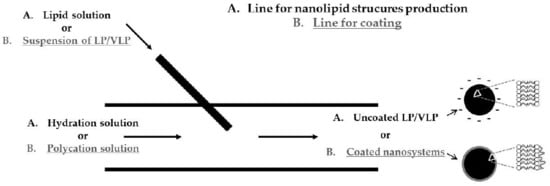
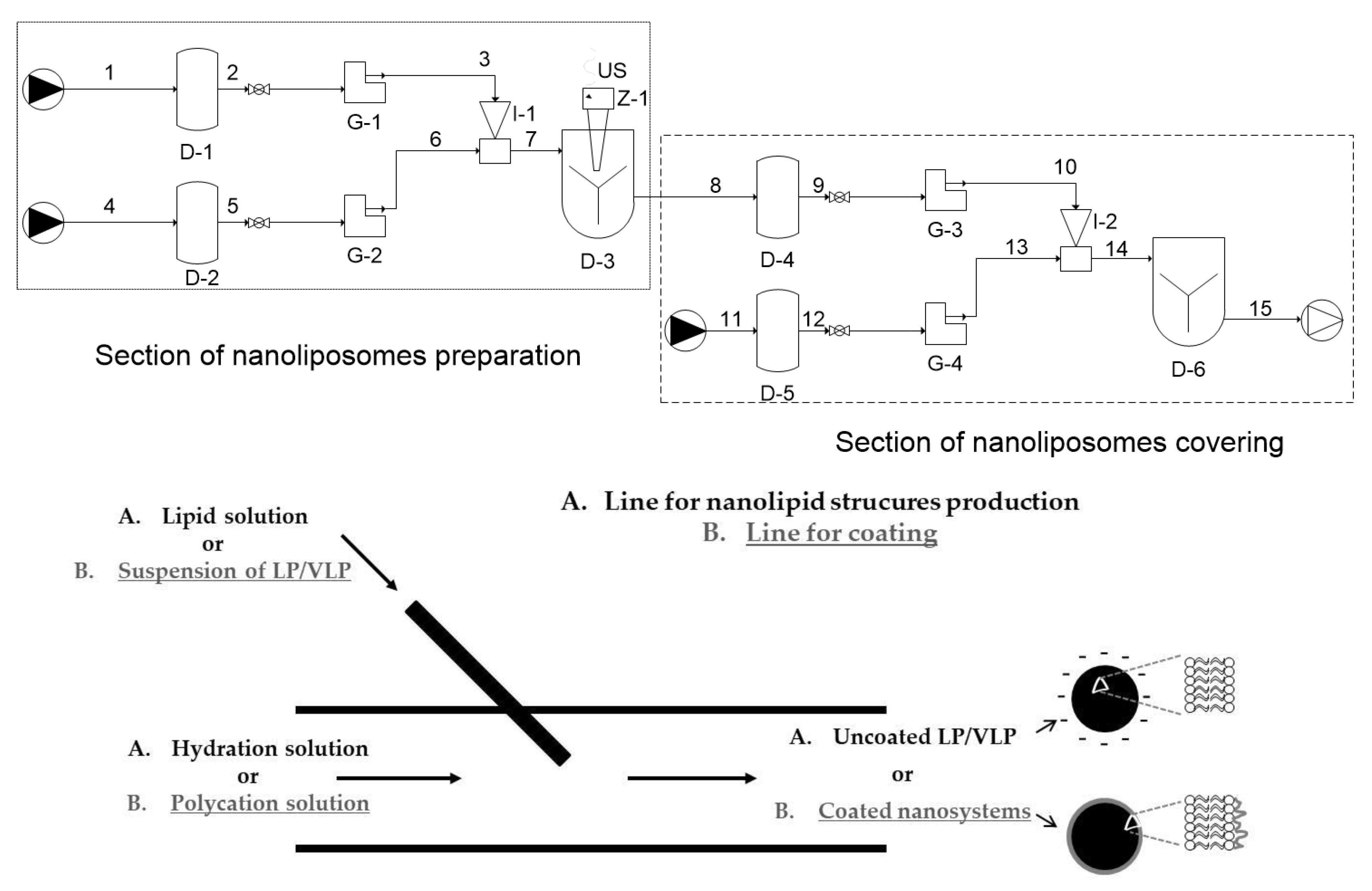
2.2.1. Production of Uncoated and Polycation-Coated Nanolipid Structures

Uncoated and polycation-coated nanolipid structures were produced by the simil-microfluidic method, detailed in previous works [2,16,17]. The same principles of interdiffusion between two phases are applied in the production and covering steps for both nanovesicles. In particular, the interdiffusion phenomena occur after the contact between aqueous and lipid phases, in the production step, and the liposome suspension and polycation solution, in the covering step [18]. In this work, for the production of uncoated nanocarriers, a lipid solution was prepared by weighing 470 mg of phosphatidylcholine (for vegan liposomes, VLP) plus 94 mg of cholesterol (for liposomes, LP) and dissolving these amounts in 10 mL of ethanol. The cholesterol (of animal origin) is usually introduced in liposome formulation to improve the stability of the liposomes [19]. Vegan liposomes were produced without cholesterol ingredient (only phosphatidylcholine). A total of 100 mL of deionized water was used as hydration solution. The lipid and hydration solutions were put in contact in the simil-microfluidic set-up by using a flowrate ratio of 1/10 for lipid/hydration solutions, as defined in [17], to obtain a hydro-alcoholic suspension containing nanometric vesicles of VLP or LP. This final suspension was first magnetically stirred for 1 h, and then one aliquot was subjected to characterization and another to coating. Nanolipid structures were negatively charged due to their constituents, so a positive charged polyelectrolyte, i.e., a polycation, was chosen to stabilize them.
Two different polycations were used for covering purposes. In particular, chitosan (from crimps shells of animal origin) was used to coat liposomes, as is typically done in the literature. A vegan alternative, Guar HC, was selected to cover vegan liposomes. Solutions with different polycation (chitosan for LP and Guar HC for VLP) concentrations, i.e., 0.0025%, 0.005%, 0.00625%, 0.0075%, 0.01% w/v, in a 0.5% (v/v) acetic acid solution, were tested in order to check the best coating for nanolipid structures. Each suspension of LP or VLP was pushed in the simil-microfluidic set-up together with the proper polycation solution at the same flow rate (25 mL/min) to obtain a suspension of chitosan-coated liposomes (CH-LP) or Guar HC-coated cholesterol-free liposomes (VLP-GHC). The obtained products were subjected first to stirring for 1 h and then to characterization. A scheme for simil-microfluidic method piping representation, including both the sections of nanoliposome preparation and nanoliposome covering, is shown in Figure 1.

2.2.2. Z-potential and Size of Nanolipid Structures

The Z-potential and size measurements of both uncoated and polycation-coated liposomes and cholesterol-free liposomes were performed by using the Zetasizer Nano ZS (Malvern, UK). The Z-potential was measured by putting an aliquot of the sample solution into a capillary cell and performing an analysis by Photon Correlation Spectroscopy (PCS). The dynamic light scattering (DLS) method was able to obtain both the numerical and intensity size distributions, together with the relevant mean size (i.e., the number mean) and Z-average (defined as the nanolipid vesicles average hydrodynamic diameter), respectively, and the size distribution (PDI). In particular, the numerical size distribution was determined by plotting the number of particles versus the particle size, and the PDI and Z-average values were calculated using the zetasizer Nano ZS software v3.30. All the samples were diluted in water. The measurements of each sample were performed in triplicate and all the results were expressed as average values with the corresponding standard deviations (SDs).

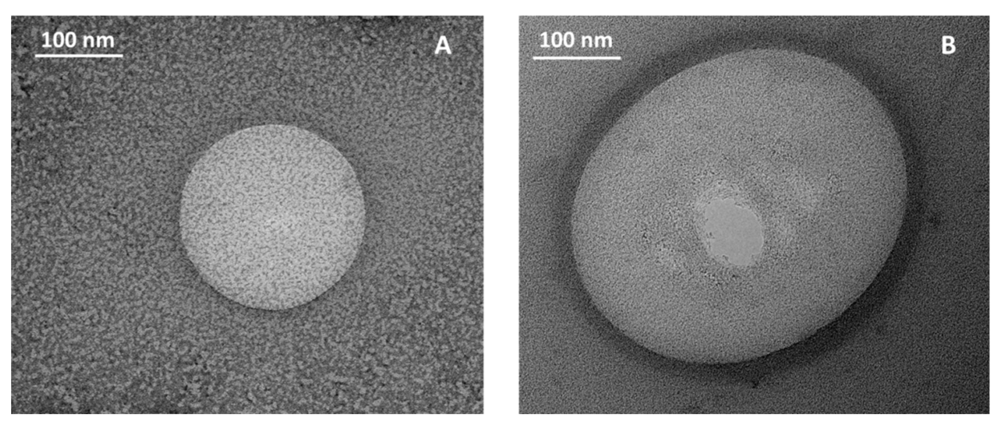
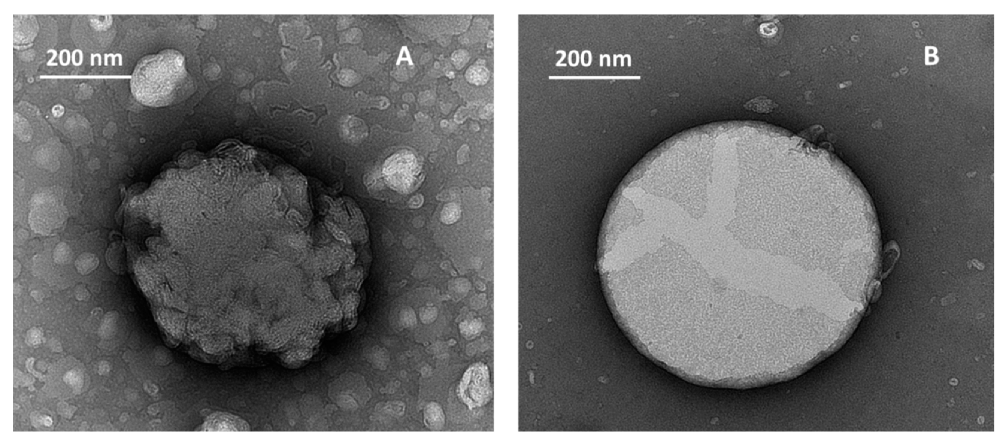
2.2.3. Transmission Electron Microscopy

Photos of uncoated liposomes and vegan liposomes, chitosan coated liposomes, and Guar HC coated vegan liposomes were taken by transmission electron microscopy (TEM) (EM 208, Philips, Hillsboro, OR, USA), equipped with a Quemesa camera (Olympus Soft Imaging Solutions). Samples were first diluted to 1:10 v/v with distilled water and then deposited on a Carbon support film on a specimen grid mesh 200 (Electron Microscopy Sciences). Before observation, they were left to air for drying, and then they were negatively stained with 1% (w/v) of uranyl acetate solution for 5 min.

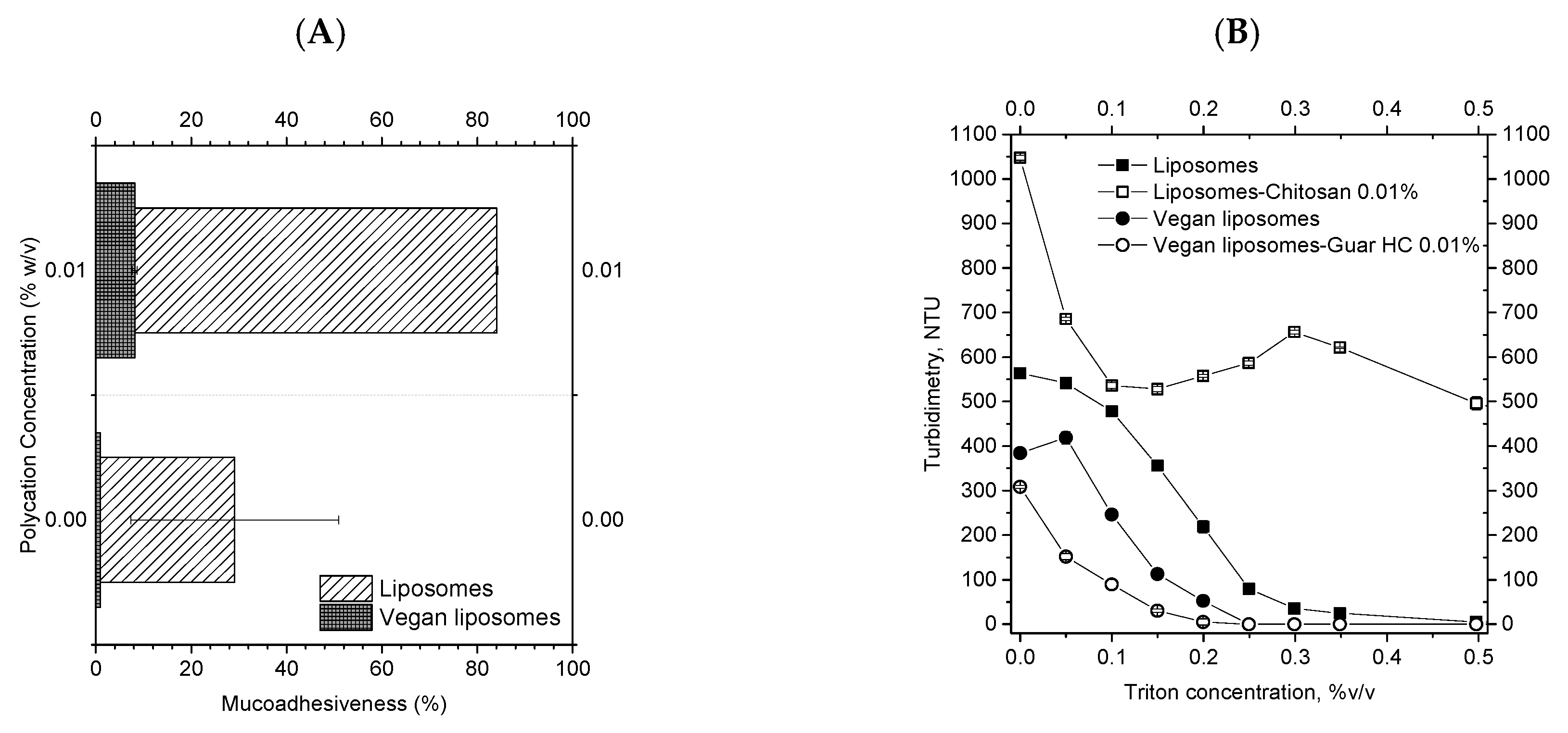
2.2.4. Mucoadhesiveness by Mucin Binding Assay

Mucoadhesiveness of uncoated and polycation-coated nanolipid vesicles was evaluated by a mucin binding assay. Briefly, an aliquot of a mucin solution in a phosphate buffer (pH 7.4, concentration of 400 μg/mL) was mixed with the same aliquot of a vesicle suspension (1:1, v/v) and left to incubate at room temperature (23 °C) for 2 h. Then, the nanolipid structures/mucin suspension was centrifuged for 1 h at a relative centrifugal force of 118,443× g at 4 °C (Beckman Optima L-90K centrifuge, SW 55 Ti rotor, Beckman Instruments, Palo Alto, CA, USA), with the aim of separating the supernatant from the pellet, i.e., the vesicles. Free mucin in the supernatant, i.e., that not absorbed by the vesicles, was detected by UV spectrophotometry (Lambda 35, Perkin Elmer, Monza, Italy) at 384 nm. The mucoadhesiveness intensity was defined as the percentage ratio between the mucin bounded to the nanolipid structures (calculated from the difference between the initial mucin concentration in the vesicles/mucin suspension of 200 μg/mL and the free mucin in the supernatant) and the initial mucin concentration (200 μg/mL). All the determinations were performed in triplicate, and the results were expressed as average values with standard deviation (SD).

2.2.5. Turbidimetry

The turbidimetry of both uncoated and polycation-coated vesicles, upon the continuous addition of a non-ionic surfactant, TritonX-100, was measured by a turbidimeter (PCE-TUM 20, PCE Instruments, Capannori, Italy) to check the vesicles’ solubilization. A total of 5 mL of the sample was diluted up to 10 mL with deionized water into a glass cuvette. Here, the sample turbidity (in Nephelometric Turbidity Units, NTU) was measured after each addition of an increasing amount of TritonX-100. The sample solution was magnetically stirred in order to obtain a homogeneous distribution and solubilisation of TritonX-100. The turbidity was evaluated until the sample was fully solubilized, i.e., up to NTU = 0. All the measurements were performed in triplicate, and the results were expressed as average values with standard deviation SD.

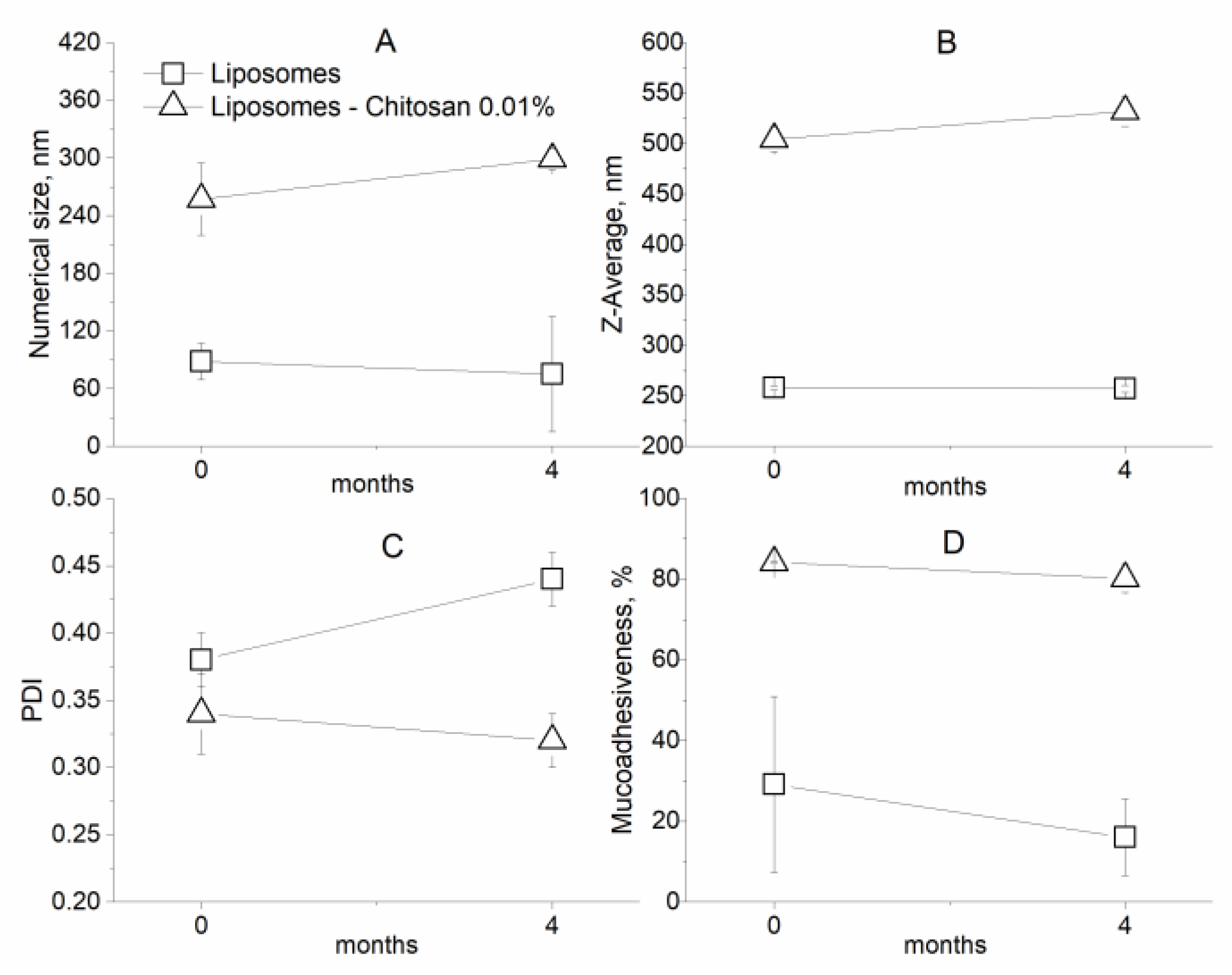
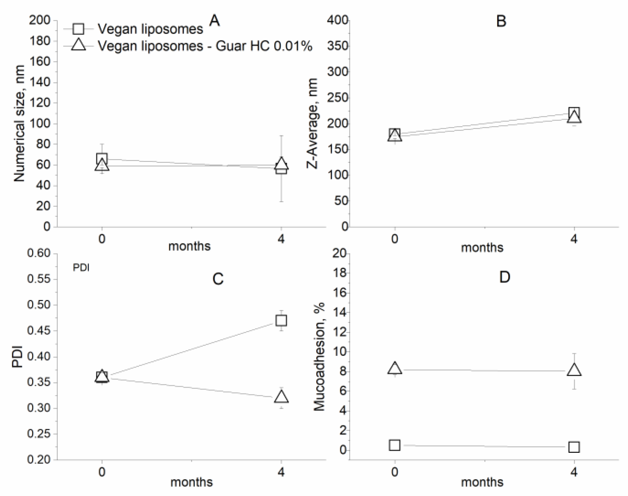
2.2.6. Stability during Storage

50 mL of uncoated and polycation-coated nanolipid vesicles were sealed and stored at 4–6 °C for 4 months to assess their stability over time. Afterwards, the samples were re-analysed for their size, PDI, zeta potential, and mucoadhesive properties.

3. Results

3.1. Adsorption of Polycations on a Nanolipid Structure Surface: Combining Experimental and Theoretical Approaches

Mixing an electrically charged polyelectrolyte with a colloidal dispersion containing oppositely charged particles is an extensively studied method to manipulate the interfacial characteristics of colloidal particles to give them improved or novel physicochemical and functional properties. Measurements of the changes in both Z-potential and mean particle diameter when a polyelectrolyte is added to a colloidal dispersion of oppositely charged particles can be used to monitor the adsorption of polyelectrolyte to the particles’ surfaces [11].

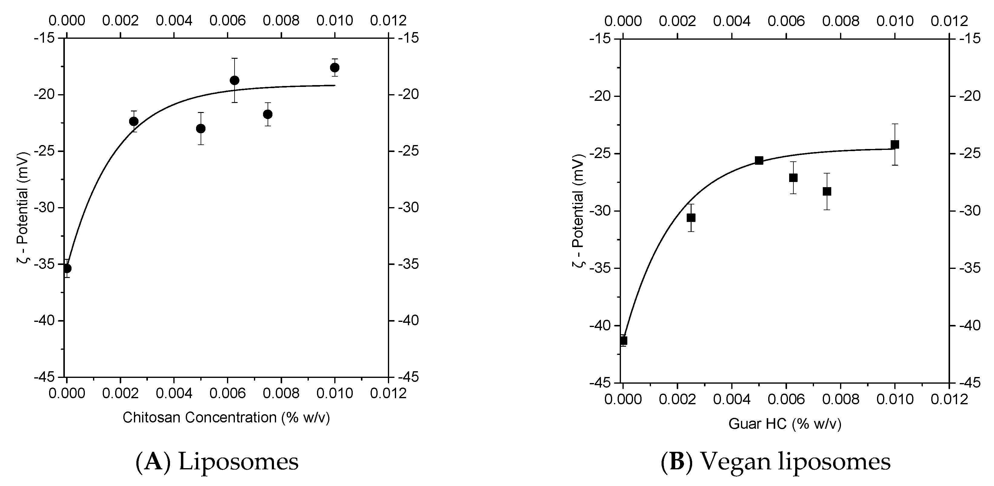
3.1.1. Experimental Approach to Nanolipid Structure Coating with a Polycation: Z-potential Evolution

In this work, the evolutions of the Z-potentials of both liposomes and vegan liposomes were recorded for different polycation concentrations, in the range 0%–0.01% w/v, (chitosan for liposomes and cationic guar gum for vegan liposomes), as shown in Figure 2.
The concentration range was chosen from observations in a previous work on the chitosan coating of liposomes [2]. In particular, it is important to keep chitosan concentrations at values under those at which coils are formed, allowing the presence of extended polymer chains able to flatly adsorb on the liposomal membrane [20]. The observed changes in surface charge suggest that, for both liposomes (Figure 2A) and vegan liposomes (Figure 2B), polycation molecules adsorbed to the vesicles’ surface until being fully coated, thereby preventing further adsorption. In particular, the Z-potential passed from −35 mV for uncoated liposomes to −18 mV after covering by using a chitosan concentration of 0.00625% w/v, and this value kept constant with concentration up to the final value of 0.01% w/v. A similar behavior was observed for vegan liposomes. The Z-potential was about −41 mV for uncoated VLP, which is more negative than the liposomes due to the absence of cholesterol [21]. The VLP Z-potential evolved to a plateau value of −24 mV after the introduction of 0.05% w/v cationic guar gum. In general, it is well known that the addition of polycations to vesicles results in a reduction in the Z-potential module, providing evidence of adsorption of polycation on the liposome’s surface [3,22].

3.1.2. Theoretical Approach to a Nanolipid Structure Coating with a Polycation: Concepts of Saturation

In general, stable colloidal dispersions are formed only in a polyelectrolyte concentration range between the saturation concentration, CSat, i.e., the minimum concentration of a polymer required to cover the oppositely charged particles, and the depletion concentration, CDep*, the ceiling for the depletion flocculation [12]. These values are largely dependent on both the particle concentration and the polyelectrolyte properties, such as the radius of gyration, molecular weight, and concentration.
It is possible to calculate the saturation concentration, CSat, from the Z-potential evolution, using an empirical model proposed by [13] for liposomes covered by chitosan:
Z ( c ) = Z Sat + ( Z 0 Z Sat ) e x p ( 3 c C Sat ) ,
where Z(c) is the zeta potential of liposomes at a chitosan concentration c, Z0 is the zeta potential at zero chitosan concentration (uncoated liposomes), and ZSat is the zeta potential at the saturation concentration CSat. In this work, the proposed model for chitosan coating of liposomes was extended to the coating of vegan liposomes by cationic guar gum. By fitting Equation (1) to the experimental data (R2 ≥ 0.95), it was possible to obtain the values of ZSat and CSat for both standard liposomes and vegan liposomes (Table 1). In particular, the saturation concentration was 0.00537% w/v of chitosan for liposomes and 0.0057% w/v of cationic guar gum for vegan liposomes.
As proposed in [13], the so-called surface coating at saturation, ΓSat (kg/m2), can be calculated using an approach similar to that used in [11] for coating emulsion droplets with a biopolymer. In particular, for emulsions, ΓSat can be calculated by the following equation:
C Sat = 3 ϕ Γ Sat r ,
where ϕ is the particle volume fraction and r is the volume–surface mean radius of the particles, i.e., the Z-average value (all measure units must be in International System of Units, SI).
Unlike emulsion droplets, lipid vesicles are characterized by a core-shell structure, with only the shell made of lipids. Thus [13], proposed to estimate the total surface area of liposomes as the area of a single liposome (a function of its average radius r, multiplied for the numbers of liposomes, n):
A = 4 n π r 2 .
The numbers of liposomes, n, can be calculated from the ratio between the total mass of phosphatidylcholine (PC) MPC,total, and the mass of PC needed to form a single vesicle, MPC,liposome, as follows:
n = M PC , total M PC , liposome = M PC , total 4 3 π ( r 3 ( r Δ r ) 3 ) ρ PC ,
where ρPC is the PC density (1015 kg/m3 at T = 298 K), and Δr is the thickness of the liposomal membrane, which is supposed to be around 4 nm [13]. Therefore, the surface coating at saturation, ΓSat, for vesicles can be written as the mass of the polycation adsorbed per unit of the surface:
Γ Sat = M Sat A = M Sat M PC , total ( r 3 ( r Δ r ) 3 ) r 2 ρ PC 3 .
By considering that the volume is the same for MSat and MPC,total, their ratio can be re-written in terms of concentrations. Thus, Equation (5) becomes:
Γ Sat = C Sat C PC , total ( r 3 ( r Δ r ) 3 ) r 2 ρ PC 3 .
Taking into account that the Z-average radii were about 253 nm and 180 nm for liposomes and vegan liposomes, respectively, the surface coating at saturation was, therefore, 1.01 × 10⁻⁷ kg/m2 for liposomes and 1.06 × 10⁻⁷ kg/m2 for vegan liposomes (Table 1).
A slightly larger amount of guar HC was required to achieve saturation. Thus, the higher surface coating for vegan liposomes is due to both the higher negative charge of the vegan liposomes and the Guar HC structure, having a single positive charge for an entire chain, different from chitosan with an amine group on each repeating unit, needing, therefore, a greater polymer amount to saturate the liposomes’ negative charge (Figure 3). Another reason for the larger amount of Guar HC needed to saturate the liposomes’ surface could be the more flexible chain of Guar HC [23], which accelerates flip-flop behaviour [24]. In particular, the adsorption of this flexible chain on vegan liposomes induces the transfer of negatively charged molecules from the internal side to the external side of the membrane, leading to the formation of clusters composed of anionic lipid molecules. This phenomenon results from the formation of loops in the adsorbed polymer (due to its flexibility), where the repulsion between the positive charges located in the loops gives rise to the formation of packing defects in the lipid bilayer and facilitates the transfer of anionic lipids from the internal monolayer to the external monolayer. In contrast, the more rigid chain of chitosan (semi-flexible) [25] forms rigid ordered layers, which do not contain extended loops witlyh uncompensated positive charges, thereby reducing the imperfection of the lipid bilayer. Therefore, chitosan has to saturate only external charges while Guar Gum has to saturate all negative charged lipid molecules.
Once the surfaces of both liposomes and vegan liposomes have become completely saturated, any additional polycation added will remain free in the continuous phase and will, consequently, generate a depletion attraction among the covered vesicles. When the depletion attraction is strong enough to overcome any repulsive interaction, large particles tend to flocculate, and it is possible to calculate the minimum amount of free polycation (CDep) required to promote depletion flocculation, as suggested by [12], for colloidal dispersions covered by oppositely charged polyelectrolytes:
C Dep = M N A ( 1 + ( 1 8 v X ) 4 v ) ,
where M is the polycation molecular weight (reported in Table 2); NA is Avogadro’s number; v is the effective molar volume of the polyelectrolyte in the solution (in m3), calculated as v = 4 π r PE 3 / 3 , with rPE as the effective radius of the polyelectrolyte molecules in the solution (Table 2); X is defined in the following equation according to [12]:
X = l n ( θ NF θ F ) 1 2 π r PE 2 ( r + 2 3 r PE ) ,
where θ F and θ NF are the volumes available to the flocculated state and the non-flocculated state, respectively. In particular, θ F was calculated by assuming that the volume fraction of particles when they are flocculated is approximately equal to the length of the depletion zone (2rPE) divided by the average distance between the surfaces of the particles, as shown in the following equation:
θ F = 2 r PE 4 π r 3 3 ϕ 2 ( r + 2 r PE ) 3 ,
θ NF = 1   θ F .
Moreover, the total amount of polycation required to promote depletion flocculation is given by Equation (11):
C Dep = ( 1 ϕ ) C Dep + C Sat .
The values of CDep* are reported in Table 2.
To produce a stable dispersion against flocculation, it is necessary that enough polycation is used to completely saturate the surfaces of the vesicles, but not enough to promote depletion flocculation. Thus, the polycation concentration must be in the range CSat < C < CDep*, particularly for liposomes covered by chitosan 0.00537 < C [% w/v] < 0.04840 and for vegan liposomes covered by cationic guar gum 0.00570 < C [% w/v] < 0.2282.
Another important factor to assure stability against aggregation for the multilayered vesicles is the time required to be completely covered by the polycation, τAds, which must be less than the time of the particle–particle collisions, τCol, i.e., τAdsCol < 1. The critical polycation concentration (CAds) is obtained when the adsorption time τAds is just equal to the time between the particle–particle collisions, τCol [12]:
C Ads = 60 Γ sat 2 r PE ϕ r 3 .
The values of CAds for both liposomes and vegan liposomes are shown in Table 2. If C > CAds, the adsorption time is faster than the collision time between the particles, and, therefore, little vesicle aggregation occurs. Therefore, it should be possible to make stable multilayer-coated vesicles without flocculation by using intermediate polycation concentrations, such as CAds < C < CDep*. Lastly, for stable covered liposomes, the chitosan concentration should be between 0.0057 and 0.0484% w/v, and for stable covered vegan liposomes, the Guar HC concentration should be between 0.0086 and 0.2282% w/v.

3.1.3. Combination of Experimental and Theoretical Approaches to Nanolipid Structure Coating with a Polycation

Combining the theoretical evaluation of CSat, CAds, and CDep* with the Z-potential evolution in Figure 2 (where it is visible that a constant value of Z-potential was reached for liposomes after adding 0.00625% w/v of chitosan, and for vegan liposomes by adding 0.005% w/v of cationic guar gum), the range of useful polycation concentrations was restricted to 0.00625%–0.01% w/v of chitosan for liposomes and 0.0089%–0.01% w/v of Guar HC for vegan liposomes.
To further evaluate the best concentration for vesicle coating, the evolution of the size number distribution (the average value is the number mean size) and size intensity distribution (proportional to vesicle weight; its average value is the Z-average) was evaluated by increasing the polycation concentration, as shown in Figure 4 and Figure 5.
In particular, the liposomes’ size (both number (Figure 4A) and Z-average (Figure 4B) values) increased by adding chitosan up to the final investigated concentration of 0.01% w/v, without visible agglomeration among the vesicles, demonstrating that the added chitosan covered the vesicles. In particular, for Z-average values, liposomes changed their sizes from 253 nm without a coating to 323 nm at 0.00625% w/v, 420 nm at 0.0075% w/v, and finally to 505 nm at 0.01% w/v of chitosan. Vegan liposomes remained largely unchanged in their size (both number (Figure 5A) and intensity (Figure 5B) values) for all the investigated cationic guar gum concentrations. The Guar HC structure, which, unlike the chitosan structure with an amine group on each repeating unit, presents only one positive charge for an entire chain. Consequently, it is expected that the covering of the vegan liposomes with cationic guar gum is partial or non-homogeneous compared to the liposomes with chitosan.
From the theoretical analyses and experimental observations of the Z-potential and size evolution of liposomes by adding chitosan, 0.01% w/v was confirmed to be the best concentration to cover liposomes without aggregation. This concentration was also chosen for Guar HC to cover vegan liposomes because, even if their size was kept unchanged with all tested polymer concentrations, the Z-potential and theoretical evaluation produced this value.

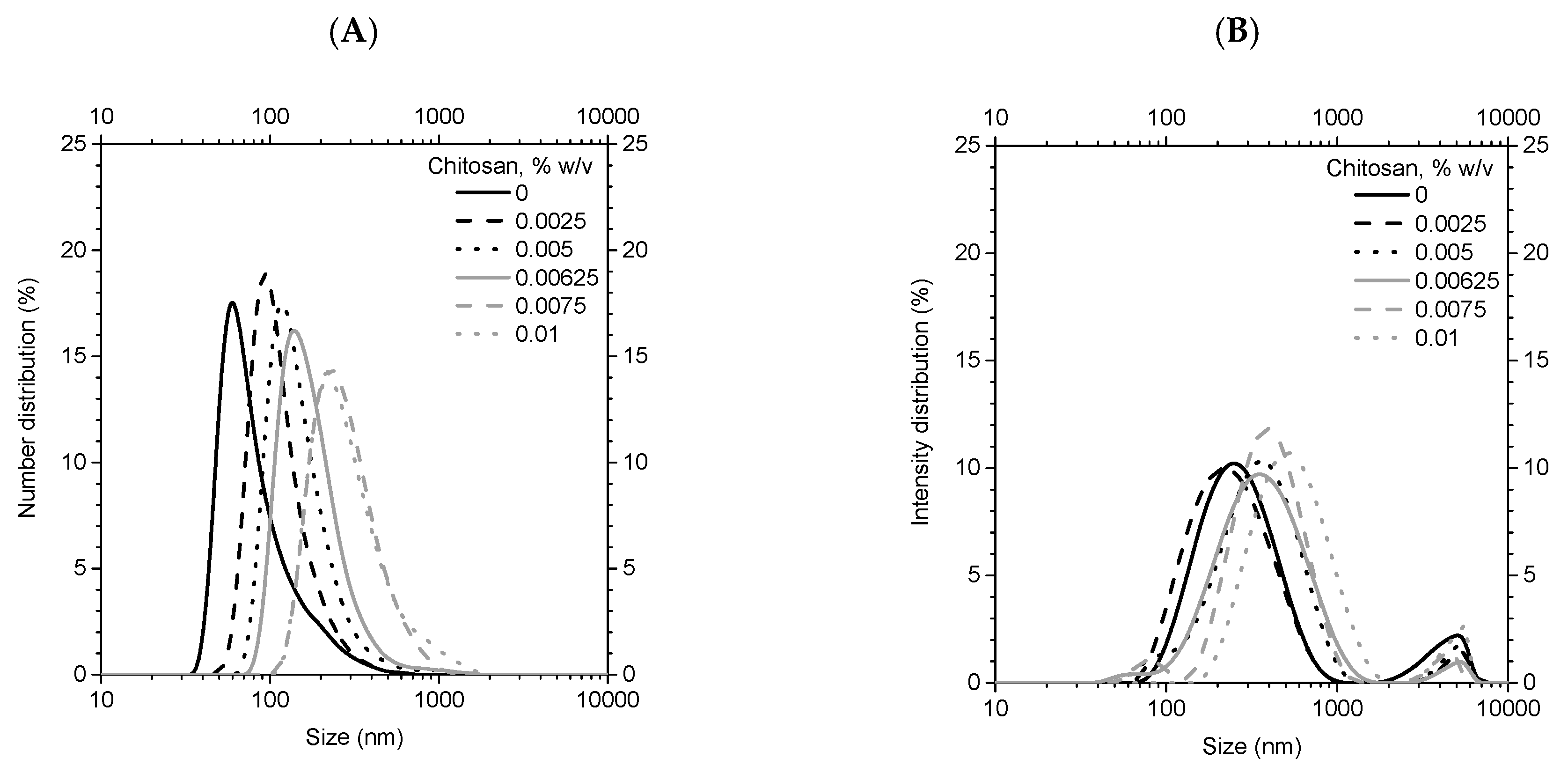
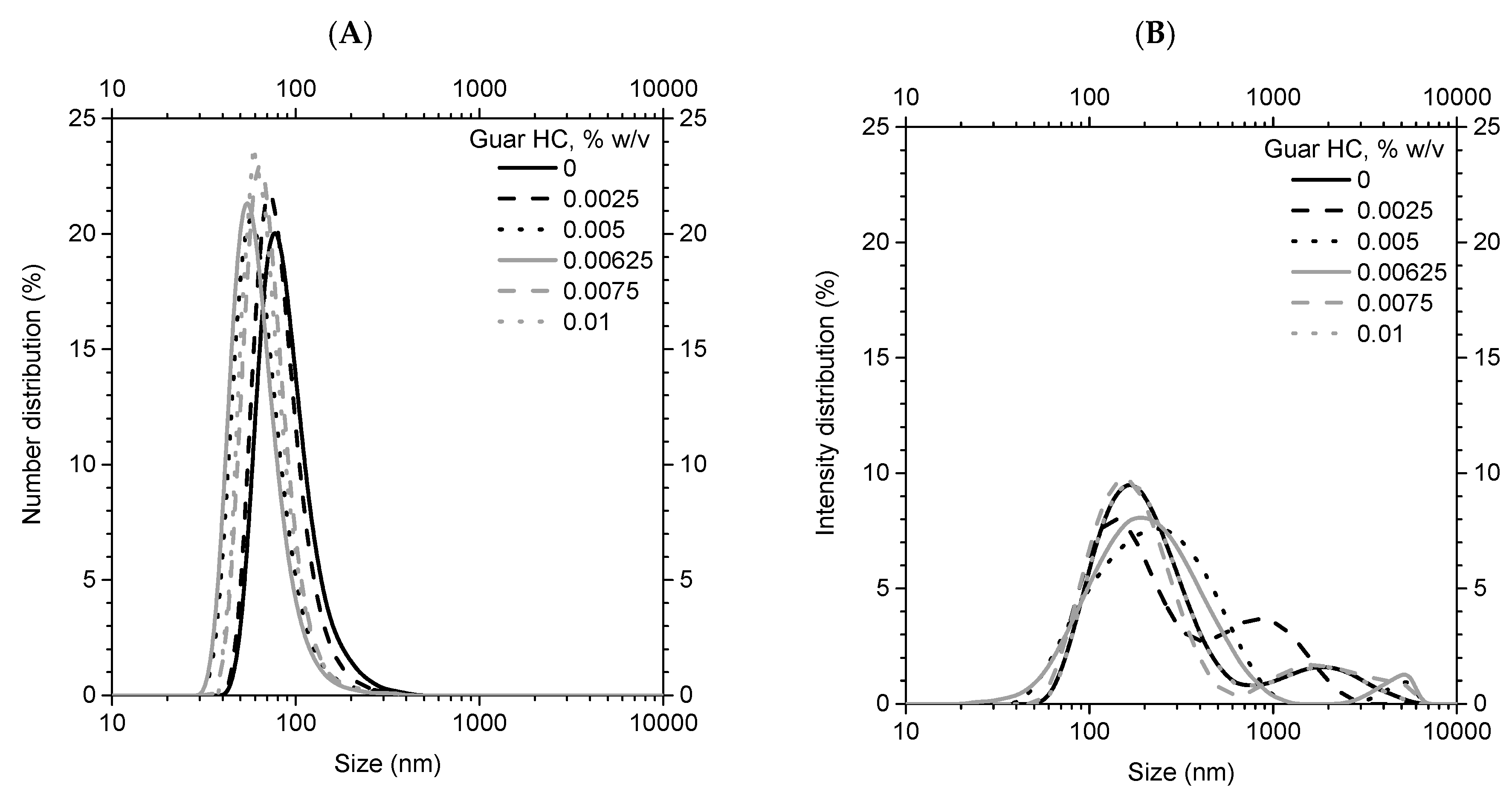
3.2. Characterization of Coated Nanolipid Structures

A summary of the size features of uncoated and coated nanolipid structures at the chosen polycation concentrations of 0.01% w/v are reported in Table 3.
Nanolipid structures coated by 0.01% polycation were characterized in terms of their morphology by TEM images, mucoadhesiveness by a mucin binding assay, and stability by turbidimetry.
TEM images of uncoated and chitosan-coated liposomes are shown in Figure 6. Uncoated ones (Figure 6A) appeared nanometric, spherical, and with a smooth surface. The chitosan coating by a simil-microfluidic technique (Figure 6B) did not alter the liposome’s shape, and the coating layer was clearly visualized on its surface, as already seen in previous works [2,16].
The TEM image of uncoated vegan liposomes (Figure 7A) showed a less defined shape (still spherical), which was due, perhaps, to the lack of the stabilizing action of cholesterol. In effect, it is known that cholesterol intercalates between lipid molecules, filling in free space, and decreasing the flexibility of surrounding lipid chains, thereby causing an increase in the mechanical rigidity of fluid membrane lipid bilayers [28]. The coating by Guar HC stabilized the vegan liposomes, as in Figure 7B, where a more defined shape of the nanolipid structure can be seen.
The mucoadhesive properties of nanolipid structures were highly improved by coating them with polycations at the chosen concentration (Figure 8A). In effect, the liposomes became very mucoadhesive passing, from about 30% of mucoadhesiveness in the uncoated form to more than 80% in the coated one. A similar result, albeit less pronounced, was observed for vegan liposomes, which showed an absence of mucoadhesiveness when uncoated and a mucoadhesiveness of about 10% when coated with Guar HC. The low mucoadhesiveness of the Guar HC-vegan liposomes’ structures compared to the Chitosan-coasted liposomes is due to the different structure of Guar HC, which, as previously reported (flip-flop effect), causes a partial/non homogenous covering. This result is reinforced by the behavior of nanocarriers during the turbidimetry test (Figure 8B). Chitosan-coated liposomes are less easily solubilized than uncoated ones thanks to their uniform layer of chitosan. On the other hand, Guar HC-coated vegan liposomes are more quickly solubilized because of their non-uniform coating, which allows the detergent to easily interact with the lipid bilayer. There is another reason for the heterogeneous dispersion of the polymer on the surface: the low Phosphatidylcholine (PC) purity between 14% and 23%, as reported in the Materials section. The low purity of PC is the cause of the vegan liposomes’ weakness (composed of only PC, without the stabilizing effect of cholesterol). This result is confirmed by the less defined shape of the vegan liposomes shown in Figure 7A. The lower stability of the vegan liposomes causes a less heterogeneous dispersion of polymera on the surface, resulting in a lower resistance to detergent and worse mucoadhesiveness properties when compared to liposomes containing cholesterol.
The evolution of size properties and mucoadhesiveness after 4 months of storage at 4 °C was observed for both standard liposomes (Figure 9) and vegan liposomes (Figure 10).
In general, the properties of both the coated and uncoated nanolipid vesicles did not change strongly, showing an acceptable stability during the 4 months of storage for both systems. However, for cholesterol containing preparations (Figure 9), uncoated liposomes were characterized by a light increase in PDI and a reduction in mucoadhesiveness after 4 months of storage, phenomena which were not observed for coated nanocarriers. This result confirms the better stability conferred by a chitosan coating. Similar results on evolution of size properties during 6 weeks of storage were reported in a previous work [2]. In vegan preparations (Figure 10), the coating with Guar HC avoided the increase of PDI during storage, also giving it improved stability.
All performed characterizations demonstrate that that coated (vegan or not) liposomes are ideal candidates for the controlled release of various ingredients. For example, chitosan coated liposomes—encapsulating nutraceuticals, with improved features in terms of stability, loading and mucoadhesiveness—have already been produced [10].

4. Conclusions

In this study, the novel simil-microfluidic technique was used in the coating process of negative charged liposomes with two kinds of polycations. In particular, chitosan-coated vesicles and a vegan alternative made by coating cholesterol-free liposomes by the vegan accepted polycation, Guar Hydroxypropyltrimonium Chloride, were produced. The study was performed by applying experimental and theoretical approaches, i.e., by experimentally evaluating the Z-potential and size evolution of (vegan or not) liposomes by using different polycation concentrations and then analyzing, starting from previous studies, saturation concepts, to detect the polycation concentrations’ range for obtaining stable covered liposomes to overcome the trial and error methodology usually used to define the best polymer concentrations for vesicle coating. The achieved results have confirmed the robustness of the investigative approach, leading to the production of fairly coated (vegan or not) nanoliposomes without aggregation phenomena.

Author Contributions

Conceptualization, A.D. and S.B.; Methodology, A.D. and S.B.; Formal Analysis, A.D. and S.B.; Investigation, A.D., S.B., and P.B.; Data Curation, A.D. and S.B.; Writing—Original Draft Preparation, A.D.; Writing—Review & Editing, A.D., A.A.B., and G.L.; Supervision, A.A.B. and G.L.

Funding

Part of the present work has been done within the funded project “Campania Oncoterapie”, POR FESR 2014–2020, D.D. n. 4 22/01/2019. CUP B61418000470007.

Acknowledgments

The authors thank the Centro di Microscopia Elettronica, University of Trieste, Italy.

Conflicts of Interest

The authors declare no conflict of interest.

References

  1. Bochicchio, S.; Dalmoro, A.; Barba, A.; d’Amore, M.; Lamberti, G. New Preparative Approaches for Micro and Nano Drug Delivery Carriers. Curr. Drug Deliv. 2017, 14, 203–215. [Google Scholar] [CrossRef] [PubMed]
  2. Bochicchio, S.; Dalmoro, A.; Bertoncin, P.; Lamberti, G.; Moustafine, R.I.; Barba, A.A. Design and production of hybrid nanoparticles with polymeric-lipid shell–core structures: Conventional and next-generation approaches. RSC Adv. 2018, 8, 34614–34624. [Google Scholar] [CrossRef]
  3. Volodkin, D.; Mohwald, H.; Voegel, J.C.; Ball, V. Coating of negatively charged liposomes by polylysine: Drug release study. J. Control. Release 2007, 117, 111–120. [Google Scholar] [CrossRef] [PubMed]
  4. Volodkin, D.; Ball, V.; Schaaf, P.; Voegel, J.C.; Mohwald, H. Complexation of phosphocholine liposomes with polylysine. Stabilization by surface coverage versus aggregation. Biochim. Et Biophys. Acta (BBA) Biomembr. 2007, 1768, 280–290. [Google Scholar] [CrossRef] [PubMed] [Green Version]
  5. Sybachin, A.; Efimova, A.; Litmanovich, E.; Menger, F.; Yaroslavov, A. Complexation of polycations to anionic liposomes: Composition and structure of the interfacial complexes. Langmuir 2007, 23, 10034–10039. [Google Scholar] [CrossRef] [PubMed]
  6. Yaroslavov, A.A.; Sybachin, A.V.; Kesselman, E.; Schmidt, J.; Talmon, Y.; Rizvi, S.A.; Menger, F.M. Liposome fusion rates depend upon the conformation of polycation catalysts. J. Am. Chem. Soc. 2011, 133, 2881–2883. [Google Scholar] [CrossRef] [PubMed]
  7. Horner, I.J.; Kraut, N.D.; Hurst, J.J.; Rook, A.M.; Collado, C.M.; Atilla-Gokcumen, G.E.; Maziarz, E.P.; Liu, X.M.; Merchea, M.M.; Bright, F.V. Effects of polyhexamethylene biguanide and polyquaternium-1 on phospholipid bilayer structure and dynamics. J. Phys. Chem. B 2015, 119, 10531–10542. [Google Scholar] [CrossRef] [PubMed]
  8. Refai, H.; Hassan, D.; Abdelmonem, R. Development and characterization of polymer-coated liposomes for vaginal delivery of sildenafil citrate. Drug Deliv. 2017, 24, 278–288. [Google Scholar] [CrossRef] [PubMed] [Green Version]
  9. Mengoni, T.; Adrian, M.; Pereira, S.; Santos-Carballal, B.; Kaiser, M.; Goycoolea, F. A Chitosan—Based Liposome Formulation Enhances the In Vitro Wound Healing Efficacy of Substance P Neuropeptide. Pharmaceutics 2017, 9, 56. [Google Scholar] [CrossRef]
  10. Dalmoro, A.; Bochicchio, S.; Lamberti, G.; Bertoncin, P.; Janssens, B.; Barba, A.A. Micronutrients encapsulation in enhanced nanoliposomal carriers by a novel preparative technology. RSC Adv. 2019, 9, 19800–19812. [Google Scholar] [CrossRef] [Green Version]
  11. Guzey, D.; McClements, D.J. Formation, stability and properties of multilayer emulsions for application in the food industry. Adv. Colloid Interface Sci. 2006, 128, 227–248. [Google Scholar] [CrossRef] [PubMed]
  12. McClements, D.J. Theoretical analysis of factors affecting the formation and stability of multilayered colloidal dispersions. Langmuir 2005, 21, 9777–9785. [Google Scholar] [CrossRef] [PubMed]
  13. Laye, C.; McClements, D.; Weiss, J. Formation of biopolymer-coated liposomes by electrostatic deposition of chitosan. J. Food Sci. 2008, 73, N7–N15. [Google Scholar] [CrossRef] [PubMed]
  14. Sandeep, C.; Deb, T.K.; Shivakumar, H. Cationic Guar Gum Poly Electrolytecomplex-Microparticles. J. Young Pharm. 2014, 6, 11. [Google Scholar] [CrossRef]
  15. Erazo-Majewicz, P.; Modi, J.J.; Xu, Z.F. Cationic, oxidized Polysaccharides in Conditioning Applications. U.S. Patent 7,589,051, 25 September 2009. [Google Scholar]
  16. Dalmoro, A.; Bochicchio, S.; Nasibullin, S.F.; Bertoncin, P.; Lamberti, G.; Barba, A.A.; Moustafine, R.I. Polymer-lipid hybrid nanoparticles as enhanced indomethacin delivery systems. Eur. J. Pharm. Sci. 2018, 121, 16–28. [Google Scholar] [CrossRef] [PubMed]
  17. Bochicchio, S.; Dalmoro, A.; Recupido, F.; Lamberti, G.; Barba, A.A. Nanoliposomes Production by a Protocol Based on a Simil-Microfluidic Approach. In Advances in Bionanomaterials; Springer: Berlin, Germany, 2018; pp. 3–10. [Google Scholar]
  18. Barba, A.A.; Lamberti, G.; d’Amore, M.; Bochicchio, S.; Dalmoro, A. Process for Preparing Nanoliposomes Comprising Micronutrients and Food Products Comprising Said Nanoliposomes. WO 2019/049186 A1, 6 september 2017. Patent WO 2019/049186 A1, 6 September 2017. [Google Scholar]
  19. Briuglia, M.L.; Rotella, C.; McFarlane, A.; Lamprou, D.A. Influence of cholesterol on liposome stability and on in vitro drug release. Drug Deliv. Transl. Res. 2015, 5, 231–242. [Google Scholar] [CrossRef] [PubMed] [Green Version]
  20. Tan, C.; Zhang, Y.; Abbas, S.; Feng, B.; Zhang, X.; Xia, S.; Chang, D. Insights into chitosan multiple functional properties: The role of chitosan conformation in the behavior of liposomal membrane. Food Funct. 2015, 6, 3702–3711. [Google Scholar] [CrossRef]
  21. Magarkar, A.; Dhawan, V.; Kallinteri, P.; Viitala, T.; Elmowafy, M.; Róg, T.; Bunker, A. Cholesterol level affects surface charge of lipid membranes in saline solution. Sci. Rep. 2014, 4, 5005. [Google Scholar] [CrossRef] [Green Version]
  22. Mady, M.M.; Darwish, M.M. Effect of chitosan coating on the characteristics of DPPC liposomes. J. Adv. Res. 2010, 1, 187–191. [Google Scholar] [CrossRef] [Green Version]
  23. Risica, D.; Dentini, M.; Crescenzi, V. Guar gum methyl ethers. Part I. Synthesis and macromolecular characterization. Polymer 2005, 46, 12247–12255. [Google Scholar] [CrossRef]
  24. Efimova, A.; Sybachin, A.; Yaroslavov, A. Effect of anionic-lipid-molecule geometry on the structure and properties of liposome-polycation complexes. Polym. Sci. Ser. C 2011, 53, 89. [Google Scholar] [CrossRef]
  25. Morris, G.A.; Castile, J.; Smith, A.; Adams, G.G.; Harding, S.E. Macromolecular conformation of chitosan in dilute solution: A new global hydrodynamic approach. Carbohydr. Polym. 2009, 76, 616–621. [Google Scholar] [CrossRef] [Green Version]
  26. Schatz, C.; Viton, C.; Delair, T.; Pichot, C.; Domard, A. Typical physicochemical behaviors of chitosan in aqueous solution. Biomacromolecules 2003, 4, 641–648. [Google Scholar] [CrossRef] [PubMed]
  27. Wang, C.; Li, X.; Li, P.; Niu, Y. Interactions between fluorinated cationic guar gum and surfactants in the dilute and semi-dilute solutions. Carbohydr. Polym. 2014, 99, 638–645. [Google Scholar] [CrossRef] [PubMed]
  28. Bhattacharya, S.; Haldar, S. Interactions between cholesterol and lipids in bilayer membranes. Role of lipid headgroup and hydrocarbon chain–backbone linkage. Biochim. Et Biophys. Acta (BBA) Biomembr. 2000, 1467, 39–53. [Google Scholar] [CrossRef]
Figure 1. Simil-microfluidic method piping representation according to the Italian UNICHIM (Association for the Unification of the Chemical Industry Sector) standard. ABOVE: Section of nanoliposome preparation: (123) lipids/ethanol feed line; (456) water feed line; (D-1 and D-2) feed tanks; (G-1 and G-2) peristaltic pumps; (I-1) injector (production section); (7) water/ethanol nanoliposome suspension; (D-3) recovering/homogenizing tank; (Z-1) sonication element for homogenization and reducing vesicle size. Section of nanoliposome covering: (8910) nanoliposome suspension; (111213) polycation solution feed line; (D-4 and D-5) feed tanks; (G-3 and G-4) peristaltic pumps; (I-2) injector (production section); (1415) coated nanoliposome suspension; (D-6) recovering/homogenizing tank. BELOW: The detail of interdiffusion between the lipid solution and the hydration solution (lines 3, 6, 7, and symbol I-1 in the UNICHIM representation) and between the liposome suspension and polycation solution (lines 13, 10, 14, and symbol I-2 in the UNICHIM representation) is given.
Coatings 09 00491 g001
Figure 2. Zeta potential evolution for unloaded liposomes (A) and vegan liposomes (B) by increasing the polymer concentration from 0% (uncoated) to 0.01% (chitosan for liposomes, A; cationic guar gum for vegan liposomes, B). Symbols are used for experimental data; the line represents the model fitting of the experimental data.
Coatings 09 00491 g002
Figure 3. (A) Chitosan structure; (B) Guar HC structure.
Coatings 09 00491 g003
Figure 4. (A) Size (left; numerical distributions) and (B) Z-average (right; intensity distribution) distributions of liposomes by increasing the chitosan concentration from 0% (uncoated) to 0.01%.
Coatings 09 00491 g004
Figure 5. (A) Size (left; numerical distributions) and (B) Z-average (right; intensity distribution) distributions of vegan liposomes by increasing the cationic guar gum concentration from 0% (uncoated) to 0.01%.
Coatings 09 00491 g005
Figure 6. TEM photos of both uncoated liposomes (A) and liposomes coated with 0.01% w/v of chitosan (B). The photos refer to larger vesicles for a better visualization of the liposome’s structure.
Coatings 09 00491 g006
Figure 7. TEM photos of both uncoated vegan liposomes (A) and those coated with 0.01% w/v of Guar HC (B). The photos refer to larger vesicles for a better visualization of the liposome’s structure.
Coatings 09 00491 g007
Figure 8. Mucoadhesiveness (A) and stability measured by turbidimetry (B) of uncoated liposomes and vegan liposomes and coated ones with 0.01% w/v of chitosan and guar HC, respectively.
Coatings 09 00491 g008
Figure 9. Numerical size (A), Z-Average (B), PDI (C), and Mucoadhesiveness (D) evolution from 0 to 4 months in storage conditions of 4 °C for liposomes (squares) and liposomes coated with 0.01% w/v of chitosan.
Coatings 09 00491 g009
Figure 10. Numerical size (A), Z-Average (B), PDI (C), and Mucoadhesiveness (D) evolution from 0 to 4 months in storage conditions of 4 °C for vegan liposomes (squares) and liposomes coated with 0.01% w/v of Guar HC.
Coatings 09 00491 g010
Table 1. The values of ZSat and CSat, from the fitting between the experimental data of Z-potential and Equation (1), with a relevant coefficient of determination and ΓSat.
ZSat, mVCSat, % w/vR2ΓSat, kg/m2
Liposomes−19.10.005370.9481.01 × 10−7
Vegan liposomes−24.50.005700.9901.06 × 10−7
Table 2. Values of CDep* and CAds for both standard liposomes and vegan liposomes, together with the values of molecular weight M and rPE of polycations.
M, kg/molrPE, mCDep*, % w/vCAds, % w/v
Liposomes–Chitosan3104 × 10−9 [26]0.04840.0057
Vegan liposomes–Guar HC6603 × 10−9 [27]0.22820.0086
Table 3. Numerical size, Z-average, and size distribution (PDI) of uncoated liposomes, vegan liposomes, and coated liposomes with 0.01% w/v of chitosan and guar HC, respectively.
Numerical Size (nm) ± SDZ-Average (nm) ± SDPDI ± SD
Liposomes88.3 ± 19.0252.8 ± 2.10.38 ± 0.02
Liposomes–Chitosan 0.01%257 ± 37.6504.4 ± 12.10.34 ± 0.03
Vegan liposomes65.9 ± 14.2179.5 ± 2.30.36 ± 0.00
Vegan liposomes–Guar HC 0.01%58.8 ± 1.3174.5 ± 2.70.36 ± 0.01

