Article

Chemical Modification of Novel Glycosidases from Lactobacillus plantarum Using Hyaluronic Acid: Effects on High Specificity against 6-Phosphate Glucopyranoside

1
Department of Biocatalysis, Institute of Catalysis (CSIC), Marie Curie 2, Campus UAM, Cantoblanco, E-28049 Madrid, Spain
2
Department of Microbial Biotechnology, Institute of Food Science, Technology and Nutrition (ICTAN-CSIC), José Antonio Novais 10, 28040 Madrid, Spain
3
Institute of Medicinal Chemistry (CSIC), Juan de la Cierva 3, E-28006 Madrid, Spain
*
Author to whom correspondence should be addressed.
Coatings 2019, 9(5), 311; https://doi.org/10.3390/coatings9050311
Submission received: 9 April 2019 / Revised: 30 April 2019 / Accepted: 7 May 2019 / Published: 9 May 2019
(This article belongs to the Special Issue Surface Chemical Modification)

Abstract

:
Three novel glycosidases produced from Lactobacillus plantarum, so called Lp_0440, Lp_2777, and Lp_3525, were isolated and overexpressed on Escherichia coli containing a His-tag for specific purification. Their specific activity was evaluated against the hydrolysis of p-nitrophenylglycosides and p-nitrophenyl-6-phosphate glycosides (glucose and galactose) at pH 7. All three were modified with hyaluronic acid (HA) following two strategies: A simple coating by direct incubation at alkaline pH or direct chemical modification at pH 6.8 through preactivation of HA with carbodiimide (EDC) and N-hydroxysuccinimide (NHS) at pH 4.8. The modifications exhibited important effect on enzyme activity and specificity against different glycopyranosides in the three cases. Physical modification showed a radical decrease in specific activity on all glycosidases, without any significant change in enzyme specificity toward monosaccharide (glucose or galactose) or glycoside (C-6 position free or phosphorylated). However, the surface covalent modification of the enzymes showed very interesting results. The glycosidase Lp_0440 showed low glycoside specificity at 25 °C, showing the same activity against p-nitrophenyl-glucopyranoside (pNP-Glu) or p-nitrophenyl-6-phosphate glucopyranoside (pNP-6P-Glu). However, the conjugated cHA-Lp_0440 showed a clear increase in the specificity towards the pNP-Glu and no activity against pNP-6P-Glu. The other two glycosidases (Lp_2777 and Lp_3525) showed high specificity towards pNP-6P-glycosides, especially to the glucose derivative. The HA covalent modification of Lp_3525 (cHA-Lp_3525) generated an enzyme completely specific against the pNP-6P-Glu (phosphoglycosidase) maintaining more than 80% of the activity after chemical modification. When the temperature was increased, an alteration of selectivity was observed. Lp_0440 and cHA-Lp_0440 only showed activity against p-nitrophenyl-galactopyranoside (pNP-Gal) at 40 °C, higher than at 25 °C in the case of the conjugated enzyme.

1. Introduction

Chemical modification of enzymes has been described as a fascinating approach for changing their catalytic properties [1,2,3,4,5]. One of the most generic strategies, which does not require previous genetic modification of the enzyme, is focused on the incorporation of molecules on protein residues chemical groups (COOH, NH2, OH) or by single chemical modification on the N terminus (the most reactive group at neutral pH) [3,4,5,6]. Both strategies have been successfully used to improve the activity or stability of enzymes in several cases [7,8,9,10]. However, very few examples have been reported, mainly in lipases, for the alteration of selectivity and, specifically, regioselectivity [9,10].
In this case, a particularly important application regarding the control of glycosidases regioselectivity [11,12] is their critical role in carbohydrate chemistry [13,14]. The process to control and modulate these enzymes for carbohydrate synthesis is a very important issue considering the key role that sugars play in a broad range of biological processes [15,16]. For example, the production of new sugars with different properties and the modification of natural products to enhance drugs’ functional properties such as solubility, pharmacokinetics, or pharmacodynamics [17,18].
From this point of view, phosphorylated carbohydrates represent an important class of sugars very relevant in biology [19]. For example, a challenging research line is obtaining Xeno nucleic acids (XNAs), synthetic nucleic acid analogues that have a different sugar backbone in comparison to DNA or RNA and could serve as building blocks for completely new genetic systems [20]. In particular, 1,5-anhydrohexitol nucleic acid (HNA) presents a glucose-mimic core with a phosphate group in C-6 [21]. However, phosphoglycosynthetic enzymes are not that widespread and need to be synthesized for their catalytic application.
Therefore, controlling the specificity of natural specific phosphoglycosidases is a very important challenge for future applications. In this work, we present the effect on regioselectivity of the chemical modification of three different glucosidases from a bacterial source, Lactobacillus plantarum, with a high selectivity to p-nitrophenyl-6-phosphate-glycopyranosides.
One of the most interesting modification of enzymes has been achieved using polymers [22]. For example, the use of tailor-made dextran polymers showed interesting results, obtaining novel and selective biocatalysts [7,8,9,10,23].
Hyaluronic acid is a well-known, widely studied polymer because of its interesting properties. It has been used as conjugate with different enzymes in biomedical applications, such as therapeutic proteins [24,25]. Structurally, is a glycopolymer based on a linear repetitive unit of a 1,4-disaccharide constituted by a glucuronic acid and a 2-acetamido-glucosamine units. As a comparison, dextran repetitive unit is a 1,3-1,6-trisaccharide (Scheme 1). Indeed, whereas the dextran must be functionalized to be applied for coating processes, HA is already functionalized due to the abundance of carboxylic groups.
Here, the covalent modification of the NH2 groups of three glycosidases from L. plantarum has been performed using hyaluronic acid to alter and improve the specificity towards p-nitrophenyl-6-phosphate-glycopyranosides against their corresponding free glycopyranosides. For that purpose, the hydrolytic activity of the different enzymes against four different substrates, 1-O-(4-nitrophenyl)-β-d-glucopyranoside (pNP-Glu), 1-O-(4-nitrophenyl)-6-O-phosphate-β-d-glucopyranoside (pNP-6P-Glu), 1-O-(4-nitrophenyl)-β-d-galactopyranoside (pNP-Gal), and 1-O-(4-nitrophenyl)-6-O-phosphate-β-d-galactopyranoside (pNP-6P-Gal) (Scheme 2) were evaluated.

2. Materials and Methods

2.1. General Description

1-O-(4-nitrophenyl)-β-d-glucopyranoside (pNP-Glu), 1-O-(4-nitrophenyl)-β-d-galactopyranoside (pNP-Gal), 1-O-(4-nitrophenyl)-6-O-phosphate-β-d-galactopyranoside (pNP-6P-Gal), N-hydroxysuccinimide (NHS), extra-low molecular weight hyaluronic acid 8000–15,000 Da (HA), acryl/bis-acrylamide 30% solution, ammonium persulfate (APS), N,N,N′,N′-tetramethylethylendiamine (TEMED), tris-base buffer, and sodium phosphate were purchased from Sigma-Aldrich (Saint Louis, MO, USA). 1-Ethyl-3-(3-dimethylaminopropyl) carbodiimide (EDC) was acquired from Tokyo Kasei (Japan). Spectrophotometric measurements were performed in a microplate reader from Biochrom (Cambridge, UK). Electrophoresis tools were acquired from Hoefer (Holliston, MA, USA).

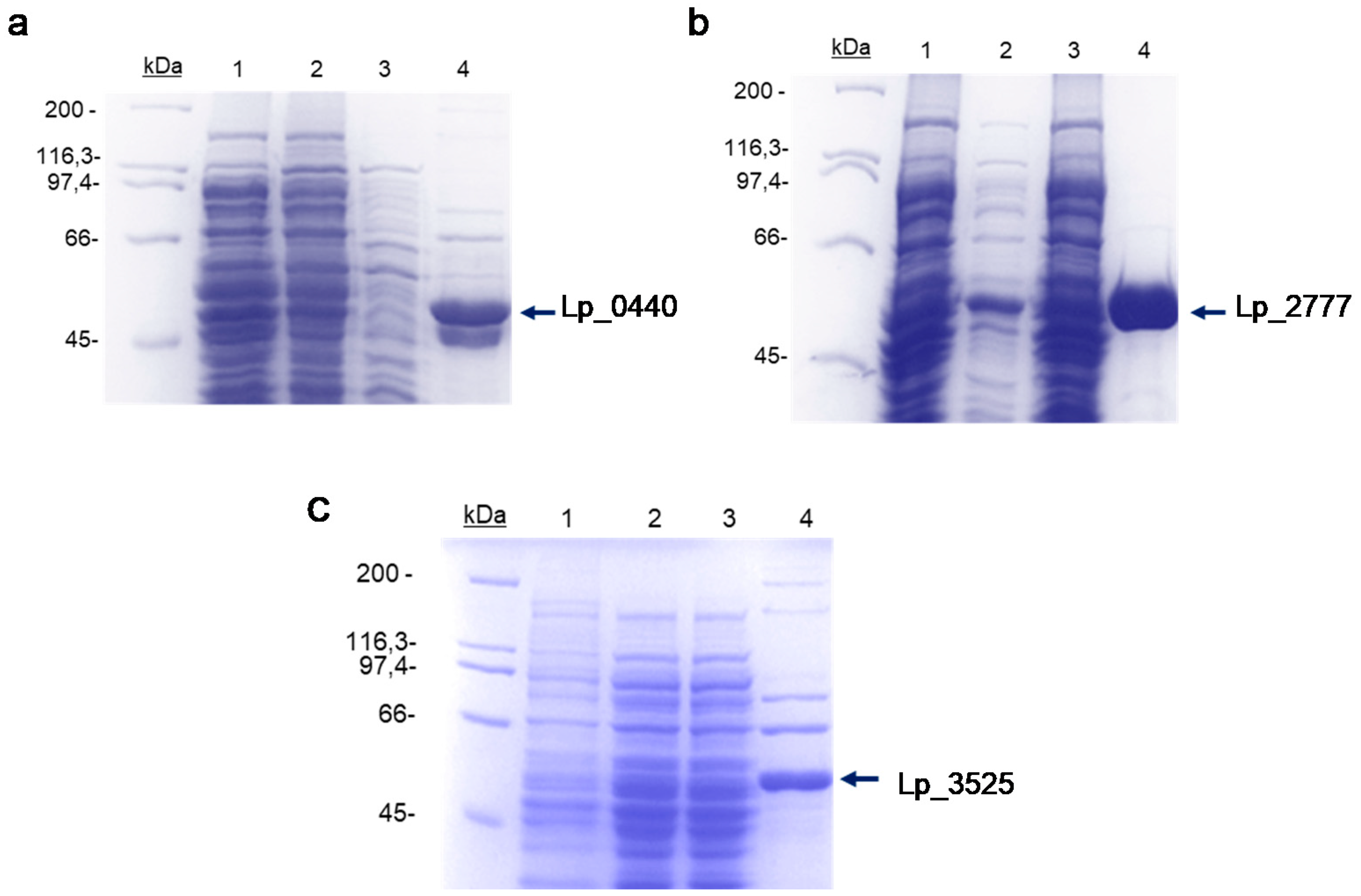
2.2. Expression of Lp_ 0440, Lp_2777, and Lp_3525 Genes in Escherichia coli and Purification of the Recombinant Proteins

The Lp_0440, Lp_2777, and Lp_3525 genes from L. plantarum WCFS1 were amplified by polymerase chain reaction (PCR). Oligonucleotides 1666 (5′ TAACTTTAAGAAGGAGATATACATATGACGATTAAAGGACGAGCGTTTC) + 1667 (5′-GCTATTAATGATGATGATGATGATGCTCAATTTCGGCACCATTTGTCGC) were used to amplify Lp_0440, primers 1591 (5′-TAACTTTAAGAAGGAGATATACATATGGCAACAACGAGTGGTTTAGA) + 1592 (5′-GCTATTAATGATGATGATGATGATGCTTCAAATCGGCCCCATTCGTC) to amplify Lp_2777, and finally, oligonucleotides 427 (5′-CATCATGGTGACGATGACGATAAGATGTCAGAGTTCCCAGAA) + 428 (5′-AAGCTTAGTTAGCTATTATGCGTACTATTTCTTTGTCAGCCCATTATGC) to amplify the Lp_3535 gene. Genbank accession number of the enzyme genes are Lp_2777 (YP_0048900399.1); Lp_3525 (YP_004891005.1); and Lp_0440 (YP_004888459.1). Advantage HD DNA polymerase (TaKaRa, Kusatsu, Japan) was used for PCR amplifications. The purified PCR products were inserted into the pURI3 (Lp_3525 gene) or pURI3-Cter (Lp_0440 and Lp_2777 genes) vectors using a restriction enzyme- and ligation-free cloning strategy [26]. These vectors produce recombinant proteins having a six-histidine affinity tag at their N-(Lp_3525) or C-terminal (Lp_0440 and Lp_2777) ends. Escherichia coli DH10B cells were transformed and the recombinant plasmids obtained (pURI3-Cter-Lp_0440, pURI3-Cter-Lp_2777, and pURI3-Lp_3525) were isolated and verified by DNA sequencing, and then used to transform E. coli BL21(DE3) cells for expression. E. coli cells were grown in lysogeny broth (LB) medium containing ampicillin (100 μg/mL), until they reached an optical density at 600 nm of 0.4 and induced by adding isopropyl β-d-1-thiogalactopyranoside (IPTG) (0.4 mM final concentration). After induction, the cells were grown at 22 °C during 20 h. The induced cells were harvested by centrifugation (8000 g for 15 min at 4 °C), resuspended in phosphate buffer (50 mM, pH 7) containing 300 mM NaCl, and disrupted by French press passages (three times at 1100 psi). Insoluble fraction of the lysates was removed by centrifugation at 47,000 g for 30 min at 4 °C. The supernatant was filtered through a 0.2-μm pore-size filter and then loaded onto a Talon Superflow resin (Clontech, Mountain View, CA, USA) equilibrated in phosphate buffer (50 mM, pH 7) containing 300 mM NaCl and 10 mM imidazole to improve the interaction specificity in the affinity chromatography step. The bound enzymes were eluted using 150 mM imidazole in the same buffer. The purity of the enzymes was determined by sodium dodecyl sulfate-polyacrylamide gel electrophoresis (SDS-PAGE) in tris-glycine buffer. Fractions containing the His6-tagged protein were pooled (fractions were dialyzed against 50 mM sodium phosphate buffer, 300 mM NaCl, pH 7 at 4 °C using dialysis membranes (OrDial D35-MWCO 3500, Orange Scientific, Braine-l’Alleud, Belgium) of 3.5 kDa pore diameter. Four changes of buffer were made to eliminate the imidazole present in the sample.) and analyzed. The three enzymes were defined as Lp_0440, Lp_2777, and Lp_3525.

2.3. Physical Modification of the New LpGs

First, 28 mg of HA were dissolved in 2.8 mL of water at pH 5 (final concentration 1% HA). Then, 200 µL of HA solution were added to 1 mL of a 0.05 mg/mL enzyme solution, and the mixture was left in agitation overnight. The novel biocatalysts were named HA-Lp_0440, HA-Lp_2777, or HA-Lp_3525, respectively.

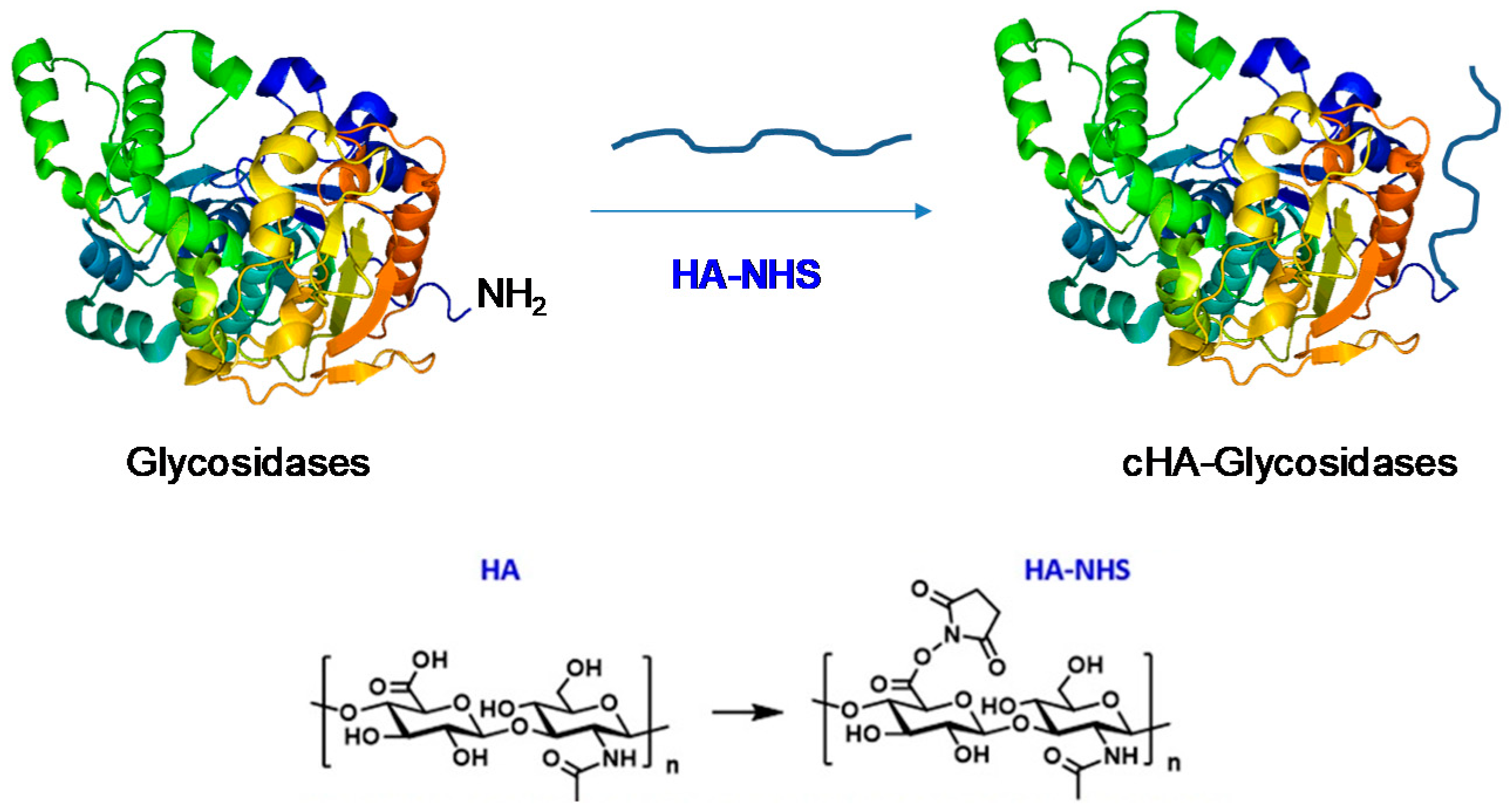
2.4. Covalent Modification of Lp Glycosidases with Hyaluronic Acid (HA)

28 mg of HA were dissolved in 2.8 mL of water at pH 5 (final concentration 1% HA). To this solution, 108 mg of EDC (10 eq) and 96 mg of NHS (15 eq) were added and pH checked (must be between 5.0 and 5.5). HA activation was left in agitation for one hour. After that, 200 µL of phosphate buffer 100 mM pH 7 was incorporated to inactivate the reagents. Next, 200 µL of activated HA were added to 1 mL of a 0.05 mg/mL enzyme solution and the mixture was left in agitation overnight. The novel biocatalysts were named as cHA-Lp_0440, cHA-Lp_2777, or cHA-Lp_3525, respectively.

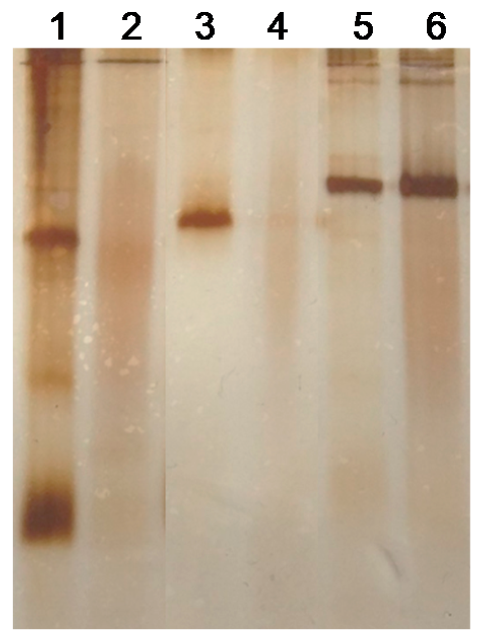
2.5. Native Electrophoresis Assay

For native PAGE, 12% separating gel and 4% stacking gel were used. Samples were mixed in Eppendorf tubes in 1:1 ratio with 2× sample buffer (25% glycerol, 62.5 mM tris-HCl pH 6.8 and 1% bromophenol blue) and the electrophoresis was performed in tris-HCl buffer pH 8.3 on ice at low voltage (around 120 V) and amperage, to prevent protein degradation. After that, silver staining was used [27].

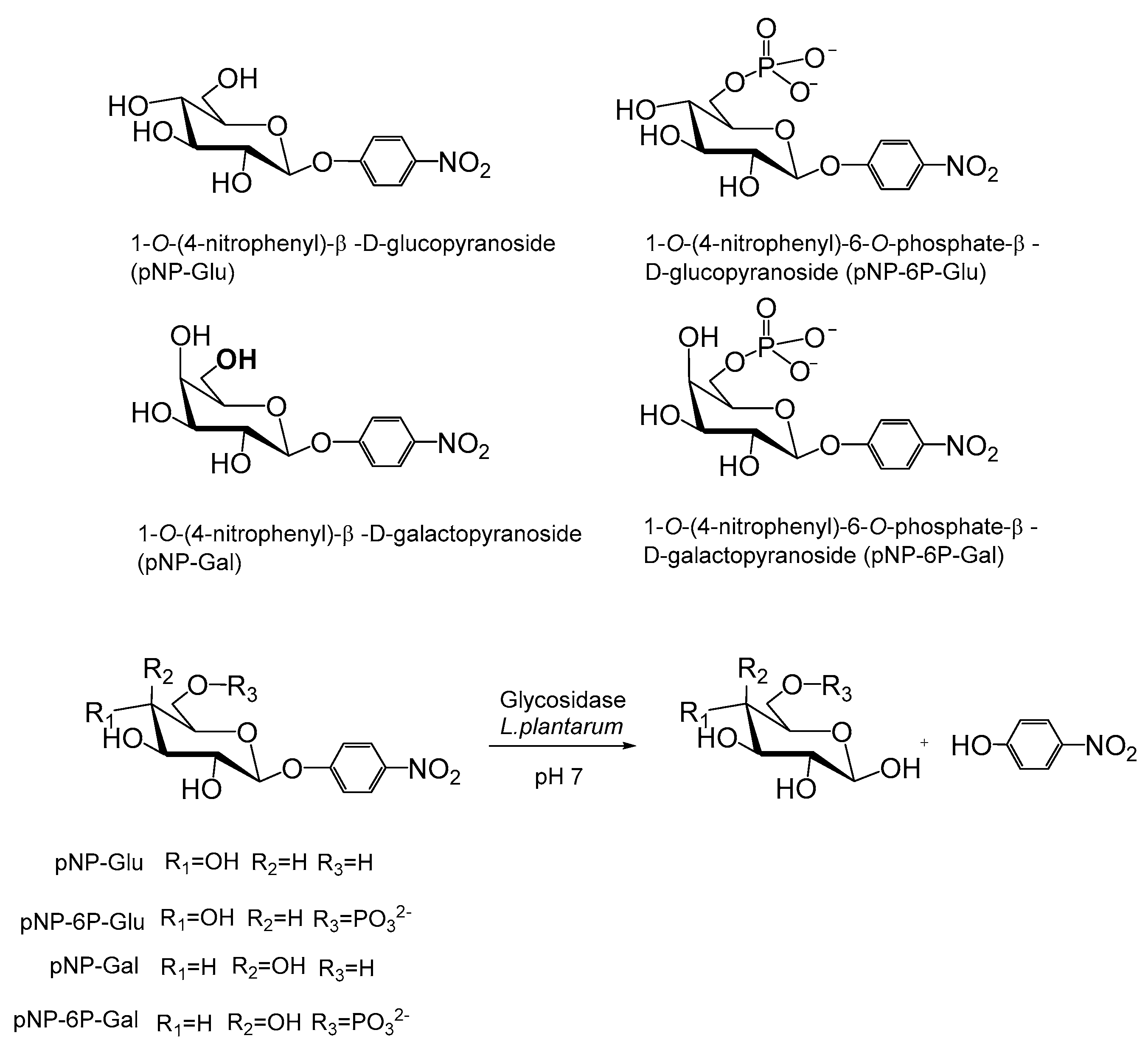
2.6. Synthesis of 4-Nitropheny,6-Phosphate-β-d-Glucopyranoside (pNP-6P-Glu)

Phosphorous oxychloride (0.74 mL) was added to a solution of 4-nitrophenyl β-d-glucopyranoside (2.6 mmol) in trimethyl phosphate (6.19 mL) and stirred for 3 h at 0 °C. Then, the pH of the reaction mixture was adjusted to 7 with aqueous ammonia 30% at 0 °C. The solvents were evaporated to dryness. The solid formed was washed with MeOH (200 mL) and the liquid phase was evaporated to dryness. The residue was washed again with AcOEt (50 mL) and Et2O (50 mL) and the liquid phase was evaporated under reduced pressure giving a white solid which was purified with HPLC semipreparative technology, using SunFire™ Prep C18 OBDTM (Waters corporation, Mildford, MA, USA) 5 μm, 19 mm × 150 mm column, to afford 404 mg of the glycoside 6-phosphate as white solid (41%). 1H NMR (500 MHz, Methanol-d4) δ 6.68 (dd, J = 9.3, 0.8 Hz, 1H), 5.71 (dd, J = 9.2, 0.8 Hz, 1H), 3.61–3.47 (m, 1H), 2.84–2.79 (m, 1H), 2.60–2.52 (m, 1H), 2.20–2.12 (m, 1H), 1.97–1.94 (m, 2H), 1.90–1.83 (m, 1H).13C NMR (126 MHz, Methanol-d4) δ 163.8, 144.0, 126.6, 117.8, 101.6, 77.7, 76.6, 74.7, 71.0, 66.7.

2.7. Enzymatic Activity Assay of Glycosidases in the Hydrolysis of p-Nitrophenyl-Glycopyranosides

Assays were monitored using a plate reader measuring at a wavelength of 405 nm. To each well, 300 µL of substrate were added and, over this, a different volume of enzyme solution was added depending of the activity of each one of them. For Lp_2777 and Lp_3525, only 10 µL (0.05 mg/mL) were needed, whereas for Lp_0440 (0.05 mg/mL), 50 µL were needed, due to its lower activity. The following substrates were tested: pNP-Glu, pNP-Gal, pNP-6P-Glu, and pNP-6P-Gal. Substrates were dissolved in phosphate buffer pH 7 to a 5 mM final concentration, except pNP-6P-Glu that was dissolved to a concentration of 1.25 mM. The enzyme activity of the different glycosidases was measured at room temperature and at 40 °C. For the latter purpose, the enzymes were incubated in a thermoblock that had been previously preheated to 40 °C. The sample was taken after 10 min and cooled at room temperature, then its activity was measured following the protocol described above.
Samples were measured at different times, obtaining 10 absorbance values, that were represented. Then, ∆Abs/min was obtained from the slope of the linear tendency showed by the data. Experiments were performed in triplicate.

3. Results and Discussion

3.1. Production and Purification of the Different Glycosidases

The proteins were identified based on their annotation in the database as 6-phospho-β-glucosidases and the three enzymes exhibited high sequence identity among them. The expression of Lp_3525 was previously analyzed [28] and the oligomeric state, enzymatic activity using salicin, cellobiose, and gentibiose as substrates, and crystallization of Lp_0440 was previously published [29].
The Lp_0440, Lp_2777, and Lp_3525 genes were cloned into the pURI3-Cter (Lp_0440 and Lp_2777 genes) and pURI3 (Lp_3525 gene) expression vectors by a ligation-free cloning strategy described previously [26]. The vectors incorporate the DNA sequence encoding hexahistidine at their N (Lp_3525)- or C-terminal (Lp_0440 and Lp_2777) ends to create a His-tagged fusion enzymes for further purification steps. Lp-3525 was cloned on the constructed pURI3 vector developed previously to produce proteins having a N-terminal poly-His-tag [30]. Later, a family vector from pURI3 were developed [26] where one of them, pURI3-Cter vector, allowed the production of recombinant proteins having poly-His-tag at the C-terminus, and Lp_2777 and Lp_0440 were cloned on it.
These genes were expressed under the control of an isopropyl-β-d-thiogalactopyranoside inducible promoter. Cell extracts were used to detect the presence of overproduced proteins by SDS-PAGE analysis (Figure 1). Overproduced proteins with an apparent molecular mass around 55 kDa were observed in cells harboring pURI3-Cter-0440 (Figure 1a), pURI3-Cter-2777 (Figure 1b), and pURI3-3525 (Figure 1c). The recombinant proteins (Lp_0440, Lp_2777, and Lp_3525) were purified by a metal affinity chromatography resin, and eluted with phosphate buffer (50 mM, pH 7) containing 300 mM NaCl and 150 mM imidazole. SDS-PAGE analysis (Figure 1) revealed the production of highly purified His6-tagged enzymes at yield from 3.4 mg protein/L culture (Lp_3525), 4.65 mg protein/L culture (Lp_0440) to 21.37 mg protein/L culture (Lp_2777).

3.2. Modification of Different LpGs by Hyaluronic Acid (HA)

The three different glycosidases (Lp_0440, Lp_2777, and Lp_3525) were specifically modified with hyaluronic acid (HA). The chemical modification was performed by the functionalized group COOH of the HA which was previously activated with N-hydroxysuccinimide (NHS) (Scheme 3). In all cases, the activated HA-NHS was added to the protein at pH 7, where mainly the most reactive group in the protein, the amino group in the N-terminus, reacted forming a covalent amide bond between enzyme and polymer (Scheme 3), producing the covalent HA (cHA)-glycosidase conjugates.
Taking into consideration that the introduction of the polymer can produce alteration in the neat charge of the protein, native PAGE of these modified glycosidases at the same concentration was performed (Figure 2).
The protein bands of the conjugates cHA-Lp_0440 and cHaLp_2777 were quite blurred with a slight lower RF, whereas the intensity of the band was conserved in the conjugate cHA-Lp_3525. Both enzymes, Lp_0440 and Lp_2777 present a His6-tag in the C-terminus of the protein, whereas in Lp_3525, this tag was located in the N-terminus. Considering the tridimensional structure of the enzyme Lp_0440 (Figure S1, Supplementary Materials), we have observed that both N and C-terminus are in the area in the protein. However, C-terminus is nearest to an alpha-helix in the protein, at 5.3 Å of distance to the Arg403 taken as reference whereas the N-terminus is at 16.3 Å (Figure S1). This alpha helix contains a number of polar aminoacid residues (Glu412, Asp401, Asp396). In the case of His6-tag at N-terminus (Lp_3525), the amino group where the polymer is anchored is far from the protein structure (Figure S2, Supplementary Materials) whereas when the His6-tag was introduced on the C-terminus (Lp_0440 and Lp_2777) the amino group on the protein from reaction is much near to the protein, being possible an interaction between the HA and tag, making maybe more difficult the electrophoretic mobility of the sample.
Also, a noncovalent strategy of the random interaction between the carboxylic groups of the polymer and amino groups on proteins (Lys) at alkaline pH was also performed. This strategy caused the interaction of the polymer on different areas of the proteins, specifically in the rich areas of Lysines (see Figure S3 in Supplementary Materials).

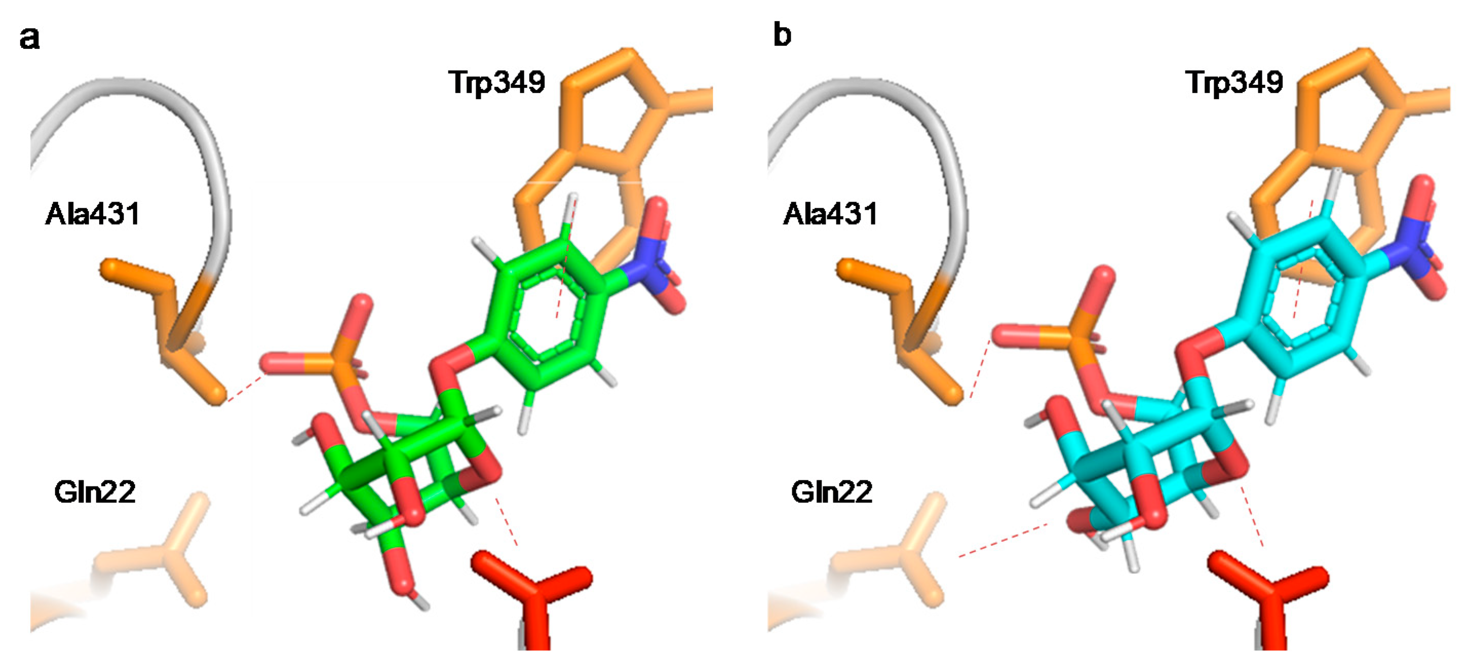
3.3. Glycosidase Activity, Specificity, and Regioselectivity of the Different Modified Glycosidases

The different nonmodified, physically and covalently modified glycosidases from L. plantarum were tested as catalysts in the hydrolysis of different p-nitrophenyl-glycopyranosides, non-phosphorylated (pNP-Glu, pNP-Gal) and C-6 phosphorylated (pNP-6P-Glu, pNP-6P-Gal) at 25 °C and pH 7 (Scheme 2).
The activity of the three different glycosidases were quite different depending on the used substrate (Table 1). Lp_0440 was a more promiscuous enzyme, being able to hydrolyze pNP-Glu, pNP-Gal, and pNP-6P-Glu. Indeed, the activity against pNP-Glu and pNP-6P-Glu was similar, and also showed two-fold less activity against pNP-Gal over pNP-Glu (Table 1, Entry 1).
A bioinformatic analysis of the crystal structure of Lp_0440 [28] showed three important aminoacidic residues clearly involved in the specificity and regioselectivity control (Figure 3). Ala431 is the responsible to interact with the phosphate molecule which is also stabilized, whereas the Gln22 is responsible to stabilize O4 in axial, controlling the regioselectivity between Gal and Glu.
Lp_0440 showed 2-times better accommodation of the Glu molecules, O in equatorial position than in axial. However, the surroundings of the active site did not affect the control of the interaction in Ala431 because the location between the hydroxyl in C-6, free or phosphorylated, presented the same accommodation, resulting in the same activity. The housing of the Gln22 enables the activity against galactopyranosides.
The modification of this enzyme with HA caused changes in activity and specificity against the different glycopyranosides. The simple physical adsorption approach caused a decrease in the activity, most notably against the pNP-6P-Glu. However, the covalent modification with HA resulted in an alteration of the modulation, directly improving the recognition of pNP-Glu more than 5 times over the pNP-6P-Glu. It was interesting that galactose activity was almost lost in the cHA-Lp_0440 variant.
The Lp_2777 enzyme was completely specific towards the p-nitrophenyl-6-P-glycopyranosides, especially pNP-6P-Glu (almost 200 times higher activity than pNP-6P-Gal). No traces of activity were observed using pNP-Glu or pNP-Gal (Table 1, Entry 5). In this case, the modification with HA caused a complete loss of activity by physical adsorption (HA-Lp_2777) and did not improve the specificity of the enzyme against any of the tested substrates in the covalent approach (cHA-Lp_2777) (Table 1, Entry 6).
A very different result was achieved in the Lp_3525 (Table 1). The enzyme was very specific for pNP-6-P-glycopyranosides and, as well as Lp_2777, showed much higher activity against pNP-6P-Glu (Table 1, Entry 7). The HA coating caused a high decrease in the activity (>90%), only conserving slight activity against pNP-6P-Glu.
However, the covalent attachment of HA generated a new enzyme (cHA-Lp_3525) highly specific, recognizing exclusively the pNP-6P-Glu, showing no activity against any other tested substrate (Table 1, Entry 9). This seems to indicate that the HA modification caused the rigidification needed in the active site as hypothesized in Figure 3a.
Furthermore, temperature effect on the activity and specificity for these enzymes was evaluated (Table 2). Short-time incubation of Lp_0440 at 40 °C showed an important shift of specificity against substrates. The enzyme recognized only the pNP-Gal at this pH, with a slight increase in activity over the one shown at 25 °C. This phenomenon was also observed in the cHA-Lp_0440 (Table 2, Entry 3), hydrolyzing exclusively pNP-Gal instead of pNP-Glu. This result could be explained by a clear alteration of the Gln22 (Figure 3) by the temperature shifting the selectivity towards the O-4 in axial (galactose) than in equatorial position (glucose) on the sugar moiety. No improvements in the specificity were observed in the other two enzymes, where Lp_3525 conserved almost all the activity whereas the modified ones suffered a high decrease (Table 2).
This mild and simple strategy to modifying for the first-time glycosidases activity and specificity could be another alternative strategy than other described in the literature to modified proteins by hyaluronic acid [31,32,33,34,35]. Most of the methodologies described for N-terminal modification of proteins using HA (e.g., slight HA oxidation or modification of HA with an acetal spacer [31,32]) are focused on biomedical applications and only few example are applied to enzymes, mainly for stability studies [31,32].

4. Conclusions

Chemical modification with hyaluronic acid has resulted in an interesting strategy to alter and modulate the specificity of three new glycosidases from L. plantarum. A very high specificity against pNP-6P-Glu has been obtained by the chemically modified cHA-Lp_3525 phosphoglycosidase. However, a more promiscuous glycosidase as Lp_0440, it was modulated its selectivity against a high specificity towards non-phosphorylated substrate after HA covalent modification.
Therefore, these results open the door to the application of this chemical modification strategy in order to control the specificity of glycosidases. This could be relevant, for example, to obtain synthetic biocatalysts needed to produce bioactive oligosaccharides.

Supplementary Materials

The following are available online at https://www.mdpi.com/2079-6412/9/5/311/s1, Figure S1: Lp_0440 structure incorporating a His6-tag in the C-terminus; Figure S2: Differences on the hyaluronic acid (HA) chemical modification of glycosidases containing a His6-tag in the N- or C-terminus; Figure S3: Lp_0440 surface tridimensional structure.

Author Contributions

C.P.-R., D.L.-T., L.P.-V., and J.C. performed the experiments; B.d.l.R. and R.M. prepared the enzymes, supervised and partially wrote the manuscript; J.M.P. supervised, conceived and designed the experiments; and J.M.P., C.P.-R., and D.L.-T. wrote the manuscript.

Funding

This work was sponsored by the Spanish Government (Project No. AGL2017-84614-C2-2-R) and CSIC Project No. CSIC-PIE 201880E011). We also thank the Ministry of Education, Youth and Sports of the Community of Madrid and the European Social Fund for a contract to C.P.-R (PEJD-2017PRE/SAL-3762) in the program of Youth Employment and the Youth Employment Initiative (YEI) 2017.

Acknowledgments

The authors thank the support by the Spanish National Research Council (CSIC).

Conflicts of Interest

The authors declare no conflict of interest.

References

  1. Spicer, C.D.; Davis, B.G. Selective chemical protein modification. Nat. Commun. 2014, 5, 4740. [Google Scholar] [CrossRef] [Green Version]
  2. MacDonald, J.I.; Munch, H.K.; Moore, T.; Francis, M.B. One-step site-specific modification of native proteins with 2-pyridinecarboxyaldehydes. Nat. Methods 2015, 11, 326–331. [Google Scholar] [CrossRef] [PubMed]
  3. Chan, A.O.Y.; Ho, C.M.; Chong, H.C.; Leung, Y.C.; Huang, J.S.; Wong, M.K.; Che, C.M. Modification of N-terminal α-amino groups of peptides and proteins using ketenes. J. Am. Chem. Soc. 2012, 134, 2589–2598. [Google Scholar] [CrossRef] [PubMed]
  4. Lai, Z.W.; Petrera, A.; Schilling, O. Protein amino-terminal modifications and proteomic approaches for N-terminal profiling. Chem. Boil. 2015, 24, 71–79. [Google Scholar] [CrossRef] [PubMed]
  5. Filice, M.; Romero, O.; Guisán, J.M.; Palomo, J.M. trans,trans-2,4-Hexadiene incorporation on enzymes for site-specific immobilization and fluorescent labeling. Org. Biomol. Chem. 2011, 9, 5535–5540. [Google Scholar] [CrossRef] [PubMed]
  6. Wu, B.; Wijma, H.J.; Song, L.; Rozeboom, H.J.; Poloni, C.; Tian, Y.; Arif, M.I.; Nuijens, T.; Quaedflieg, P.J.L.M.; Szymanski, W.; et al. Versatile peptide C-terminal functionalization via a computationally engineered peptide amidase. ACS Catal. 2016, 6, 5405–5414. [Google Scholar] [CrossRef]
  7. Díaz-Rodríguez, A.; Davis, B.G. Chemical modification in the creation of novel biocatalysts. Chem. Boil. 2011, 15, 211–219. [Google Scholar] [CrossRef]
  8. Romero, O.; Filice, M.; de las Rivas, B.; Carrasco-Lopez, C.; Klett, J.; Morreale, A.; Hermoso, J.A.; Guisan, J.M.; Abian, O.; Palomo, J.M. Semisynthetic peptide-lipase conjugates for improved biotransformations. Chem. Commun. 2012, 48, 9053–9055. [Google Scholar] [CrossRef]
  9. Gutarra, M.L.E.; Romero, O.; Abian, O.; Torres, F.A.G.; Freire, D.M.G.; Castro, A.M.; Guisan, J.M.; Palomo, J.M.; Freire, D.M.G. Enzyme surface glycosylation in the solid phase: Improved activity and selectivity of Candida antarctica lipase B. Chem. Cat. Chem. 2011, 3, 1902–1910. [Google Scholar] [CrossRef]
  10. Callaghan, C.; Redmond, M.; Filice, M.; Alnoch, R.C.; Mateo, C.; Palomo, J.M. Biocatalytic process optimization for the production of high-added-value 6-O-hydroxy and 3-O-hydroxy glycosyl building blocks. Chem. Cat. Chem. 2017, 9, 2536–2543. [Google Scholar] [CrossRef]
  11. Perugino, G.; Trincone, A.; Rossi, M.; Moracci, M. Oligosaccharide synthesis by glycosynthases. Trends Biotechnol. 2004, 22, 31–37. [Google Scholar] [CrossRef]
  12. Ajisaka, K.; Yamamoto, Y. Control of the regioselectivity in the enzymatic syntheses of oligosaccharides using glycosidases. Trends Glycosci. Glycotechnol. 2002, 14, 1–11. (In Japanese) [Google Scholar] [CrossRef]
  13. García, C.; Hoyos, P.; Hernáiz, M.J. Enzymatic synthesis of carbohydrates and glycoconjugates using lipases and glycosidases in green solvents. Biocatal. Biotrans. 2018, 36, 131–140. [Google Scholar] [CrossRef]
  14. Harit, V.K.; Ramesh, N.G. Amino-functionalized iminocyclitols: Synthetic glycomimetics of medicinal interest. RSC Adv. 2016, 6, 109528–109607. [Google Scholar] [CrossRef]
  15. Seeberger, P.H.; Werz, D.B. Synthesis and medical applications of oligosaccharides. Nat. Cell Boil. 2007, 446, 1046–1051. [Google Scholar] [CrossRef]
  16. Murrey, H.E.; Hsieh-Wilson, L.C. The chemical neurobiology of carbohydrates. Chem. Rev. 2008, 108, 1708–1731. [Google Scholar] [CrossRef]
  17. Campbell, C.T.; Yarema, K.J. Large-scale approaches for glycobiology. Genome Boil. 2005, 6, 236. [Google Scholar] [CrossRef]
  18. Filice, M.; Ubiali, D.; Fernandez-Lafuente, R.; Fernandez-Lorente, G.; Guisán, J.M.; Palomo, J.M.; Terreni, M. A chemo-biocatalytic approach in the synthesis of β-O-naphtylmethyl-N-peracetylated lactosamine. J. Mol. Catal. B Enzym. 2008, 52, 106–112. [Google Scholar] [CrossRef]
  19. Chen, L.; Huang, G. The antioxidant activity of derivatized cushaw polysaccharides. Int. J. Boil. Macromol. 2019, 128, 1–4. [Google Scholar] [CrossRef]
  20. Pinheiro, V.B.; Taylor, A.I.; Cozens, C.; Abramov, M.; Renders, M.; Zhang, S.; Chaput, J.C.; Wengel, J.; Peak-Chew, S.-Y.; McLaughlin, S.H.; et al. Synthetic genetic polymers capable of heredity and evolution. Science 2012, 336, 341–344. [Google Scholar] [CrossRef] [Green Version]
  21. Herdewijn, P. Nucleic acids with a six-membered ‘carbohydrate’ mimic in the backbone. Chem. Biodivers. 2010, 7, 1–59. [Google Scholar] [CrossRef]
  22. Wright, T.A.; Page, R.C.; Konkolewicz, D. Polymer conjugation of proteins as a synthetic post-translational modification to impact their stability and activity. Polym. Chem. 2019, 10, 434–454. [Google Scholar] [CrossRef]
  23. Romero, O.; Rivero, C.W.; Guisan, J.M.; Palomo, J.M.; Leite, L. Novel enzyme-polymer conjugates for biotechnological applications. PeerJ 2013, 1, e27. [Google Scholar] [CrossRef]
  24. Montagner, I.M.; Merlo, A.; Carpanese, D.; Pietà, A.D.; Mero, A.; Grigoletto, A.; Loregian, A.; Renier, D.; Campisi, M.; Zanovello, P.; et al. A site-selective hyaluronan-interferonα2a conjugate for the treatment of ovarian cancer. J. Control. Release 2016, 236, 79–89. [Google Scholar] [CrossRef] [Green Version]
  25. Huang, G.; Huang, H. Application of hyaluronic acid as carriers in drug delivery. Drug Deliv. 2018, 25, 766–772. [Google Scholar] [CrossRef] [Green Version]
  26. Curiel, J.A.; Rivas, B.D.L.; Mancheño, J.M.; Muñoz, R. The pURI family of expression vectors: A versatile set of ligation independent cloning plasmids for producing recombinant His-fusion proteins. Protein Expr. Purif. 2011, 76, 44–53. [Google Scholar] [CrossRef] [Green Version]
  27. Chevallet, M.; Luche, S.; Rabilloud, T. Silver staining of proteins in polyacrylamide gels. Nat. Protoc. 2006, 1, 1852–1858. [Google Scholar] [CrossRef] [Green Version]
  28. Marasco, R.; Muscariello, L.; Varcamonti, M.; De Felice, M.; Sacco, M. Expression of the bglH gene of Lactobacillus plantarum is controlled by carbon catabolite repression. J. Bacteriol. 1998, 180, 3400–3404. [Google Scholar]
  29. Michalska, K.; Tan, K.; Li, H.; Hatzos-Skintges, C.; Bearden, J.; Babnigg, G.; Joachimiak, A. GH1-family 6-P-β-glucosidases from human microbiome lactic acid bacteria. Acta Crystallogr. Sect. D Boil. Crystallogr. 2013, 69, 451–463. [Google Scholar] [CrossRef]
  30. De las Rivas, B.; Curiel, J.A.; Mancheño, J.M.; Muñoz, R. Expression vectors for enzyme restriction- and ligation-independent cloning for producing recombinant His-fusion proteins. Biotechnol. Prog. 2007, 23, 680–686. [Google Scholar] [CrossRef]
  31. Mero, A.; Pasqualin, M.; Campisi, M.; Renier, D.; Pasut, G. Conjugation of hyaluronan to proteins. Carbohydr. Polym. 2013, 92, 2163–2170. [Google Scholar] [CrossRef]
  32. Yang, J.-A.; Kim, E.-S.; Kwon, J.H.; Kim, H.; Shin, J.H.; Yun, S.H.; Choi, K.Y.; Hahn, S.K. Transdermal delivery of hyaluronic acid–Human growth hormone conjugate. Biomaterials 2012, 33, 5947–5954. [Google Scholar] [CrossRef]
  33. Mero, A.; Campisi, M. Hyaluronic acid bioconjugates for the delivery of bioactive molecules. Polymers 2014, 6, 346–369. [Google Scholar] [CrossRef]
  34. Martínez-Sanz, E.; Ossipov, D.A.; Hilborn, J.; Larsson, S.; Jonsson, K.B.; Varghese, O.P. Bone reservoir: Injectable hyaluronic acid hydrogel for minimal invasive bone augmentation. J. Control. Release 2011, 152, 232–240. [Google Scholar] [CrossRef]
  35. Yang, J.-A.; Park, K.; Jung, H.; Kim, H.; Hong, S.W.; Yoon, S.K.; Hahn, S.K. Target specific hyaluronic acid–Interferon alpha conjugate for the treatment of hepatitis C virus infection. Biomaterials 2011, 32, 8722–8729. [Google Scholar] [CrossRef]
Scheme 1. Different natural polysaccharides.
Coatings 09 00311 sch001
Scheme 2. Hydrolysis of p-nitrophenyl-glycopyranosides catalyzed by glycosidases.
Coatings 09 00311 sch002
Figure 1. The sodium dodecyl sulfate-polyacrylamide gel electrophoresis (SDS-PAGE) analysis of the purification of different glycosidases from Lactobacillus plantarum WCFS1. (a) Lp_0440, (b) Lp_2777, and (c) Lp_3525. Lane 1: Soluble cell extracts of Escherichia coli BL21 (DE3) (pURI3-Cter for a,b) or (pURI3 for c). Lane 2: E. coli BL21 (DE3) (pURI3-Cter-0440 for (a), pURI3-Cter-2777 for (b), pURI3-Cter-3525 for (c). Lane 3: Flowthrough. Lane 4: Protein eluted after His affinity resin and dialysis. The gel was stained with Coomassie blue. Molecular mass markers are located on the left (SDS-PAGE Standards, Bio-Rad, Hercules, CA, USA).
Coatings 09 00311 g001
Scheme 3. The chemical modification with NHS-activated hyaluronic acid (8–14 kDa) of the different glycosidases.
Coatings 09 00311 sch003
Figure 2. Native PAGE of the different modified cHA-glycosidades. Lp_0440 (Lane 1), cHA-Lp_0440 (Lane 2), Lp_2777 (Lane 3), cHA-Lp_2777 (Lane 4), Lp_3525 (Lane 5), cHA-Lp_3525 (Lane 6).
Coatings 09 00311 g002
Figure 3. Lp_0440 in complex with (a) pNP-6P-Glu and (b) pNP-6P-Gal. Glu180 (active site) is marked in red, pNP-6P-Glu in green, and pNP-6P-Gal in blue. The protein structure was obtained from the Protein Data Bank (PDB code: 3qom) and the pictures were created using Pymol v. 0.99.
Coatings 09 00311 g003
Table 1. The hydrolysis of p-nitrophenyl-glycopyranosides catalyzed by glycosidases from L. plantarum at 25 °C a.
EntryBiocatalystActivity (∆Abs/min) a
pNP-Glu bpNP-6P-Glu cpNP-Gal bpNP-6P-Gal b
1Lp_044039.20 ± 1.2039.1 ± 1.1015.60
2HA-Lp_04405.80 ± 0.203.30 ± 0.105.10
3cHA-Lp_044013.10 ± 0.391.90 ± 0.1070
4Lp_27770270 ± 801.4 ± 0.07
5HA-Lp_27770000
6cHA-Lp_2777080 ± 200.5 ± 0.04
7Lp_35250335 ± 10012.4 ± 0.38
8HA-Lp_3525020 ± 0.6000
9cHA-Lp_35250252 ± 7.500
a Activity values ×10−3; b Conditions: Substrates were dissolved in phosphate buffer pH 7 to a 5 mM final concentration, and 10 µL of enzyme solution (0.05 mg/mL) were added, except for Lp_0440, where 50 µL (0.05 mg/mL) were used; c Conditions: Substrates were dissolved in phosphate buffer pH 7 to a 1.25 mM final concentration., and 10 µL of enzyme solution (0.05 mg/mL) were added, except for Lp_0440, where 50 µL (0.05 mg/mL) were used.
Table 2. Hydrolysis of p-nitrophenyl-glycopyranosides catalyzed by glycosidases from L. plantarum at 40 °C a.
EntryBiocatalystActivity (∆Abs/min) a
pNP-Glu bpNP-6P-Glu cpNP-Gal bpNP-6P-Gal b
1Lp_04400017 ± 0.500
2HA-Lp_0440001.6 ± 0.130
3cHA-Lp_04400011.4 ± 0.340
4Lp_2777069 ± 203.2 ± 0.01
5HA-Lp_27770000.7 ± 0.07
6cHA-Lp_277708.7 ± 0.2600.8 ± 0.08
7Lp_35250300 ± 905.5 ± 0.17
8HA-Lp_35250000
9cHA-Lp_352500.3 ± 0.0303 ± 0.15
a Activity values ×10−3; b Conditions: Substrates were dissolved in phosphate buffer pH 7 to a 5 mM final concentration, and 10 µL of enzyme solution (0.05 mg/mL) were added, except for Lp_0440, where 50 µL (0.05 mg/mL) were used; c Conditions: Substrates were dissolved in phosphate buffer pH 7 to a 1.25 mM final concentration, and 10 µL of enzyme solution (0.05 mg/mL) were added, except for Lp_0440, where 50 µL (0.05 mg/mL) were used.

