Article

Super-Hydrophobic Co–Ni Coating with High Abrasion Resistance Prepared by Electrodeposition

1
National Center for Materials Service Safety, University of Science and Technology Beijing, Beijing 100083, China
2
Shandong Provincial Key Laboratory for High Strength Lightweight Metallic Materials, Advanced Materials Institute, Qilu University of Technology (Shandong Academy of Sciences), Jinan 250000, China
*
Authors to whom correspondence should be addressed.
These authors contributed equally to this work.
Coatings 2019, 9(4), 232; https://doi.org/10.3390/coatings9040232
Submission received: 19 February 2019 / Revised: 23 March 2019 / Accepted: 29 March 2019 / Published: 2 April 2019
(This article belongs to the Special Issue Superhydrophobic Coatings for Corrosion and Tribology)

Abstract

:
Although super-hydrophobic surfaces have great application prospects in industry, their preparation cost and mechanical durability have limited their practical utilization. In this work, we presented a new low-cost process preparation for super-hydrophobic Co–Ni coating on carbon steel substrate via an electrodeposition route. The deposited Co–Ni coating with cauliflower-shaped micro-nano structures exhibited high super-hydrophobic properties with water contact angles over 161° after modification with 1H,1H,2H,2H-Perfluorooctyltrichlorosilane (PFTEOS). Evaluated by the linear abrasion methods, the super-hydrophobic coating can maintain super-hydrophobicity after abrasion distance of 12 m under the applied pressure of 5 kPa, which was attributed to the high cobalt content of the Co–Ni coating. Moreover, electrochemical tests showed that the super-hydrophobic Co–Ni coatings exhibited a good anti-corrosion performance thus providing an adequate protection to the carbon steel substrates.

1. Introduction

Biomimetic materials with hierarchical structures, which are compatible with environmental requirements, have become a major concern in the scientific community recently [1,2]. It is well known that many naturally occurring surfaces, including lotus leaves and water striders, possess super-hydrophobic and self-cleaning properties, having water contact angles (WCA) greater than 150° and a small contact angle hysteresis of less than 10° [3,4]. Super-hydrophobic coatings have received extensive research attentions due to their potential applications in corrosion protection [5], anti-icing [6], water/oil separation [7] and drag reduction [8]. Generally speaking, the wetting property of a solid surface is determined by the combination of chemical compositions and microstructures. To fabricate the super-hydrophobic surface, a large number of techniques have been developed, such as etching [9], femtosecond laser machining [10], chemical vapor deposition [11], anodic oxidation [12] and colloidal coating [13]. Certain fabrication techniques to develop super-hydrophobic surfaces have been limited in industrial applications because they are relatively expensive and time-consuming. Generally, the electrodeposition method represents a lower cost technique being well adapted towards industrial applications so that it is considered suitable to be applied to build large-area super-hydrophobic surfaces [14].
Until now, in spite of the various preparation techniques, the commercialization of super-hydrophobic surfaces have been significantly limited due to their poor mechanical abrasion resistance and surface chemical stability, as well as the fragility of their microscopic roughness features [15]. Mechanical strength and durability of super-hydrophobic coatings are, therefore, of considerable focus in recent studies [15,16]. The robust super-hydrophobic coating of contact angle above 163° was fabricated by She et al. on a magnesium alloy substrate via the combination of electrodeposition and chemical modification technique [17]. After mechanical abrasion for 0.7 m with the 800 grit SiC sandpapers under applied pressure of 1.2 kPa, the as-prepared sample with pinecone-like hierarchical structure could maintain the contact angle above 150° [17]. Electrodeposition of Mg–Mn–Ce magnesium plate in the ethanol solution of cerium nitrate and myristic acid was used by Liu et al. to construct a super-hydrophobic surface with a maximum contact angle of 158° that lose the super-hydrophobicity after abrasion for 0.4 m under the pressure of 1.3 kPa [18]. A super-hydrophobic Ni coating with pinecone-like hierarchical micro-nanostructure was prepared by Su et al. [19], which involved electroplating in a Watts bath and heat-treatment with triethoxysilane (AC-FAS). The deposit was found to possess super-hydrophobicity and a good mechanical abrasion resistance after mechanical abrasion against 800 grit SiC sandpaper for 1.0 m under the pressure of 4.80 kPa [19]. Jain et al. [20] prepared a durable copper-based super-hydrophobic surface with cauliflower shaped fractal morphology via an electrodeposition route which lost its non-wetting nature after being dragged on 800 grit sandpaper for a distance of 2.0 m under the applied pressure of 3 kPa. Tam et al. fabricated super-hydrophobic nanocrystalline Ni–PTFE composite coating by co-electrodeposition process [21]. On the 800 grit sandpaper, the water contact angle of the Ni–PTFE composite coating can remain above 150° after 50 m of abrasion under the applied pressure of 2.0 kPa [21]. Until now, the abrasion resistance of super-hydrophobic surfaces is still not satisfactory, thus more research attention should be paid to improve its mechanical durability.
In this work, robust super-hydrophobic Co–Ni coating with cauliflower-shaped micro-nano structures was fabricated on carbon steel via a low-cost electrochemical deposition process. In the linear abrasion test, the super-hydrophobic properties can be maintained after abrasion distance of 12 m under the applied pressure of 5 kPa, which was significantly improved compared with the previous reports [19,20]. Moreover, electrochemical tests demonstrated that the super-hydrophobic Co–Ni coating possessed good corrosion resistance for carbon steel substrate.

2. Materials and Methods

Nickel chloride (NiCl2), cobalt chloride (CoCl2) and boric acid (H3BO3) were bought from Sino pharm Chemical Reagent Co., Ltd. (Beijing, China). The low surface energy material (1H,1H,2H,2H-Perfluorooctyltrichlorosilane, PFTEOS) was provided from Beijing Bai Ling Wei Technology Co., Ltd. (Beijing, China). The chemical reagents were of analytical grade and used as-received.
A conventional three electrode configuration was used to prepare the samples. Carbon steel substrate in a square shape was used as working electrode. A platinum sheet with a size of 30 × 30 mm2 worked as counter electrode while saturated calomel electrode (SCE) was used as the reference electrode. Before electrochemical deposition, the carbon steel substrate was sealed in epoxy with an exposed surface area of 1 cm2. Afterwards, it was mechanically polished down to 2000 grit size by SiC sandpapers and rinsed with deionized water. The mixed solution was selected based on our previous report [22] and improved for the purpose of abrasion resistance. The solutions with CoCl2 (0.1 mol/L), NiCl2 (0.03 mol/L) and H3BO3 (0.1 mol/L) were prepared and used directly. The cyclic voltammograms of carbon steel were recorded in the solution of CoCl2 0.1 mol/L + H3BO3 0.1 mol/L, NiCl2 0.03 mol/L + H3BO3 0.1 mol/L, CoCl2 0.1 mol/L + NiCl2 0.03 mol/L + H3BO3 0.1 mol/L with a scan rate of 10 mV/s. The Co–Ni coatings were deposited at a constant potential of −1.0, −1.4 and −1.7 V for 3000 s at room temperature in mixed solution CoCl2 0.1 mol/L + NiCl2 0.03 mol/L + H3BO3 0.1 mol/L. After electrochemical deposition, the as-prepared samples were treated with 5 wt.% PFTEOS ethanol solution for 1 h at room temperature, then rinsed and dried for investigations.
To test the wetting property of the as-prepared samples, the water contact angle and water sliding angle (WSA) were measured by an Automatic Contact Angle Meter (Model SL150 Series, USA KINO, Boston, MA, USA) at ambient temperature using 5 and 12 μL distilled water, respectively. During the measurement of water sliding angle, the inclination of the slope was controlled by the cornering device. For accuracy, the different samples were measured under the same condition for three times in order to obtain the average values reported. The surface morphologies of all samples were obtained by scanning electron microscope (SEM, EVO MA 25/LS 25, Carl Zeiss, Opokochen, Germany). The chemical compositions and chemical states of the top coating surface were characterized by EDS (X-flash-Detector 5010, Bruker, Karlsruhe, Germany) and X-ray photoelectron spectrometer (XPS, Kratos Axis Ultra DLD, Kratos Analytical, Hadano, Japan). The linear abrasion test with 800 grit SiC sandpaper was carried out to evaluate the mechanical durability of the as-prepared samples [23]. The laser scanning confocal microscopy (LSCM, LEXTOLS4000 OLYMPUS, Tokyo, Japan) was employed to investigate the surface roughness of the as-deposited coatings.
The electrochemical corrosion resistance was evaluated by a conventional three electrode system, in which the as-prepared sample, a platinum plate and a saturated calomel electrode (SCE) were used as the working electrode, counter electrode and reference electrode, respectively. The test was conducted with an electrochemical workstation (Gamry-Reference 3000, Gamry Instruments, Warminster, PA, USA) in 3.5 wt.% NaCl solution at room temperature. The potentiodynamic polarization curves were recorded from −1.3 to 0.0 V with a scan rate of 0.5 mV/s. The Tafel extrapolation method was used to extract the corrosion potential (Ecorr) and corrosion current density (Icorr) from the polarization curves. Electrochemical impedance spectroscopy (EIS) measurements were performed at open circuit potential (OCP) under the employed amplitude signal of 5 mV in the frequency range from 105 to 10−2 Hz. Software ZSimpWin (version 3.3) was utilized to fit the obtained impedance data.

3. Results and Discussion

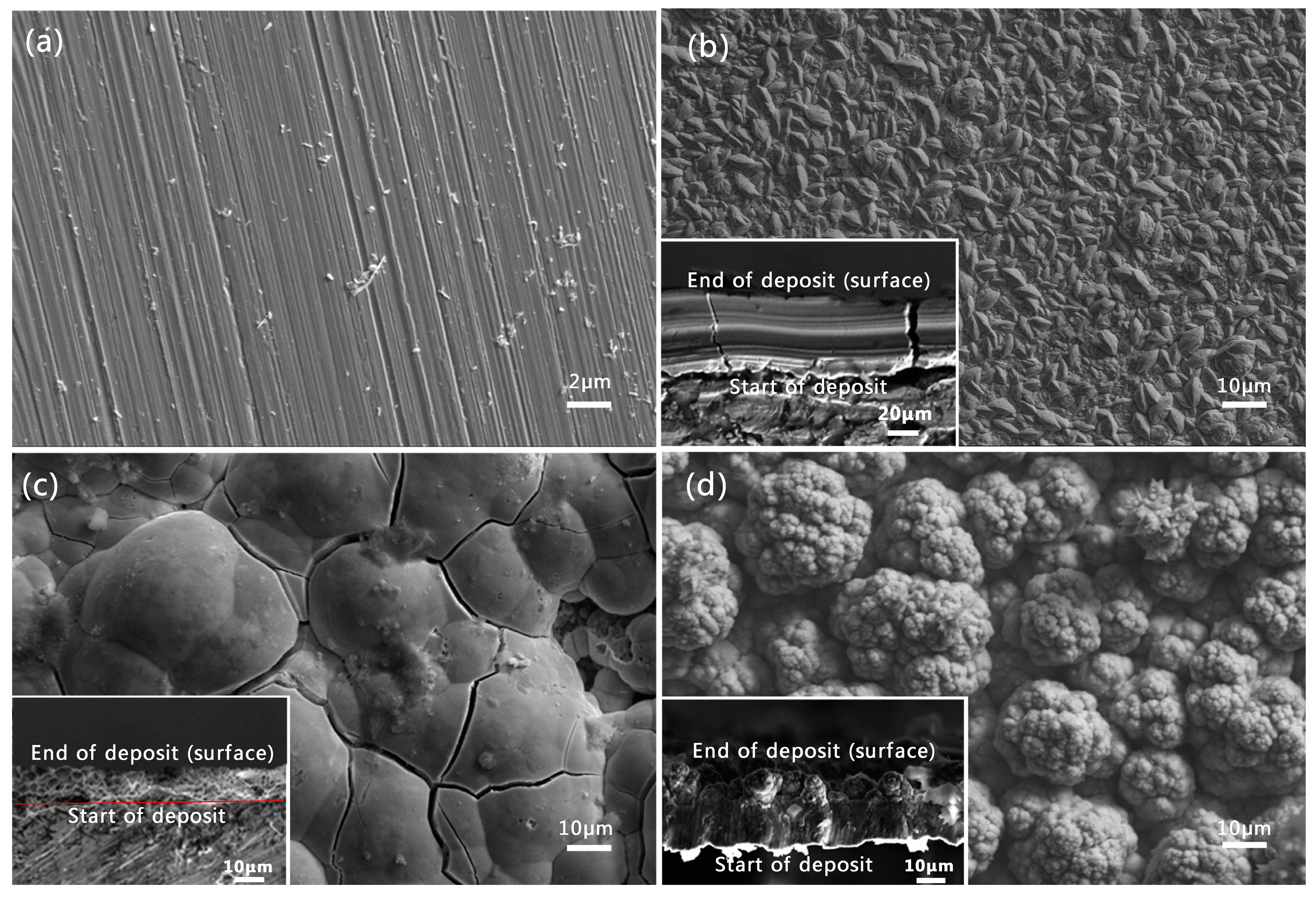
3.1. Surface Morphology of the As-Deposited Co–Ni Coating

In order to determine the applied deposition potentials, the cyclic voltammograms of carbon steel were recorded with a scan rate of 10 mV/s in different solutions (Figure S1). Accordingly, the applied potentials for metal electrodeposition should be more negative than its equilibrium potential. The higher over-potential will lead to larger deposition driving force, which will favor the formation of hierarchical structures. Figure 1 shows typical morphologies of the bare carbon steel and electrodeposited Co–Ni coating under different applied potentials for 3000 s in the mixed solution with CoCl2 (0.1 mol/L), NiCl2(0.03 mol/L) and H3BO3 (0.1 mol/L). For the bare carbon steel, the polished surface displayed lots of scratches (Figure 1a). After electrochemical deposition under the applied potential at −1.0 V for 3000 s in the above-mentioned solution, the carbon steel surface was covered by uniform granular structures containing average size of sub-micrometer in diameter and the as-polished scratches were covered completely (Figure 1b). As shown in the cross-section view, the thickness of the coating is around 26 μm for the deposition time of 3000 s, and the EDS result showed that the cobalt content is around 93.8%, which is higher than that of the initial electrolyte (77%, CoCl2 0.1 mol/L NiCl2 0.03 mol/L). After the deposition at −1.4 V for 3000 s, the coating surface evidenced lots of spherical humps with an average size of 33 μm and a large number of cracks (Figure 1c), which may be attributed to the internal stress generated during the electrochemical deposition process. The cross-section image revealed that the thickness of the coating was around 40 μm and the large spherical humps grew from the thin layer composed of small irregular crystals. Increasing the applied potential to −1.7 V, the cauliflower-shaped micro-nano structures with multiscale fractal nature were obtained (Figure 1d). The EDS result shows that the cobalt content decreased to around 77.7% as presented in Table 1, which is similar to that of the initial electrolyte (77%). The anomalous Co–Ni deposition behavior can be attributed to the formation of Co hydroxyl precipitate, which could hinder the subsequent Ni deposition at the solid/electrolyte interface [24]. Under different thermodynamic and kinetic conditions, the various external crystal morphologies including powder [25], film [26] and dendrite shapes [27,28] could be generated during the electrodeposition process. In our case, increasing the over-potential can favor the development of protrusions in the direction of increasing concentration, therefore leading to the formation of cauliflower-shaped micro-nano structures.

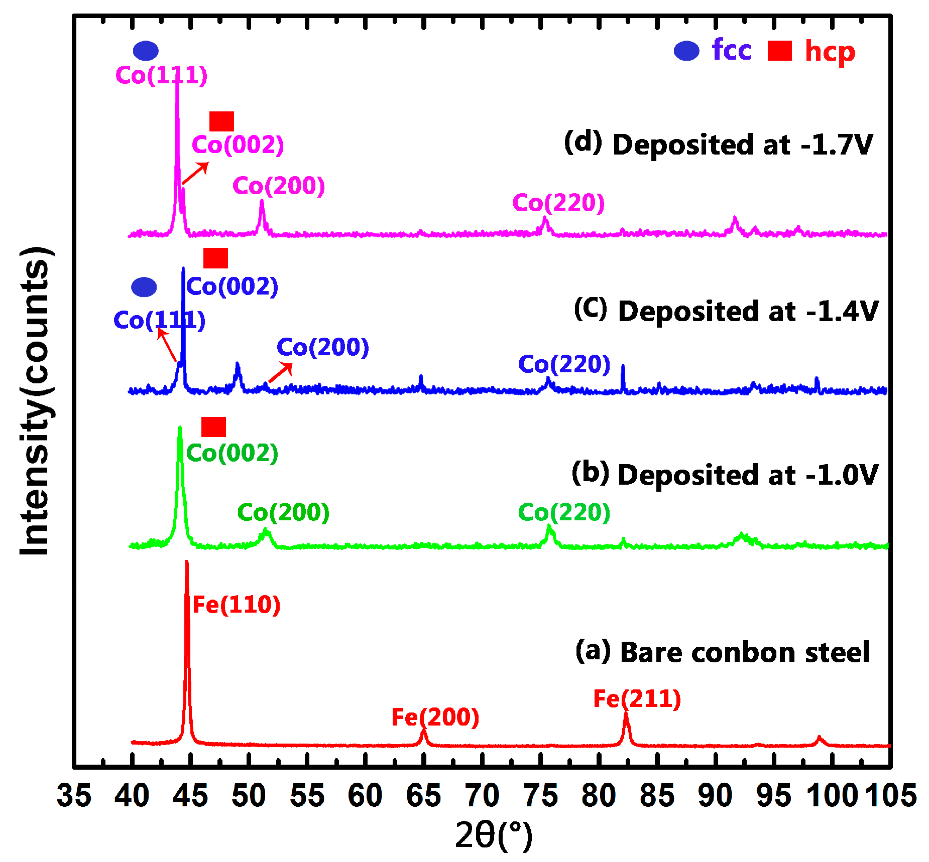
3.2. Structure of As-Deposited Co–Ni Coating

To identify the crystal structure of the deposited coatings, the XRD technique was conducted. Figure 2 shows the XRD patterns of the bare carbon steel substrate and Co–Ni coatings prepared at the applied potentials of −1.0, −1.4 and −1.7 V, respectively. For the bare carbon steel substrate, the diffraction peaks in 2θ of 44.5°, 65° and 82° can be attributed to the existence of Fe (JCPDS file No. 870721) (Figure 2a). After electrochemical deposition, the diffraction peaks located at 44.5°, 75.9°, and 84.1° correspond to the peaks of (002), (110), and (103) crystalline faces of close-packed hexagonal (hcp) Co (JCPDS file No. 040850) (Figure 2b–d); the diffraction peaks near 44.2°, 51.5°, and 75.9° can be assigned to the peaks of (111), (200), and (220) crystalline faces of face-centered cube (fcc) Co (JCPDS file No. 897093) (Figure 2b–d). In the XRD patterns, no pure nickel can be detected, revealing the formation of homogeneous solid solutions of Co–Ni alloy under the deposition conditions [29,30]. When deposited at the applied potential of −1.0 V, the Co–Ni coating shows completely a hexagonal structure with the cobalt content of around 93.8% according to the EDS results (Table 1). By increasing the applied deposition potential, the cobalt content decreases. As the cobalt content on the coating obtained at the applied potential of −1.7 V decreases to around 77.7%, the face-centered cubic structure becomes dominant.

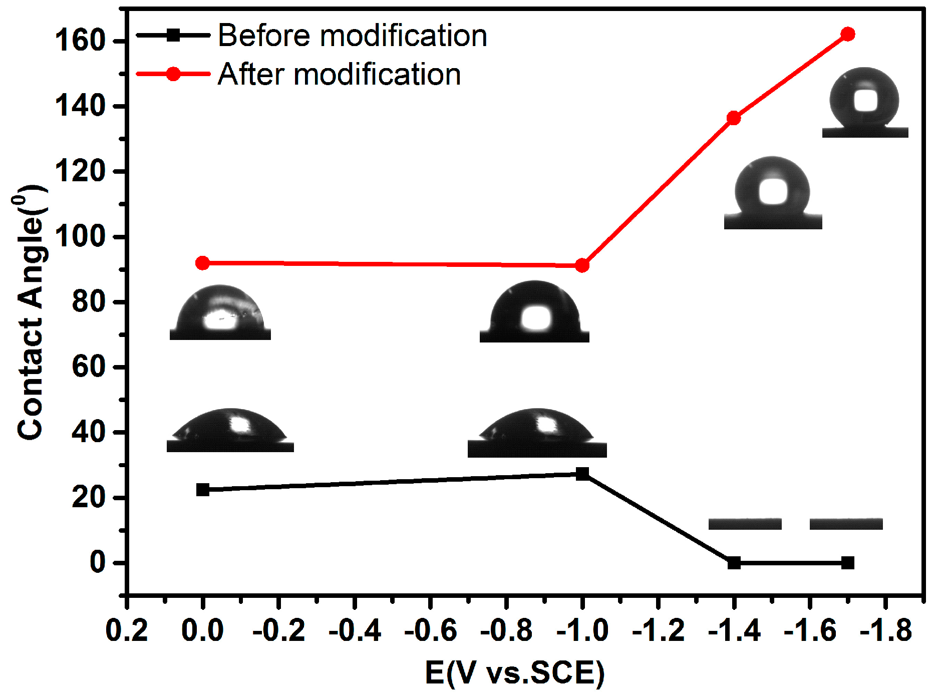
3.3. Surface Wetting Property of As-Deposited Co–Ni Coatings

Before the wetting property measurement of the as-deposited Co–Ni coatings, the surface roughness of these coatings was evaluated by the laser scanning confocal microscopy. As shown in the Figure S2, the Co–Ni coating deposited at −1.4 V showed the Ra of around 1.71 μm, which was higher than that of Co–Ni coating deposited at −1.0 V. Meanwhile, the Co–Ni coating with cauliflower-shaped micro-nano structures deposited at −1.7 V showed the highest surface roughness with Ra of 7.77 μm. By adjusting the applied potential to a more negative direction, hierarchical structures with higher surface roughness can be favored to be generated.
To examine the surface wetting property of the as-prepared Co–Ni coatings, the water contact angle tests were conducted. Figure 3 depicts the contact angle variations of the Co–Ni coatings with the applied potentials before and after modification by PFTEOS in ethanol solution. For bare carbon steel substrate after polishing, and Co–Ni coating deposited at the applied potential of −1.0 V, the contact angles were around 20°. After modification by PFTEOS, the water contact angle reached 95° and the surfaces displayed a hydrophobic property. After electrodeposition at the applied potentials ranging from −1.4 to −1.7 V, the water contact angles of the Co–Ni coatings with spherical humps structures and cauliflower-shaped micro-nano structures decreased to almost zero, showing the super-wetting properties of deposited Co–Ni coatings. However, after modification by 5 wt.% PFTEOS in ethanol solution for 1 h at room temperature, the water contact angle increases drastically to 140° for the sample with spherical humps structures, and to 161° for the sample with cauliflower-shaped micro-nano structures deposited at −1.7 V, respectively.
The above results indicate that the wetting properties of the deposited hierarchical Co–Ni coating with cauliflower-shaped micro-nano structures converted from super-wetting to super-hydrophobic behaviors during the PFTEOS modification process. The super-hydrophobic behavior on Co–Ni coating with cauliflower-shaped micro-nano structures was endowed with a high surface roughness and low surface energy materials achieved by the combination of electrodeposition process at higher overpotential and surface modification by PFTEOS. The super-hydrophobicity of the as-deposited Co–Ni coating can be explained by the Cassie-Baxter model. Based on the Cassie-Baxter Equation [31]:
cos θ = f sl ( cos θ y + 1 ) 1
where, θy and θ are Young’s contact angle and liquid–gas contact angle, fsl is the contact area fraction of solid–liquid interface, the area fraction of water-air interface calculated is around 94%, suggesting that the water droplet was sustained by the heterogeneous composite surface consisting of Co–Ni cauliflower-shaped micro-nano structures and air cushion among these structures.

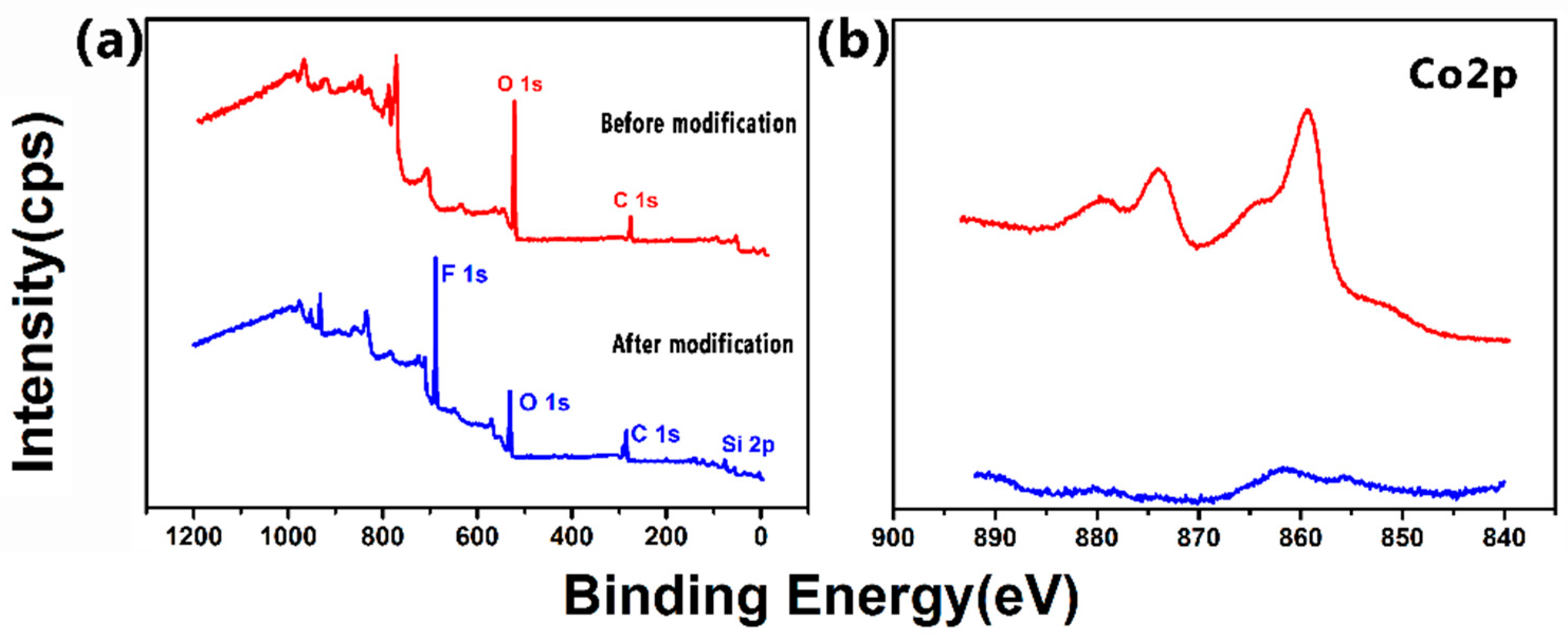
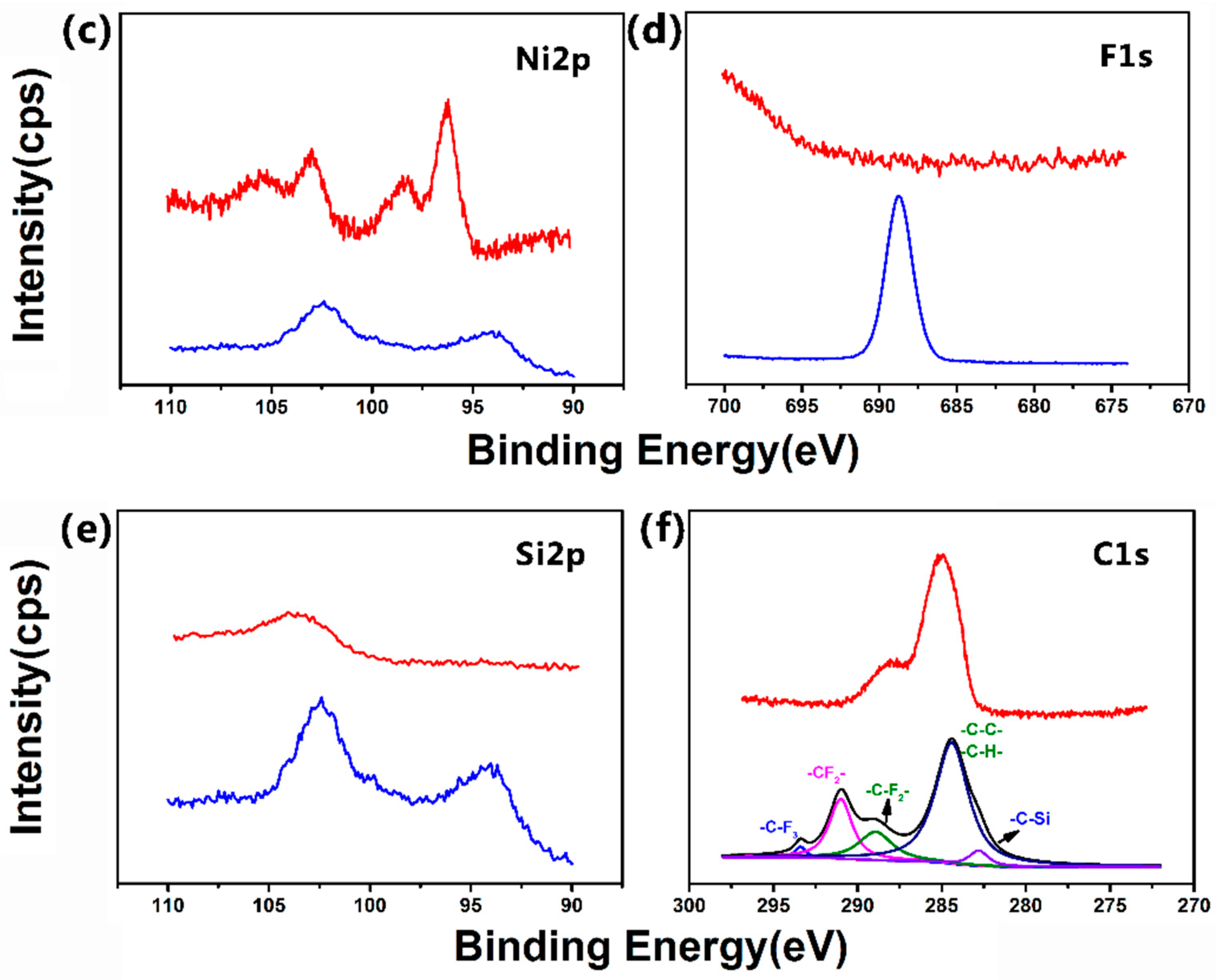
3.4. Surface Composition of the Super-Hydrophobic Co–Ni Coating

X-ray photoelectron spectroscopy (XPS) was utilized to confirm the adsorption of the PFTEOS molecules on the Co–Ni coating with cauliflower-shaped micro-nano structures deposited at the applied potential of –1.7 V. As can be seen in Figure 4, before PETEOS treatment, the strong signals of Co 2p and Ni 2p core levels reveals that the formation of Co–Ni electrodeposits (Figure 4a–c). After treatment by PFTEOS ethanol solution for 1 h, strong signal of F 1s and Si 2p core levels can be observed (Figure 4a,d,e). Figure 4f depicted the high-resolution C 1s core level spectra. The peaks at binding energy of 293.6, 291.1 and 288.7 eV can be ascribed to the –C–F3 group, −C–F2− group and –C−CF2– group respectively [19,32]. These results demonstrated that the PFTEOS molecules adsorbed successfully on the Co–Ni coating with cauliflower-shaped micro-nano structures after treatment. Accordingly, the Co–Ni coating with cauliflower-shaped micro-nano structures prepared by electrochemical deposition exhibits higher surface roughness Ra of 7.77 μm. After PFTEOS treatment, the fluorinated components with low surface energy endowed the rough Co–Ni coating super-hydrophobic properties, which is similar to the observed lotus leaves with dual micro and nano-scale structure covered by a wax layer [3].

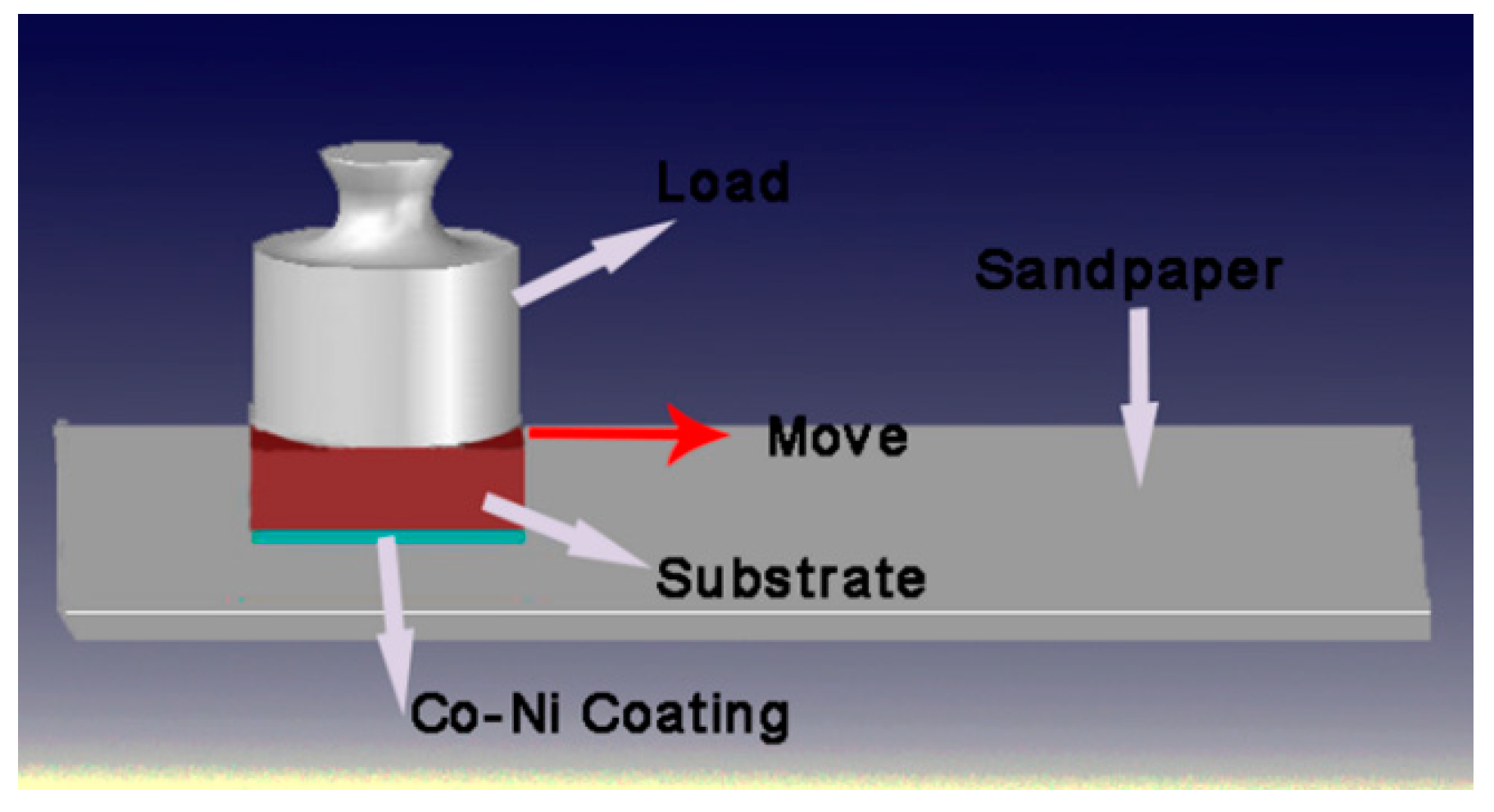
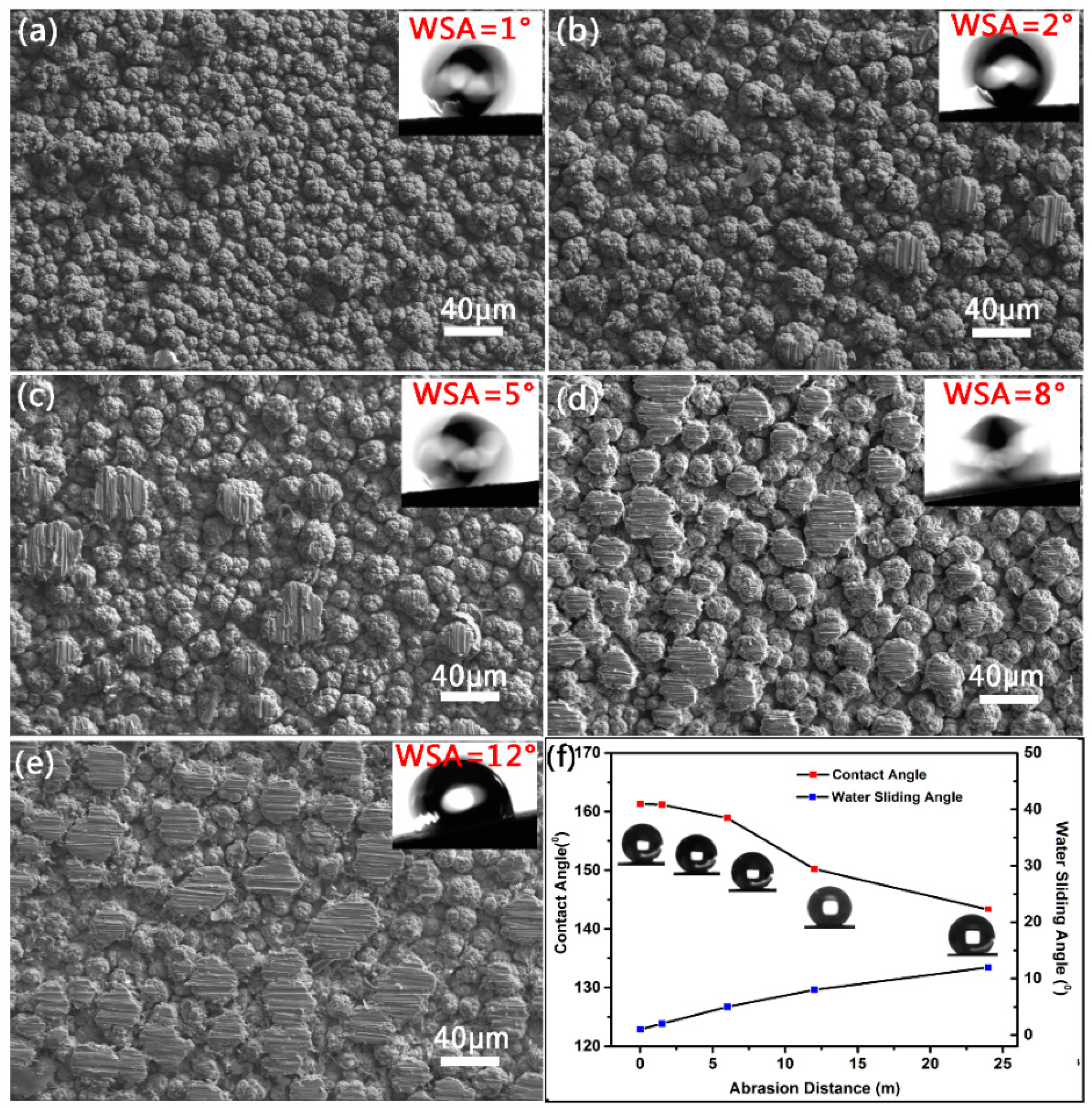
3.5. Abrasion Resistance of the Super-Hydrophobic Co–Ni Coating

Due to the special features (such as dual micro-nano structures and modification by low surface energy materials) which is essential for fabricating super-hydrophobic surfaces, such surfaces are susceptible to mechanical abrasion. At present, to enhance the abrasion resistance of super-hydrophobic coatings has become the main concern for their practical applications [23]. Recently, Ras et al. [23] suggested that linear abrasion should be adopted to assess the mechanical durability of the super-hydrophobic coating because this wear-test method is accessible to most lab researches, applicable in most industrial production, and able to generate a large uniformly surface suitable for wetting measurements. Therefore, in this work the linear abrasion test was conducted, which is shown in Figure 5. The 800 grit SiC sandpaper was placed face up and used as abrasion surface. The super-hydrophobic Co–Ni coating was tested under the applied pressure of 5 kPa at a speed of 5 mm/s.
Figure 6 depicts the SEM images for the super-hydrophobic Co–Ni coating deposited at −1.7 V before and after linear abrasion tests and the relationship between contact angles and abrasion distance. As shown in Figure 6a, the Co–Ni coating with cauliflower-shaped micro-nano structures shows contact angle of 161° and water sliding angle of 1° before linear abrasion tests, exhibiting greater super-hydrophobicity. After abrasion distance of 1.5 m under the applied pressure of 5 kPa, few prominent cauliflower-shaped micro-nano structures were worn and obvious scratches appeared on the top of these structures (Figure 6b). It is obvious that the cauliflower-shaped micro-nano structures were well preserved and the water contact angle maintained around 160° with water sliding angle of 2°. After increasing the abrasion distance to 6 m, large cauliflower-shaped structures were worn more seriously, and the water contact angle dropped to 158° and the water sliding angle increased to 5° (Figure 6c). When abrasion distance reaching 12 m, all the large cauliflower-shaped microstructures appeared different degrees of wear and the water contact angle dropped dramatically to near 150° (Figure 6d). With the increase of abrasion distance, the water sliding angle increases to 8° after 12 m of abrasion. When the abrasion distance increased to 24 m, the large cauliflower-shaped microstructures were almost completely worn away. The water contact angle dropped to near 143° and the sliding angle increased to 12° (Figure 6f). With the increase of abrasion distance, the wetting properties of deposited hierarchical Co–Ni coating with cauliflower-shaped micro-nano structures transited from super-hydrophobic behaviors to hydrophobic behaviors.
Moreover, the mechanical durability, which was tested under the similar conditions by the linear abrasion method, of the electrodeposited super-hydrophobic coatings including nickel [19], copper [20] and cobalt [33] was summarized in Table 2. As compared to the electrodeposited coatings in the literatures, our Co–Ni coating with cauliflower-shaped micro-nano structures exhibits longer abrasion distance before the water contact angle drops to 150° except the Ni–PTFE composite coating tested under lower pressure. When the as-prepared Co–Ni coating loses its super-hydrophobic properties, the water sliding angle increases to 8°, which is better than other electrodeposited coatings. These results demonstrated that our prepared super-hydrophobic Co–Ni coating has great abrasion resistance. And the great abrasion resistance can be attributed to the high cobalt content which possesses higher hardness. This comparison implies that, to further increase the abrasion resistance of super-hydrophobic coating, the co-electrodeposition of Co–Ni coating with PTFE particles maybe a good choice.

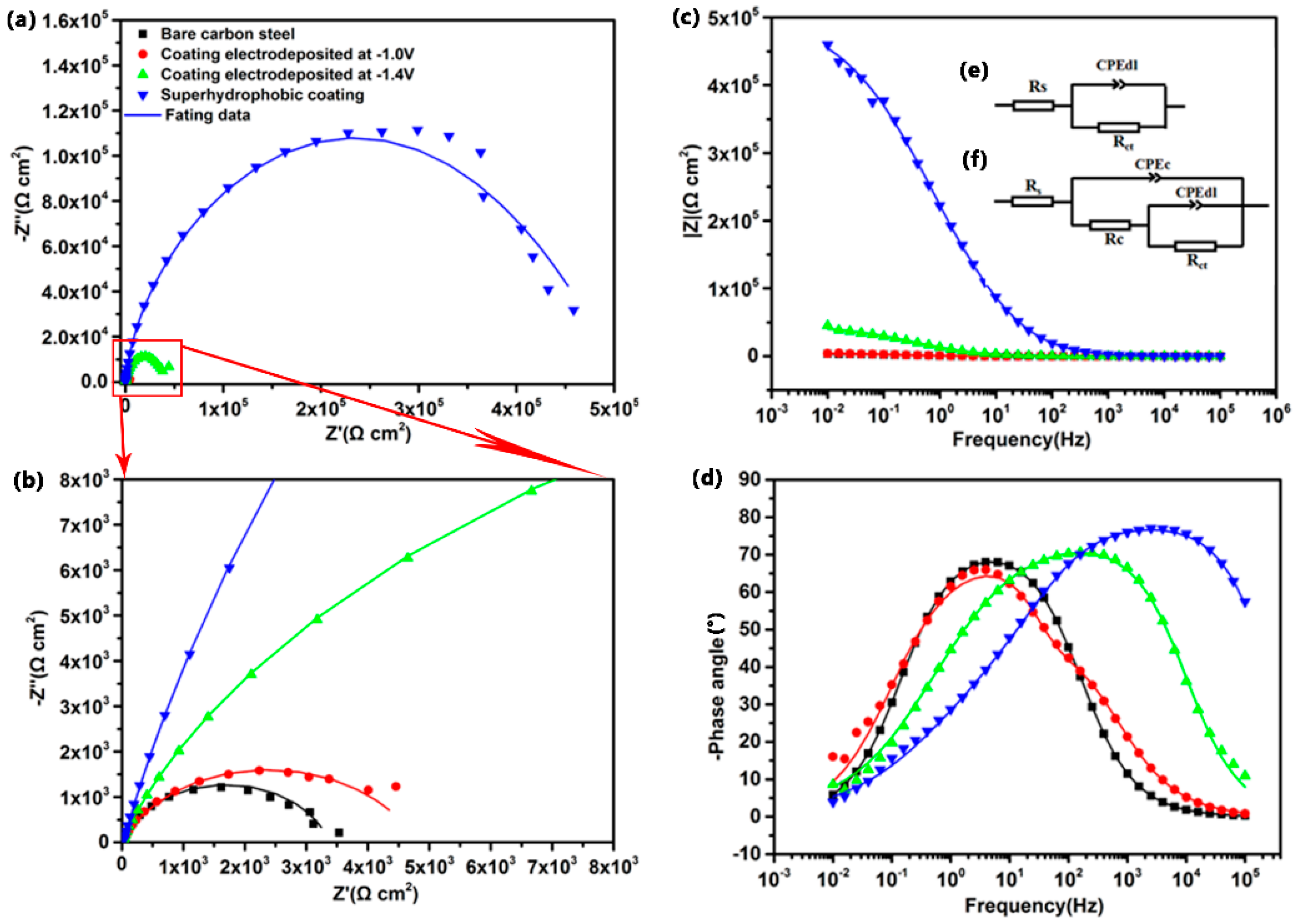
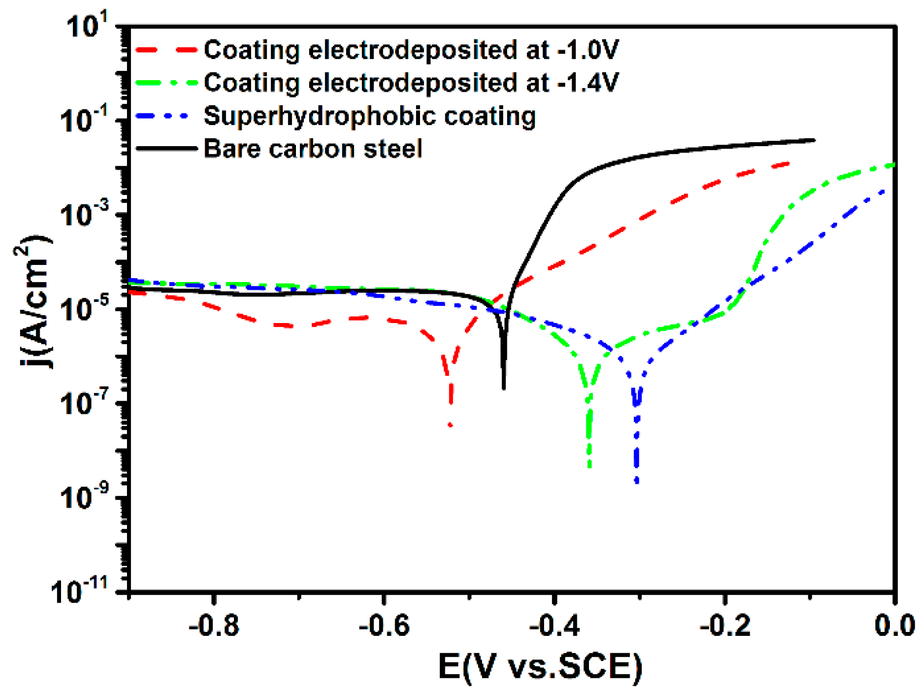
3.6. Corrosion Resistance of the Super-Hydrophobic Co–Ni Coating

The anti-corrosion performance of the super-hydrophobic Co–Ni coating was evaluated by electrochemical methods. Figure 7 depicts the potentiodynamic polarization curves of carbon steel substrate, Co–Ni coatings deposited under the applied potentials of −1.0 and −1.4 V, and super-hydrophobic Co–Ni coating obtained at the applied potentials of −1.7 V with a sweep rate of 0.5 mV/s in 3.5 wt.% NaCl solution after immersion for 1 h, respectively. Tafel extrapolation method was applied to extract the corrosion potential (Ecorr) and corrosion current density (Icorr), which was summarized in Table 3.
Revealed from the Figure 7, the carbon steel substrate displayed negative corrosion potential (Ecorr = −459 mV vs. SCE) with high corrosion current density (Icorr = 1.23 × 10⁻⁵ A/cm2). After electrodeposition under the applied potential of −1.0 V, the corrosion potential of Co–Ni coating shifted negatively to −522 mV, which indicated that the Co–Ni coating with uniform granular structures displayed a higher corrosion susceptibility compared with the carbon steel substrate. The corrosion potential of Co–Ni coating deposited at −1.4 V shifted 100 mV toward positive direction compared with carbon steel with lower corrosion current density of 1.50 × 10⁻⁶ A/cm2. For the super-hydrophobic Co–Ni coating deposited at −1.7 V, the corrosion potential and corrosion current density are −303 mV and 5.87 × 10⁻⁷ A/cm2 respectively. The corrosion current density of as-prepared super-hydrophobic Co–Ni coating was 20 times lower than that of bare carbon steel substrate, demonstrating the significantly improved anti-corrosion performance of the super-hydrophobic Co–Ni coating with cauliflower-shaped micro-nano structures.
To further evaluate the corrosion resistance of the superhydrophobic Co–Ni coating, electrochemical impedance spectroscopy (EIS) was applied. Figure 8 displays the EIS results of the carbon steel substrate, Co–Ni coatings deposited at −1.0 and −1.4 V, and super−hydrophobic Co–Ni coating at −1.7 V in the form of Nyquist plots and Bode plots. From Figure 8a,b, it can be noted that the super−hydrophobic Co–Ni coating exhibits a larger semicircle in Nyquist plots, revealing a higher charge transfer resistance compared with other Co–Ni coatings. Figure 8c shows the corresponding impedance modulus versus frequency plot. The impedance modulus value of the super−hydrophobic Co–Ni coating (4.6 × 10⁵ Ω cm2) is larger than those of the Co–Ni coating deposited at −1.4 V (4.5 × 10⁴ Ω cm2), Co–Ni coating deposited at −1.0 V (4.2 × 10³ Ω cm2) and carbon steel substrate (3.5 × 10³ Ω cm2) at the frequency of 0.01 Hz. The higher impedance modulus at low frequency domain could be attributed to the super−hydrophobic behavior of the Co–Ni coating, which barriers the infiltration of aggressive medium liquid into the substrate, indicating a better anti−corrosion performance of the as−prepared super−hydrophobic Co–Ni coating. The impedance modulus of the coatings decreased with the increase of the frequency, finally reached to a value of around 60 Ω cm2 at the high frequency domain which indicated that the coatings could allow the aggressive species inside the electrolyte infiltrate the coatings easily. As it can be seen from Figure 8d, two time constants exist within the testing frequency range for the super−hydrophobic Co–Ni coating, corresponding to the electrochemical process at high frequency range and the film/metal interface at low frequency. For the other Co–Ni coatings and carbon steel substrate, only one time constant can be observed, which reveals only one electrochemical process occurring within the testing frequency range.
In order to further understand the electrochemical process, equivalent circuit models were applied to fit EIS results. The simple Randles circuit shown in Figure 8e was used to analyze the EIS results of carbon steel substrate, where Rs and Rct stand for the solution resistance and the charge transfer resistance respectively, the constant phase element CPEdl models the non-ideal capacitance at solid/electrolyte interface as a result of the inhomogeneous current distribution. According to the literature, the impedance of CPE was defined as follows:
ZCPE = 1/Y0(jω)n
where ZCPE represents CPE impedance, Y0 represents modulus, ω represents angular frequency and n is the exponent of the CPE varying between 0 and 1. The equivalent electrical circuit shown in Figure 8f was used to analyze the EIS results of the Co–Ni coatings deposited at −1.0 and −1.4 V as well as the super-hydrophobic Co–Ni coating deposited at −1.7 V, where Rc and CPEc represent the coating resistance and non-ideal capacitance at coating/electrolyte interface, respectively. The electrochemical parameters acquired through simulating with the equivalent circuit are presented in Table 4. From the obtained results, the Rct value of the super-hydrophobic Co–Ni coating is three orders of magnitude higher than that of carbon steel substrate. Generally, the inhibition efficiency (η) was computed with the equation as follows [35]: η(%) = (1 − Rctb/Rctc) × 100%, where the Rctb and Rctc are the charge transfer resistance of bare carbon steel and the super-hydrophobic Co–Ni coating, respectively. Based on the values in Table 4, the inhibition efficiency of the as-deposited super-hydrophobic Co–Ni coating can be calculated as 99.3%, suggesting that the electrochemical charge transfer process of super-hydrophobic Co–Ni coating is very difficult to occur under the tested conditions.
The EIS results were in agreement with the potentiodynamic polarization curves, which indicates that the as-prepared super-hydrophobic Co–Ni coating with cauliflower-shaped micro-nano structures has a larger charge transfer resistance and exhibits excellent anti-corrosion performance. The lower corrosion probability and corrosion rate of the super-hydrophobic Co–Ni coating could be attributed to the trapped air among the hierarchical cauliflower-shaped micro-nano structures, which can work as corrosion barrier effectively through limiting the lower contact area of solid coating with the electrolyte solution at the interface, therefore providing better protection for the carbon steel substrate.

4. Conclusions

Mechanical durability and corrosion resistance are of great importance for the real industrial applications of super-hydrophobic surfaces. In our work, a super-hydrophobic coating with a contact angle around 161° was successfully fabricated on a carbon steel substrate by electrochemical deposition. The super-hydrophobicity of the constructed coating derived from the cauliflower-shaped micro-nanostructures and the low-surface-energy PFTEOS layer with fluorinated components. In addition, the Co–Ni coatings possessing different surface microstructures were prepared using electrochemical deposition by varying the applied potentials at room temperature. The as-prepared super-hydrophobic Co–Ni coating with cauliflower-shaped micro-nanostructures exhibited excellent mechanical abrasion resistance. The linear abrasion test revealed that the super-hydrophobic properties were maintained well after abrasion distance of 12 m under the applied pressure of 5 kPa, which was significantly improved by the increase of cobalt content in the Co–Ni coating. Furthermore, electrochemical tests indicated that the super-hydrophobic Co–Ni coating showed lower corrosion current density and a higher charge transfer resistance as a result of limiting the contact area of the solid coating with the electrolyte solution at the interface, providing excellent protection for the carbon steel. In summary, through a combination of electrodeposition and the modification by low surface energy materials, this facile and low-cost preparation process offers an effective technique and promises the practical applications of super-hydrophobic coatings.

Supplementary Materials

The following are available online at https://www.mdpi.com/2079-6412/9/4/232/s1, Figure S1: Cyclic voltammograms of carbon steel obtained with a scan rate of 10 mV/s in the solution of CoCl2 0.1 mol/L + H3BO3 0.1 mol/L, NiCl2 0.03 mol/L + H3BO3 0.1 mol/L, CoCl2 0.1 mol/L + NiCl2 0.03 mol/L + H3BO3 0.1 mol/L; Figure S2: Surface roughness of Co–Ni coatings deposited under the applied potentials of (a) −1.0 V, (b) −1.4 V, and (c) −1.7 V for 3000 s in the mixed solution at room temperature.

Author Contributions

Conceptualization, Y.X.; Methodology, Y.X.; Software, S.W.; Validation, S.W. and P.B.; Formal Analysis, S.W.; Investigation, S.W. and P.B.; Resources, S.W.; Data Curation, G.Z.; Writing—Original Draft Preparation, S.W.; Writing—Review and Editing, Y.X. and Y.J.; Visualization, Y.X.; Supervision, Y.J.; Project Administration, Y.X.; Funding Acquisition, Y.X.

Funding

This research was funded by the Fundamental Research Funds for the Central Universities China (Project ID: FRF-TP-18-009A1).

Conflicts of Interest

The authors declare no conflict of interest.

References

  1. Sanchez, C.; Arribart, H.; Guille, M.M.G. Biomimetism and bioinspiration as tools for the design of innovative materials and systems. Nat. Mater. 2005, 4, 277–288. [Google Scholar] [CrossRef] [PubMed]
  2. Serrà, A.; Zhang, Y.; Sepúlveda, B.; Gómez, E.; Nogués, J.; Michler, J.; Philippe, L. Highly active ZnO-based biomimetic fern-like microleaves for photocatalytic water decontamination using sunlight. Appl. Catal. B Environ. 2019, 248, 129–146. [Google Scholar] [CrossRef]
  3. Liu, K.; Jiang, L. Bio-inspired self-cleaning surfaces. Annu. Rev. Mater. Res. 2012, 42, 231–263. [Google Scholar] [CrossRef]
  4. Liu, K.; Yao, X.; Jiang, L. Recent developments in bio-inspired special wettability. Chem. Soc. Rev. 2010, 39, 3240. [Google Scholar] [CrossRef] [PubMed]
  5. Vazirinasab, E.; Jafari, R.; Momen, G. Application of superhydrophobic coatings as a corrosion barrier: A review. Surf. Coat. Technol. 2018, 341, 40–56. [Google Scholar] [CrossRef]
  6. Eberle, P.; Tiwari, M.K.; Maitra, T.; Poulikakos, D. Rational nanostructuring of surfaces for extraordinary icephobicity. Nanoscale 2014, 6, 4874–4881. [Google Scholar] [CrossRef] [PubMed]
  7. Ma, Q.; Cheng, H.; Fane, A.G.; Wang, R.; Zhang, H. Recent development of advanced materials with special wettability for selective oil/water separation. Small 2016, 12, 2186–2202. [Google Scholar] [CrossRef]
  8. Pu, X.; Li, G.; Huang, H. Preparation, anti-biofouling and drag-reduction properties of a biomimetic shark skin surface. Biol. Open 2016, 5, 389–396. [Google Scholar] [CrossRef]
  9. Liao, R.; Zuo, Z.; Guo, C.; Yuan, Y.; Zhuang, A. Fabrication of superhydrophobic surface on aluminum by continuous chemical etching and its anti-icing property. Appl. Surf. Sci. 2014, 317, 701–709. [Google Scholar] [CrossRef]
  10. Long, J.; Fan, P.; Gong, D.; Jiang, D.; Zhang, H.; Li, L.; Zhong, M. Superhydrophobic surfaces fabricated by femtosecond laser with tunable water adhesion: From lotus leaf to rose petal. ACS Appl. Mater. Interfaces 2015, 7, 9858–9865. [Google Scholar] [CrossRef]
  11. Yu, J.; Qin, L.; Hao, Y.; Kuang, S.; Bai, X.; Chong, Y.; Zhang, W.; Wang, E. Vertically aligned boron nitride nanosheets: Chemical vapor synthesis, ultraviolet light emission, and superhydrophobicity. ACS Nano 2010, 4, 414–422. [Google Scholar] [CrossRef]
  12. Jeong, C.; Choi, C.H. Single-step direct fabrication of pillar-on-pore hybrid nanostructures in anodizing aluminum for superior superhydrophobic efficiency. ACS Appl. Mater. Interfaces 2012, 4, 842–848. [Google Scholar] [CrossRef] [PubMed]
  13. Lee, M.; Kwak, G.; Yong, K. Wettability control of ZnO nanoparticles for universal applications. ACS Appl. Mater. Interfaces 2011, 3, 3350–3356. [Google Scholar] [CrossRef]
  14. Tam, J.; Palumbo, G.; Erb, U. Recent advances in superhydrophobic electrodeposits. Materials 2016, 9, 151. [Google Scholar] [CrossRef]
  15. Verho, T.; Bower, C.; Andrew, P.; Franssila, S.; Ikkala, O.; Ras, R.H.A. Mechanically durable superhydrophobic surfaces. Adv. Mater. 2011, 23, 673–678. [Google Scholar] [CrossRef]
  16. Peng, C.; Chen, Z.; Tiwari, M.K. All-organic superhydrophobic coatings with mechanochemical robustness and liquid impalement resistance. Nat. Mater. 2018, 17, 355–360. [Google Scholar] [CrossRef] [PubMed]
  17. She, Z.; Li, Q.; Wang, Z.; Li, L.; Chen, F.; Zhou, J. Researching the fabrication of anticorrosion superhydrophobic surface on magnesium alloy and its mechanical stability and durability. Chem. Eng. J. 2013, 228, 415–424. [Google Scholar] [CrossRef]
  18. Liu, Q.; Chen, D.; Kang, Z. One-step electrodeposition process to fabricate corrosion-resistant superhydrophobic surface on magnesium alloy. ACS Appl. Mater. Interfaces 2015, 7, 1859–1867. [Google Scholar] [CrossRef] [PubMed]
  19. Su, F.; Yao, K. Facile fabrication of superhydrophobic surface with excellent mechanical abrasion and corrosion resistance on copper substrate by a novel method. ACS Appl. Mater. Interfaces 2014, 6, 8762–8770. [Google Scholar] [CrossRef]
  20. Jain, R.; Pitchumani, R. Facile fabrication of durable copper-based superhydrophobic surfaces via electrodeposition. Langmuir 2018, 34, 3159–3169. [Google Scholar] [CrossRef]
  21. Tam, J.; Jiao, Z.; Lau, J.C.F.; Erb, U. Wear stability of superhydrophobic nano Ni–PTFE electrodeposits. Wear 2017, 374, 1–4. [Google Scholar] [CrossRef]
  22. Xue, Y.; Wang, S.; Zhao, G.; Taleb, A.; Jin, Y. Fabrication of Ni–Co coating by electrochemical deposition with high super-hydrophobic properties for corrosion protection. Surf. Coat. Technol. 2019, 363, 352–361. [Google Scholar] [CrossRef]
  23. Tian, X.; Verho, T.; Ras, R.H.A. Moving superhydrophobic surfaces toward real-world applications. Science 2016, 352, 142–143. [Google Scholar] [CrossRef] [PubMed]
  24. Chung, C.K.; Chang, W.T. Effect of pulse frequency and current density on anomalous composition and nanomechanical property of electrodeposited Ni–Co films. Thin Solid Films 2009, 517, 4800–4804. [Google Scholar] [CrossRef]
  25. Maksimović, V.M.; Lačnjevac, U.Č.; Stoiljković, M.M.; Pavlović, M.G.; Jović, V.D. Morphology and composition of Ni–Co electrodeposited powders. Mater. Charact. 2011, 62, 1173–1179. [Google Scholar] [CrossRef]
  26. Yang, Y.; Cheng, Y.F. Electrolytic deposition of Ni–Co–SiC nano-coating for erosion-enhanced corrosion of carbon steel pipes in oilsand slurry. Surf. Coat. Technol. 2011, 205, 3198–3204. [Google Scholar] [CrossRef]
  27. Rafailović, L.D.; Minić, D.M.; Karnthaler, H.P.; Wosik, J.; Trišović, T.; Nauer, G.E. Study of the dendritic growth of Ni–Co alloys electrodeposited on Cu substrates. J. Electrochem. Soc. 2010, 157, D295. [Google Scholar] [CrossRef]
  28. Jain, R.; Pitchumani, R. Fractal model for wettability of rough surfaces. Langmuir 2017, 33, 7181–7190. [Google Scholar] [CrossRef]
  29. ASM Handbook: Volume 3: Alloy Phase Diagrams, 10th ed.; ASM International: Novelty, OH, USA, 1992.
  30. Wang, N.; Hang, T.; Shanmugam, S.; Li, M. Preparation and characterization of nickel–cobalt alloy nanostructures array fabricated by electrodeposition. CrystEngComm 2014, 16, 6937–6943. [Google Scholar] [CrossRef]
  31. Zhang, B.; Zhu, Q.; Li, Y.; Hou, B. Facile fluorine-free one step fabrication of superhydrophobic aluminum surface towards self-cleaning and marine anticorrosion. Chem. Eng. J. 2018, 352, 625–633. [Google Scholar] [CrossRef]
  32. Wang, P.; Li, T.; Zhang, D. Fabrication of non-wetting surfaces on zinc surface as corrosion barrier. Corros. Sci. 2017, 128, 110–119. [Google Scholar] [CrossRef]
  33. Li, W.; Kang, Z. Fabrication of corrosion resistant superhydrophobic surface with self-cleaning property on magnesium alloy and its mechanical stability. Surf. Coat. Technol. 2014, 253, 205–213. [Google Scholar] [CrossRef]
  34. Tan, C.; Li, Q.; Cai, P.; Yang, N.; Xi, Z. Fabrication of color-controllable superhydrophobic copper compound coating with decoration performance. Appl. Surf. Sci. 2015, 328, 623–631. [Google Scholar] [CrossRef]
  35. Qing, Y.; Yang, C.; Yu, Z.; Zhang, Z.; Hu, Q.; Liu, C. Large-area fabrication of superhydrophobic zinc surface with reversible wettability switching and anticorrosion. J. Electrochem. Soc. 2016, 163, D385–D391. [Google Scholar] [CrossRef]
Figure 1. SEM images of (a) the bare carbon steel and the Co–Ni coatings under the applied potential of (b) −1.0 V, (c) −1.4 V, and (d) −1.7 V for 3000 s in the mixed solution at room temperature. The insets show the corresponding cross section.
Coatings 09 00232 g001
Figure 2. XRD patterns of (a) the bare carbon steel surface and the Co–Ni coatings for 3000 s under the applied potential of (b) −1.0 V, (c) −1.4 V, and (d) −1.7 V in the mixed solution at room temperature.
Coatings 09 00232 g002
Figure 3. The water contact angles of the samples deposited at different potentials before and after modification by PFTEOS.
Coatings 09 00232 g003
Figure 4. XPS spectra before and after modification of the Co–Ni coating deposited under the applied potential at −1.7 V in the mixed solution: (a) XPS survey spectra; (b) Co 2p spectra; (c) Ni 2p spectra; (d) F 1s spectra; (e) Si 2p spectra; and (f) high resolution C 1s spectra.
Coatings 09 00232 g004aCoatings 09 00232 g004b
Figure 5. A scheme for linear abrasion test.
Coatings 09 00232 g005
Figure 6. SEM images for (a) super-hydrophobic Co–Ni coating before abrasion and after abrasion under the applied pressure of 5 kPa for (b) 1.5 m, (c) 6 m, (d) 12 m, and (e) 24 m; (f) The water contact angle and water sliding angle variations on these surface with the abrasion distance. The insert images in (a) to (e) are the profiles of a water droplet sliding on the Co–Ni coating with different water sliding angles.
Coatings 09 00232 g006
Figure 7. Potentiodynamic polarization curves of the bare carbon steel, Co–Ni coating deposited at −1.0 and −1.4 V, and super-hydrophobic Co–Ni coating deposited at −1.7 V after immersion in 3.5 wt.% NaCl aqueous solution for 1h with a scan rate of 0.5 mV/s.
Coatings 09 00232 g007
Figure 8. EIS results of the bare carbon steel, Co–Ni coating deposited at −1.0 and −1.4 V, and super-hydrophobic Co–Ni coating deposited at −1.7 V in 3.5 wt.% NaCl solution after immersion for 1 h: (a) Nyquist plots; (b) high-resolution Nyquist plot in (a) marked with red box; (c) bode −|Z| versus frequency plots; and (d) bode-phase angle versus frequency plots. (e) Equivalent circuits for simulating EIS results of bare carbon steel. (f) Equivalent circuits for simulating EIS results of the Co–Ni coating deposited at −1.0 and −1.4 V, and the super-hydrophobic Co–Ni coating deposited at −1.7 V.
Coatings 09 00232 g008
Table 1. EDS results of the deposited Co–Ni coatings under different applied potentials for 3000 s in the mixed solution.
PotentialsCoating
Ni (at.%)Co (at.%)
−1.0 V6.293.8
−1.4 V19.081.0
−1.7 V22.377.7
Table 2. Abrasion distances leading to the loss of super-hydrophobic properties. Abrasive medium: 800 grit SiC sandpaper.
MaterialsPressure (kPa)Abrasion Length (m)Initial WCA (°)Final WCA (°)Initial WSA (°)Final WSA (°)
Co–Ni coating (this work)5.012.016115018
Ni–PTFE [21]2.050.0156150352
Cu [20]3.02.0162143318
Ni [19]4.81.0162150315
Mg−Mn−Ce [18]1.30.41601502Not given
Co [33]1.51.1156148140
Cu compound [34]1.20.71631401Not given
Table 3. Extracted corrosion potential (Ecorr) and current density (Icorr) from the potentiodynamic polarization measurements presented in Figure 6.
SampleEcorr (mV)Icorr (A/cm2)Corrosion Rate (mm/a)
Bare carbon steel−459.41.23 × 10−50.137
Co–Ni coating at −1.0 V−522.75.08 × 10−60.057
Co–Ni coating at −1.4 V−359.41.50× 10−60.017
Super−hydrophobic coating−303.35.87 × 10−70.0066
Table 4. The impedance parameters of bare carbon steel, Co–Ni coating deposited at −1.0 and−1.4 V, and super-hydrophobic Co–Ni coating deposited at −1.7 V extracted by fitting the EIS results recorded in 3.5 wt.% NaCl solution (Figure 8) with the corresponding equivalent circuits. The units of Rs, Rct, Rc and CPE are Ω·cm2, kΩ·cm2, kΩ·cm2 and Ω−1·sn·cm−2, respectively.
Fitted ParametersRsCPEdlndlRctCPEcncRc
Bare carbon steel8.421.08 × 10−30.6592.40
Co–Ni coating deposited at −1.0 V10.753.59 × 10−50.9834.652.83 × 10−40.7290.116
Co–Ni coating deposited at −1.4 V11.683.06 × 10−50.62926.41.00 × 10−50.81616.1
Super−hydrophobic coating23.821.43 × 10−60.582369.11.78 × 10−70.86898.9

