Article

Sub-Ablative Femtosecond Laser Modification of the Nonlinear Optical Response of Amorphous TiO2 Thin Films


1
Georgi Nadjakov Institute of Solid State Physics, Bulgarian Academy of Sciences, 72 Tzarigradsko Chaussee Blvd, 1784 Sofia, Bulgaria
2
Institute of Optical Materials and Technologies “Acad. Yordan Malinovski”, Bulgarian Academy of Sciences, “Acad. G. Bontchev” Str. 109, 1113 Sofia, Bulgaria
3
Rostislaw Kaischew Institute of Physical Chemistry, Bulgarian Academy of Sciences, Akad. G. Bonchev Str., bl. 11, 1113 Sofia, Bulgaria
*
Author to whom correspondence should be addressed.
Coatings 2026, 16(2), 220; https://doi.org/10.3390/coatings16020220
Submission received: 8 January 2026 / Revised: 2 February 2026 / Accepted: 6 February 2026 / Published: 8 February 2026

Highlights

What are the main findings?
  • Sub-ablative femtosecond laser irradiation induces systematic changes in the nonlinear optical response of amorphous TiO2 coatings.
  • Effective nonlinear refractive index and effective nonlinear absorption coefficient were evaluated by the z-scan technique under femtosecond excitation.
  • A reversal of the sign of the effective nonlinear refractive index is observed after femtosecond laser processing.
What are the implications of the main findings?
  • The observed changes in the nonlinear optical response may be associated with laser-induced modification of defect states in amorphous TiO2 films.
  • Laser-induced densification is considered as an additional possible contribution to the modified nonlinear optical response.
  • The results demonstrate the potential of femtosecond laser processing for post-deposition engineering of functional optical coatings.

Abstract
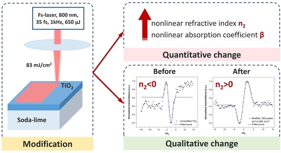
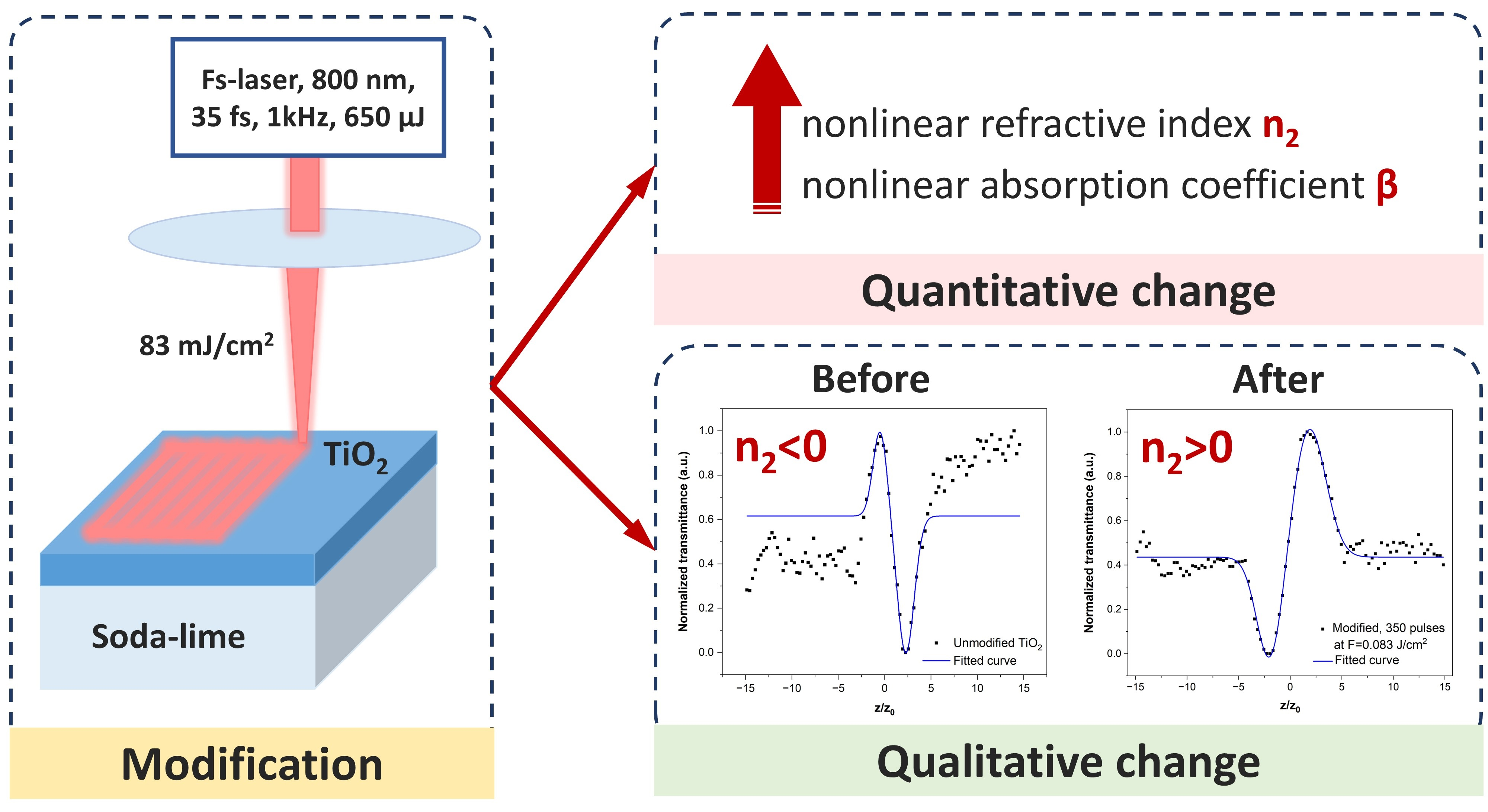
Femtosecond laser processing has emerged as a promising post-deposition method for tailoring the properties of dielectric thin films, offering localized modification without thermal damage. This study investigates the effect of sub-ablative femtosecond laser irradiation on the nonlinear optical response of a TiO2 single-layer coating deposited on soda-lime glass by electron-beam evaporation. The coating was modified using 35 fs pulses at 800 nm delivered at a repetition rate of 1 kHz and a fluence of 0.083 J/cm2 while varying the number of pulses per spot. The effective nonlinear refractive index (n2,eff) and effective nonlinear absorption coefficient (βeff) were measured using the z-scan technique with femtosecond excitation. The as-deposited TiO2 coating exhibited a negative effective nonlinear refractive index, signifying a self-defocusing nonlinear response, while femtosecond laser irradiation leads to pronounced changes in the effective nonlinear parameters. An increase in the magnitude of both effective nonlinear coefficients and a reversal of the sign of the effective nonlinear refractive index are experimentally observed after irradiation with higher pulse numbers. These findings provide experimental evidence that sub-ablative femtosecond laser processing can be used as a post-deposition tool to control the nonlinear optical response of TiO2 thin films.

Graphical Abstract

1. Introduction

Femtosecond laser pulses have emerged as a powerful tool for precise, controllable modification of a wide range of materials, owing to their combination of ultrashort pulse duration and extremely high peak intensities, which enable highly localized energy deposition with minimal thermal diffusion. These characteristics make femtosecond lasers particularly attractive for processing dielectric materials, where excitation across wide band gaps relies predominantly on nonlinear optical processes, such as multiphoton absorption and avalanche ionization. This allows structural and electronic modifications to be induced with minimal heat-affected zones and high spatial precision, in contrast to nanosecond or longer laser pulses [1,2,3].
Thin dielectric and oxide films play a central role in modern photonics and laser technologies, serving as functional layers in optical coatings, waveguides, resonators, and nonlinear optical elements [4,5]. The performance of such components depends not only on their linear optical properties but also on their nonlinear optical response, which influences beam propagation, self-focusing, optical limiting, and laser-induced damage processes [6].
Titanium dioxide (TiO2) is among the most widely used oxide materials in optical and optoelectronic applications owing to its high refractive index, wide band gap, chemical stability, and optical transparency in the visible and near-infrared spectral ranges. TiO2 thin films are employed in optical coatings, waveguides, resonators, and nonlinear optical devices, as well as in photovoltaics and photocatalysis [7,8]. This material can exist in amorphous or crystalline phases (anatase, rutile, or brookite), with its optical and electronic properties strongly influenced by phase composition, defect density, and stoichiometry [9].
The nonlinear optical properties, including the nonlinear refractive index n2 and the nonlinear absorption coefficient β, are highly relevant to applications such as ultrafast optical switching, optical limiting, and beam shaping. Transition-metal oxides such as TiO2 exhibit enhanced optical nonlinearity due to the high polarizability of oxide ions and metal cations [10]. Recent studies have demonstrated that defects engineering, doping, nanocomposites, plasmon-mediated effects, and annealing can further enhance the nonlinear optical response of TiO2-based materials [11,12,13,14,15,16]. Nevertheless, these methods typically involve compositional modifications or thermal processing, thereby limiting their applicability to post-deposition processing and to spatially selective tuning of functional optical coatings. As a result, there is a growing interest in alternative post-deposition approaches that enable controlled tuning of nonlinear optical properties without altering the material composition or deposition process.
Femtosecond laser modification is a promising alternative method for tailoring the physicochemical properties of TiO2 coatings through controlled, contactless, and spatially selective processing. Previous works have demonstrated that femtosecond laser irradiation below the material’s ablation threshold enables non-thermal structural modifications, defect mitigation, densification, and improvements in mechanical properties of oxide thin films while keeping the surface intact [17,18]. The present study explores another aspect of femtosecond laser modification of oxide thin films, focusing on their optical nonlinear properties. To the authors’ knowledge, the current literature on this subject shows a significant gap regarding the direct relationship between sub-ablative femtosecond laser modification and its influence on the nonlinear optical properties of transition-metal oxides thin films.
The present study investigates the effect of sub-ablative femtosecond laser modification on the nonlinear optical response of TiO2 thin films. By operating below the ablation threshold and with minimal incubation effect, the approach aims to selectively alter the internal electronic and structural properties of the coating without causing surface damage or creating scattering centers that could impair optical performance. The findings provide new insights into how femtosecond laser irradiation affects the nonlinear optical response of TiO2 coatings, thereby advancing the laser-assisted engineering of nonlinear photonic materials.

2. Materials and Methods

2.1. Sample Preparation

The titanium dioxide (TiO2) single-layer coating was deposited on a soda-lime glass substrate with a size of 75 × 38 mm and a thickness of 1 mm (2947-75x38, Corning Incorporated, Corning, NY, USA) by electron-beam evaporation in a high-vacuum deposition system (Symphony 9; Tecport Optics Inc., Orlando, FL, USA). Before deposition, the substrate was ultrasonically cleaned in a detergent solution, deionized water, and isopropyl alcohol. The cleaned substrate was mounted on a stationary substrate holder without rotation and inserted into the system. Additionally, ion etching was performed under a vacuum of 3 × 10⁻⁵ Torr for 2.5 min using Argon ions (Ar+) to clean the substrate surface and improve the adhesion of the deposited material. During deposition, the pressure in the vacuum chamber was approximately 8 × 10⁻⁵ Torr, and no additional substrate heating or ion-assisted processes were applied; the substrate temperature increased solely due to radiative heating from the electron-beam source. This deposition regime was intentionally chosen to limit adatom mobility and promote the formation of a low-density amorphous TiO2 layer. The deposition rate was maintained at approximately 2 Å/s, controlled in situ using a quartz crystal microbalance (QCM) connected to an IC6 thickness controller (Inficon, Bad Ragaz, Switzerland). An oxygen (O2) background was introduced during deposition at a flow rate of 10 sccm to ensure stoichiometric oxidation of the evaporated material and to minimize oxygen deficiency in the growing film. The as-prepared sample was divided into 28 squares of 1 × 1 cm2 arranged in a grid (Figure 1). The first row was left unmodified and used as a reference, while the remaining squares were irradiated with varying numbers of laser pulses per spot under sub-ablative and minimal incubation conditions.

2.2. Characterization of the Coating

The surface morphology of the unmodified and laser-modified TiO2 coating was examined using three-dimensional optical profilometry with an optical microscope—3D Optical profiler Zeta-20 (Zeta Instruments, Milpitas, CA, USA). Measurements were taken with a 100× objective. The vertical scanning range was 13–15 µm, sampled with 200 focus steps, resulting in a Z-step size of 0.063–0.073 µm. For each measurement, three separate surface areas were analyzed to evaluate surface uniformity. Additionally, larger surface areas were imaged with a 20× magnification over a field of view of 476 × 357 µm. All analyses were conducted at room temperature.
Atomic force microscopy (AFM) measurements were carried out using the MFP-3D system (Asylum Research, Oxford Instruments, Santa Barbara, CA 93117, USA). The experiments were performed in ambient air conditions at room temperature. Silicon cantilevers (AC160TS) with a resonance frequency of 300 kHz and a nominal spring constant of 26 N/m were employed. Surface morphometrical parameters, including roughness values, were evaluated using Igor Pro software (version 6.37).
Before the femtosecond laser modification, the linear optical transmittance of the TiO2 coating was measured over the UV-Vis-NIR spectral region (320–1750 nm) via a spectrophotometer Perkin Elmer Lambda 1050 (PerkinElmer, Shelton, CT, USA).
X-ray diffraction (XRD) analysis was carried out to evaluate the structural states and phase compositions of the unmodified and laser-modified thin films using an Empyrean diffractometer (Malvern Panalytical, Almelo, The Netherlands) equipped with a Cu anode X-ray tube and a PIXcel3D detector. The measurements were performed at an accelerating voltage of 45 kV and a tube current of 40 mA. Diffraction patterns were collected in the 2θ range from 20° to 70° using a step size of 0.05° (2θ) and an exposure time of 200 s.
Fourier transform infrared spectroscopy in attenuated total reflection (ATR-FTIR) was used to analyze the chemical composition of the coatings. A Vertex 70 spectrophotometer (Bruker, Rosenheim, Germany) with the standard PIKE MIRacle ATR accessory (Pike Technologies, Madison, WI, USA) was employed to acquire the spectra. The ATR accessory featured a ZnSe reflection prism with three internal reflections covered by a diamond plate. Spectra were recorded in the mid-infrared (MIR) range (4000–600 cm−1), limited by the transmission edge-cut of the ZnSe crystal, averaging 64 interferograms at a resolution of 4 cm−1 at room temperature. All spectral data were visualized using OriginPro Version 2025b (10.2) (OriginLab Corporation, Northampton, MA, USA).

2.3. Experimental Setup and Conditions
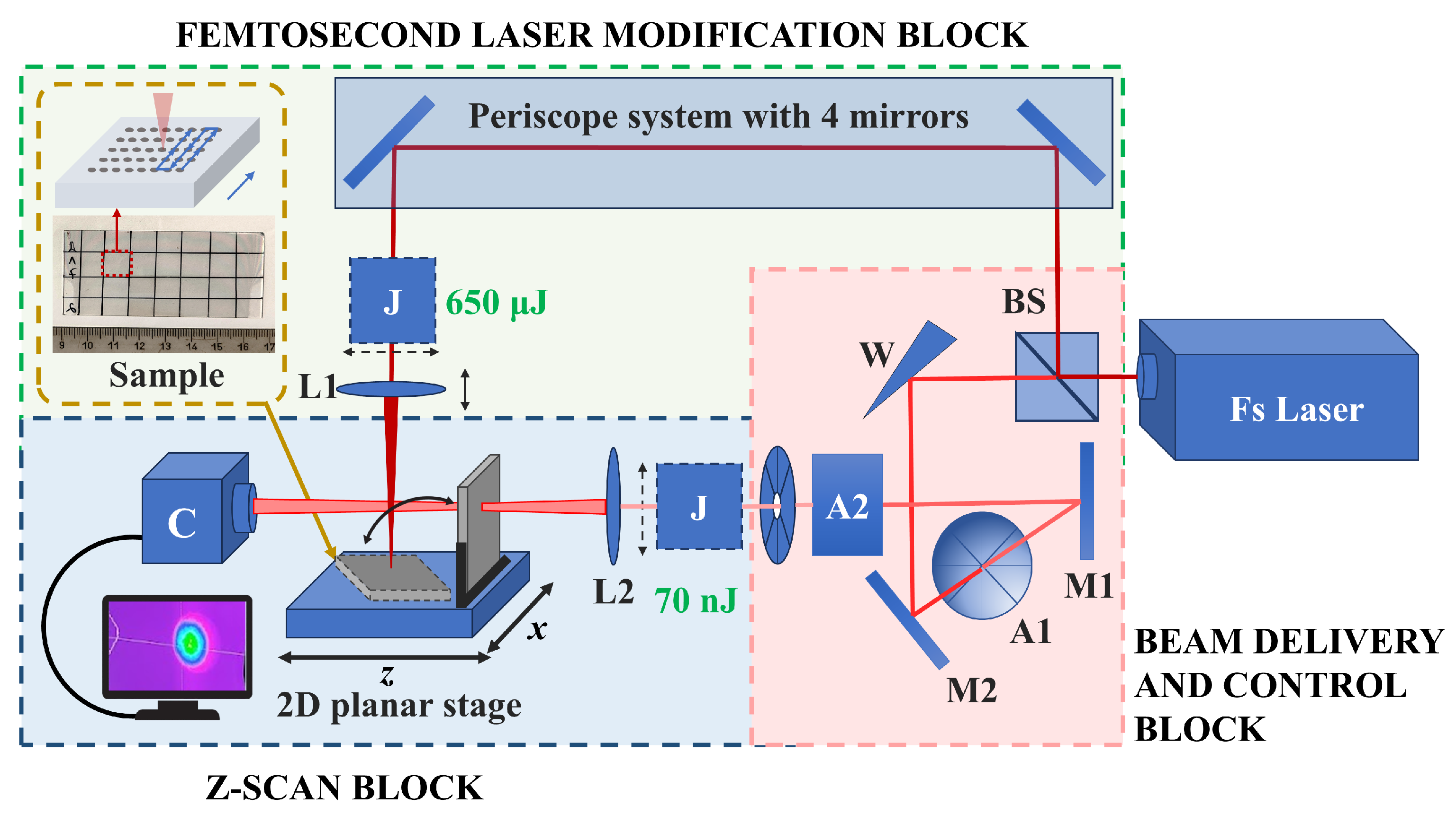
The experimental setups for femtosecond laser surface modification and z-scan measurements of nonlinear optical parameters were constructed in parallel and operated using a single laser source, as schematically illustrated in Figure 1. A titanium–sapphire femtosecond laser system, Spitfire Ace (Spectra-Physics, Milpitas, CA, USA), was used as the laser source. The system operated at a central wavelength of λ = 800 nm, a repetition rate of ν = 1 kHz, and a pulse duration of τ = 35 fs. The laser beam was directed into a beam delivery and control block, where a beam splitter (BS) divided the beam into two optical paths, guiding it either toward the laser modification block or toward the z-scan measurement block.
Figure 1. Schematic representation of the experimental setup for femtosecond laser surface modification and z-scan measurements.
Coatings 16 00220 g001
The femtosecond laser modification setup was optimized to ensure stable beam propagation and flexible control of the laser spot size at the sample surface. The beam was directed through a periscope system consisting of four low-dispersion reflective mirrors, ensuring proper alignment and height adjustment before focusing. A power meter (J) was placed in the beam path to monitor the incident pulse energy continuously. Focusing was achieved using a lens with a focal length of f = 100 mm (L1), mounted on a vertically adjustable holder that allowed micrometric movement along the optical axis. This configuration enabled precise tuning of the laser spot diameter on the sample surface. The sample was intentionally positioned slightly in front of the focal plane to avoid strong nonlinear self-focusing, plasma shielding, and excessive ablation. All laser modification experiments were conducted at a constant pulse energy of 650 µJ. By keeping the pulse energy constant while varying the laser spot size by moving the lens along the optical axis, the applied laser fluence was adjusted. After a series of tests, the optimal laser spot diameter was set to 1 mm. This corresponds to a fluence of F = 0.083 J/cm2, which remains significantly below the reported damage threshold of 0.55 J/cm2 observed under similar conditions elsewhere [19].
The sample was mounted on a Planar DL two-axis mechanical bearing direct-drive linear stage system (Aerotech, Pittsburgh, PA, USA), which was used for both laser modification and z-scan measurements. The stage offers a travel range of 100 mm × 100 mm, a positioning accuracy of ±0.4 µm, a flatness of ±1 µm, and a maximum translation speed of 500 mm/s, allowing for quick and precise sample positioning. During laser modification, the translational stage was programmed to move in a serpentine pattern, covering each grid square uniformly. The translation speeds along both axes were adjusted to control the number of pulses incident on a single spot, thereby tuning the cumulative irradiation dose. The interline distance was chosen to match the laser spot diameter, minimizing pulse overlap between adjacent scan lines.
The z-scan block was designed to measure the effective nonlinear refractive index (n2,eff) and effective nonlinear absorption coefficient (βeff), following the original concept presented in [20], but modified by replacing the standard aperture with a laser beam profiler camera and using a femtosecond laser. After the beam splitter, the laser beam travelled through a system of low-dispersion group-velocity mirrors and energy attenuation elements to reduce pulse broadening and enable precise control of the incident energy. The optical setup included an optical wedge (W) for coarse energy attenuation, a pair of low-dispersion reflective mirrors (M1 and M2), and additional attenuators (A1 and A2) for exact energy adjustment. The beam was then focused with a lens having a focal length of f = 250 mm (L2). The pulse energy measured before the focusing lens was E = 70 nJ, which was experimentally found to be optimal for reliable z-scan measurements while preventing sample damage and thermal effects. The experimental sample was mounted in a holder perpendicular to the laser beam and moved along the optical (z) axis using the same Aerotech translational stage. Transmission was recorded as a function of the sample’s position relative to the focal plane, following the standard z-scan method. The spatial profile of the laser beam was monitored using a laser beam profiler camera (C), model BGPUSB-L11059 (Ophir Optronics, Jerusalem, Israel). This silicon CCD detector operated over the wavelength range 190–1100 nm, with an image format of 35 mm × 24 mm and a sensor resolution of 4008 × 2672 pixels. It offered a dynamic range of 59 dB, a high signal-to-noise ratio, beam size detection from 90 µm to 23.8 mm, and a full-resolution frame rate of 3.1 fps. The camera was positioned 720 mm from the focusing lens and 470 mm from the focal point, allowing accurate characterization of beam quality and spatial stability during the experiments. The graphs shown are averaged over 10 measurements.

2.4. Z-Scan Method

The nonlinear optical properties of the investigated coating were estimated using the z-scan technique, which is a well-established and sensitive method for measuring the nonlinear refractive index (n2) and the nonlinear absorption coefficient (β) of the materials. The technique was initially introduced by Sheik-Bahae et al. [20] and has since been widely applied to various materials owing to its simplicity and high accuracy [21,22,23,24,25]. The principle of the z-scan method is well described in [26].
In thin-film-on-substrate geometries, the measured z-scan response represents a superposition of nonlinear effects originating from the thin film, the underlying substrate, and their interfaces. The substrate may contribute to the detected nonlinear phase shift and absorption through its own nonlinear refractive index and nonlinear absorption, particularly when the interaction length in the substrate is significantly larger than the film thickness. In addition, multiple internal reflections and interference effects at the air-film and film-substrate interfaces can locally modify the intensity distribution within the thin film, potentially affecting the magnitudes of the extracted nonlinear parameters. Since these substrate- and interface-related contributions are not explicitly accounted for in the standard Z-scan formalism, they can introduce uncertainty in the absolute values of the nonlinear coefficients [27,28]. Therefore, the extracted nonlinear refractive index and nonlinear absorption coefficient are reported as effective values (n2,eff and βeff) that describe the overall nonlinear response of the film-substrate system.
The normalized transmittance is typically expressed as a function of the reduced coordinate z/z0, where z is the distance of the sample from the focal plane, z0 = (πω02)/λ is the Rayleigh length of the beam, ω0 is the beam waist radius at the focus, and λ is the laser wavelength.
The effective nonlinear refractive index n2 was calculated using the following expression:
n 2 , e f f = λ T p v 4 L π ω 0 2 2 P ,
where ΔTp−v is the peak-to-valley transmittance difference (dimensionless), L is the optical path length of the sample, and P is the peak power of the laser pulse.
The effective nonlinear absorption coefficient β was determined using the following formula:
β e f f = 2 π T ω 0 2 2 P L   .
The detailed mathematical approach is outlined elsewhere [29]. During calculations, the SI units for all variables in Equations (1) and (2) are used. The uncertainty associated with the reported values was estimated based on the fitting sensitivity of the z-scan curves and the relevant experimental parameters used in the analysis. Accordingly, confidence intervals for the extracted effective nonlinear coefficients were calculated using standard error propagation.

3. Results

3.1. Morphological, Structural, and Linear Optical Characterization

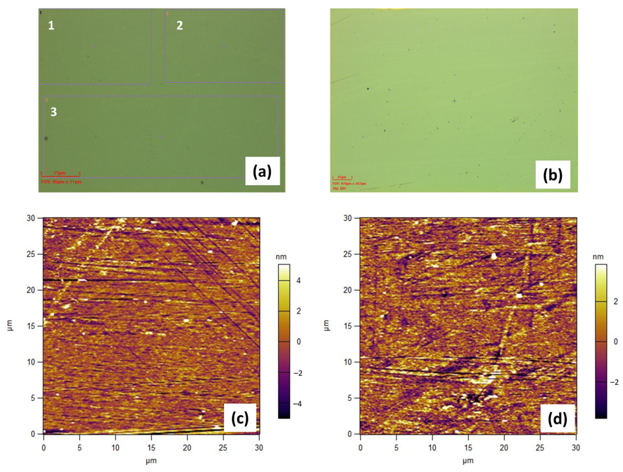
The surface morphology of the unmodified and femtosecond laser-modified TiO2 coating was examined by three-dimensional optical profilometry and atomic force microscopy (AFM), as summarized in Figure 2. Optical profilometry images acquired at 100× magnification (Figure 2a) reveal that the as-deposited coating exhibits an arithmetic mean roughness of Sa ≈ 18 nm and a root mean square roughness of Sq ≈ 23 nm. After laser processing with 350 pulses per spot, the corresponding roughness values remain nearly unchanged (Sa ≈ 17 nm, Sq ≈ 21 nm), indicating that femtosecond laser irradiation does not induce surface roughening or morphological degradation. Additional measurements at lower magnification (20×) over larger surface areas (Figure 2b) revealed the presence of moderate micro-defects randomly distributed across the surface originating from the deposition process and confirmed the absence of laser-induced surface structures, periodic patterns, or ablation-related features, demonstrating that the modification occurs in a sub-ablative regime.
Complementary AFM measurements conducted over surface areas of 30 × 30 µm further confirmed the preservation of nanoscale surface texture (Figure 2c,d). The RMS roughness values decrease slightly from 2.28 nm for the unmodified coating to 1.84 nm for the coating modified with 350 pulses, suggesting that the laser treatment does not introduce nanoscale damage and may even promote minor surface smoothing. Overall, both profilometry and AFM results confirmed that the morphological integrity of the coating is maintained after femtosecond laser irradiation.
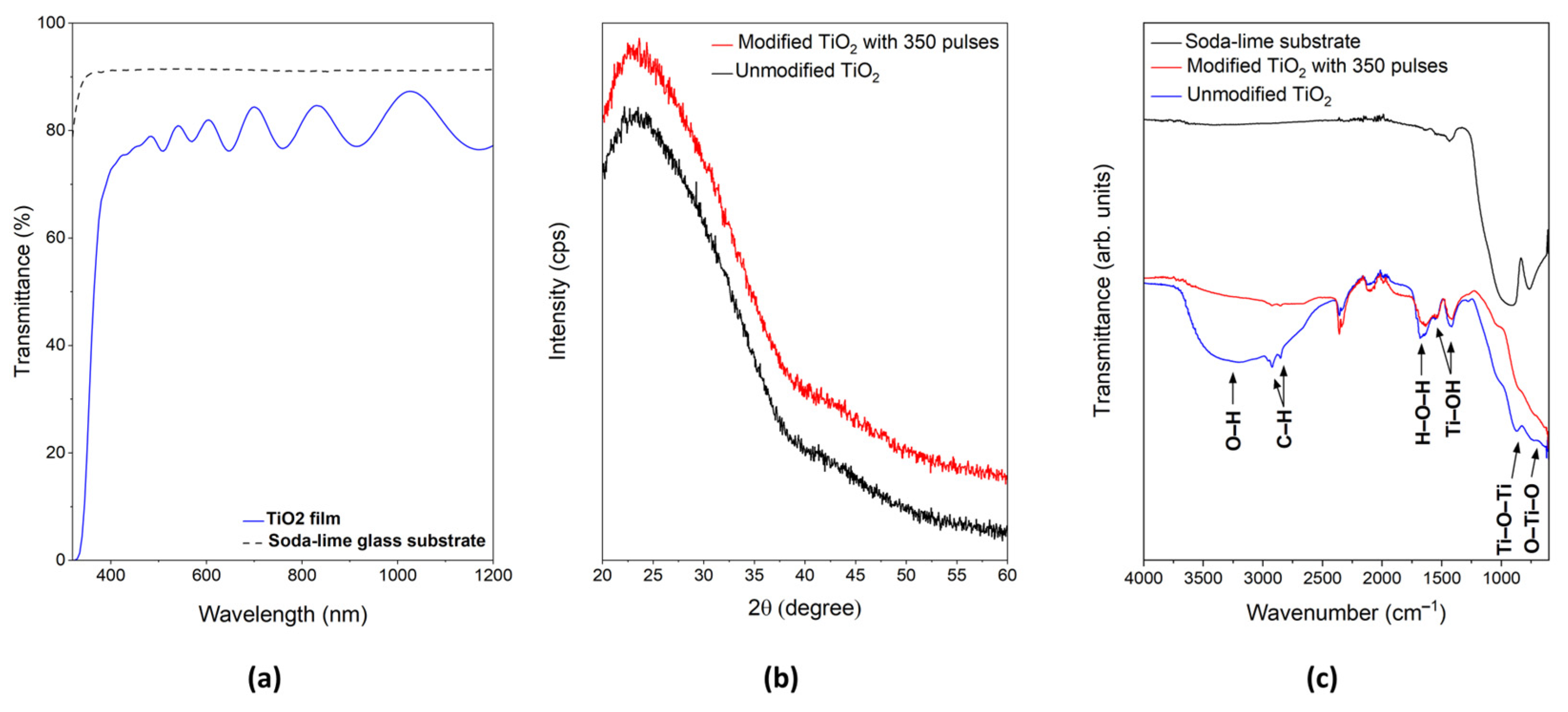
The linear optical transmittance in the UV-Vis-NIR range of the unmodified TiO2 coating and the soda-lime glass substrate is shown in Figure 3a. The substrate exhibits a nearly constant transmittance of 90%–92%, while the coating shows a lower transmittance, fluctuating between 76%–88%, consistent with values reported in the literature [30]. The coating exhibits a steep increase in transmittance above 330 nm, indicating negligible absorption in the visible and near-infrared regions. The pronounced interference fringes in the Vis-NIR region confirm the smoothness and uniformity of the deposited film [31].
X-ray diffraction patterns of the unmodified coating and the coating modified with 350 pulses are presented in Figure 3b. Both diffractograms are dominated by a broad diffuse halo in the 2θ range of 20–35°, with no detectable diffraction peaks associated with the crystalline phases of TiO2 (anatase or rutile). This confirms that the femtosecond laser processing under the applied sub-ablative conditions does not induce crystallization or phase transformation and that the amorphous structure of the TiO2 film is preserved.
The ATR-FTIR spectra of the as-deposited and the modified TiO2 coating are shown in Figure 3c. The spectrum of the soda-lime glass substrate is included for comparison. These spectra provide information about surface functional groups. The broad band between 600 cm−1 and 900 cm−1 indicates Ti–O stretching vibrations and vibrations in the O–Ti–O lattice. The H–O–H bending and O–H stretching vibrations, attributed to adsorbed water and hydroxyl ions on the surface, appear as broad bands around 1650 cm−1 and in the 3000–3600 cm−1 range, respectively. Peaks at 2926 and 2852 cm−1, associated with C–H functional groups, likely result from various organic compounds adsorbed on the surface. The absorption bands around 1553 and 1420 cm−1 are assigned to Ti–OH vibrations [32,33]. After laser modification, a noticeable reduction in the intensity of O–H related bands is observed, together with subtle changes in the broad Ti–O/Ti–O–Ti vibrational region, indicating laser-induced changes in surface chemistry.

3.2. Nonlinear Optical Measurements

Before conducting z-scan measurements on the sample, initial calibration measurements were performed on fused silica samples with known thickness as reference materials to ensure the setup’s accuracy. These calibration results created a reliable baseline for determining average values and assessing data repeatability. The corresponding z-scan curves obtained for fused silica are presented in the Supplementary Materials. They exhibited the expected symmetric profile around the focal position (z = 0). The z-scan trace linked to the nonlinear absorption (Figure S1a) showed a well-defined characteristic transmittance minimum, while the trace associated with the nonlinear refraction (Figure S1b) presented a typical peak–valley shape (pre-focal transmittance minimum followed by a post-focal maximum), corresponding to a positive nonlinear refractive index and a self-focusing effect, as reported in other studies [34]. The calculated values of n2 and β shown in Table 1 were consistent with commonly reported values for fused silica under femtosecond excitation [35,36], confirming the reliability and accuracy of the experimental setup.
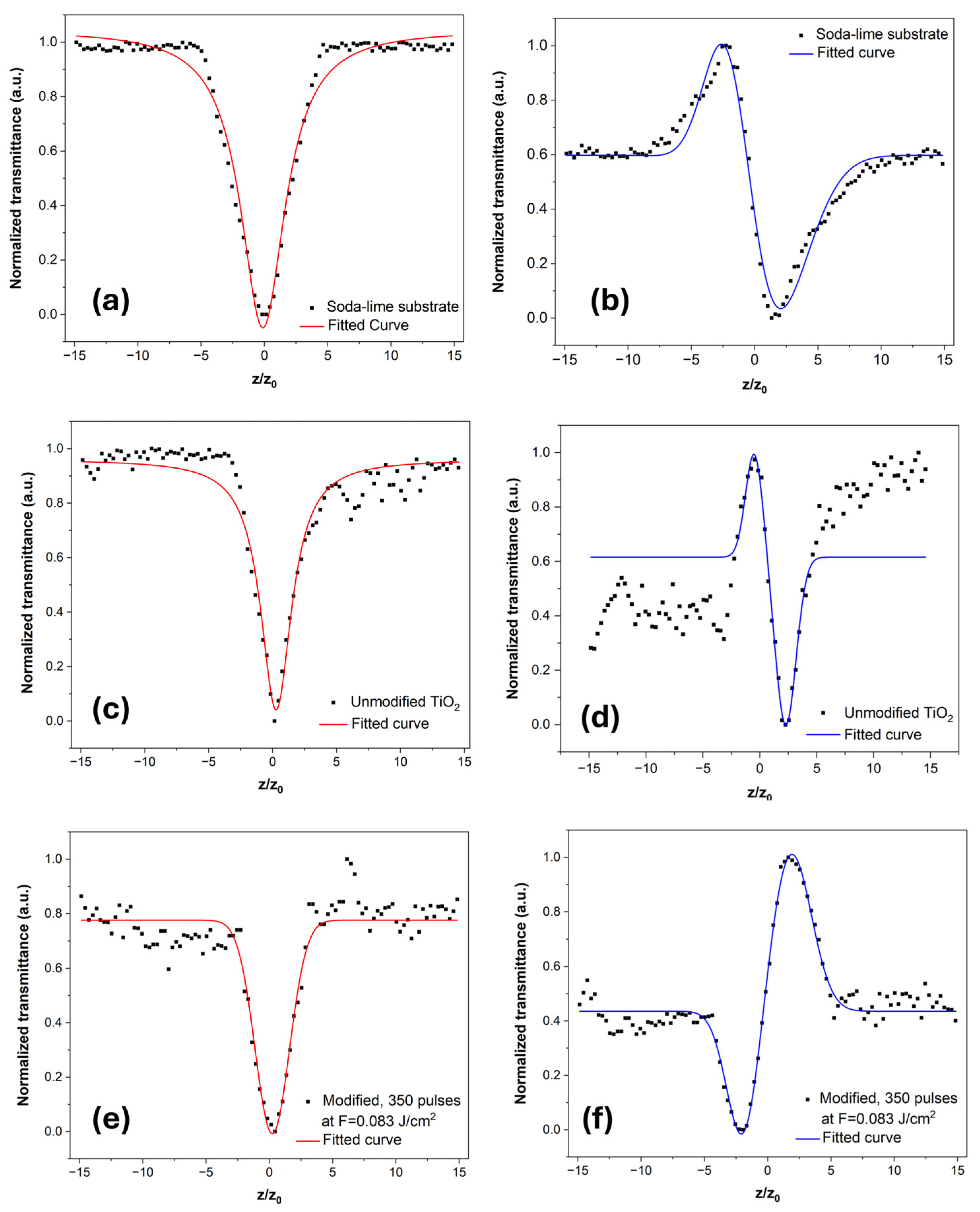
Figure 4 illustrates the experimentally measured z-scan curves (black squares) of the nonlinear optical properties of the samples. The corresponding theoretical fitted curves, derived from the equations in [20] are displayed as solid lines on the graphs.
To assess the influence of the substrate, z-scan measurements were additionally performed on a bare soda-lime glass under the same experimental conditions (Figure 4a,b). The extracted nonlinear optical parameters, summarized in Table 1, are consistent with previously reported values for soda-lime glass in the femtosecond regime [37,38]. The substrate exhibits a pronounced effective nonlinear absorption coefficient, while the effective nonlinear refractive index remains comparable in magnitude to that measured for the unmodified TiO2 coating. Such behavior is consistent with thin-film-on-substrate z-scan measurements, where the substantially longer interaction length in the thick substrate can significantly contribute to the measured effective response [20,27,28]. In this context, the parameter βeff should be regarded as an effective intensity-dependent loss coefficient for the substrate, which may include contributions beyond pure multiphoton absorption, such as propagation- and scattering-related effects that are not explicitly separated by the standard z-scan model [20,39]. In contrast, the nonlinear refraction z-scan trace indicates a similar contribution to the effective refractive nonlinearity in the thin-film-on-substrate geometry. While the substrate contribution may also be affected by femtosecond laser irradiation, it is expected to remain comparable across the coated surface under identical processing and measurement conditions. Therefore, the systematic variations in n2,eff and βeff with increasing pulse number are likely dominated by changes within the TiO2 coating.
For the unmodified TiO2 coating, the nonlinear absorption z-scan curve (Figure 4c) also revealed pronounced effective nonlinear absorption. The nonlinear refraction z-scan curve (Figure 4d) exhibited a peak–valley configuration characteristic of a negative effective nonlinear refractive index. It indicated a self-defocusing nonlinear response (a pre-focal peak followed by a post-focal valley). These findings align with previous studies on the optical nonlinearity of TiO2, which similarly report negative n2 values [30,40]. In those works, the open-aperture z-scan traces displayed transmittance peaks, attributed to saturable absorption associated with absorption tails or defect-related one-photon transitions. On the other hand, the present measurements showed a valley-type absorption profile with a positive effective nonlinear absorption coefficient β, suggesting that multiphoton and/or excited-state absorption processes dominate under the applied conditions.
Compared to fused silica and bare substrate, the TiO2 layer showed increased noise and slight asymmetry in the z-scan traces, which could be attributed to thin-film interference effects, structural inhomogeneities, and substrate-film coupling [11,41,42]. The calculated effective nonlinear optical parameters for the as-deposited TiO2 film, presented in Table 1, are several orders of magnitude lower than those reported in other studies. This difference could be explained by the amorphous and relatively low-density nature of the film, compared to the predominantly crystalline TiO2 phases investigated previously, as well as by variations in experimental conditions and z-scan configurations [30,36,40,43].
Following femtosecond laser modification, significant changes in the nonlinear optical response of the TiO2 coating are observed. Representative z-scan traces for the coating modified with 350 pulses per spot are shown in Figure 4e,f. The nonlinear absorption trace exhibits a deeper transmittance minimum, indicating enhanced effective nonlinear absorption, while the nonlinear refraction trace displays an inverted peak–valley configuration compared to the unmodified film, suggesting a sign reversal of the effective nonlinear refractive index and a transition from a self-defocusing to a self-focusing nonlinear response.
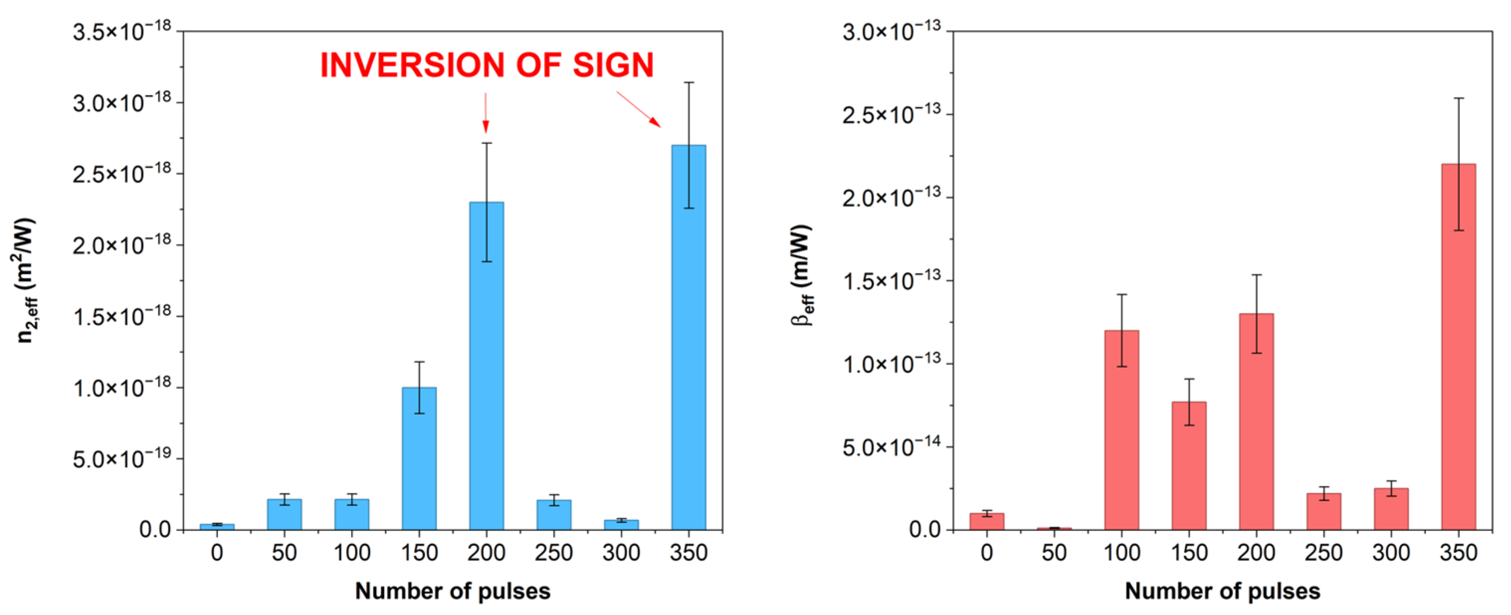
The evolution of the effective nonlinear parameters with the number of pulses per spot is shown in Figure 5. Both n2,eff and βeff exhibit a non-monotonic dependence on pulse number. Compared to the as-deposited coating, pronounced changes in both nonlinear coefficients are observed after irradiation with 200 and 350 pulses, such as increase in magnitude and sign inversion of the effective nonlinear refractive index, as presented in Table 1. At intermediate pulse numbers (250 and 300 pulses), reduced values of both parameters are observed.

4. Discussion

The overall nonlinear optical response in thin metal oxide films is often complex and depends heavily on factors such as the laser pulse duration, excitation wavelength, material morphology, and the presence of defects or impurities [44]. In the case of femtosecond laser irradiation, energy deposition occurs on ultrashort time-scales (~10−15 s), which are significantly shorter than the characteristic electron-phonon coupling time (~10−13–10−12 s) [6]. Consequently, the laser-matter interaction is dominated by electronic excitation rather than thermal lattice heating. Under these conditions, nonlinear refraction and absorption arise primarily from the instantaneous electronic Kerr effect, while additional contributions may originate from sub-bandgap states, defect-related electronic transitions, and short-range structural rearrangements.
The non-monotonic dependence of n2,eff and βeff on the number of applied pulses (Figure 5) indicates that sub-ablative femtosecond laser modification does not lead to a simple linear scaling of the third-order nonlinear response with irradiation dose. Instead, multiple nonlinear contributions may coexist, and their relative influence evolves as the cumulative pulse number increases.
A particularly notable feature is the inversion of the sign of the effective nonlinear refractive index observed after laser processing with higher pulse numbers. Similar sign reversals have been reported in the literature and have been attributed to different annealing conditions [45] or to laser-induced photochemical alteration of the nanoclusters [46]. Generally, femtosecond laser-induced sign changes in the third-order nonlinear response have been linked to modifications of the electronic structure of the material, including changes in defect-related absorption, redistribution of sub-bandgap states, and band-gap shifts, which alter the balance between competing nonlinear contributions [47,48,49].
The reduced n2,eff and βeff values obtained at 250 and 300 pulses may be influenced by substrate-related or parasitic linear contributions inherent to thin-film z-scan measurements. Such effects can potentially distort the nonlinear signatures [42].
One plausible contribution to the observed evolution of the nonlinear optical response is the modification of defect-related electronic states induced by femtosecond laser irradiation. Defects are intrinsic to optical thin films, originating from the film deposition process, growth conditions, and subsequent post-deposition treatments [50]. They play a crucial role in determining the material’s linear and nonlinear optical properties by introducing localized electronic states and structural disorder, such as distortions and vacancies, which give rise to intermediate electronic levels within the band gap [51,52]. As a result, defect states may strongly influence light-matter interaction processes, particularly under high-intensity laser excitation.
In addition to defect-related effects, laser-induced structural relaxation and densification represent another plausible mechanism contributing to the observed nonlinear optical modulation. Femtosecond laser irradiation below the ablation threshold can promote localized densification in amorphous oxide networks by reducing free volume and increasing short-range order in the initially low-density amorphous TiO2 film through ultrafast electronic excitation followed by rapid energy relaxation [53,54,55]. The ATR-FTIR results revealed spectral changes indicative of laser-induced dihydroxylation, which are consistent with structural relaxation and possible densification effects in the amorphous TiO2 network. According to Tamura et al., surface hydroxyl groups originate from under-coordinated oxide ions in structurally disordered oxides, and their removal leads to improved charge neutralization and stabilization of the oxide network without requiring crystallization [56]. In line with this, Ponton et al. demonstrated that partial dihydroxylation in amorphous oxide films is directly associated with local densification and increased short-range order, detected by FTIR analysis [57]. Consequently, a progressive increase in local structural order within the amorphous phase remains a plausible explanation for the enhanced nonlinear optical response, in agreement with previous reports [58].
While laser-induced phase transitions and crystallization in TiO2 thin films have been primarily reported under MHz or CW laser irradiation, where thermal accumulation drives the process [59,60], the present study employed femtosecond laser irradiation at a lower repetition rate of 1 kHz. The interpulse separation of approximately 1 ms prevents significant heat accumulation between successive pulses. Under these conditions, the incubation effect is likely dominated by defect accumulation, electronic excitation, and local structural changes rather than by purely thermal mechanisms [61]. Consistently, XRD analysis confirms the absence of long-range structural ordering after laser processing. From an application perspective, maintaining the amorphous nature of optical coatings is essential, as crystallization can introduce light scattering, refractive-index inhomogeneities, mechanical stress, and long-term instability, thereby degrading optical performance [62].

5. Conclusions

This study demonstrates that sub-ablative femtosecond laser irradiation is an effective post-deposition method for modifying the nonlinear optical properties of TiO2 coatings. The as-deposited coating exhibits a self-defocusing nonlinear response, while laser processing induces pronounced modifications of the third-order nonlinearity, including an enhancement of both the effective nonlinear refractive index and nonlinear absorption, as well as an inversion of the sign of the nonlinear refractive index.
The pulse-dependent evolution of the nonlinear parameters reveals a non-monotonic behavior, indicating the involvement of multiple contributing mechanisms that evolve with cumulative irradiation. Importantly, these modifications are achieved without inducing surface damage or crystallization, preserving the amorphous structure and morphological integrity, which is a critical requirement for high-performance optical coatings.
Overall, the present study provides new experimental insight into the controlled modulation of third-order optical nonlinearity in oxide thin films using femtosecond laser processing. This is a promising post-deposition method for the engineering of functional optical coatings for nonlinear photonic applications, including optical limiting, ultrafast all-optical switching, and high-power laser systems.

Supplementary Materials

The following supporting information can be downloaded at: https://www.mdpi.com/article/10.3390/coatings16020220/s1, Figure S1: Z-scan measurements of the (a) nonlinear absorption coefficient of fused silica as a reference material and the (b) nonlinear refractive index of fused silica. The black squares represent the experimentally measured normalized transmittance, while the solid lines correspond to the theoretical fits.

Author Contributions

Conceptualization, V.A., G.Y. and E.I.; methodology, V.A. and G.Y.; validation, G.Y. and S.K.; formal analysis, G.Y., K.S. and I.M.; investigation, V.A., G.Y., K.S., T.T., I.M., V.S., V.K. and I.Z.; resources, G.Y., I.M. and T.T.; data curation, V.A., G.Y., K.S., S.K., T.T. and I.M.; writing—original draft preparation, V.A.; writing—review and editing, V.A. and G.Y.; visualization, V.A. and G.Y.; supervision, G.Y. and E.I.; project administration, V.A. and E.I.; funding acquisition, V.A. and E.I. All authors have read and agreed to the published version of the manuscript.

Funding

This research was funded by the BULGARIAN NATIONAL SCIENCE FUND under the National Scientific Program “Petar Beron i NIE”, grant number KP-06-DB/9, “Femtosecond laser modification of different materials (FLAMAT)”. The APC was funded by the BULGARIAN NATIONAL SCIENCE FUND, grant number KP-06-DB/9. Deposition of coatings was funded by the BULGARIAN NATIONAL SCIENCE FUND, grant number № KΠ-06-H57/5, 2021–2024.

Institutional Review Board Statement

Not applicable.

Informed Consent Statement

Not applicable.

Data Availability Statement

The original data presented in the study are openly available in Zenodo at https://doi.org/10.5281/zenodo.18300654.

Acknowledgments

Research equipment of the Distributed Research Infrastructure INFRAMAT, as part of the Bulgarian National Roadmap for Research Infrastructures and supported by the Bulgarian Ministry of Education and Science, was used in this investigation.

Conflicts of Interest

The authors declare no conflicts of interest. The funders had no role in the design of the study; in the collection, analyses, or interpretation of data; in the writing of the manuscript; or in the decision to publish the results.

Abbreviations

The following abbreviations are used in this manuscript:
UV-Vis-NIRUltraviolet–Visible–Near Infrared
ATR-FTIRFourier Transform Infrared in attenuated total reflection
AFMAtomic force microscopy
RMSRoot Mean Square
QCMQuartz crystal microbalance
CCDCharge-Coupled Device
SIInternational System of Units
CWContinuous Wave

References

  1. Zhang, B.; Wang, Z.; Tan, D.; Qiu, J. Ultrafast Laser-Induced Self-Organized Nanostructuring in Transparent Dielectrics: Fundamentals and Applications. PhotoniX 2023, 4, 24. [Google Scholar] [CrossRef] [Scilit]
  2. Yang, C.; Ji, C.; Feng, S.; Liu, Y.; Wei, W.; Long, Y. Ultrafast Laser-Matter Interaction Mechanisms and Applications in Functional Device Fabrication: Recent Advances and Perspectives. Appl. Phys. Rev. 2025, 12, 031325. [Google Scholar] [CrossRef] [Scilit]
  3. Jia, Y.; Chen, F. Recent Progress on Femtosecond Laser Micro-/Nano-Fabrication of Functional Photonic Structures in Dielectric Crystals: A Brief Review and Perspective. APL Photonics 2023, 8, 090901. [Google Scholar] [CrossRef] [Scilit]
  4. Hossain, M.I.; Aissa, B.; Khandakar, A.; Thomas, K.; Rahman, A.; Mansour, S. Exploring the Optical and Morphological Properties of Metal Oxide Thin Films Produced via Reactive Electron Beam Evaporation. Cogent Eng. 2024, 11, 2338144. [Google Scholar] [CrossRef] [Scilit]
  5. Selvi, K.T.; Sagadevan, S. Recent Developments in Optoelectronic and Photonic Applications of Metal Oxides. In Metal Oxides for Optoelectronics and Optics-Based Medical Applications; Sagadevan, S., Podder, J., Mohammad, F., Eds.; Elsevier: Amsterdam, The Netherlands, 2022; pp. 33–57. ISBN 9780323858243. [Google Scholar] [CrossRef] [Scilit]
  6. Bonse, J.; Krüger, J. Structuring of Thin Films by Ultrashort Laser Pulses. Appl. Phys. 2022, 129, 14. [Google Scholar] [CrossRef] [Scilit]
  7. Dar, R.A.; Ansari, M.Z. Impact of Ion Beam Irradiation on Optical and Structural Properties of Nanocomposite ZnO Thin Films. Radiat. Eff. Defects Solids 2024, 179, 1462–1477. [Google Scholar] [CrossRef] [Scilit]
  8. Özmenteş, R.; Hassanien, A.S. Characterizations and Optical Discussions of Thermally Evaporated Titanium Dioxide Thin Films. J. Opt. 2024, 54, 1322–1340. [Google Scholar] [CrossRef] [Scilit]
  9. Jia, T.; Zhang, J.; Wu, J.; Wang, D.; Liu, Q.; Qi, Y.; Hu, B.; He, P.; Pan, W.; Qi, X. Synthesis Amorphous TiO2 with Oxygen Vacancy as Carriers Transport Channels for Enhancing Photocatalytic Activity. Mater. Lett. 2020, 265, 127465. [Google Scholar] [CrossRef] [Scilit]
  10. Dimitrov, V.; Sakka, S. Linear and Nonlinear Optical Properties of Simple Oxides. II. J. Appl. Phys. 1996, 79, 1741–1745. [Google Scholar] [CrossRef] [Scilit]
  11. Sahoo, S.; Surbhi, K.; Bhakta, S.; Das, R.; Sahoo, P.K. Influence of Defects on the Linear and Nonlinear Optical Properties of Cu-Doped Rutile TiO2 Microflowers. Phys. Chem. Chem. Phys. 2024, 26, 10191–10201. [Google Scholar] [CrossRef] [Scilit]
  12. Kandasamy, M.; Seetharaman, A.; Lakshmy, S.; Arjunan, N.; Manickavasakam, K.; Shetty, M.; Kanchana, S.; Qin, J.; Jothivenkatachalam, K.; Chakraborty, B. Defect Engineered N-S Codoped TiO2 Nanoparticles for Photocatalytic and Optical Limiting Applications: Experimental and DFT Insights. Spectrochim. Acta A Mol. Biomol. Spectrosc. 2024, 310, 123846. [Google Scholar] [CrossRef] [Scilit]
  13. Pang, C.; Li, R.; Huang, J.; Li, Z.; Chen, M.; Dong, N.; Wang, J.; Ren, F.; Chen, F. Ultrafast Electron Transfer Dynamics in Ag/TiO2 Nanocomposite for Tailoring of Optical Nonlinearity. Appl. Surf. Sci. 2021, 539, 148258. [Google Scholar] [CrossRef] [Scilit]
  14. Dong, L.; Chu, H.; Li, Y.; Zhao, S.; Li, D. Enhanced Optical Nonlinearity and Ultrafast Carrier Dynamics of TiO2/CuO Nanocomposites. Compos. B Eng. 2022, 237, 109860. [Google Scholar] [CrossRef] [Scilit]
  15. Banerjee, D.; Moram, S.S.B.; Byram, C.; Rathod, J.; Jena, T.; Podagatlapalli, G.K.; Soma, V.R. Plasmon-Enhanced Ultrafast and Tunable Thermo-Optic Nonlinear Optical Properties of Femtosecond Laser Ablated TiO2 and Silver-Doped TiO2 Nanoparticles. Appl. Surf. Sci. 2021, 569, 151070. [Google Scholar] [CrossRef] [Scilit]
  16. Alsharif, M.A.; Alqurashi, R.S.; Hamdalla, T.A. The Role of Annealing in Optimizing the Electrical, Linear, and Nonlinear Optical Properties ZIf-8@TiO2 Films for Possible Use in Photonic and Optoelectronic Devices. Phys. Scr. 2024, 99, 065983. [Google Scholar] [CrossRef] [Scilit]
  17. Zhi, X.; Li, X.; Yuan, S.; Wang, D.; Wang, K. Mechanic Properties Modification of SiO2 Thin Films by Femtosecond Laser. Optik 2022, 251, 168404. [Google Scholar] [CrossRef] [Scilit]
  18. Yuan, K.; Geng, F.; Zhang, Q.; Li, Y. Femtosecond Laser Strengthening of Electron-Beam Deposited SiO2 Thin Film on Fused Silica Substrates. Thin Solid Film. 2023, 780, 139959. [Google Scholar] [CrossRef] [Scilit]
  19. Yao, J.; Fan, Z.; Jin, Y.; Zhao, Y.; He, H.; Shao, J. Investigation of Damage Threshold to TiO2 Coatings at Different Laser Wavelength and Pulse Duration. Thin Solid Film. 2008, 516, 1237–1241. [Google Scholar] [CrossRef] [Scilit]
  20. Sheik-Bahae, M.; Said, A.A.; Wei, T.-H.; Hagan, D.J.; Van Stryland, E.W. Sensitive Measurement of Optical Nonlinearities Using a Single Beam. IEEE J. Quantum Electron. 1990, 26, 760–769. [Google Scholar] [CrossRef] [Scilit]
  21. Saha, A. Nonlinear Optical Properties of Chalcone Derivatives-a Short Review. Mater. Today Proc. 2022, 64, 605–610. [Google Scholar] [CrossRef] [Scilit]
  22. Jeyaseeli, J.R.; Jaikumar, P.; Girisun, T.C.S.; Philominal, A. Third Order Nonlinear Optical Properties of Undoped and Bi-Doped ZnO-ZrO2 Nanocomposites. J. Mol. Struct. 2025, 1321, 139918. [Google Scholar] [CrossRef] [Scilit]
  23. Zhang, Y.X.; Wang, Y.H. Nonlinear Optical Properties of Metal Nanoparticles: A Review. RSC Adv. 2017, 7, 45129–45144. [Google Scholar] [CrossRef] [Scilit]
  24. Singh, V.; Kharangarh, P.R.; Kumar, P.; Singh, D.; Sanjay; Ghosh, A.; Kumar, S. Measurements of Third-Order Optical Nonlinearity Using Z-Scan Technique: A Review. AIP Conf. Proc. 2019, 2142, 140035. [Google Scholar] [CrossRef] [Scilit]
  25. Ajami, A.; Ovsianikov, A.; Liska, R.; Baudis, S. Z-Scan Technique: A Review from Conventional Z-Scan to White Light Z-Scan. Appl. Phys. B 2024, 130, 138. [Google Scholar] [CrossRef] [Scilit]
  26. Van Stryland, E.W.; Sheik-Bahae, M. Z-Scan. In Characterization Techniques and Tabulations for Organic Nonlinear Optical Materials; Kuzyk, M.C., Dirk, C.W., Eds.; Routledge: New York, NY, USA, 2018; pp. 655–692. ISBN 9781351461818. [Google Scholar] [CrossRef] [Scilit]
  27. Petrov, D.V. Reflection Z-Scan Technique for the Study of Nonlinear Refraction and Absorption of a Single Interface and Thin Film. J. Opt. Soc. Am. B 1996, 13, 1491. [Google Scholar] [CrossRef] [Scilit]
  28. Tognazzi, A.; Franceschini, P.; Tran, T.N.L.; Chiasera, A.; Vincenti, M.A.; Cino, A.C.; Akozbek, N.; Scalora, M.; De Angelis, C. Z-Scan Theory for Thin Film Measurements: Validation of a Model beyond the Standard Approach Using ITO and HfO2. Opt. Mater. X 2023, 19, 100242. [Google Scholar] [CrossRef] [Scilit]
  29. Yankov, G.; Stefanov, I.; Dimitrov, K.; Piroeva, I.; Dimowa, L.T.; Tarassov, M.P.; Shivachev, B.L.; Yoneda, H.; Petrov, T. Measurement of Nonlinear Refractive Index and Multiphoton Absorption by the Subpicosecond z -Scan Method of Tellurite Multicomponent Glassy Matrixes Having Nonlinear Susceptibility. Phys. Scr. 2013, T157, 014026. [Google Scholar] [CrossRef] [Scilit]
  30. Niu, R.; Cui, L.; Wang, W. Nonlinear Optical Properties of Indium-Doped Single-Phased TiO2 Thin Films. J. Korean Phys. Soc. 2022, 81, 311–316. [Google Scholar] [CrossRef] [Scilit]
  31. Nakaruk, A.; Lin, C.Y.W.; Channei, D.; Koshy, P.; Sorrell, C.C. Fe-Doped and Mn-Doped Titanium Dioxide Thin Films. J. Solgel Sci. Technol. 2012, 61, 175–178. [Google Scholar] [CrossRef] [Scilit]
  32. Kunnamareddy, M.; Diravidamani, B.; Rajendran, R.; Singaram, B.; Varadharajan, K. Synthesis of Silver and Sulphur Codoped TiO2 Nanoparticles for Photocatalytic Degradation of Methylene Blue. J. Mater. Sci. Mater. Electron. 2018, 29, 18111–18119. [Google Scholar] [CrossRef] [Scilit]
  33. Toubal, B.; Elkourd, K.; Bouab, R.; Abdelaziz, O. The Impact of Copper–Cerium (Cu–Ce) Addition on Anatase-TiO2 Nanostructured Films for Its Inactivation of Escherichia Coli and Staphylococcus Aureus. J. Solgel Sci. Technol. 2022, 103, 549–564. [Google Scholar] [CrossRef] [Scilit]
  34. Zhang, L.; Ren, H.; Ma, H.; Shi, Z.; Yang, Y.; Yuan, Q.; Feng, X.; Ma, Y.; Chen, B. Determining the Nonlinear Refractive Index of Fused Quartz by Femtosecond Laser Z-Scan Technology. In Optical Measurement Technology and Instrumentation; Han, S., Tan, J., Eds.; SPIE: Bellingham, WA, USA, 2016; p. 101551N. [Google Scholar] [CrossRef] [Scilit]
  35. Ma, Y.; Zhang, L.; Shi, Z.; Ren, H.; Yang, Y.; Ma, H.; Yuan, Q.; Feng, X.; Chen, B. Femtosecond Z-Scan Measurements of the Nonlinear Refractive Index of Fused Silica. In Proceedings of the 2017 International Conference on Optical Instruments and Technology: Optoelectronic Measurement Technology and Systems; Zhu, J., Xu, K., Dong, L., Tam, H.-Y., Xiao, H., Eds.; SPIE: Bellingham, WA, USA, 2018; p. 91. [Google Scholar] [CrossRef] [Scilit]
  36. Boyd, R.W. Nonlinear Optics, 4th ed.; Academic Press: Cambridge, MA, USA, 2020. [Google Scholar]
  37. Shehata, A.; Ali, M.; Schuch, R.; Mohamed, T. Experimental Investigations of Nonlinear Optical Properties of Soda-Lime Glasses and Theoretical Study of Self-Compression of Fs Laser Pulses. Opt. Laser Technol. 2019, 116, 276–283. [Google Scholar] [CrossRef] [Scilit]
  38. Lin, Z.; Xu, L.; Wu, J.; Zhang, X.; Ren, M.; Zhu, L.; Hayasaki, Y.; Zhang, H.; Wang, P. Investigation of the Influence of Nonlinear Absorption Properties on Glass Modification Induced by Femtosecond Laser Irradiation. J. Non. Cryst. Solids 2026, 672, 123867. [Google Scholar] [CrossRef] [Scilit]
  39. Sutherland, R.L. Handbook of Nonlinear Optics, 2nd ed.; CRC Press: Boca Raton, FL, USA, 2003; ISBN 9781135541194. [Google Scholar]
  40. Long, H.; Chen, A.; Yang, G.; Li, Y.; Lu, P. Third-Order Optical Nonlinearities in Anatase and Rutile TiO2 Thin Films. Thin Solid Film. 2009, 517, 5601–5604. [Google Scholar] [CrossRef] [Scilit]
  41. Hales, J.M.; Hagan, D.J.; Jen, A.K.-Y.; Perry, J.W.; Li, Z.; Jang, S.-H.; Van Stryland, E.W.; Hu, H.; Ensley, T.R.; Benis, S. Nonlinear Refraction and Absorption Measurements of Thin Films by the Dual-Arm Z-Scan Method. Appl. Opt. 2019, 58, D28–D33. [Google Scholar] [CrossRef] [Scilit]
  42. Patterson, B.M.; White, W.R.; Robbins, T.A.; Knize, R.J. Linear Optical Effects in Z-Scan Measurements of Thin Films. Appl. Opt. 1998, 37, 1854. [Google Scholar] [CrossRef] [Scilit] [PubMed]
  43. Lin, S.S.; Hung, Y.H.; Chen, S.C. Optical Properties of TiO2 Thin Films Deposited on Polycarbonate by Ion Beam Assisted Evaporation. Thin Solid Film. 2009, 517, 4621–4625. [Google Scholar] [CrossRef] [Scilit]
  44. Gu, B.; Wang, H.-T. Linear and Nonlinear Optical Properties of Ferroelectric Thin Films. In Ferroelectrics―Physical Effects; Lallart, M., Ed.; InTech: London, UK, 2011. [Google Scholar]
  45. Chouhan, R.; Baraskar, P.; Agrawal, A.; Gupta, M.; Sen, P.K.; Sen, P. Effects of Oxygen Partial Pressure and Annealing on Dispersive Optical Nonlinearity in NiO Thin Films. J. Appl. Phys. 2017, 122, 025301. [Google Scholar] [CrossRef] [Scilit]
  46. Osborne, D.H., Jr.; Haglund, R.F., Jr.; Gonella, F.; Garrido, F. Laser-Induced Sign Reversal of the Nonlinear Refractive Index of Ag Nanoclusters in Soda-Lime Glass. Appl. Phys. B 1998, 66, 517–521. [Google Scholar] [CrossRef] [Scilit]
  47. Yamamoto, H.; Tanaka, S.; Naito, T.; Hirao, K. Nonlinear Change of Refractive Index of Co3O4 Thin Films Induced by Semiconductor Laser (λ = 405 nm) Irradiation. Appl. Phys. Lett. 2002, 81, 999–1001. [Google Scholar] [CrossRef] [Scilit]
  48. Kim, V.V.; Bundulis, A.; Grube, J.; Ganeev, R.A. Variation of the Sign of Nonlinear Refraction of Carbon Disulfide in the Short-Wavelength Region. Opt. Mater. Express 2022, 12, 2053. [Google Scholar] [CrossRef] [Scilit]
  49. Lapointe, J.; Bérubé, J.-P.; Ledemi, Y.; Dupont, A.; Fortin, V.; Messaddeq, Y.; Vallée, R. Nonlinear Increase, Invisibility, and Sign Inversion of a Localized Fs-Laser-Induced Refractive Index Change in Crystals and Glasses. Light Sci. Appl. 2020, 9, 64. [Google Scholar] [CrossRef] [Scilit]
  50. Cheng, X.; Wang, Z. Defect-Related Properties of Optical Coatings. Adv. Opt. Technol. 2014, 3, 65–90. [Google Scholar] [CrossRef] [Scilit]
  51. Souza, J.C.; Ribeiro, R.A.P.; da Trindade, L.G.; de Oliveira, R.C.; Costa, L.D.; de Oliveira, M.C.; de Lazaro, S.R.; Sambrano, J.R.; Mendonça, C.R.; de Boni, L.; et al. Unconventional Disorder by Femtosecond Laser Irradiation in Fe2O3. ACS Omega 2021, 6, 28049–28062. [Google Scholar] [CrossRef] [Scilit]
  52. Limaye, M.V.; Chen, S.C.; Lee, C.Y.; Chen, L.Y.; Singh, S.B.; Shao, Y.C.; Wang, Y.F.; Hsieh, S.H.; Hsueh, H.C.; Chiou, J.W.; et al. Understanding of Sub-Band Gap Absorption of Femtosecond-Laser Sulfur Hyperdoped Silicon Using Synchrotron-Based Techniques. Sci. Rep. 2015, 5, 11466. [Google Scholar] [CrossRef] [Scilit]
  53. Itoh, K.; Watanabe, W.; Nolte, S.; Schaffer, C.B. Ultrafast Processes for Bulk Modification of Transparent Materials. MRS Bull. 2006, 31, 620–625. [Google Scholar] [CrossRef] [Scilit]
  54. Huser, T.; Risbud, S.; Chan, J.W.; Krol, D.M. Structural Changes in Fused Silica after Exposure to Focused Femtosecond Laser Pulses. Opt. Lett. 2001, 26, 1726–1728. [Google Scholar] [CrossRef] [Scilit]
  55. Lancry, M.; Poumellec, B. UV Laser Processing and Multiphoton Absorption Processes in Optical Telecommunication Fiber Materials. Phys. Rep. 2013, 523, 207–229. [Google Scholar] [CrossRef] [Scilit]
  56. Tamura, H.; Mita, K.; Tanaka, A.; Ito, M. Mechanism of Hydroxylation of Metal Oxide Surfaces. J. Colloid Interface Sci. 2001, 243, 202–207. [Google Scholar] [CrossRef] [Scilit]
  57. Ponton, S.; Dhainaut, F.; Vergnes, H.; Samelor, D.; Sadowski, D.; Rouessac, V.; Lecoq, H.; Sauvage, T.; Caussat, B.; Vahlas, C. Investigation of the Densification Mechanisms and Corrosion Resistance of Amorphous Silica Films. J. Non. Cryst. Solids 2019, 515, 34–41. [Google Scholar] [CrossRef] [Scilit]
  58. Rachana, K.; Nagaraja, K.K.; Poornesh, P.; Jagadeesh Chandra, R.B.; Pramodini, S. Third-Order Nonlinear Optical Properties of TiO2 Nano Colloidal Synthesized via Sol–Gel and Hydrothermal Methods: A Comparison. Mater. Sci. Eng. B 2025, 319, 118312. [Google Scholar] [CrossRef] [Scilit]
  59. Hoppius, J.S.; Bialuschewski, D.; Mathur, S.; Ostendorf, A.; Gurevich, E.L. Femtosecond Laser Crystallization of Amorphous Titanium Oxide Thin Films. Appl. Phys. Lett. 2018, 113, 071904. [Google Scholar] [CrossRef] [Scilit]
  60. Benavides, J.A.; Trudeau, C.P.; Gerlein, L.F.; Cloutier, S.G. Laser Selective Photoactivation of Amorphous 2 Films to Anatase and/or Rutile Crystalline Phases. ACS Appl. Energy Mater. 2018, 1, 3607–3613. [Google Scholar] [CrossRef] [Scilit]
  61. Yin, J.; Lin, Z.; Ji, L.; Hong, M.; Yin, J.; Lin, Z.; Ji, L.; Hong, M. Femtosecond Laser Micro/Nano-Processing via Multiple Pulses Incubation. Opto Electron. Technol. 2025, 1, 250003. [Google Scholar] [CrossRef] [Scilit]
  62. Liang, X.; Liu, B.; Yuan, Q.; Lin, X.; Ren, S.; Zhao, S.; Yin, X.; Fei, S. Fine Control of Optical Properties of Nb2O5 Film by Thermal Treatment. Micromachines 2024, 15, 1453. [Google Scholar] [CrossRef] [Scilit] [PubMed]
Figure 2. Morphological characterization of the TiO2 thin film: (a) Optical profilometry image of the unmodified surface acquired at 100× magnification, showing the three independent regions of interest used for surface roughness evaluation; (b) Optical profilometry image of the TiO2 coating modified with 350 femtosecond laser pulses, recorded at 20× magnification over a larger field of view; (c) AFM topography map of the unmodified TiO2 coating; (d) AFM topography map of the TiO2 coating after femtosecond laser modification with 350 pulses.
Coatings 16 00220 g002
Figure 3. (a) UV-Vis-NIR transmittance spectra of the as-deposited TiO2 coating and the soda-lime glass substrate in the wavelength range 320–1200 nm; (b) X-ray diffraction (XRD) patterns of the unmodified and modified with 350 pulses TiO2 coating; (c) ATR-FTIR spectra of the soda-lime substrate, unmodified TiO2 coating, and laser-modified TiO2 coating.
Coatings 16 00220 g003
Figure 4. Z-scan measurements of: (a) nonlinear absorption coefficient of soda-lime glass substrate; (b) nonlinear refractive index of soda-lime glass substrate; (c) effective nonlinear absorption coefficient of unmodified TiO2 coating; (d) effective nonlinear refractive index of unmodified TiO2 coating; (e) effective nonlinear absorption coefficient of modified TiO2 coating with 350 femtosecond laser pulses at a fluence of 0.083 J/cm2; (f) effective nonlinear refractive index of modified TiO2 coating with 350 femtosecond laser pulses at a fluence of 0.083 J/cm2. The black squares represent the experimentally measured normalized transmittance, while the solid lines correspond to the theoretical fits.
Coatings 16 00220 g004
Figure 5. Evolution of the effective nonlinear refractive index n2,eff and the effective nonlinear absorption coefficient βeff as a function of the number of femtosecond laser pulses per spot.
Coatings 16 00220 g005
Table 1. Calculated values of the effective nonlinear refractive index n2,eff and the effective nonlinear absorption coefficient βeff for fused silica as a reference material, the unmodified TiO2 coating, and the modified TiO2 coating with 200 and 350 pulses, where an inversion of the nonlinearity sign is experimentally observed. The reported confidence intervals correspond to the estimated experimental uncertainty calculated via error propagation.
Materialn2,eff ± Δn2
[m2/W]
βeff ± Δβ
[m/W]
Fused silica(5.7 ± 1.0) × 10−20(4.7 ± 0.8) × 10−14
Soda-lime substrate−(9.1 ± 1.7) × 10−20(4 ± 0.7) × 10−13
Unmodified TiO2 coating−(4 ± 0.7) × 10−20(1 ± 0.2) × 10−14
Modified TiO2 coating with 200 pulses(2.3 ± 0.4) × 10−18(1.3 ± 0.2) × 10−13
Modified TiO2 coating with 350 pulses(2.7 ± 0.4) × 10−18(2.2 ± 0.4) × 10−13
Disclaimer/Publisher’s Note: The statements, opinions and data contained in all publications are solely those of the individual author(s) and contributor(s) and not of MDPI and/or the editor(s). MDPI and/or the editor(s) disclaim responsibility for any injury to people or property resulting from any ideas, methods, instructions or products referred to in the content.