Article

Strontium-Doped Ti3C2Tx MXene Coatings on Titanium Surfaces: Synergistic Osteogenesis Enhancement and Antibacterial Activity Evaluation

State Key Laboratory of Oral Diseases & National Center for Stomatology & National Clinical Research Center for Oral Diseases & Department of Implantology, West China Hospital of Stomatology, Sichuan University, Chengdu 610041, China
*
Author to whom correspondence should be addressed.
Coatings 2025, 15(7), 847; https://doi.org/10.3390/coatings15070847 (registering DOI)
Submission received: 16 June 2025 / Revised: 13 July 2025 / Accepted: 17 July 2025 / Published: 19 July 2025
(This article belongs to the Section Surface Coatings for Biomedicine and Bioengineering)

Abstract

To improve implant osseointegration while preventing infection, we developed a strontium (Sr)-doped Ti3C2Tx MXene coating on titanium, aiming to synergistically enhance bone integration and antibacterial performance. MXene is a family of two-dimensional transition-metal carbides/nitrides whose abundant surface terminations endow high hydrophilicity and bioactivity. The coating was fabricated via anodic electrophoretic deposition (40 V, 2 min) of Ti3C2Tx nanosheets, followed by SrCl2 immersion to incorporate Sr2+. The coating morphology, phase composition, chemistry, hydrophilicity, mechanical stability, and Sr2+ release were characterized. In vitro bioactivity was assessed with rat bone marrow mesenchymal stem cells (BMSCs)—with respect to viability, proliferation, migration, alkaline phosphatase (ALP) staining, and Alizarin Red S mineralization—while the antibacterial efficacy was evaluated against Staphylococcus aureus (S. aureus) via live/dead staining, colony-forming-unit enumeration, and AlamarBlue assays. The Sr-doped MXene coating formed a uniform lamellar structure, lowered the water-contact angle to ~69°, and sustained Sr2+ release (0.36–1.37 ppm). Compared to undoped MXene, MXene/Sr enhanced BMSC proliferation on day 5, migration by 51%, ALP activity and mineralization by 47%, and reduced S. aureus viability by 49% within 24 h. Greater BMSCs activity accelerates early bone integration, whereas rapid bacterial suppression mitigates peri-implant infection—two critical requirements for implant success. Sr-doped Ti3C2Tx MXene thus offers a simple, dual-function surface-engineering strategy for dental and orthopedic implants.

1. Introduction

The long-term success of dental implants largely depends on the bone integration ability of the implant surface [1,2]. However, traditional surface-modification methods such as surface roughening, while enhancing bone bonding, may also increase the risk of bacterial adhesion and biofilm formation [3]. Studies show that while rough surfaces on implants can accelerate bone integration, they also provide a favorable environment for bacterial growth and can trigger peri-implant infections [4]. Implant-associated infections are primarily caused by pathogens such as Staphylococcus aureus (S. aureus) and Porphyromonas gingivalis, which adhere to and form biofilms on the surface, potentially leading to bone resorption and implant failure [5]. Therefore, developing implant surface coatings that simultaneously promote bone integration and possess antimicrobial properties is crucial for improving the clinical efficacy of implants [6,7].
MXene is a class of two-dimensional transition-metal carbides/nitrides that has emerged in recent years, with titanium carbide Ti3C2Tx being the most representative [8]. Tx represents abundant surface functional groups (–OH, =O, –F, and, rarely, –Cl) opposed to the surface of the Ti layer during the production of MXene by chemically etching the MAX phase [9]. Ti3C2Tx MXene possesses a large specific surface area and excellent hydrophilicity and conductivity, showing great potential for application in tissue engineering and biomedical fields [10]. Using MXene as an implant coating is expected to impart both biological activity and antimicrobial properties to the surface. On the one hand, studies have demonstrated that MXene exhibits excellent antibacterial activity. The sharp edges of its nanosheets can disrupt bacterial membranes and chemical damage by inducing oxidative stress, inducing the production of reactive oxygen species (ROS) that effectively kill bacteria [11]. On the other hand, MXene can promote bone regeneration. Studies has shown that depositing MXene on titanium surfaces significantly enhances the adhesion and proliferation of bone marrow mesenchymal stem cells (BMSCs) and accelerates osteogenic differentiation [12]. The negatively charged functional groups on the MXene surface easily adsorb positively charged adhesive proteins, enhancing hydrophilicity and providing adhesion sites for cells, thus promoting the adhesion and anchorage of osteogenesis-related cells [13]. Furthermore, MXene coatings themselves can regulate cell signaling pathways (e.g., activating Wnt/β-catenin) and upregulate osteogenic gene expression to enhance osteogenic differentiation [14]. In conclusion, MXene is a promising candidate for implant surface coatings, expected to achieve the dual functions of promoting bone formation and antimicrobial effects through its hydrophilic surface and unique two-dimensional structure. However, the osteogenic regeneration capability of pure MXene coatings remains limited. Therefore, further modification of MXene coatings through chemical or microscopic physical morphology changes is necessary to achieve better osteogenic effects.
Strontium (Sr) is an essential element in human bone tissue. Studies have shown that appropriate concentrations of Sr2+ have dual regulatory effects on bone metabolism: they promote osteoblast proliferation and differentiation while inhibiting osteoclast activity, thus enhancing new bone formation and reducing bone resorption [15]. Based on this characteristic, strontium ions are often introduced onto the surface of bone implant materials to enhance osteogenic performance [16]. Studies have incorporated varying amounts of Sr into titanium oxide porous coatings and found that, without altering the coating morphology, it significantly promotes the proliferation of osteoblast precursor cells and enhances alkaline phosphatase activity [17]. Zhang et al. [18] incorporated Sr into TiO2 coatings through micro-arc oxidation and similarly observed significant improvements in cell adhesion and proliferation, as well as a marked enhancement of the coating’s osteogenic activity. Additionally, Sr2+ has anti-inflammatory effects and can increase new bone mineralization, making it a promising candidate for implant surface modification [19]. However, simple Sr-doped coatings may lack antibacterial properties, and while antibacterial elements (such as Ag and Cu) can inhibit bacteria, excessive amounts may affect bone tissue healing [20]. Therefore, combining osteogenic Sr with MXene materials that have physical/chemical antibacterial properties has the potential to synergistically improve bone integration and anti-infection performance.
Based on the above background, this study combines two-dimensional Ti3C2Tx MXene material with Sr2+ functional ions and prepares Sr-doped MXene surface coatings on pure titanium substrates through electrophoretic deposition and immersion doping methods (Figure 1). The negatively charged functional groups on the MXene surface effectively adsorb and load Sr2+, thereby enhancing its osteogenic activity while maintaining the hydrophilic adhesion-promoting and physical antibacterial properties of the MXene coating, providing a synergistic antibacterial effect. This study systematically characterizes the morphology, structure, elemental composition, hydrophilicity, and mechanical stability of the resulting composite coating, and evaluates its effects on BMSC proliferation, migration, and osteogenic differentiation through in vitro experiments. Additionally, the antibacterial performance of the coating against the common pathogenic bacterium S. aureus in peri-implantitis is also tested. This composite coating provides a new strategy for the design of novel implant surface coatings and for addressing peri-implantitis.

2. Materials and Methods

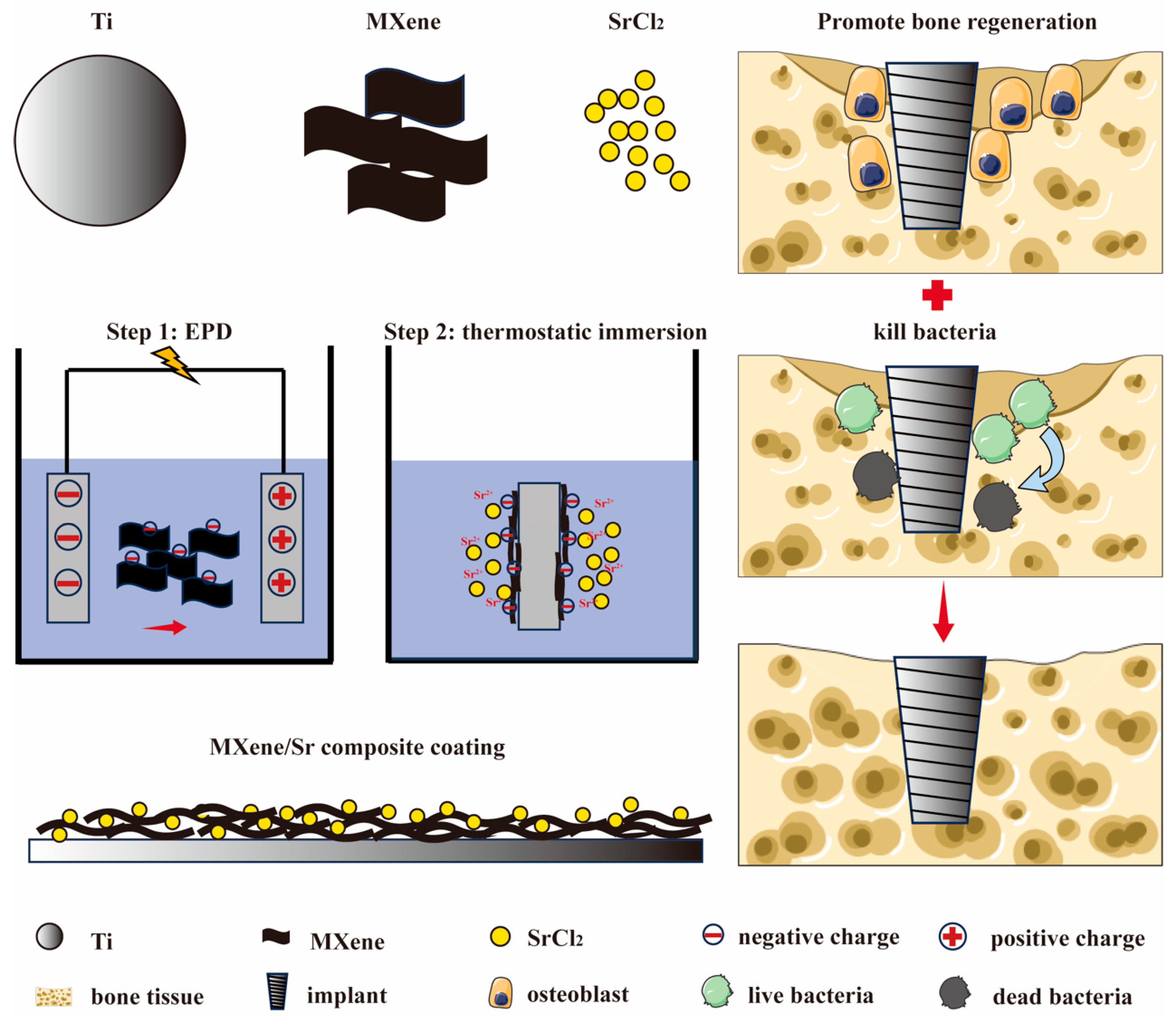
2.1. Coating Preparation

Pure titanium sheets (10 mm × 10 mm × 1 mm, Arit Electromechanical Equipment Co., Ltd., Wuhu, China) were polished sequentially using 600–1200 mesh alumina sandpaper in a gradient manner until the surface exhibited a glossy finish. The titanium sheets were then ultrasonically cleaned for 15 min in acetone (China National Pharmaceutical Group, Beijing, China), anhydrous ethanol (China National Pharmaceutical Group, China), and ultrapure water, respectively. After drying, the samples were sealed in ethanol (Ti control group). The treated titanium sheets were used as the anode and placed in a Ti3C2Tx MXene suspension (YiYi Technology Co., Jiling, China) for electrophoretic deposition (EPD), with graphite sheets as the electrode. The MXene suspension was prepared by dispersing Ti3C2Tx nanosheets in deionized water (concentration 0.15 mg/mL). The EPD parameters were set to a constant voltage of 40 V, with a deposition time of 2 min. Afterward, the samples were thoroughly washed with ultrapure water and naturally dried, resulting in MXene-coated titanium sheet samples (MXene group). The MXene-coated samples were then immersed in a 10 mg/mL strontium chloride (China National Pharmaceutical Group, China) aqueous solution and subjected to oscillation at 45 °C for 12 h, allowing Sr2+ ions to diffuse into and load onto the coating. Afterward, the samples were rinsed and dried, resulting in Sr-doped MXene coating samples (MXene/Sr group).

2.2. Material Characterization

Field-emission scanning electron microscopy (SEM, JEOL JSM-7900F, Tokyo, Japan) was used to observe the microstructure of the surface and cross-section of the samples from each group and to compare the uniformity and thickness of the coating. X-ray diffraction (XRD, PANalytical Empyrean, Overijssel, The Netherlands) was employed to analyze the phase composition and crystal structure changes of the coatings within the diffraction angle range of 2θ = 5–80°. Micro-Raman spectroscopy (Raman, Thermo Scientific DXR, Waltham, MA, USA) was used to detect the characteristic vibrational peaks of the Ti3C2Tx MXene coating to evaluate the integrity of the coating structure after Sr2+ incorporation. X-ray photoelectron spectroscopy (XPS, Thermo Fischer, ESCALAB 250Xi, Waltham, MA, USA) was utilized to analyze the elemental composition and chemical bonding states of the coatings, comparing the changes in elements between MXene/Sr and MXene coatings, particularly focusing on the form and content of Sr elements. A contact angle measurement instrument (Dataphysics OCA20, Filderstadt, Germany) was used to record the static water-contact angles of the surfaces of the samples at room temperature to assess the effect of the coating on hydrophilicity. A nano-scratch tester (Nano Scratch Tester, Anton Paar, Austria) was used to conduct scratch tests on the samples by gradually increasing the load. It recorded the normal load (critical load) and the corresponding frictional force, thereby making it possible to evaluate the coating’s bonding strength and scratch resistance to the substrate. Additionally, to detect the release behavior of Sr2+ ions, the MXene/Sr samples were placed in centrifuge tubes containing 5 mL phosphate-buffered saline (PBS) and stored at 37 °C. At 1, 3, 7, and 14 days, the solution was collected and replenished with an equal volume of fresh PBS, and the Sr2+ concentrations in the PBS at each time point were measured using inductively coupled plasma mass spectrometry (ICP-MS, Agilent 7500cx, Beijing, China). The cumulative release curve was plotted based on the Sr2+ concentration in ppm.

2.3. In Vitro Cell Compatibility Evaluation

2.3.1. Cell Culture

Rat bone marrow mesenchymal stem cells (BMSCs, STM-CE-2310, Yaji Biotechnology Co., Ltd., Shanghai, China) were used in vitro experiments. The cells were cultured in α-MEM medium (αMEM, GIBCO, Waltham, MA, USA), which was supplemented with 10% fetal bovine serum (FBS) (Four Seasons Qing Company, Hangzhou, China) and 1% penicillin-streptomycin mixed antibiotics (Hyclone, Logan, UT, USA), under constant culture conditions of 37 °C and 5% CO2. When the culture reached approximately 80% confluence, the cells were dissociated using 0.25% trypsin–0.02% EDTA (Invitrogen, Waltham, MA, USA). After centrifugation at 1000 r/min for 5 min, the cells were resuspended in fresh α-MEM medium for subsequent experiments.

2.3.2. Cytotoxicity

Each group of samples was placed in a 24-well plate, with 2 × 10⁴ BMSCs cells seeded per well. The cells were cultured for 24 h at 37 °C and 5% CO2. Afterward, a Calcein-AM/PI (propidium iodide) live-dead cell double-staining kit (Sigma-Aldrich, St. Louis, MO, USA) was used for fluorescence staining. Each well received 1 mL of working staining solution containing 2 μM Calcein-AM and 4 μM PI, and was incubated in the dark for 30 min. The cells were then observed and imaged under a fluorescence microscope (Chemidoc MP, BIO-RAD, Kaki Bukit, Singapore). Live cells were stained green with Calcein-AM, while dead cells were stained red with PI.

2.3.3. Transwell Migration Assay

Ti, MXene, and MXene/Sr-coated titanium sheets were placed in 24-well plates, and 1 mL of α-MEM containing 10% FBS was added. The samples were incubated at 37 °C with 5% CO2 for 72 h. After incubation, the supernatant was collected and sterilized by filtration through a 0.22 μm filter to prepare the conditioned medium for each group. Then, 200 μL of serum-free α-MEM containing BMSCs (2 × 10⁴ cells/well) was added to the upper chamber of the Transwell insert (Corning Inc., Corning, NY, USA), and the corresponding conditioned medium was added to the lower chamber. The samples were incubated at 37 °C with 5% CO2 for 24 h. After fixation with 4% paraformaldehyde for 15 min, the cells were stained with 0.1% crystal violet for 10 min. The Transwell membrane was placed flat on a glass slide and captured under a microscope. The number of cells that migrated through the membrane was counted, and the migration rate was calculated.

2.3.4. Cell Proliferation

Cell proliferation was assessed using the Cell Counting Kit-8 (CCK-8, APE-BIO, Houston, TX, USA) to evaluate the effect of the materials on osteoblast proliferation. Each group was placed in 24-well plates, and 2 × 10⁴ BMSCs cells were seeded per well. The cells were cultured at 37 °C with 5% CO2 for 1, 3, and 5 days. The medium was replaced with fresh medium every 2 days. At the predetermined time points, 350 μL of a working solution prepared by mixing 10% CCK-8 reagent with 90% regular medium was added to each well, with a blank control well. After incubating at 37 °C for 1 h, 100 μL of the supernatant from each well was transferred to a 96-well plate. The absorbance was then measured at 450 nm using a microplate reader.

2.4. In Vitro Osteogenic Differentiation Detection of Cells

After 2 days of basal culture, cells on each group were subjected to osteogenic differentiation induction in vitro. The osteogenic induction medium was α-MEM complete medium supplemented with a final concentration of 10 mM β-glycerophosphate (China National Pharmaceutical Group, China), 100 nM dexamethasone (China National Pharmaceutical Group, China), and 50 μg/mL ascorbic acid (China National Pharmaceutical Group, China). The samples were then cultured in osteogenic induction medium

2.4.1. ALP Staining

After 7 days of induction, the samples were washed twice with PBS and then fixed with 650 μL of 4% paraformaldehyde at 4 °C for 30 min. The samples were stained by adding 5-bromo-4-chloro-3-indolyl phosphate/nitro blue tetrazolium (BCIP/NBT) alkaline phosphatase staining working solution (Biotime Biotechnology, Shanghai, China) and incubated at 37 °C in the dark for 30 min. Alkaline phosphatase (ALP) activity causes the BCIP hydrolysis product to react with NBT, forming an insoluble blue-violet precipitate. After staining, the working solution was removed, and the samples were washed lightly with PBS. Imaging was performed under an optical microscope (BX51, Olympus, Tokyo, Japan). The images were quantitatively analyzed using ImageJ Fiji 2.14.0 software: the file was converted to 8-bit; an equal-diameter circular ROI was applied to each photo; thresholding with the Default algorithm (35–255) produced a binary mask; and integrated density was obtained.

2.4.2. ARS Staining

After 21 days of osteogenic induction differentiation, the medium was aspirated, and the cells were gently washed three times with pre-warmed PBS at 37 °C (5 min each time). The cells were then fixed with 4% paraformaldehyde for 10 min. Alizarin Red S (ARS) staining solution (Yisheng Biotechnology, Shanghai, China) was added to ensure the samples were covered, and the samples were incubated in a 37 °C incubator for 30 min. After thorough washing with distilled water, the samples were observed and imaged under an optical microscope. The images were quantitatively analyzed using ImageJ Fiji 2.14.0 software: an equal-diameter circular ROI was applied to each photo, red deposits were isolated with the Color Threshold tool (Hue 5–25, Saturation > 50, Brightness > 50), and the positive area was quantified with the Analyze Particles.

2.5. In Vitro Antibacterial Test

2.5.1. Bacterial Culture

S. aureus (ATCC 29213, MicroBioLogics, San Diego, CA, USA) was streaked on an LB agar plate (Yisheng Biotechnology, China) and incubated at 37 °C for 24 h. Single colonies with a regular shape were picked and transferred into 3 mL pre-sterilized LB liquid medium. The culture was then incubated overnight at 37 °C with 150 rpm agitation. Afterward, 1 mL of sterilized LB medium was centrifuged (5000 rpm, 5 min) to collect the bacteria, and the bacterial suspension was adjusted to a concentration of 107 CFU/mL for subsequent antibacterial experiments.

2.5.2. Live/Dead Bacterial Staining Observation

The bacterial suspension (50 μL, 107 CFU/mL) was evenly applied to the surface of the samples and incubated at 37 °C for 24 h. After incubation, the sample surfaces were gently washed 1–2 times with PBS. The samples were then covered with a staining working solution (BBcellProbe®N01/PI) prepared according to the instructions of the live/dead staining kit (BestBio, Shanghai, China) and incubated in the dark for 15 min. After staining, the samples were observed under a fluorescence microscope (Chemidoc MP, BIO-RAD, Kaki Bukit, Singapore) to observe the distribution of live (green) and dead (red) bacteria.

2.5.3. Bacterial Cell Integrity Observation

The prepared bacterial suspension (50 μL, 107 CFU/mL) was evenly applied to the surface of the samples and incubated at 37 °C for 24 h in a constant temperature incubator. After incubation, the samples were immediately fixed in 2.5% glutaraldehyde solution at 4 °C overnight. After fixation, the samples were dehydrated sequentially using 30%, 50%, 70%, 90%, and 100% ethanol gradient solutions for 15 min each. The samples were then treated with hexamethyldisilazane solution and allowed to air-dry. Subsequently, SEM was used to observe the morphological changes in the bacteria.

2.5.4. Agar Re-Culturing and Colony Counting

The bacterial suspension (50 μL, approximately 1 × 10⁷ CFU/mL) was evenly applied to the surface of the samples and incubated at 37 °C for 24 h. After incubation, the attached bacteria were detached by sonication for 1 min. Each group’s eluted bacterial suspension was serially diluted tenfold with 0.9% NaCl. Then, 100 μL of the diluted solution was plated onto LB agar plates. After spreading with a disposable L-shaped spreader, the plates were incubated at 37 °C for 18 h. Colonies were quantified using ImageJ software.

2.5.5. Bacterial Activity Quantification

The bacterial metabolic activity was quantitatively measured using the AlamarBlue assay kit (Thermo Scientific, Waltham, MA, USA), reflecting the bacterial survival rate. The bacterial suspension (50 μL, 107 CFU/mL) was evenly applied to the surface of the samples and incubated at 37 °C for 24 h. After incubation, 500 μL of 10% AlamarBlue working solution was added to each sample, and incubation continued at 37 °C in the dark for 2 h. Finally, 100 μL of the culture medium was transferred to a black 96-well plate. The fluorescence intensity (FI) was measured at an excitation wavelength of 560 nm and an emission wavelength of 590 nm. Relative survival rate (%) = FI (experiment)/FI (control) × 100%, where FI (experiment) is the fluorescence intensity of the MXene and MXene/Sr groups, and FI (control) is the fluorescence intensity of the Ti group.

2.6. Data Statistics and Analysis

All quantitative results are presented as “mean ± standard deviation”. Prior to the group comparison, normality was tested with the Shapiro–Wilk test and homogeneity of variances with the Levene test in GraphPad Prism 9.0. For comparisons of multiple groups, one-way analysis of variance (ANOVA) was used, followed by Tukey’s post-hoc test for intergroup difference assessment. Statistical analysis was performed using GraphPad Prism 9.0. The significance threshold was set at p < 0.05.

3. Results and Discussion

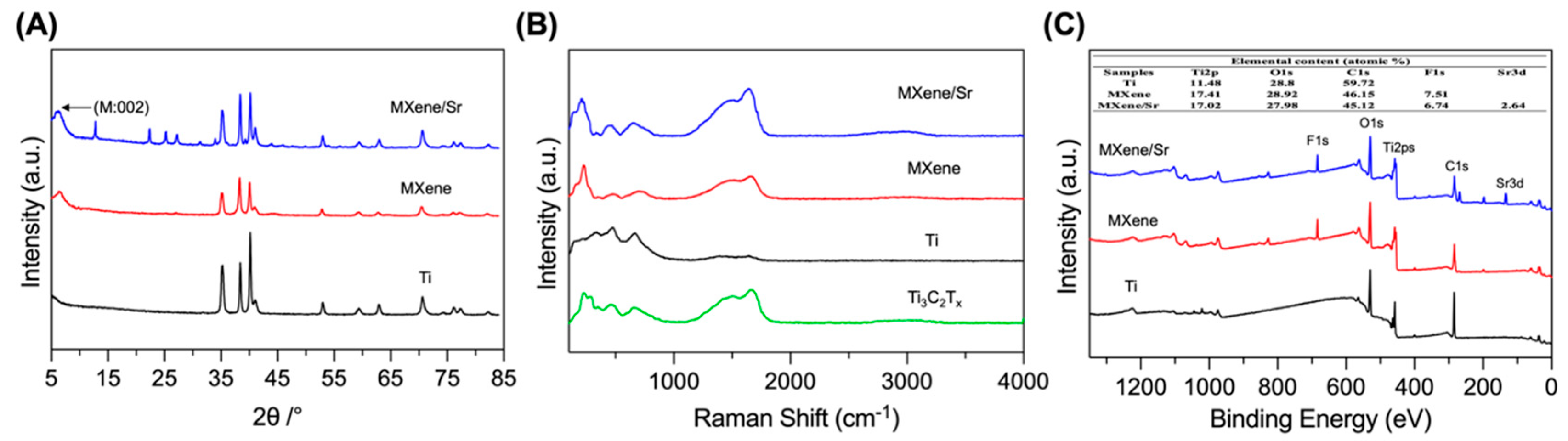
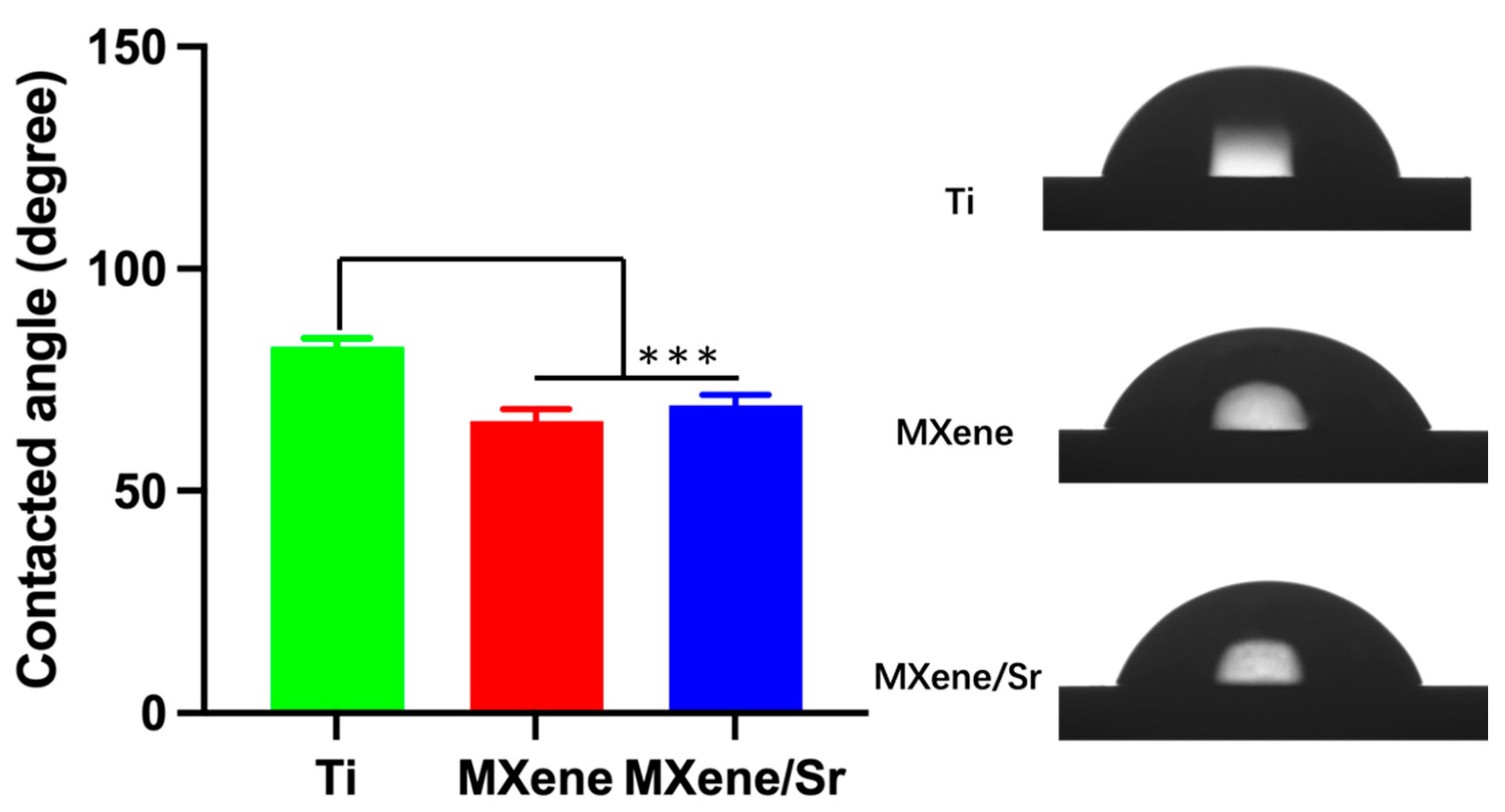
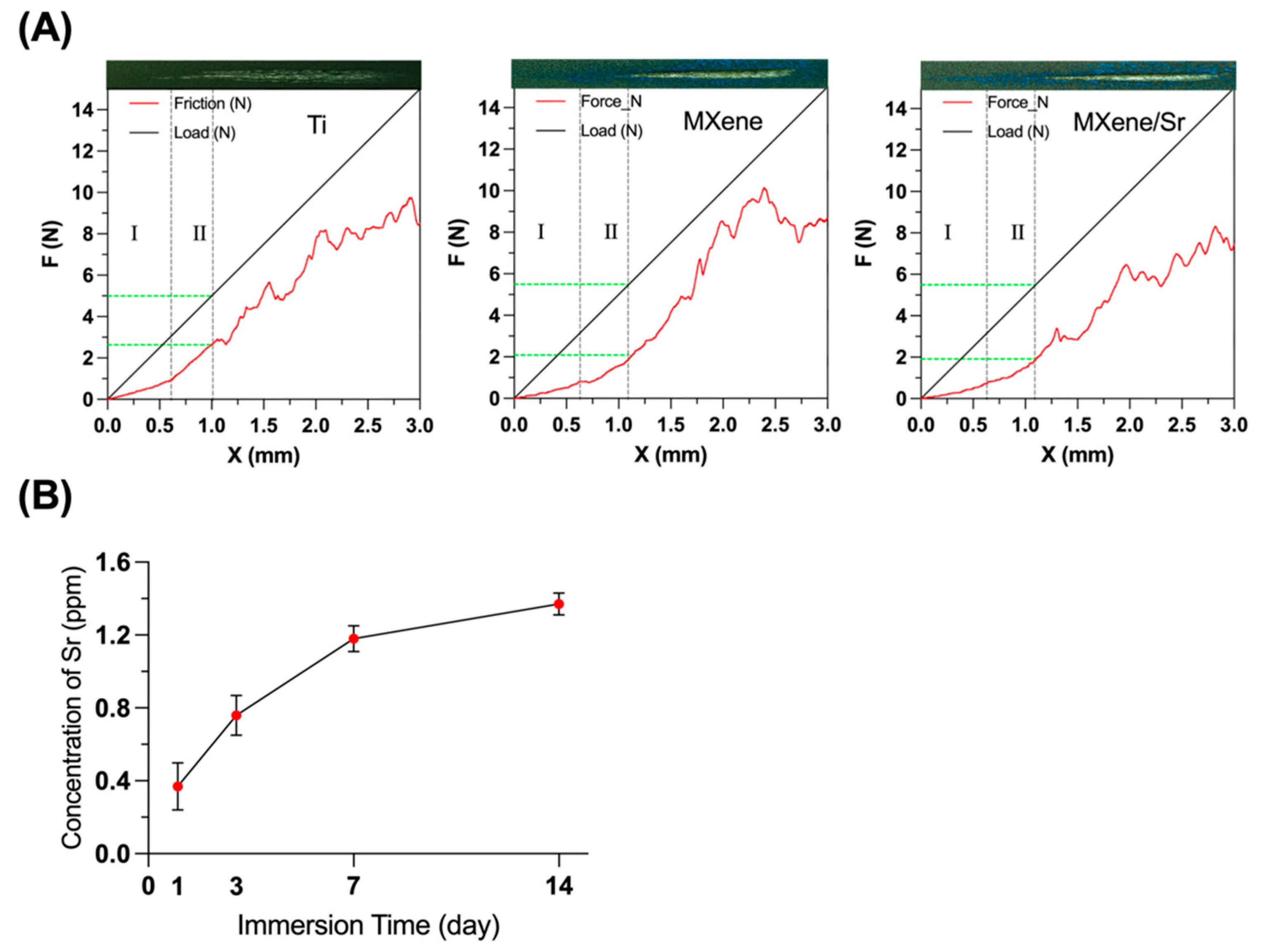
3.1. Material Characterization

Figure 2A shows digital optical images of the Ti, MXene, and MXene/Sr samples. The coated surface became considerably darker than the bare Ti, mainly owing to the deposition of Ti3C2Tx. SEM results (Figure 2B) revealed that the treated pure titanium surface was relatively flat. In contrast, deposition of the MXene coating produced a wavy, uneven surface topography, which was attributable to the uniform accumulation of Ti3C2Tx sheets. After immersion in the SrCl2 solution, the surface morphology of the MXene/Sr group remained similar to that of the Sr-free MXene group. This indicated that the incorporation of Sr2+ did not compromise the integrity or continuity of the MXene layer. Cross-sectional SEM images further showed that both MXene and MXene/Sr samples formed distinct, tightly adherent lamellar structures on the titanium substrate, with a thickness on the micrometer scale. This agreed with previous reports of self-assembled architectures produced by electrophoretic MXene deposition on metal substrates and demonstrated the good reproducibility of the present fabrication strategy in terms of morphology control and interfacial bonding [21].
Figure 3A presents XRD patterns of the Ti, MXene, and MXene/Sr groups. All coatings exhibit the characteristic reflections of α-Ti [22]. For the MXene sample, a broadened Ti3C2Tx (002) peak appears at 2θ ≈ 7.0°, confirming successful formation of an MXene layer by electrophoretic deposition [23]. The MXene/Sr spectrum retains the same (002) reflection with an identical profile, indicating that incorporation of Sr2+ does not disrupt the primary MXene structure. Additional reflections at 12.8° and in the 25°–30° range for MXene/Sr correspond to Sr-containing phases, suggesting that Sr2+ is introduced mainly by intercalation or coordination with surface terminations. Raman spectra (Figure 3B) likewise reveal the characteristic Ti3C2Tx bands (~200, 400, and 600 cm−1) for both MXene and MXene/Sr coatings, in excellent agreement with the literature [24], further demonstrating that the coating was successfully fabricated and that Sr immersion has a minimal impact on MXene structural stability. A slight shift in the 600–700 cm−1 region of the MXene/Sr spectrum is likely associated with vibrational modes of Sr compounds [25]. Collectively, the XRD and Raman results confirm that electrophoretic deposition followed by immersion achieves gentle, functional Sr2+ doping while preserving the layered integrity of MXene.
X-ray photoelectron spectroscopy (XPS) offered nanometer-scale surface sensitivity (~10 nm) and chemical-state resolution, making it indispensable for confirming Sr incorporation and elucidating the surface chemistry of the MXene coating. Survey spectra (Figure 3C) showed that MXene coating consisted mainly of Ti, C, and O elements, with a small amount of F detected (originating from the –F terminal groups on the MXene sheets); for the MXene/Sr group, distinct Sr 3d5/2 and Sr 3d3/2 doublet signals appeared at approximately 134 eV and 132 eV, respectively, which were absent in the MXene group, confirming that Sr had been successfully introduced to the surface of the MXene coating. These binding energies are consistent with the chemical states of SrCO3 or Sr–O bonds [26], suggesting that Sr2+ coordinates with surface functional groups of MXene to form Sr(OH)2-like surface species. Atomic-percent data show that pristine Ti contains 11.48% Ti 2p, 28.80% O 1s, and 59.72% C 1s. After electrophoretic deposition, the MXene coating exhibits an elevated Ti 2p content (17.41%) together with O 1s (28.92%) and F 1s (7.51%), reflecting the stable −O/−F terminations of Ti3C2Tx. Subsequent Sr functionalization yields 2.64% Sr 3d in the MXene/Sr coating.
Surface wettability, quantified by the static water-contact angle, was closely linked to early protein adsorption, osteogenic cell attachment, and even bacterial adhesion; a lower angle (higher hydrophilicity) generally favored bone integration and hampered bacterial colonization [27,28]. After modification with the MXene coating, the hydrophilicity of the titanium surface was significantly improved, and the effect of Sr doping on hydrophilicity was minimal. The static water-contact angle test results (Figure 4) showed that the contact angle of the pure titanium surface was approximately 82°, indicating moderate hydrophilicity/hydrophobicity. After deposition of the MXene coating, the contact angle decreased to approximately 65°, indicating an increase in hydrophilicity (p < 0.01 compared to Ti). The contact angle of the MXene/Sr composite coating was approximately 69°, slightly higher than that of the MXene group but still significantly lower than pure titanium (p < 0.05). The increased hydrophilicity of the coating can be attributed to the polar functional groups (–OH/–O, etc.) present on the MXene sheet surface, which increase the surface free energy. Additionally, the nanoscale wrinkle structure of the coating also contributes to its hydrophilicity [29]. Similar reductions reported for Ti3C2 coatings by Huang [30] corroborate this behavior.
Robust coating–substrate adhesion was crucial for load-bearing implants; delamination during implantation would negate any surface function. In adhesion tests, the nano-scratch test (Figure 5A) showed that the pure titanium substrate failed at approximately 5.05 N of normal load. In contrast, the critical load of the MXene coating increased to about 5.49 N, and the MXene/Sr coating was approximately 5.46 N, slightly higher than the uncoated Ti. Although the MXene/Sr group was slightly lower than the MXene group, the difference between the two was very small. This indicated that Sr doping did not weaken the adhesion strength between the coating and the substrate. Additionally, the friction force recorded during the scratching process was lower in the coating groups (especially the MXene/Sr group, at approximately 1.92 N) compared to the pure titanium group (2.64 N), possibly because the MXene sheets reduce direct friction between the probe and the metal [31]. Overall, the composite coating not only significantly improved surface hydrophilicity but also maintained good adhesion strength and mechanical stability with the substrate at a moderate thickness.
Controlled release of Sr2+ was biologically relevant because micromolar levels (≈0.2–1.5 ppm) had been shown to stimulate osteoblast proliferation, enhance ALP activity, and upregulate osteogenic genes, whereas excessive doses could be cytotoxic [32,33]. The MXene/Sr coating exhibited a biphasic profile (Figure 5B): initially, 0.36 ppm of Sr2+ was released on day 1, which increased to 0.76 ppm on day 3, further rising to 1.18 ppm on day 7, and reaching about 1.37 ppm on day 14, after which the curve plateaued. Statistical analysis indicated that the 0–3 day period represented the rapid release phase (average release rate ≈ 0.25 ppm/day), while days 7–14 entered a slower phase (≈0.03 ppm/day). Notably, during days 1–7, the Sr2+ concentration released from the coating ranged from 0.3 to 1.2 ppm, which is within a range conducive to cell proliferation and differentiation. In summary, the MXene/Sr coating can stably provide sustained Sr2+ release in the early stages of implantation, laying the foundation for long-term biological effects of the coating.

3.2. In Vitro Cell Biocompatibility Evaluation

3.2.1. Cytotoxicity and Proliferation

Live/dead staining results (Figure 6A) showed that there were no obvious red dead cells in any group, and the cell morphology showed no significant changes. This indicated that the coating materials had negligible cytotoxicity to BMSCs. The number of live cells in the MXene and MXene/Sr groups was slightly higher than in the Ti group. In the cell proliferation experiment, the CCK-8 quantification results (Figure 6B) showed that on day 1, the proliferation level of cells in each group was similar. On day 3, the OD values of the MXene and MXene/Sr groups were slightly higher than that of the Ti group, and on day 5, the differences became more evident. The OD value of the Ti group on day 5 was approximately 0.83; the MXene group reached 0.97 (p < 0.05 compared to Ti); and the MXene/Sr group was the highest at approximately 1.11 (p < 0.05 compared to MXene). These results indicate that the composite coating significantly promotes cell proliferation, likely due to the excellent biocompatibility and hydrophilicity of MXene, with its surface functional groups (–OH/–O, etc.) facilitating cell adhesion and expansion [34,35]. Moreover, Sr has been shown to promote the proliferation of osteoblasts and MSCs, potentially by activating osteogenic signaling pathways (such as Wnt/β-catenin and MAPK/Runx2 pathways), thus enhancing the proliferative potential of cells [36]. Therefore, the synergistic effect of MXene and Sr2+ promoted the proliferation of BMSCs, which is beneficial for osseointegration around the implant.

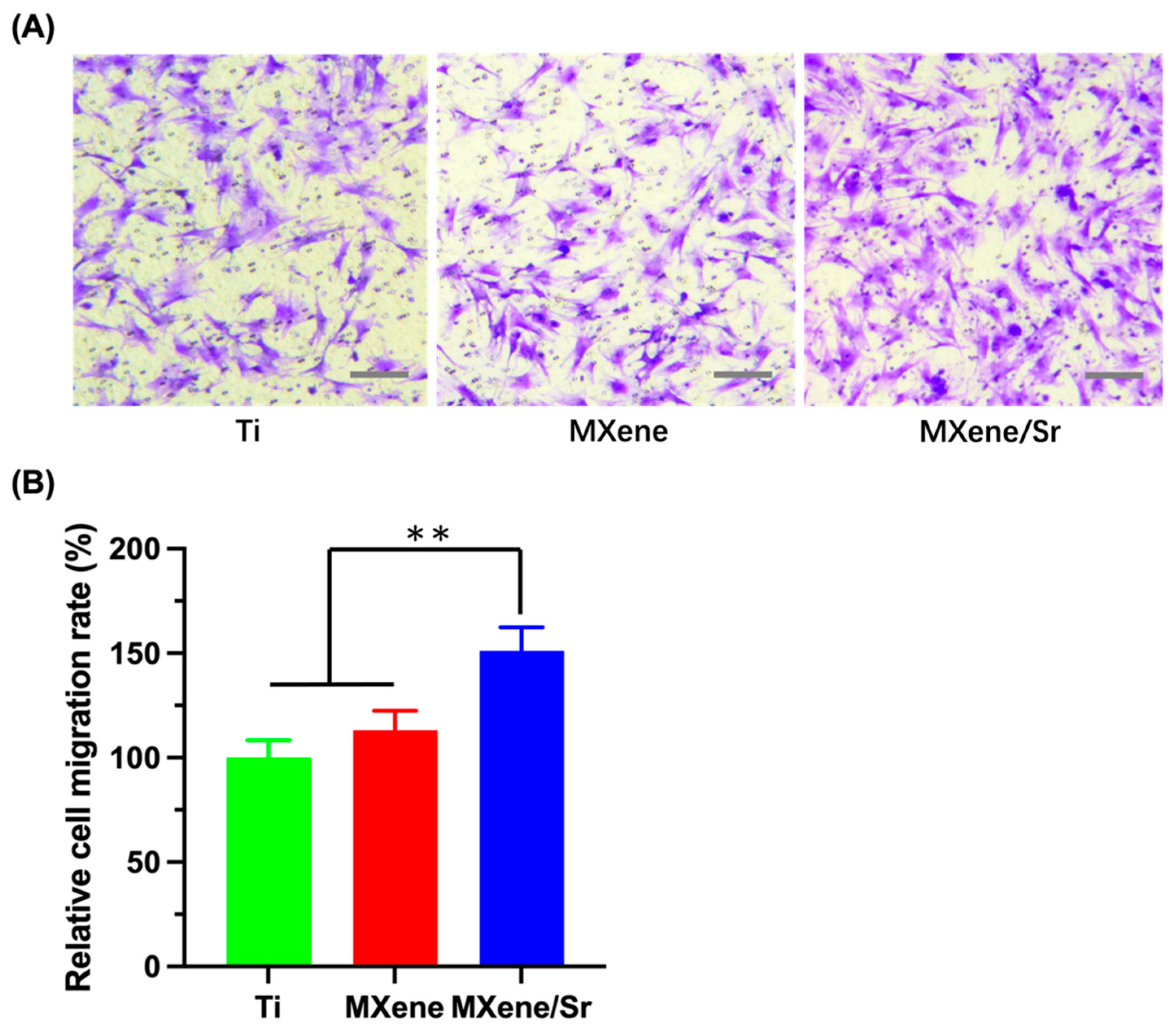
3.2.2. Transwell Migration Assay

Transwell migration assays revealed intergroup differences in BMSC chemotaxis after 72 h of conditioning (Figure 7A). Crystal-violet micrographs showed the highest density of transmigrated cells in the MXene/Sr group. Quantitatively (Figure 7B), the migration rate of the Ti group was set at 100 ± 8.27%, rising to 113.23 ± 9.21% for MXene and further to 151.23 ± 11.23% for MXene/Sr—significantly higher than both Ti and MXene. Thus, MXene extract alone modestly promoted BMSC migration, whereas continuous Sr2+ release in the MXene/Sr group markedly enhanced chemotaxis, conferring superior cell-recruitment capability and reinforcing BMSC motility. Mechanistically, Sr2+ exerts multiple regulatory effects on intracellular pathways. Studies show that Sr2+ up-regulates Wnt/β-catenin signaling and down-regulates the osteogenic inhibitor sclerostin, while activation of the calcium-sensing receptor (CaSR) triggers downstream cascades that phosphorylate Akt and increase nuclear β-catenin, thereby promoting cell homing [37,38,39]. Sr2+ also activates the PI3K/Akt pathway, further enhancing BMSC responsiveness to chemotactic gradients [40]. Collectively, the MXene/Sr composite coating is expected to improve osteogenic cell recruitment and thus accelerate early peri-implant bone regeneration.

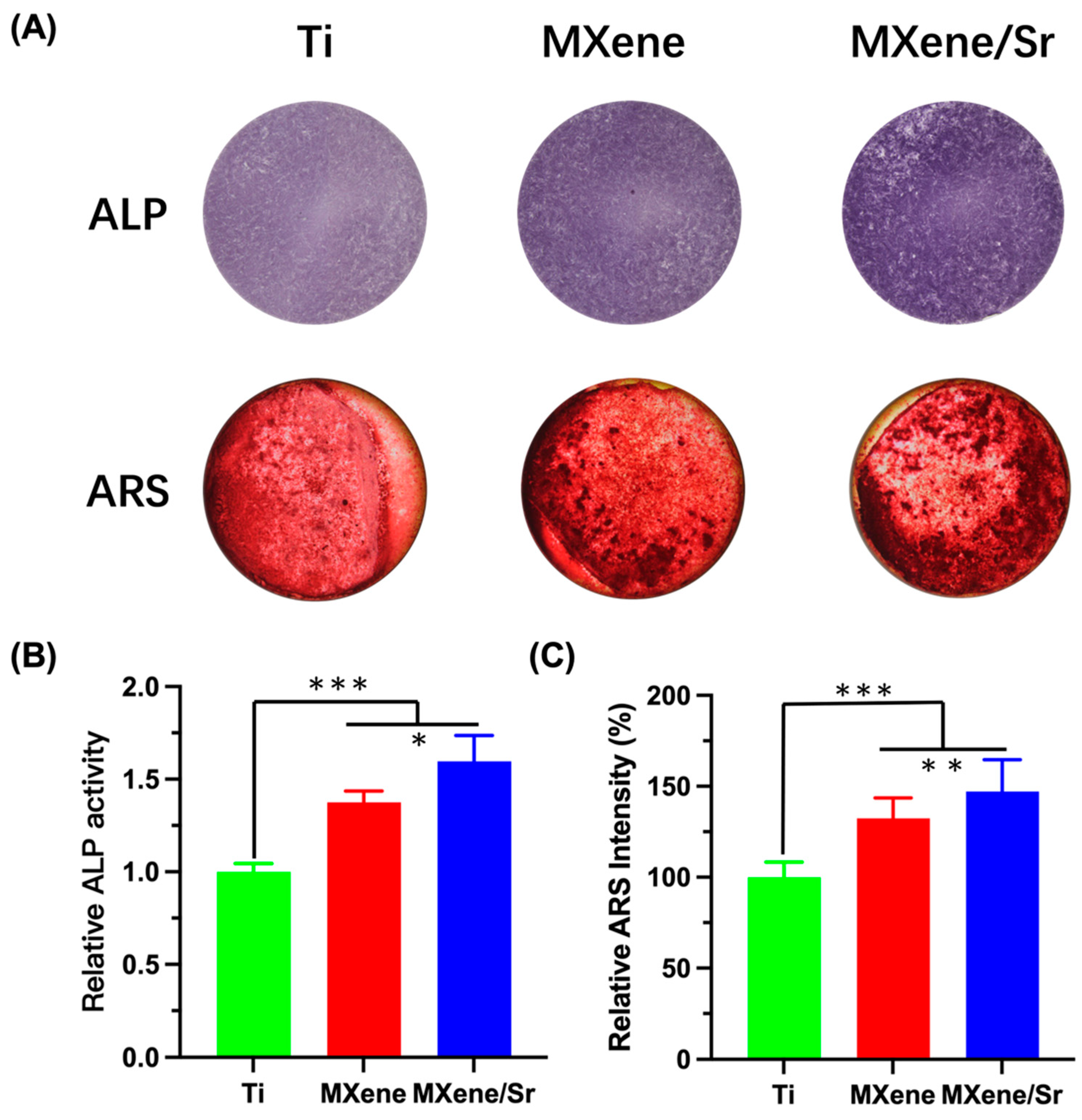
3.3. In Vitro Osteogenic Differentiation of Cells

In early osteogenic differentiation, ALP activity on day 7 showed a trend of MXene/Sr > MXene > Ti across the groups. Staining results showed that the MXene/Sr group exhibited the deepest ALP staining and the largest precipitation area (Figure 8A); in the quantitative results (Figure 8B), the relative ALP activity of MXene (1.37 ± 0.06) was significantly higher than that of Ti (1.00 ± 0.05), indicating that the MXene coating itself significantly promoted ALP production. After incorporating Sr2+, the relative ALP activity in the MXene + Sr group further increased to 1.60 ± 0.14. This result is consistent with qualitative observations: after Sr doping, the purple precipitates were more widespread and denser, indicating enhanced ALP activity and early differentiation capability. Previous studies have reported that Sr-containing coatings can promote ALP activity and the expression of osteogenesis-related genes in BMSCs [41]. Geng et al. [42] observed that Sr-doped Ti coatings significantly increased ALP staining and upregulated the expression of ALP and other osteogenic markers in in vitro experiments. Similarly, Xing et al. [43] observed the highest ALP activity in the Sr group at day 7 in their Sr-doped calcium phosphate coatings. Therefore, in this study, the MXene coating showed potential in promoting early osteogenesis, and the additional release of Sr2+ further amplified this effect, likely by promoting the activation of transcription factors such as Runx2 to accelerate osteogenesis [36,44].
The 21-day Alizarin Red S staining results of late-stage osteogenesis are shown in Figure 7A. The control group exhibited faint Alizarin Red staining, with scattered calcium nodule areas. In contrast, the MXene group showed a significant increase in the number of surface-stained calcium nodules, with larger areas and a darker color. In the MXene/Sr group, Alizarin Red staining was significantly deeper, and the area of calcium salt deposition and calcium nodules further increased, showing a clustered distribution. Subsequent ARS quantitative analysis (Figure 8C) showed that, compared to pure Ti, the optical density of MXene was 132.33 ± 11.2%, while that of MXene/Sr was 147.23 ± 15.2%. This result suggests that MXene-Sr can effectively promote late-stage osteogenic differentiation of cells. Reports from the literature indicate that Sr-doped materials significantly increase bone-like nodule formation and mineralization area [45,46,47]. Bizelli-Silveira et al. [48] found that high concentrations of Sr promoted more bone nodule formation in BMSCs. In this study, the enhanced mineralization effect observed may be due to the sustained release of Sr, which provides long-term activation of osteogenic signaling. Moreover, the hydrophilicity and functional groups on the MXene coating surface improved cell adhesion and the efficiency of mineralized matrix formation [49]. The combined effects resulted in more abundant calcification deposition.
In summary, the Sr-doped Ti3C2Tx MXene coating elicited a concerted enhancement of BMSC functions—proliferation, chemotactic migration, ALP expression, and extracellular-matrix mineralization—thereby laying a solid cellular foundation for osseointegration. Its osteo-inductive action could be rationalized in three inter-dependent domains: (i) the layered MXene architecture offered a hydrophilic, mildly electronegative interface that mimicked native bone, enriched adsorbed adhesive proteins, and promoted integrin-mediated cell anchorage [12]; (ii) the release of Sr2+ engaged the calcium-sensing receptor (CaSR) on osteoblast-lineage cells and activated the PLC–IP3/PKC and Ras/MAPK–ERK1/2 cascades that upregulated Runx2, ALP, collagen I, and osteocalcin, thereby accelerating matrix maturation [50]; (iii) electroconductive MXene sheets, together with Sr2+, stabilized cytoplasmic β-catenin and triggered canonical Wnt/β-catenin signaling in BMSCs, further amplifying osteogenic gene transcription and mineral deposition [51,52]. Collectively, these physicochemical and signaling synergies were expected to accelerate bone bonding and reinforce the interfacial integrity between the coated surface and surrounding tissue.

3.4. In Vitro Antibacterial Evaluation

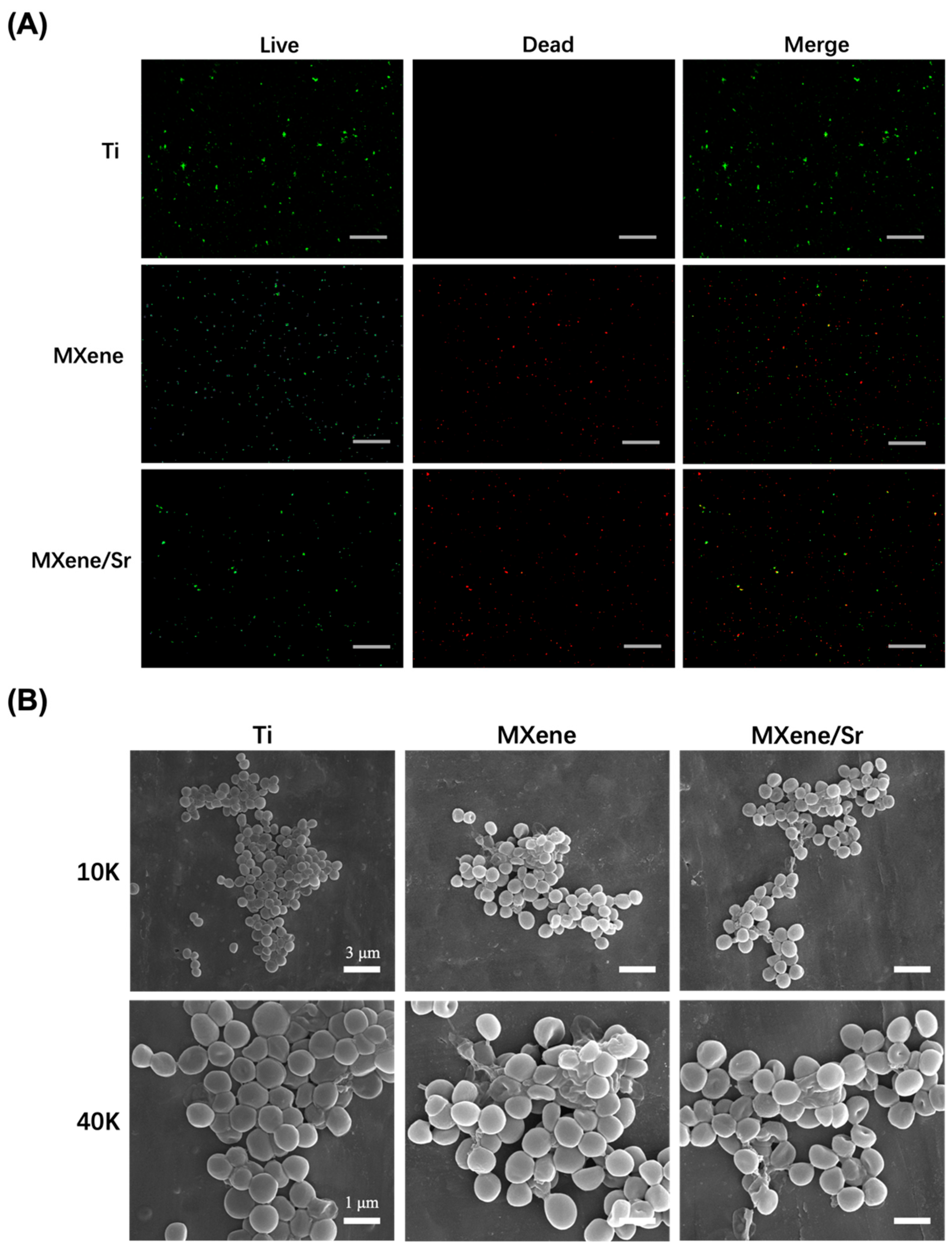
3.4.1. Bacterial Integrity Analysis

As shown in Figure 9A, Ti surfaces were almost completely covered by continuous green fluorescence with only sporadic red dots, indicating that S. aureus remained highly viable on inert titanium. In contrast, MXene samples exhibited markedly fewer, discretely distributed green signals and a greater abundance of red-stained (dead) bacteria, demonstrating that the two-dimensional Ti3C2Tx MXene effectively reduced bacterial viability. The MXene/Sr samples displayed imaging features nearly identical to those of MXene, with similarly diminished green fluorescence and widespread red staining. SEM observations (Figure 9B) revealed dense, clustered cocci with smooth walls on pristine Ti, indicative of vigorous adhesion and intact morphology. On MXene, bacterial numbers declined and many cells showed shrinkage, indentation, or slight collapse, evidencing envelope damage. The MXene/Sr surface harbored comparable cell densities to MXene, with wrinkled membranes and ruptured contours to a similar extent. Collectively, these data confirm that electrophoretic Ti3C2Tx coatings markedly suppress initial S. aureus colonization and trigger local membrane disruption.
The underlying contact-active antibacterial mechanism can be summarized in three stages. First, bacteria are adsorbed onto the highly hydrophilic Ti3C2Tx surface under static conditions [53]. Second, the Ti3C2Tx sheets, bearing abundant –O, –OH, and –F terminations, are negatively charged; electrostatic repulsion between Ti3C2Tx and the positively charged wall teichoic acids in the bacterial generates stresses sufficient to exceed the mechanical strength of the cell wall, producing nanometer-scale perforations that expand into characteristic Gram-positive wall defects [54,55]. Finally, once the peptidoglycan is breached, strong Coulomb attraction between negatively charged Ti3C2Tx and the positively charged head-groups of phosphatidylcholine further compromises membrane integrity. The conductive 2D sheets act as “electron bridges” across the insulating lipid bilayer, initiating trans-membrane electron transfer, membrane depolarization, metabolic disruption, and, ultimately, apoptosis or necrosis [56]. At the Sr2+ release levels in this study, additional synergistic antibacterial effects were minimal, with no further reduction in adhesion or enhancement of membrane disintegration.

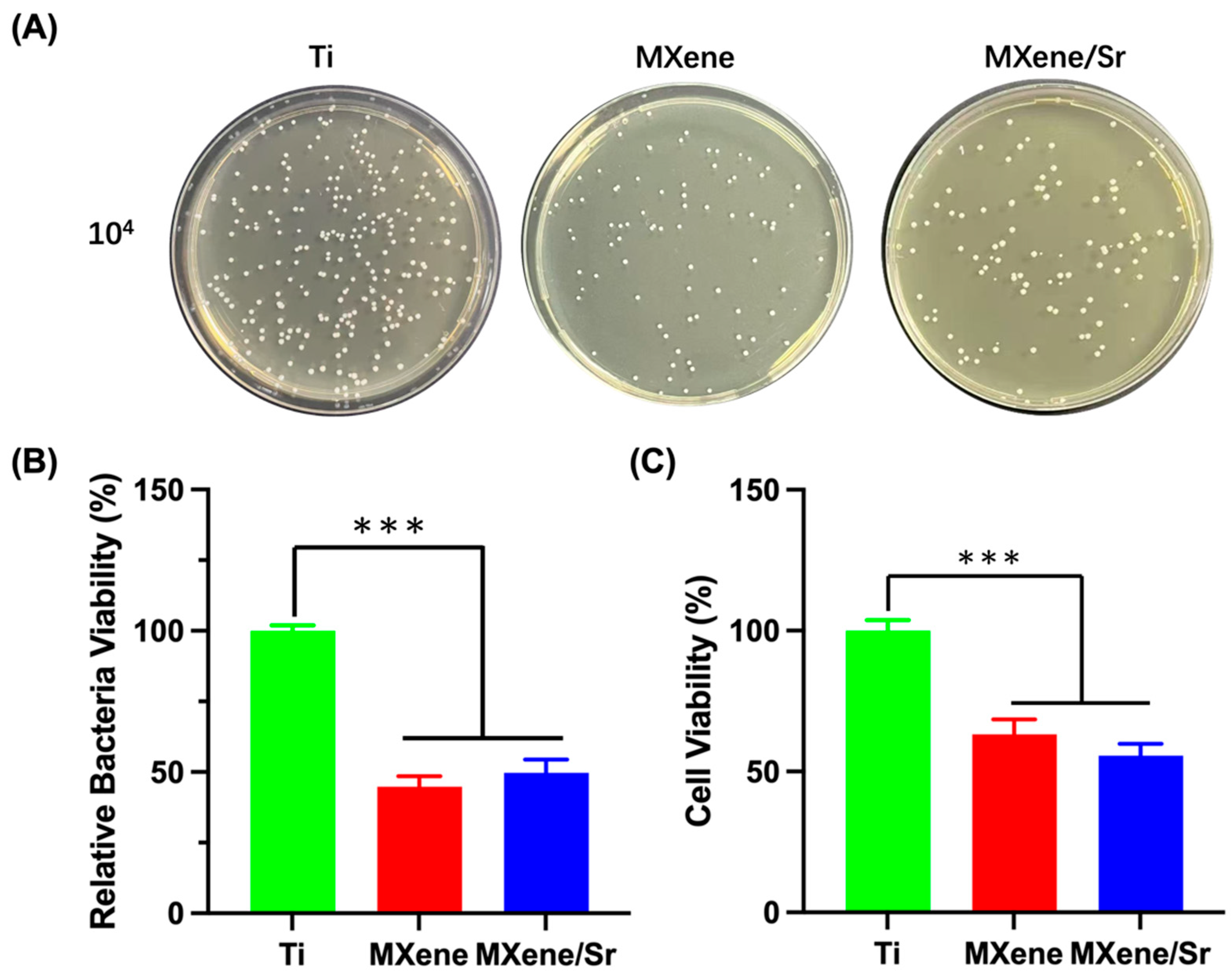
3.4.2. Agar Culture and Quantitative Assessment of Bacterial Viability

Agar-plate enumeration (Figure 10A,B) showed that the MXene group reduced S. aureus survival to 44.8 ± 3.7%, demonstrating effective inhibition of bacterial growth and proliferation. The MXene/Sr samples yielded a relative survival rate of 49.7 ± 4.7%, which is not statistically different from MXene alone. Thus, electrophoretically deposited MXene achieved nearly 50% antibacterial efficacy without external stimuli, and Sr incorporation did not provide additional killing within 24 h. Subsequent AlamarBlue assays quantified the metabolic viability on each surface (Figure 10C): bacteria on MXene and MXene/Sr coatings showed relative survival rates of 63.2 ± 3.2% and 57.2 ± 2.8% (p < 0.01), respectively, both significantly lower than control, with no significant difference between the two coatings. This trend indicates that while Sr2+ provides a slight synergistic effect, the primary antibacterial activity originates from the MXene itself. Notably, AlamarBlue measures metabolic activity, which can differ from colony counts: in the MXene group, metabolic activity was about 63% of control versus a 45% survival rate, implying that some damaged cells retain residual metabolism. Previous studies have likewise shown that Ti3C2Tx is highly bactericidal to both Gram-negative and Gram-positive strains [11,57]. Rasool et al. [58] reported > 98% inactivation within minutes at high Ti3C2Tx concentrations, with SEM evidence of severe membrane damage. Another study found 24 h inhibition rates of 73% and 67% against B. subtilis and E. coli, respectively [59]. Our ≈ 53% value aligns with these trends.
The mechanism is attributable to MXene’s physicochemical traits. Two-dimensional Ti3C2Tx sheets mechanically lacerate bacterial membranes; their negative functional groups electrostatically repel the negative cell wall, curbing adhesion and provoking membrane damage [60,61]. Contact with Ti3C2Tx also induces ROS generation, exacerbating oxidative damage and cellular disintegration [62]. By contrast, trace Sr2+ offers little short-term bactericidal benefit; systematic reviews note minimal Sr release within 24 h for most Sr-functionalized samples, yielding limited immediate antibacterial action [63]. The ~0.3–0.45 ppm Sr2+ released here matches reports that low levels have negligible antibacterial potency. Even so, MXene endows the composite coating with solid baseline antibacterial properties, aiding suppression of bacterial growth and thus supporting control of peri-implant infections.
In summary, the Sr-doped Ti3C2Tx MXene composite coating developed in this study exhibits dual osteogenic-enhancing and antibacterial functions, offering a new surface-modification strategy for dental and orthopedic implants. However, this study had several limitations. First, the 24-h antibacterial assessment might not fully capture long-term bacterial colonization or the development of persistent biofilms on implant surfaces, phenomena that typically arise over several days to weeks. In future work, we plan to extend the duration of the antibacterial experiments to monitor biofilm formation, thereby providing a model that more closely mimics chronic infection scenarios. Second, our experiments were limited to short-term in vitro assays, which might not fully reflect the long-term behavior of implants in vivo. Factors present in the physiological environment—such as serum proteins, immune responses, and mechanical stress—could influence Sr2+ release and the coating’s interactions with cells and bacteria. Therefore, future studies should include in vivo evaluations to confirm sustained osseointegration benefits and infection prevention. Additionally, exploring complementary strategies (e.g., co-doping with other osteogenic or antimicrobial elements, or further improving the coating’s adhesion) would be worthwhile to enhance the coating’s overall performance in clinical applications.

4. Conclusions

Surface technologies that simultaneously accelerated osseointegration and resist infection were pivotal to improving the success of dental and orthopedic implants. In this study, a Sr-doped Ti3C2Tx MXene coating was successfully fabricated on titanium, and its synergistic osteogenic and antibacterial properties were thoroughly evaluated in vitro. Characterization confirmed that the MXene/Sr composite retained the layered architecture and primary chemistry of pristine MXene without structural compromise. The composite surface became markedly more hydrophilic (contact angle reduced by ≈15–20°) and exhibited high critical-load resistance and mechanical stability. In vitro assays demonstrated excellent biocompatibility: the combined effects of MXene and Sr2+ markedly enhanced BMSC proliferation, migration, and osteogenic differentiation. Antibacterial tests showed that MXene effectively suppressed S. aureus adhesion and growth, reducing bacterial viability by almost 50% within 24 h, with pronounced morphological and metabolic damage. Collectively, the Sr-doped Ti3C2Tx MXene coating provides dual functionality—promoting bone regeneration and imparting antibacterial activity—and thus offers a promising strategy for the surface modification of dental and orthopedic implants.

Author Contributions

Conceptualization, Y.L.; methodology, Y.L.; investigation, Y.L.; data curation, Y.L.; formal analysis, Y.L.; writing–original draft, Y.L.; supervision, A.M.; project administration, A.M.; funding acquisition, A.M.; writing–review and editing, A.M.; correspondence, A.M. All authors have read and agreed to the published version of the manuscript.

Funding

This study was supported by the Key Research and Development Program of Sichuan Province (Grant No. 2024YFFK0294).

Institutional Review Board Statement

Not applicable.

Informed Consent Statement

Not applicable.

Data Availability Statement

The data that support the findings of this study are available upon request from the corresponding author. The data are not publicly available due to privacy restrictions.

Acknowledgments

The authors gratefully acknowledge the technical and experimental support provided by the core facility staff at the State Key Laboratory of Oral Diseases and West China Hospital of Stomatology, Sichuan University.

Conflicts of Interest

The authors declare no conflict of interest.

References

  1. Lu, X.; Wu, Z.; Xu, K.; Wang, X.; Wang, S.; Qiu, H.; Li, X.; Chen, J. Multifunctional Coatings of Titanium Implants Toward Promoting Osseointegration and Preventing Infection: Recent Developments. Front. Bioeng. Biotechnol. 2021, 9, 783816. [Google Scholar] [CrossRef] [PubMed]
  2. Zhu, G.; Wang, G.; Li, J.J. Advances in implant surface modifications to improve osseointegration. Mater. Adv. 2021, 2, 6901–6927. [Google Scholar] [CrossRef]
  3. Abu Alfaraj, T.; Al-Madani, S.; Alqahtani, N.S.; Almohammadi, A.A.; Alqahtani, A.M.; AlQabbani, H.S.; Bajunaid, M.K.; A Alharthy, B.; Aljalfan, N. Optimizing Osseointegration in Dental Implantology: A Cross-Disciplinary Review of Current and Emerging Strategies. Cureus 2023, 15, e47943. [Google Scholar] [CrossRef] [PubMed]
  4. Mandracci, P.; Mussano, F.; Rivolo, P.; Carossa, S. Surface Treatments and Functional Coatings for Biocompatibility Improvement and Bacterial Adhesion Reduction in Dental Implantology. Coatings 2016, 6, 7. [Google Scholar] [CrossRef]
  5. Alves, C.H.; Russi, K.L.; Rocha, N.C.; Bastos, F.; Darrieux, M.; Parisotto, T.M.; Girardello, R. Host-microbiome interactions regarding peri-implantitis and dental implant loss. J. Transl. Med. 2022, 20, 425. [Google Scholar] [CrossRef] [PubMed]
  6. Dong, H.; Liu, H.; Zhou, N.; Li, Q.; Yang, G.; Chen, L.; Mou, Y. Surface Modified Techniques and Emerging Functional Coating of Dental Implants. Coatings 2020, 10, 1012. [Google Scholar] [CrossRef]
  7. Kumaravel, V.; Nair, K.M.; Mathew, S.; Bartlett, J.; Kennedy, J.E.; Manning, H.G.; Whelan, B.J.; Leyland, N.S.; Pillai, S.C. Antimicrobial TiO2 nanocomposite coatings for surfaces, dental and orthopaedic implants. Chem. Eng. J. 2021, 416, 129071. [Google Scholar] [CrossRef] [PubMed]
  8. Luo, D.; Zhang, H.-Q.; Xuanyuan, X.-Y.; Deng, D.; Lu, Z.-M.; Liu, W.-S.; Li, M. MXene-Derived Multifunctional Biomaterials: New Opportunities for Wound Healing. Biomater. Res. 2025, 29, 0143. [Google Scholar] [CrossRef] [PubMed]
  9. Bhardwaj, S.K.; Singh, H.; Khatri, M.; Kim, K.-H.; Bhardwaj, N. Advances in MXenes-based optical biosensors: A review. Biosens. Bioelectron. 2022, 202, 113995. [Google Scholar] [CrossRef] [PubMed]
  10. Liao, M.; Cui, Q.; Hu, Y.; Xing, J.; Wu, D.; Zheng, S.; Zhao, Y.; Yu, Y.; Sun, J.; Chai, R. Recent advances in the application of MXenes for neural tissue engineering and regeneration. Neural Regen. Res. 2024, 19, 258–263. [Google Scholar] [CrossRef] [PubMed]
  11. Chen, H.; Wang, Y.; Chen, X.; Wang, Z.; Wu, Y.; Dai, Q.; Zhao, W.; Wei, T.; Yang, Q.; Huang, B.; et al. Research Progress on Ti3C2Tx-Based Composite Materials in Antibacterial Field. Molecules 2024, 29, 2902. [Google Scholar] [CrossRef] [PubMed]
  12. Zhang, J.; Fu, Y.; Mo, A. Multilayered Titanium Carbide MXene Film for Guided Bone Regeneration. Int. J. Nanomed. 2019, 14, 10091–10103. [Google Scholar] [CrossRef] [PubMed]
  13. Li, J.; Fan, Z.; Guan, Z.; Ruan, J. Injectable MXene/Ag-HA composite hydrogel for enhanced alveolar bone healing and mechanistic study. Front. Bioeng. Biotechnol. 2024, 12, 1485437. [Google Scholar] [CrossRef] [PubMed]
  14. Zhu, Y.; Liu, H.; Wu, P.; Chen, Y.; Deng, Z.; Cai, L.; Wu, M. Multifunctional injectable hydrogel system as a mild photothermal-assisted therapeutic platform for programmed regulation of inflammation and osteo-microenvironment for enhanced healing of diabetic bone defects in situ. Theranostics 2024, 14, 7140–7198. [Google Scholar] [CrossRef] [PubMed]
  15. Wirsig, K.; Kilian, D.; von Witzleben, M.; Gelinsky, M.; Bernhardt, A. Impact of Sr2+ and hypoxia on 3D triple cultures of primary human osteoblasts, osteocytes and osteoclasts. Eur. J. Cell Biol. 2022, 101, 151256. [Google Scholar] [CrossRef] [PubMed]
  16. Li, Y.; Qi, Y.; Gao, Q.; Niu, Q.; Shen, M.; Fu, Q.; Hu, K.; Kong, L. Effects of a micro/nano rough strontium-loaded surface on osseointegration. Int. J. Nanomed. 2015, 10, 4549–4563. [Google Scholar]
  17. Wang, H.; Ma, Y.; Li, J.; Zhou, C.; Xu, A.; Xu, Y.; He, F. Modulating autophagy by strontium-doped micro/nano rough titanium surface for promotion of osteogenesis and inhibition of osteoclastogenesis. Colloids Surf. B Biointerfaces 2022, 210, 112246. [Google Scholar] [CrossRef] [PubMed]
  18. Zhang, Y.; Zhu, Y.; Lu, D.; Dong, W.; Bi, W.; Feng, X.; Wen, L.; Sun, H.; Qi, M. Evaluation of osteogenic and antibacterial properties of strontium/silver-containing porous TiO2 coatings prepared by micro-arc oxidation. J. Biomed. Mater. Res. B Appl. Biomater. 2021, 109, 505–516. [Google Scholar] [CrossRef] [PubMed]
  19. Abdalla, M.M.; Sayed, O.; Lung, C.Y.K.; Rajasekar, V.; Yiu, C.K.Y. Applications of Bioactive Strontium Compounds in Dentistry. J. Funct. Biomater. 2024, 15, 216. [Google Scholar] [CrossRef] [PubMed]
  20. Pan, C.; Liu, T.; Yang, Y.; Liu, T.; Gong, Z.; Wei, Y.; Quan, L.; Yang, Z.; Liu, S. Incorporation of Sr2+ and Ag nanoparticles into TiO2 nanotubes to synergistically enhance osteogenic and antibacterial activities for bone repair. Mater. Des. 2020, 196, 109086. [Google Scholar] [CrossRef]
  21. Jang, J.H.; Oh, J.S.; Lee, E.J.; Han, C.M. Electophoretically Deposition of Ti3C2 on Titanium Surface for Hard Tissue Implant Applications. Coatings 2021, 11, 761. [Google Scholar] [CrossRef]
  22. Jin, G.; Cao, H.; Qiao, Y.; Meng, F.; Zhu, H.; Liu, X. Osteogenic activity and antibacterial effect of zinc ion implanted titanium. Colloids Surf. B Biointerfaces 2014, 117, 158–165. [Google Scholar] [CrossRef] [PubMed]
  23. Allen-Perry, K.; Straka, W.; Keith, D.; Han, S.; Reynolds, L.; Gautam, B.; Autrey, D.E. Tuning the Magnetic Properties of Two-Dimensional MXenes by Chemical Etching. Materials 2021, 14, 694. [Google Scholar] [CrossRef] [PubMed]
  24. Xie, X.; Zhu, Y.; Li, F.; Zhou, X.; Xue, T. Preparation and characterization of Ti3C2Tx with SERS properties. Sci. China Technol. Sci. 2019, 62, 1202–1209. [Google Scholar] [CrossRef]
  25. Zhang, Y.; Liu, L.; Li, M.; Wang, S.; Fu, J.; Yang, M.; Yan, C.; Liu, Y.; Zheng, Y. Dose-dependent enhancement of in vitro osteogenic activity on strontium-decorated polyetheretherketone. Sci. Rep. 2025, 15, 3063. [Google Scholar] [CrossRef] [PubMed]
  26. Zhang, P.; Cao, J.; Yang, Z.; Wu, Z.; Wu, L. Adsorptive Removal of Sr (Ii) by Means of Multilayer Titanium Carbon Nitrogen (Ti3cntx) Mxene: Box-Behnken Modeling Design and Experimental Study. SSRN 2022. [Google Scholar] [CrossRef]
  27. Barberi, J.; Spriano, S. Titanium and Protein Adsorption: An Overview of Mechanisms and Effects of Surface Features. Materials 2021, 14, 1590. [Google Scholar] [CrossRef] [PubMed]
  28. Shao, H.; Ma, M.; Wang, Q.; Yan, T.; Zhao, B.; Guo, S.; Tong, S. Advances in the superhydrophilicity-modified titanium surfaces with antibacterial and pro-osteogenesis properties: A review. Front. Bioeng. Biotechnol. 2022, 10, 1000401. [Google Scholar] [CrossRef] [PubMed]
  29. Taloub, N.; Henniche, A.; Liu, L.; Li, J.; Rahoui, N.; Hegazy, M.; Huang, Y. Improving the mechanical properties, UV and hydrothermal aging resistance of PIPD fiber using MXene (Ti3C2(OH)2) nanosheets. Compos. Part B Eng. 2019, 163, 260–271. [Google Scholar] [CrossRef]
  30. Huang, S.; Fu, Y.; Mo, A. Electrophoretic-deposited MXene titanium coatings in regulating bacteria and cell response for peri-implantitis. Front. Chem. 2022, 10, 991481. [Google Scholar] [CrossRef] [PubMed]
  31. Qiu, K.; Zhang, Y.; Wang, L.; Wu, M.; Jin, J.; Shi, W. N-Functionalized Ti2C MXene as a High-Performance Adsorbent for Strontium Ions: A First-Principles Study. J. Phys. Chem. C 2023, 127, 11167–11175. [Google Scholar] [CrossRef]
  32. Su, T.-R.; Huang, T.-H.; Kao, C.-T.; Ng, H.Y.; Chiu, Y.-C.; Hsu, T.-T. The Calcium Channel Affect Osteogenic Differentiation of Mesenchymal Stem Cells on Strontium-Substituted Calcium Silicate/Poly-ε-Caprolactone Scaffold. Processes 2020, 8, 198. [Google Scholar] [CrossRef]
  33. Qiao, L.; Zheng, X.; Xie, C.; Wang, Y.; Ye, L.; Zhao, J.; Liu, J. Bioactive Materials in Vital Pulp Therapy: Promoting Dental Pulp Repair Through Inflammation Modulation. Biomolecules 2025, 15, 258. [Google Scholar] [CrossRef] [PubMed]
  34. Rozmysłowska-Wojciechowska, A.; Mitrzak, J.; Szuplewska, A.; Chudy, M.; Woźniak, J.; Petrus, M.; Wojciechowski, T.; Vasilchenko, A.S.; Jastrzębska, A.M. Engineering of 2D Ti3C2 MXene Surface Charge and its Influence on Biological Properties. Materials 2020, 13, 2347. [Google Scholar] [CrossRef] [PubMed]
  35. Wychowaniec, J.K.; Litowczenko, J.; Tadyszak, K.; Natu, V.; Aparicio, C.; Peplińska, B.; Barsoum, M.W.; Otyepka, M.; Scheibe, B. Unique cellular network formation guided by heterostructures based on reduced graphene oxide—Ti3C2Tx MXene hydrogels. Acta Biomater. 2020, 115, 104–115. [Google Scholar] [CrossRef] [PubMed]
  36. Liu, X.; Huang, H.; Zhang, J.; Sun, T.; Zhang, W.; Li, Z. Recent Advance of Strontium Functionalized in Biomaterials for Bone Regeneration. Bioengineering 2023, 10, 414. [Google Scholar] [CrossRef] [PubMed]
  37. Liu, Z.; Yu, Z.; Chang, H.; Wang, Y.; Xiang, H.; Zhang, X.; Yu, B. Strontium-containing α-calcium sulfate hemihydrate promotes bone repair via the TGF-β/Smad signaling pathway. Mol. Med. Rep. 2019, 20, 3555–3564. [Google Scholar] [CrossRef] [PubMed]
  38. Yang, F.; Yang, D.; Tu, J.; Zheng, Q.; Cai, L.; Wang, L. Strontium enhances osteogenic differentiation of mesenchymal stem cells and in vivo bone formation by activating Wnt/catenin signaling. Stem Cells 2011, 29, 981–991. [Google Scholar] [CrossRef] [PubMed]
  39. Yu, H.; Liu, Y.; Yang, X.; He, J.; Zhong, Q.; Guo, X. The anti-inflammation effect of strontium ranelate on rat chondrocytes with or without IL-1β in vitro. Exp. Ther. Med. 2022, 23, 208. [Google Scholar] [CrossRef] [PubMed]
  40. Rybchyn, M.S.; Slater, M.; Conigrave, A.D.; Mason, R.S. An Akt-dependent increase in canonical Wnt signaling and a decrease in sclerostin protein levels are involved in strontium ranelate-induced osteogenic effects in human osteoblasts. J. Biol. Chem. 2011, 286, 23771–23779. [Google Scholar] [CrossRef] [PubMed]
  41. Alves Côrtes, J.; Dornelas, J.; Duarte, F.; Messora, M.R.; Mourão, C.F.; Alves, G. The Effects of the Addition of Strontium on the Biological Response to Calcium Phosphate Biomaterials: A Systematic Review. Appl. Sci. 2024, 14, 7566. [Google Scholar] [CrossRef]
  42. Geng, T.; Wang, Y.; Lin, K.; Zhang, C.; Wang, J.; Liu, Y.; Yuan, C.; Wang, P. Strontium-doping promotes bone bonding of titanium implants in osteoporotic microenvironment. Front. Bioeng. Biotechnol. 2022, 10, 1011482. [Google Scholar] [CrossRef] [PubMed]
  43. Xing, H.; Li, R.; Wei, Y.; Ying, B.; Li, D.; Qin, Y. Improved Osteogenesis of Selective-Laser-Melted Titanium Alloy by Coating Strontium-Doped Phosphate with High-Efficiency Air-Plasma Treatment. Front. Bioeng. Biotechnol. 2020, 8, 367. [Google Scholar] [CrossRef] [PubMed]
  44. Wang, H.; Hsu, Y.C.; Wang, C.; Xiao, X.; Yuan, Z.; Zhu, Y.; Yang, D. Conductive and Enhanced Mechanical Strength of Mo2Ti2C3 MXene-Based Hydrogel Promotes Neurogenesis and Bone Regeneration in Bone Defect Repair. ACS Appl. Mater. Interfaces 2024, 16, 17208–17218. [Google Scholar] [CrossRef] [PubMed]
  45. Kermani, F.; Mollazadeh Beidokhti, S.; Baino, F.; Gholamzadeh-Virany, Z.; Mozafari, M.; Kargozar, S. Strontium- and Cobalt-Doped Multicomponent Mesoporous Bioactive Glasses (MBGs) for Potential Use in Bone Tissue Engineering Applications. Materials 2020, 13, 1348. [Google Scholar] [CrossRef] [PubMed]
  46. Martín-del-Campo, M.; Sampedro, J.G.; Flores-Cedillo, M.L.; Rosales-Ibañez, R.; Rojo, L. Bone Regeneration Induced by Strontium Folate Loaded Biohybrid Scaffolds. Molecules 2019, 24, 1660. [Google Scholar] [CrossRef] [PubMed]
  47. Nie, R.; Sun, Y.; Lv, H.; Lu, M.; Huangfu, H.; Li, Y.; Zhang, Y.; Wang, D.; Wang, L.; Zhou, Y. 3D printing of MXene composite hydrogel scaffolds for photothermal antibacterial activity and bone regeneration in infected bone defect models. Nanoscale 2022, 14, 8112–8129. [Google Scholar] [CrossRef] [PubMed]
  48. Bizelli-Silveira, C.; Abildtrup, L.A.; Spin-Neto, R.; Foss, M.; Søballe, K.; Kraft, D.C.E. Strontium enhances proliferation and osteogenic behavior of bone marrow stromal cells of mesenchymal and ectomesenchymal origins in vitro. Clin. Exp. Dent. Res. 2019, 5, 541–550. [Google Scholar] [CrossRef] [PubMed]
  49. Jang, J.-H.; Lee, E.-J. Influence of MXene Particles with a Stacked-Lamellar Structure on Osteogenic Differentiation of Human Mesenchymal Stem Cells. Materials 2021, 14, 4453. [Google Scholar] [CrossRef] [PubMed]
  50. Wang, L.; Jiang, S.; Zhou, J.; Gholipourmalekabadi, M.; Cao, Y.; Lin, K.; Zhuang, Y.; Yuan, C. From hard tissues to beyond: Progress and challenges of strontium-containing biomaterials in regenerative medicine applications. Bioact. Mater. 2025, 49, 85–120. [Google Scholar] [CrossRef] [PubMed]
  51. Zhang, J.; Tang, S.; Ding, N.; Ma, P.; Zhang, Z. Surface-modified Ti3C2 MXene nanosheets for mesenchymal stem cell osteogenic differentiation via photothermal conversion. Nanoscale Adv. 2023, 5, 2921–2932. [Google Scholar] [CrossRef] [PubMed]
  52. Sun, T.; Ma, D.; Song, Y.; Hu, J.; Yang, Z.; Wang, X.; Zhang, J. Effects of 0.01 mM strontium on human periodontal ligament stem cell osteogenic differentiation via the Wnt/β-catenin signaling pathway. J. Int. Med. Res. 2025, 53, 3000605251315024. [Google Scholar] [CrossRef] [PubMed]
  53. Yang, J.; Bao, W.; Jaumaux, P.; Zhang, S.; Wang, C.; Wang, G. MXene-based composites: Synthesis and applications in rechargeable batteries and supercapacitors. Adv. Mater. Interfaces 2019, 6, 1802004. [Google Scholar] [CrossRef]
  54. Rauch, C.; Leigh, J. Theoretical evaluation of wall teichoic acids in the cavitation-mediated pores formation in Gram-positive bacteria subjected to an electric field. Biochim. Et Biophys. Acta (BBA) Gen. Subj. 2015, 1850, 595–601. [Google Scholar] [CrossRef] [PubMed]
  55. Alimohammadi, F.; Sharifian Gh, M.; Attanayake, N.H.; Thenuwara, A.C.; Gogotsi, Y.; Anasori, B.; Strongin, D.R. Antimicrobial Properties of 2D MnO(2) and MoS(2) Nanomaterials Vertically Aligned on Graphene Materials and Ti(3)C(2) MXene. Langmuir 2018, 34, 7192–7200. [Google Scholar] [CrossRef] [PubMed]
  56. Ou, L.; Song, B.; Liang, H.; Liu, J.; Feng, X.; Deng, B.; Sun, T.; Shao, L. Toxicity of graphene-family nanoparticles: A general review of the origins and mechanisms. Part. Fibre Toxicol. 2016, 13, 57. [Google Scholar] [CrossRef] [PubMed]
  57. Santos, X.; Álvarez, M.; Videira-Quintela, D.; Mediero, A.; Rodríguez, J.; Guillén, F.; Pozuelo, J.; Martín, O. Antibacterial Capability of MXene (Ti3C2Tx) to Produce PLA Active Contact Surfaces for Food Packaging Applications. Membranes 2022, 12, 1146. [Google Scholar] [CrossRef] [PubMed]
  58. Rasool, K.; Helal, M.; Ali, A.; Ren, C.E.; Gogotsi, Y.; Mahmoud, K.A. Antibacterial Activity of Ti3C2Tx MXene. ACS Nano 2016, 10, 3674–3684. [Google Scholar] [CrossRef] [PubMed]
  59. Rasool, K.; Mahmoud, K.A.; Johnson, D.J.; Helal, M.; Berdiyorov, G.R.; Gogotsi, Y. Efficient Antibacterial Membrane Based on Two-Dimensional Ti3C2Tx (MXene) Nanosheets. Sci. Rep. 2017, 7, 1598. [Google Scholar] [CrossRef] [PubMed]
  60. Wu, D.; Zhao, R.; Chen, Y.; Wang, Y.; Li, J.; Fan, Y. Molecular insights into MXene destructing the cell membrane as a “nano thermal blade”. Phys. Chem. Chem. Phys. 2021, 23, 3341–3350. [Google Scholar] [CrossRef] [PubMed]
  61. Rajavel, K.; Shen, S.; Ke, T.; Lin, D. Achieving high bactericidal and antibiofouling activities of 2D titanium carbide (Ti3C2Tx) by delamination and intercalation. 2D Mater. 2019, 6, 035040. [Google Scholar] [CrossRef]
  62. Ye, S.; Zhang, H.; Lai, H.; Xu, J.; Yu, L.; Ye, Z.; Yang, L. MXene: A wonderful nanomaterial in antibacterial. Front. Bioeng. Biotechnol. 2024, 12, 1338539. [Google Scholar] [CrossRef] [PubMed]
  63. Alshammari, H.; Bakitian, F.; Neilands, J.; Andersen, O.Z.; Stavropoulos, A. Antimicrobial Properties of Strontium Functionalized Titanium Surfaces for Oral Applications, A Systematic Review. Coatings 2021, 11, 810. [Google Scholar] [CrossRef]
Figure 1. Schematic illustration of the two-step fabrication and multifunctional performance of the Sr-doped Ti3C2Tx MXene coating. First, few-layer Ti3C2Tx MXene nanosheets are electrophoretically deposited onto a polished titanium substrate (Step 1: EPD). Next, the MXene-coated titanium is immersed in an SrCl2 solution under thermostatic conditions, allowing Sr2+ ions to be adsorbed and loaded onto the negatively charged MXene terminals (Step 2: immersion).
Coatings 15 00847 g001
Figure 2. (A) Digital optical images of the Ti, MXene, and MXene/Sr samples; (B) Surface and cross-section morphologies of various samples. The scale bar is 1 μm.
Coatings 15 00847 g002
Figure 3. (A) XRD patterns of Ti, MXene, and MXene/Sr; (B) Raman spectra of the samples; (C) XPS full spectra and relative surface element content of various samples.
Coatings 15 00847 g003
Figure 4. Water-contact angle measurements of the samples, n = 3, *** p < 0.01.
Coatings 15 00847 g004
Figure 5. (A) Load/fiction (N)-displacement (mm) curve and upper apex are surface topography after being scratched under load of the samples. (B) Cumulative Sr2+ Release of the MXene/Sr sample, n = 3.
Coatings 15 00847 g005
Figure 6. (A) Live/dead double staining of BMSCs after seeding on various samples for 24 h. Live cells were stained fluorescent green and dead cells were stained red. The scale bar is 100 μm; (B) OD450 values of cells cultured on each sample surface for 1, 3, and 5 days, n = 5, * p < 0.05, *** p < 0.001.
Coatings 15 00847 g006
Figure 7. (A) Micrographs of cells migrating across the Transwell membrane. The scale bar is 100 μm; (B) Quantitative analysis of BMSC migration rates, n = 3; ** p < 0.01.
Coatings 15 00847 g007
Figure 8. (A) ALP (7D) and Alizarin Red S (21D) staining of BMSCs on each sample (B) Quantitative analysis of relative ALP activity in BMSCs on each sample surface. Ti group used as baseline; n = 3; * p < 0.05, *** p < 0.001. (C) Quantitative analysis of relative optical density of ARS-stained BMSCs on each sample surface. Ti group used as baseline; n = 3; ** p < 0.01, *** p < 0.001.
Coatings 15 00847 g008
Figure 9. (A) Live/dead fluorescent staining images of S. aureus adhered to various samples. Live bacteria were stained fluorescent green and dead red. The scale bar is 100 μm. (B) Morphological observation by SEM of S. aureus on various sample surfaces at different magnifications.
Coatings 15 00847 g009
Figure 10. (A) Photographs of recultivated S. aureus colonies on agar culture plates, diluted to 104 CFU/mL; (B) counting analysis of relative survival rate of bacterial colonies. n = 5, *** p < 0.001; (C) Bacterial cell viability of S. aureus. n = 5, *** p < 0.001.
Coatings 15 00847 g010
Disclaimer/Publisher’s Note: The statements, opinions and data contained in all publications are solely those of the individual author(s) and contributor(s) and not of MDPI and/or the editor(s). MDPI and/or the editor(s) disclaim responsibility for any injury to people or property resulting from any ideas, methods, instructions or products referred to in the content.

