Article

Preparation of Superhydrophobic Hydroxyapatite Coating on AZ31 Mg Alloy by Combining Micro-Arc Oxidation and Liquid-Phase Deposition

1
Chengdu Aircraft Industry Group Co., Ltd., Chengdu 610092, China
2
School of Materials and Energy, University of Electronic Science and Technology of China, Chengdu 611731, China
*
Author to whom correspondence should be addressed.
Coatings 2025, 15(6), 675; https://doi.org/10.3390/coatings15060675
Submission received: 30 April 2025 / Revised: 26 May 2025 / Accepted: 30 May 2025 / Published: 1 June 2025

Abstract

Magnesium as a biodegradable metal implant has garnered attention. Nevertheless, its rapid degradation rate and insufficient osseointegration restrict its clinical applications. In order to enhance the corrosion resistance and bioactivity of magnesium alloys, superhydrophobic hydroxyapatite (HA) layers were synthesized on micro-arc oxidized (MAO)-treated AZ31B magnesium alloy through liquid-phase deposition. This study examined the surface morphology, phase composition, bonding strength, wettability, electrochemical properties, and in vitro mineralization of the synthesized coatings. The study results demonstrated that the improved corrosion resistance of composite coatings in Hank’s solution is due to the formation of a protective HA layer. The inclusion of the MAO coating significantly enhances the bonding strength between the hydroxyapatite (HA) layer and the bare magnesium alloy. The concentration of NaH2PO4 affects both the microstructure and wettability. The composite coating exhibited excellent osseointegration capabilities, with new HA layers observed after immersing the samples in simulated body fluid (SBF) solution for three days. These findings suggest that the combination of MAO and solution treatment presents a promising method for enhancing biocompatibility and reducing magnesium degradation, thus making it a viable option for biodegradable implant applications.

1. Introduction

Magnesium alloys are increasingly recognized as highly promising materials in the biomedical field owing to their unique advantages [1,2]. Primarily, magnesium is naturally present in the human body, which plays an important role in numerous biological functions. Their biocompatibility renders magnesium alloys suitable for medical implants [3,4]. A distinguishing feature of magnesium alloys is their biodegradability, which enables implants to gradually dissolve and can be absorbed or excreted by the body, thus eliminating the need for secondary surgery to remove the implant [5,6]. Magnesium alloys possess mechanical properties that more closely resemble natural bone compared to other metals such as titanium or stainless steel. This reduces the risk of stress shielding, a phenomenon where the implant assumes excessive load, resulting in bone weakening [7]. Furthermore, the degradation process releases magnesium ions, which can stimulate bone growth and healing, rendering them particularly beneficial for orthopedic applications [8]. Magnesium alloys can be tailored to adjust their degradation rates and mechanical properties by modifying their composition and processing methods, thereby offering flexibility for various biomedical applications [9].
Although magnesium alloy biomedical materials offer numerous advantages, they also present specific challenges and limitations that must be addressed. A primary concern is the rapid and often unpredictable degradation rate of magnesium alloys in physiological environments, which could impair implant integrity before the tissue has adequately healed. This rapid degradation can also result in the accumulation of hydrogen gas pockets around the implant site, potentially causing inflammation and impeding the healing process [10]. Moreover, magnesium and its alloys are generally regarded as non-bioactive, indicating that they do not inherently promote specific biological responses such as bone growth or tissue integration [11].
To address these challenges, different surface modification methods, such as chemical conversion coatings [12], electrodeposition [13], and micro-arc oxidation (MAO) [14], among others, are being investigated to improve the bioactivity and corrosion resistance of magnesium alloys. Among these techniques, MAO has attracted significant attention. MAO coatings create a stable ceramic layer on the surface of the magnesium alloy, which can significantly elevate the corrosion resistance of magnesium alloy and prolong its functional lifespan in the body [15]. However, MAO coatings typically exhibit a porous surface structure, which can serve as initiation sites for corrosion and compromise the protective nature of the coating. These defects may permit bodily fluids to reach the underlying magnesium alloy, potentially resulting in localized degradation. Therefore, it is crucial to seal the micro-pores and micro-cracks in the MAO coating to further enhance its anticorrosion performance. This can be achieved by integrating the MAO coating with additional complementary processes [16]. The combination of a hydroxyapatite (HA) layer and an MAO coating has emerged as an effective way of improving the corrosion resistance and bioactivity of MAO coatings on magnesium alloys [17,18]. HA layers are famed for their unique properties, such as promoting bone cell attachment and proliferation, thereby facilitating bone ingrowth and integration with the implant. The MAO coating is essential for creating a strong and durable bond with the substrate alloy within the composite structure. Not only does it form a robust connection, but it also cultivates an environment that fosters the formation of the HA layer within the structure. This specially designed environment facilitates the in situ generation of HA layers, ensuring that they integrate seamlessly. The HA layer is extremely efficient in sealing tiny flaws that may be present within the MAO coating.
Li et al. developed a distinct, double-layered MAO/HA coating. They achieved this by sealing the micro-pores in the MAO coating using a solution treatment, thereby significantly enhancing its corrosion resistance [19]. Tang et al. implemented a straightforward and eco-friendly steam sealing technique that significantly improved the corrosion resistance of the MAO coating on AZ31 alloy. This study also revealed the connection between wettability and osteogenesis [20]. Wang et al. produced needle-shaped fluoridated hydroxyapatite on the surface of the MAO layer. During degradation testing in simulated bodily fluids, the composite coating was found to have a strong ability to induce hydroxyapatite deposition on its surface [21]. Li et al. designed Ca-P nanostructured coatings on the surface of MAO coatings using chemical deposition. The Ca-P nanostructured coatings grew and completely covered the MAO coating. The morphology and thickness of Ca-P nanostructured coatings can be regulated by varying the deposition time [22]. Zhang et al. used a liquid-phase deposition technique to construct a Ca-P sealing coating on the outer surface of the MAO coating [23]. Farshid et al. devised bilayer HA/MAO coatings on WE43 alloy and revealed that these coatings exhibited higher adhesion strength and BMP-2 viability compared to random coatings. Moreover, the wettability of bilayer HA/MAO coatings can enhance osteo-immunomodulatory activity [24]. Recent studies involving MAO/HA bilayer coatings have clearly demonstrated that tuning the surface architecture and chemical compositions can modulate wettability, osteoclastogenesis, and corrosion resistance [25]. Unfortunately, the impact of a changing chemical environment on the morphology, wettability, and performance of the composite coating is seldom reported.
In this study, MAO/HA composite coatings were successfully fabricated on biodegradable AZ31 Mg alloy substrates using micro-arc oxidation and liquid-phase deposition (Scheme 1). Subsequently, the wettability and corrosion resistance of the coatings were evaluated to elucidate the relationship between corrosion performance and coating structure.

2. Experiments

The surface modification process for a AZ31 magnesium alloy involves the following two steps: (i) high-potential anodization of the alloy to generate an MAO ceramic coating and (ii) solution treatment in C10H12N2O8Na2Ca and NaH2PO4 to form a hydroxyapatite (HA) coating.

2.1. Preparation of MAO Coating

AZ31 magnesium alloy disks were machined to dimensions of ϕ16 × 6 mm. The disks were ground using abrasive papers, ultrasonically cleaned with (CH3)2CO and H2O, and subsequently dried. A fresh electrolyte was made by mixing reagent-grade chemicals, specifically 6 g/L NaOH and 12 g/L Na3PO4·12H2O, in deionized water for the micro-arc oxidation (MAO) process. The applied voltage was set to 400 V, with a frequency of 1000 Hz, a duty cycle of 10%, and an oxidation duration of 10 min. The temperature of the electrolyte was kept at 30 °C using a cooling system. Samples prepared via micro-arc oxidation were designated as MAO coatings.

2.2. Liquid-Phase Deposition

In the subsequent phase, hydroxyapatite (HA) coatings were applied to the MAO-coated samples via liquid-phase deposition. The precursor solution was prepared by dissolving Na2HPO4 and EDTACa in H2O. The pH was adjusted to 9 by adding 0.5 mol/L NaOH, and the Ca/P ratio was maintained at 1.67 by adjusting the concentrations of NaH2PO4 and EDTACa. The magnesium alloy coated with MAO was immersed in the solution at 90 °C for eight hours. The concentrations of Na2HPO4 were controlled at 0.05, 0.1, and 0.15 mmol, respectively, and the resulting samples were labeled as MAO-LPD-1, MAO-LPD-2, and MAO-LPD-3. Following the reaction, the samples were rinsed with H2O and dried.

2.3. Morphologies and Characterizations

The microscopic morphologies of the coated samples were examined via scanning electron microscopy (SEM, FEI, QUANTA-200F). Elemental concentrations were determined using an energy-dispersive X-ray spectrometer (EDS, Oxford Model 7537). The phase composition of the coated samples was analyzed using thin-film X-ray diffraction (TFXRD, Philip X’Pert) with Cu Kα radiation at a glancing angle of 2°. Additionally, Fourier-transform infrared spectroscopy (FT-IR, Bruker Vector 22, Berlin, Germany) was employed to analyze the phase and structure of the specimens. The coating thickness was measured with an eddy current-based thickness gauge (CTG-10, Time Company, China). The surface roughness of the specimens (Ra) was optically measured using a TR200 roughness gauge (Time Company, China). The static contact angles of the various surfaces were measured at 25 °C and 50% relative humidity using the sessile drop method. This was conducted using a 1 µL water droplet within a telescopic goniometer (OCA20, Germany). The average contact angle values were calculated from measurements taken at five different positions, and images were captured using a digital camera.

2.4. Bonding Strength Tests

The interfacial bond strength between the composite coating and the AZ31 magnesium alloy substrate was evaluated using an adhesion–tension test, in accordance with ASTM C633 [26]. Sample preparation involved removing the coating from one side and roughening the exposed substrate. Both sides of the prepared samples were then affixed to cylindrical steel jigs (10 mm diameter) using J-39 adhesive. For each sample type, the reported bonding strength represents the average of five independent measurements.

2.5. Electrochemical Tests

Corrosion measurements were performed using a PARSTAT 2273 potentiostat in a standard three-electrode configuration. The configuration comprised a saturated Ag/AgCl reference electrode, a platinum counter electrode, and a working electrode with an active area of 1 cm2. Polarization tests were carried out in Hank’s solution maintained at 37 °C. Prior to testing, the samples were immersed in Hank’s solution for 1 h to stabilize the open circuit potential (OCP). A potential range from −0.5 V to +0.5 V (vs. OCP) was applied at a scan rate of 1 mV/s. The Tafel extrapolation method was utilized to estimate the corrosion current density (Icorr) and corrosion potential. The polarization resistance (Rp) was calculated using the Stern–Geary equation. Electrochemical impedance spectroscopy (EIS) measurements were conducted over a frequency range of 100 kHz to 10 mHz, with a perturbation amplitude of 10 mV. To ensure reproducibility, all electrochemical tests were conducted in triplicate.

2.6. In Vitro Mineralization Tests

The specimens were vertically immersed in simulated body fluid (SBF) contained within a glass beaker to assess their in vitro biomineralization behavior. A volume-to-sample area ratio of 20 mL/cm2 was maintained. The samples were soaked for 3 days. After the immersion test, the samples were removed from the SBF, rinsed with deionized water, and dried with warm air. The samples’ composition and surface microstructure were subsequently analyzed using X-ray diffraction (XRD) and scanning electron microscopy (SEM).

3. Results and Discussions

3.1. Surface Morphologies and Elemental Ratio of the Coatings

Figure 1 illustrates the surface morphologies of the MAO, MAO-LPD-1, MAO-LPD-2, and MAO-LPD-3 coatings. The surface morphology of the MAO-coated Mg (Figure 1a) exhibits a characteristic structure with uniformly distributed agglomerated solid particles and micro-pores. These holes are formed when molten oxide and gas bubbles are expelled from the micro-arc discharge channels during the MAO process [27]. These pores potentially compromise the long-term corrosion resistance of the MAO coating by allowing corrosive fluids to penetrate. Fortunately, the structure provides additional sites for HA nucleation, enhancing interface binding during solution treatment. In the MAO-LPD-1 sample, the porous structure is similar to that of the MAO sample. However, the micropores are smaller, and the solid particles have increased in size (Figure 1b). With increased NaH2PO4 concentration, the synthesized coating consists of dandelion flower-like clusters formed by numerous nanoscale needle-like crystals. The layer is uniform and nearly free of pores or cracks, as shown in Figure 1c. This homogeneity prevents contact between the MAO coating and corrosive fluids. At a NaH2PO4 concentration of 1.5 mmol, the composite coating surface is uniformly and completely covered with numerous bar-like structures. The high-magnification SEM image inset in Figure 1d reveals a surface morphology featuring numerous nano-plates with thicknesses ranging from approximately 1–3 μm and lengths between 10 and 20 μm. It is evident that the concentration of NaH2PO4 significantly affects the morphology.
EDS analysis shows that the composite coatings contain the elements Mg, O, P, and Ca (Figure 2). Contrasted with the MAO coating, the Mg content in the composite coatings is significantly lower due to the formation of solution treatment deposit layers. It can be deduced that Mg originates from the MAO coating or Mg substrate and deposits into the outer layer. The Ca elements originate from the solution. From the EDS results, the relative concentrations of Mg and O decrease with the increase of NaH2PO4 concentration in the solution. There is an increase in the relative concentration of P. The relative concentration of Ca increases initially, followed by a decrease. This trend is related to the deposition layer formation process. The Ca/P atomic ratio is calculated and shown in Figure S1. The Ca/P atomic ratio is 1.91, 1.15, and 0.77 for the MAO-LPD-1, MAO-LPD-2, and MAO-LPD-3 coatings, respectively. Notably, the NaH2PO4 concentration in the solution significantly affects the Ca/P atomic ratio. This variation is due to the differing deposition rates of calcium and phosphorus ions. Additionally, the high ionization tendency of magnesium (Mg) and the release of Mg2⁺ ions may result in the replacement of calcium (Ca2⁺) ions. This study shows that Mg2⁺ ions are released from the PEO layer when MgO dissolves.

3.2. Cross-Sectional Morphology and EDS Analysis

The thickness of the composite coatings formed at different NaH2PO4 concentrations via MAO and solution treatment is shown in Figure S2. The thickness of the MAO coating was roughly 13.5 ± 0.7 μm. Measurements of the composite coatings revealed thicknesses of approximately 15.2 µm, 20.8 µm, and 22.4 µm for the MAO-LPD-1, MAO-LPD-2, and MAO-LPD-3, respectively. This increase is attributed to the formation of the HA layer. The cross-sectional images and corresponding EDS mappings of the MAO and MAO-LPD-2 samples are presented in Figure 3. According to its cross-section morphology (Figure 3a), the MAO coating comprises a double layer: a loose, porous outer layer and a dense inner layer. The micropores in the outer layer are closely associated with residual discharge channels and entrapped gas pores. Figure 3b displays a cross-sectional view of the MAO-LPD-2 coatings. The pores in the outer layer of the MAO coating are filled by HA synthesized through solution treatment despite the complex interior shapes of these pores. The LPD layer exhibits a high degree of density and uniformity, exhibiting a close proximity to the MAO ceramic layer. Since the treatment solution contacts the entire surface of the MAO coating, including the pores, the HA layer demonstrates an effective sealing capability. Given the excellent sealing properties and bioactivity of the HA layer, the MAO-LPD composite coating is expected to exhibit improved corrosion resistance and enhanced bioactivity. EDS area scanning (indicated in Figure 3b) for the MAO coating and MAO-LPD-2 coating is shown in Figure 3c and Figure 3d, respectively. Contrasted with the MAO coating, the Mg content decreased markedly, while the Ca and C contents increased significantly. The Ca element originates from the solution, while the C element may derive from CO2 dissolved in the water. This suggests the formation of carbonate-substituted hydroxyapatite, aligning with previous reports [26].
Figure 4 presents the EDS analysis of the MAO-LPD-2 coating cross-section. Near the coating/substrate interface, the concentration of P gradually increases from the substrate towards the coating, whereas the concentration of Mg exhibits an opposite trend. A reduction in Mg concentration is observed at the interface between the MAO coating and the LPD layer with decreasing depth. However, the Ca content increases sharply in this interfacial region. The concentration of P remains relatively constant throughout the coatings. The variations in Ca concentration within the MAO coating indicate that the solution penetrated into the coating, depositing in the pores and micro-cracks, thereby sealing them.
Figure 5 presents the XRD patterns of MAO, MAO-LPD-1, MAO-LPD-2, and MAO-LPD-3. Figure 5a shows the XRD patterns of MAO-treated Mg. Besides the diffraction peaks from Mg, only peaks corresponding to MgO are detected on the MAO substrate. No P-containing phase is identified in the XRD measurements, likely due to its low content and nanocrystalline or amorphous structure. After the solution treatment, HA diffraction peaks (JCPDS No. 54-0022) appear in all coatings, indicating that an HA layer was successfully synthesized on the MAO surface [28]. As the concentration of NaH2PO4 in the solution increases, the XRD patterns of the MAO-LPD samples remain similar to that of the HA sample, suggesting that the NaH2PO4 concentration does not significantly impact the original HA coatings. Concurrently, the corresponding intensities of Mg and MgO decrease significantly with increasing NaH2PO4 concentration, indicating a probable increase in the thickness of the composite coatings.
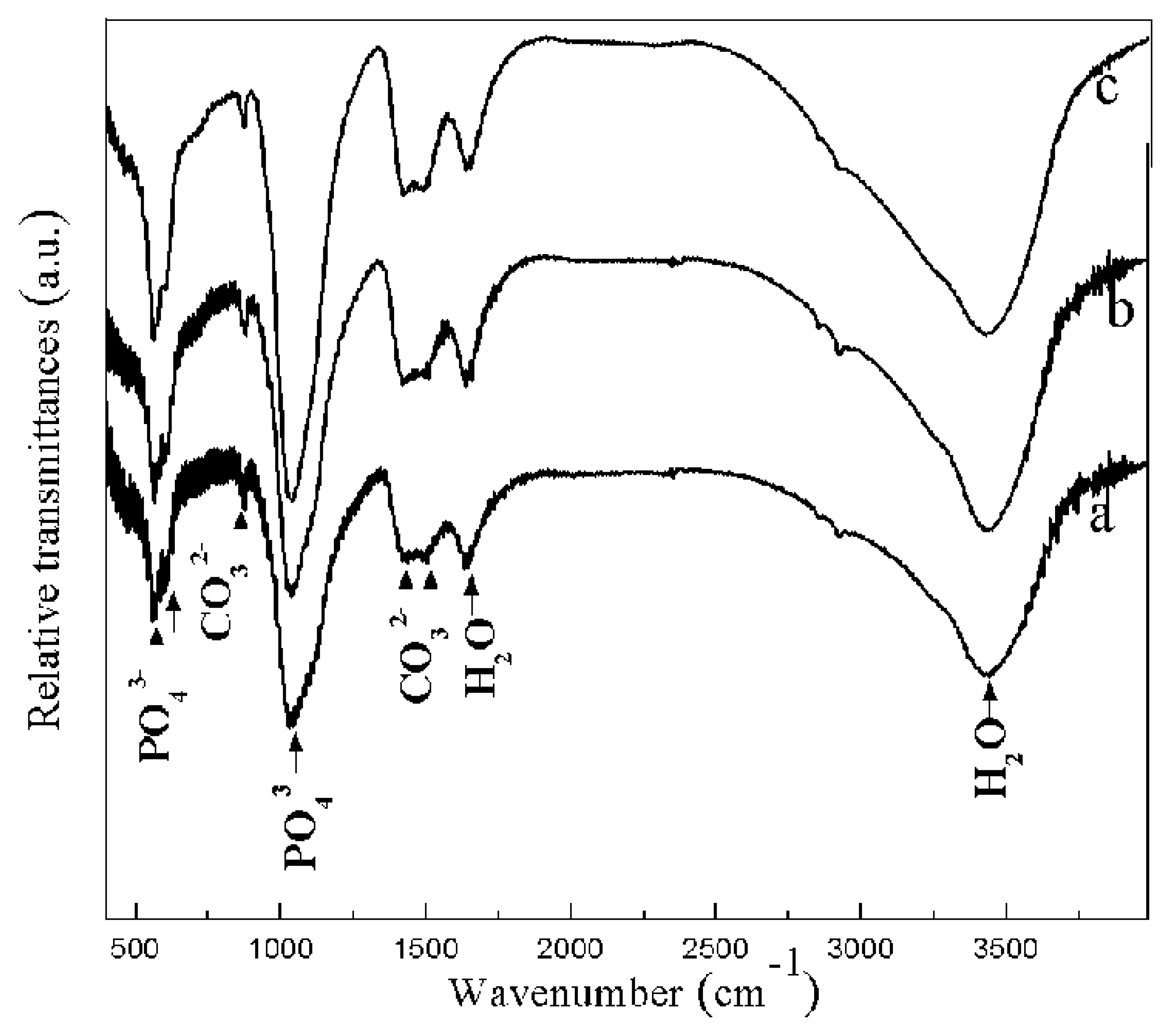
Figure 6 presents the FT-IR spectra of the composite coatings treated with a combination of MAO and solution treatment. The spectra reveal absorption peaks at 1021 cm−1 and 598 cm−1 corresponding to the vibration modes of the P-O bond in the phosphate group, indicating the formation of an apatite structure [29]. The presence of H2O in the coating is indicated by two peaks: a broad absorption band at 3437 cm−1 and a bending mode at 1646 cm−1 [30]. Additionally, absorption peaks at 860 cm−1, 1448.0 cm−1, and 1414.5 cm−1 are associated with the symmetric bending and asymmetric stretching vibrations of CO32− [31]. The presence of the CO32− absorption peak is likely due to the reaction of CO2 in the air with the solution, suggesting the presence of B-type HCA, wherein some PO43− groups are substituted by CO32− groups [32].
Figure S3 shows the surface roughness (average Ra values) of the MAO and composite coatings. It is abundantly clear that the surface roughness of the MAO coating significantly increases after the solution treatment. The composite coatings have undergone an increase in surface roughness which is attributed to solution deposition layers on the surface of the MAO coating; furthermore, this observation aligns well with the changes in surface morphologies depicted in Figure 1. As the concentration of NaH2PO4 in the solution increases, the Ra value of the composite coatings initially rises and then declines, reaching a maximum when the concentration of NaH2PO4 is 0.1 mmol/L.
The wetting behavior of the samples was examined using water contact angle (CA) measurements. Figure 7 presents photographs of 1 μL water droplets on the coated samples. It is evident that the surface of the MAO-coated magnesium alloy is hydrophilic, exhibiting a CA of approximately 87°. This high hydrophilicity, combined with surface defects, makes the MAO film more susceptible to corrosive environments. Upon solution treatment with varying concentrations of NaH2PO4, the contact angles for MAO-LPD-1, MAO-LPD-2, and MAO-LPD-3 were found to be 118°, 161°, and 152°, respectively. Both MAO-LPD-2 and MAO-LPD-3 coatings exhibited superhydrophobic characteristics. The consistent changes in contact angles, along with SEM results, indicate that the highly textured morphology is crucial for water repellency. The dandelion flower-like structure, composed of needle-like nanorods, creates a hydrophobic surface, as shown in Figure S4. Furthermore, the superhydrophobic structure can block contact with corrosive agents, thereby providing improved corrosion resistance.
The bonding strength of implant materials is crucial for their clinical applications. This strength is assessed by the durability of the coatings on the modified implants [33]. Figure 8a highlights the importance of the bonding strength between the coating and the substrate. The control sample, with an LPD coating, exhibits an average bonding strength of 15 MPa. For the MAO-LPD composite coatings, the MAO interlayer significantly enhances the bonding strength between the LPD coating and the AZ31 magnesium alloy substrate. Specifically, the bonding strengths for MAO-LPD-1, MAO-LPD-2, and MAO-LPD-3 are 172%, 110%, and 75% higher, respectively, compared to the LPD coating alone. This increased bonding strength is attributed to the porous outer layer of the MAO coating, which interlocks with the LPD layer, enhancing mechanical interlocking.
Figure 8b illustrates the typical morphology of the MAO-LPD-2 coating, revealing two distinct regions, labeled A and B. These regions represent different planes of crack propagation within the composite coatings and adhesive joints. The majority of the fracture surface corresponds to plane A where the original morphology of the MAO layer has been retained and most of the LPD layer has peeled off, indicating that the tensile fracture occurs at the interface between the MAO and LPD layers. Region B exhibits fewer micropores due to the sealing effect of the LPD layer and the adhesive.
Energy-dispersive X-ray (EDX) elemental maps of the fracture surface at the boundary between Regions A and B are presented in Figure 8c,d. Region A exhibits high concentrations of magnesium and low concentrations of calcium, while Region B exhibits the opposite, reflecting the higher calcium content in the LPD layer compared to the MAO layer. The substantial carbon content in Region B indicates the presence of epoxy adhesive, characteristic of cohesive failure within the coating manifesting as spot flaking on the coating surface.
Metal implants are susceptible to corrosion and the release of metal ions upon contact with tissues and body fluids, which can degrade their performance. The parameters of corrosion resistance in metal implants are critical for determining their efficiency and performance. The corrosion resistance of the AZ31 alloy, MAO, MAO-LPD-1, MAO-LPD-2, and MAO-LPD-3 was evaluated using potentiodynamic polarization tests in Hank’s solution, with the corresponding polarization curves presented in Figure 9. From these curves, the corrosion potentials (Ecorr) and corrosion current densities (Icorr) of the samples were derived and summarized in Table 1.
The MAO coating exhibited a more positive corrosion potential than the uncoated magnesium alloy, with solution treatment further enhancing the coating’s corrosion potential. The MAO treatment significantly reduced the Icorr of the magnesium alloy from 86.7 μA/cm2 to 5.23 μA/cm2, demonstrating a marked improvement in corrosion resistance. This enhancement is attributed to the MAO coating’s ability to effectively impede the penetration of corrosive media, thereby improving the corrosion resistance of the magnesium alloy.
After the PEO samples were treated with the solution, there was a clear improvement in the Ecorr values for all of the samples, MAO-LPD-1 (−83 mV), MAO-LPD-2 (−77 mV), and MAO-LPD-3 (−65 mV), compared to the MAO coating (−95 mV). This shift toward more positive Ecorr values indicates an enhancement in the electrochemical stability of the samples. Furthermore, the Icorr of the samples following solution treatment was significantly reduced, approximately one to two orders of magnitude lower than that of the MAO coating, illustrating the additional improvement in corrosion resistance conferred by the solution treatment.
Moreover, as the NaH2PO4 concentration increases, the corrosion current density progressively decreases. The MAO-LPD-3 coating exhibited the lowest Icorr value and the highest Ecorr value, suggesting that it provides the most efficient protection for the Mg alloy. These results highlight the superior corrosion resistance of the MAO-LPD-2 and MAO-LPD-3 coatings, which can be attributed to the coatings’ hydrophobic properties and the occlusion of most micro-pores in the MAO layer, thereby improving the overall corrosion protection.
To thoroughly investigate the electrochemical properties of the samples, electrochemical impedance spectroscopy (EIS) was performed using Hank’s solution. Figure 10 presents the Nyquist plots for bare AZ magnesium, the MAO-coated sample, MAO-LPD-1, MAO-LPD-2, and MAO-LPD-3 coatings. The experimental data are represented by the symbols, while the fitted data are shown by the solid lines. The Nyquist plots clearly show that the radius of the capacitive loop for the MAO-treated samples is significantly larger than that of the pure magnesium alloy. Furthermore, the radius of the capacitive loop for the solution-treated samples is considerably larger than that of the MAO-coated samples. As the concentration of NaH2PO4 in the solution increased, the capacitive loop of the MAO-LPD composite coatings continued to expand. This observation suggests that higher NaH2PO4 concentrations in the solution enhanced the sealing effect of the MAO coating and also confirms the MAO-LPD composite coatings’ ability to effectively prevent the infiltration of corrosive media, thereby significantly improving the corrosion resistance of magnesium alloys. In general, a larger radius of the capacitive loop corresponds to higher resistance, indicating a greater resistance to electron exchange. For metal surfaces, having a lower electron exchange capacity means less corrosion.
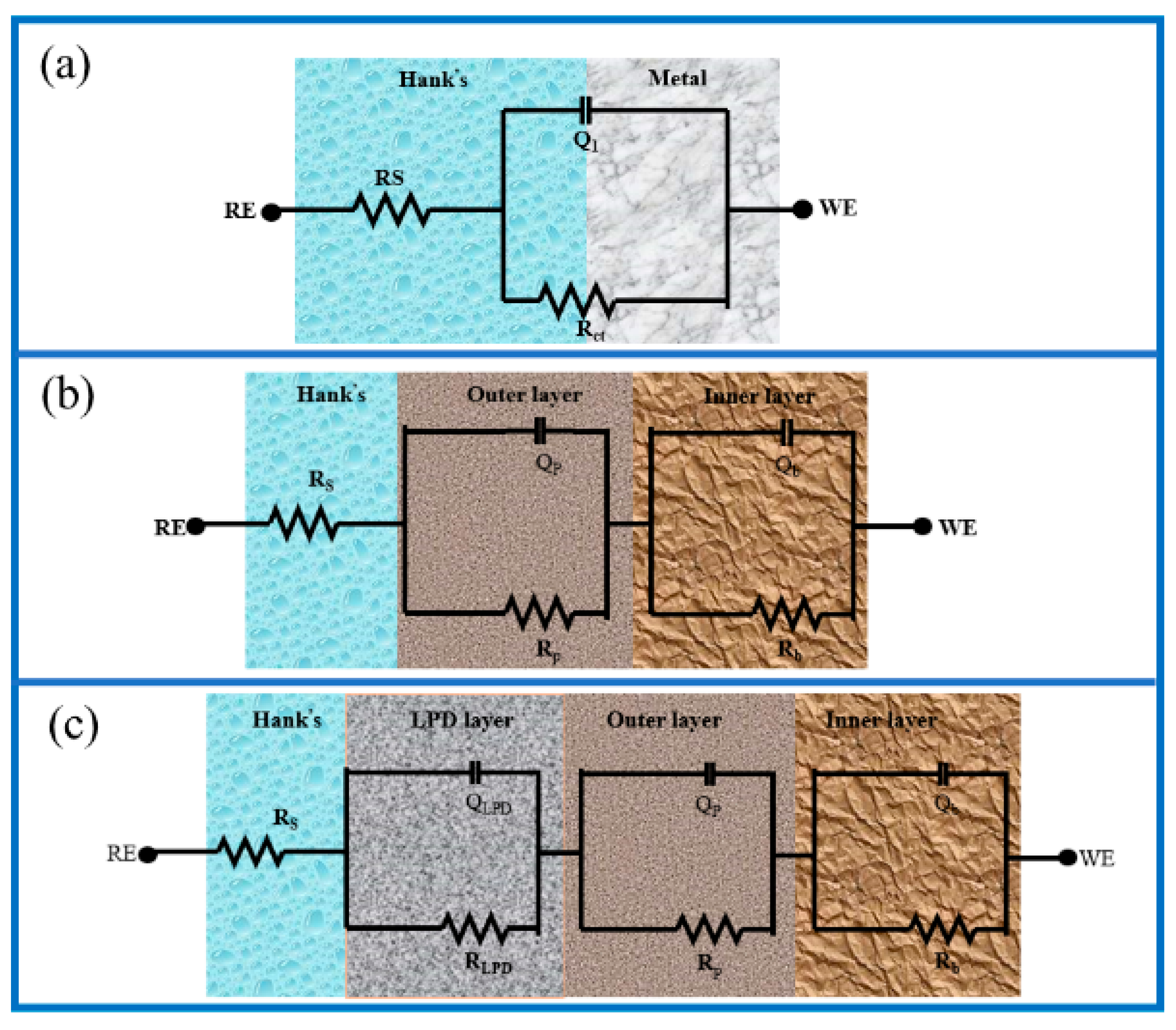
Figure 11 shows the equivalent circuit models that were used to fit the experimental data to different types of electrode/electrolyte interfaces. Capacity-depressed semicircles in the high-frequency range are exhibited by the Nyquist curves of the bare Mg alloy, and the Nyquist plots were simulated by the equivalent circuit shown in Figure 11a. The Nyquist plot of MAO-coated Mg is characterized by a high-frequency capacitive loop, followed by a low-frequency capacitive loop. The outer coating layer’s properties are represented by the high-frequency capacitive loop, while the inner coating layer’s properties are represented by the low-frequency capacitive loop [34]. With regard to the MAO-treated specimen, two possibilities emerge with respect to the number of time constants in the EIS results, i.e., two or three time constants, which may be ascribed to disparities in electrical parameters and electrolyte composition [35]. Discerning between the external and internal layers is frequently arduous due to the relatively feeble barrier properties of the external layer. Occasionally, the outer and inner layers come together, or the time constant linked to the outer layer cannot be measured [36]. In this study, two well-defined time constants emerge at medium and low frequencies, which are regarded as representations of the responses of the PEO layer and the double electric layer, respectively. Thus, a two-time-constant model, as shown in Figure 11b, was employed to fit the EIS results. Following liquid-phase deposition, a new time constant appears at high frequencies. This is associated with the deposition layer. Consequently, three time constants are distributed across the high-, medium-, and low-frequency ranges. The time constants at elevated and moderate frequencies are indicative of the responses exhibited by the deposition layer and the MAO outer layer, correspondingly. The electrochemical activity at the double layer between the MAO inner layer and the Mg substrate is reflected by the time constant at low frequencies. As shown in Figure 2, SEM observations have confirmed the presence of two protective layers on the magnesium surface. In accordance with the architecture of the coatings, the equivalent circuits employed for the purpose of fitting the impedance data are demonstrated in Figure 11c. In Figure 11, Rs represents the resistance of the electrolyte between the working and reference electrodes; in both circuits, Q2−n and Q3−n model the electrical properties of the outer and inner layers of the MAO coating, respectively; R2 is the resistance of the outer layer saturated by the electrolyte; and R3 represents the resistance of the inner layer of the MAO coating. For the MAO-LPD-1, MAO-LPD-2, and MAO-LPD-3 samples, R1 and Q1−n were added to the circuit model to describe the sealing treatment’s effectiveness and obtain the best fit. The fitting results are provided in Table 2.
To mitigate the scatter effect at the electrode/electrolyte interface, constant phase elements (CPEs), represented by the symbol Q, were employed as a replacement for capacitance. The formula for CPE impedance, in which Y0 represents the CPE constant and n is the empirical exponent, is defined as follows: if n = 1, Q exhibits purely capacitive behavior, whilst if n = 0, Q exhibits purely resistive behavior. The behavior of CPE is typically attributed to factors such as distributed surface reactivity, surface inhomogeneity, roughness, or electrode porosity [37,38].
The Rs values of the samples tested in solutions are comparable due to the consistent composition of the solutions and the fixed positions of the working electrode and reference electrode during the tests. The MAO impedance fitting results from the circuit diagram clearly indicate an increase in the resistance of the MAO coating. Specifically, the resistance value increased by two orders of magnitude, thereby enhancing corrosion protection through the formation of a ceramic oxide coating on the Mg alloy surface. Although the MAO coating surface contains numerous micropores, these pores are not interconnected and the inner MAO layer consists of dense MgO, thereby providing effective corrosion protection for the Mg alloy. Additionally, in MAO coatings the Rb values were found to exceed their corresponding Rp values. This is attributed to the dense and flawless morphology of the inner layer of the coatings, while the outer layer displays a more porous structure.
The additional components, R1 with resistance values of 972, 2873, and 3223 Ω·cm2, were provided by the MAO-LPD-1, MAO-LPD-2, and MAO-LPD-3 samples. Furthermore, the R2 values of MAO-LPD-2 and MAO-LPD-3 are approximately three and four orders of magnitude greater than those of the MAO-coated Mg, respectively. This suggests that sealing the pores in the MgO layer with HA effectively prevents the penetration of corrosive solutions into the coatings. Additionally, it is noteworthy that the fitted Q2−n values of the coatings follow the order MAO > MAO-LPD-1 > MAO-LPD-2 > MAO-LPD-3. This indicates that MAO-LPD-3 coatings have a dielectric constant lower than that of the MAO-LPD-1 coating and significantly lower than that of the MAO coating. It is well-established that more porous and defective coatings exhibit higher dielectric constants. Therefore, the significantly lower Q3−n of the MAO-LPD-3 coating is attributed to its much more compact structure in comparison to the MAO coating, making it more effective in preventing corrosion of the underlying Mg. Additionally, the lower Q3−n value of MAO-LPD-3-coated Mg compared to MAO-coated Mg, also indicates superior anticorrosion performance, as suggested in other studies. The R3 value increased with increasing NaH2PO4 concentration in the solution, whereas the Q3−n value decreased with increasing NaH2PO4 concentration. This indicates that the performance of the inner layer of the coating is enhanced by solution treatment to a certain extent, providing improved corrosion resistance to the magnesium alloy. This conclusion is consistent with the results of polarization curve measurements and morphological analysis.
Mineralization is a critical factor in enhancing implant–tissue integration post-implantation for biomaterials. Previous research typically evaluates the bioactivity of biomaterials indirectly by examining apatite formation on their surfaces in simulated body fluid (SBF) [39]. Apatite formation has traditionally been considered a predictor of favorable bioactivity. Figure 12 presents SEM images of the surface morphologies of the MAO-LPD-1, MAO-LPD-2, and MAO-LPD-3 samples after immersion in SBF for 3 days. After 3 days of immersion, a substantial number of irregular spherical deposits appeared on the surface of the MAO-LPD-1 sample (Figure 12a), leading to the disappearance of its porous structure. The MAO-LPD-2 sample exhibited a flower-like morphology with a spherical structure after 3 days of immersion: the layers of these “flowers” covered the entire surface. For the MAO-LPD-3 sample, its entire surface was densely covered by cluster-like particles (Figure 12c), with significant deposition of irregular spherical matter. Notably, the ability to induce new deposition formation not only demonstrated improved surface bioactivity but also enhanced long-term corrosion resistance as a self-repair process could occur, wherein the previous HA layer degraded and a new HA layer simultaneously formed.

4. Conclusions

Hydroxyapatite layers have been successfully deposited on an MAO-coated AZ31B Mg alloy via solution treatment in an aqueous NaH2PO4 solution. The microstructures of composite coatings prepared at different NaH2PO4 concentrations have been studied. Wettability and bonding strength were investigated. The corrosion behavior and in vitro mineralization of the samples have also been analyzed.
The following conclusions were drawn:
  • The composite coatings comprise an inner MAO coating and an outer HA layer. The solution penetrates the porous structure of the MAO coating, deposits within it, and subsequently fuses with the porous layer.
  • Solution treatment enhances the hydrophobicity of the MAO coatings. A maximum contact angle of 161° is achieved at NaH2PO4 concentration of 0.1 mmol/L. This effect is attributed to the formation of dandelion-like clusters on the surface.
  • The bonding strength of the composite coatings is enhanced by the incorporation of the MAO coating. All composite coatings exhibit a tensile strength exceeding 20 MPa.
  • Electrochemical measurements revealed that the composite coatings exhibit significantly greater corrosion resistance than bare and MAO-coated magnesium, owing to the sealing effect and enhanced hydrophobicity.
  • The composite coatings demonstrate superior apatite-forming bioactivity. The formation of new hydroxyapatite on the surface of the composite coatings was observed after 3 days of immersion in SBF.
Therefore, the MAO-LPD composite coatings developed in this study possess excellent mechanical properties, corrosion resistance, and bioactivity, providing a promising strategy for the clinical application of biodegradable magnesium alloys.

Supplementary Materials

The following supporting information can be downloaded at: https://www.mdpi.com/article/10.3390/coatings15060675/s1, Figure S1. The variation of the Ca/P ratios of the coatings; Figure S2. Variation of thickness of the coatings; Figure S3. Variation of roughness of the coatings; Figure S4. Schematic diagram of structural superhydrophobic.

Author Contributions

Conceptualization, Y.H. and H.T.; methodology, Y.H.; validation, Y.H., X.L. and H.T.; investigation, Y.H., Y.Y. and F.J.; writing—original draft preparation, Y.H.; writing—review and editing, Y.H., X.L. and H.T. All authors have read and agreed to the published version of the manuscript.

Funding

This research received no external funding.

Institutional Review Board Statement

Not applicable.

Informed Consent Statement

Not applicable.

Data Availability Statement

Data are contained within the article and Supplementary Materials.

Conflicts of Interest

Authors Yanqing Hu and Xin Liang were employed by Chengdu Aircraft Industry Group Co., Ltd. The remaining authors declare that the research was conducted in the absence of any commercial or financial relationships that could be construed as a potential conflict of interest.

References

  1. Kim, J.; Gilbert, J.L.; Lv, W.W.; Du, P.; Pan, H. Reduction reactions dominate the interactions between Mg alloys and cells: Understanding the mechanisms. Bioact. Mater. 2025, 45, 363–387. [Google Scholar] [CrossRef]
  2. Staiger, M.P.; Pietak, A.M.; Huadmai, J.; Dias, G. Magnesium and its alloys as orthopedic biomaterials: A review. Biomaterials 2006, 27, 1728–1734. [Google Scholar] [CrossRef]
  3. Zhang, Y.; Xu, J.; Ruan, Y.C.; Yu, M.K.; O’Laughlin, M.; Wise, H.; Chen, D.; Tian, L.; Shi, D.; Wang, J.; et al. Implant-derived magnesium induces local neuronal production of CGRP to improve bone-fracture healing in rats. Nat. Med. 2016, 22, 1160–1169. [Google Scholar] [CrossRef]
  4. Yang, J.; Koons, G.L.; Cheng, G.; Zhao, L.; Mikos, A.G.; Cui, F.-Z. A review on the exploitation of biodegradable magnesium-based composites for medical applications. Biomed. Mater. 2018, 13, 022001. [Google Scholar] [CrossRef]
  5. Zhao, D.; Witte, F.; Lu, F.; Wang, J.; Li, J.; Qin, L. Current status on clinical applications of magnesium-based orthopaedic implants: A review from clinical translational perspective. Biomaterials. 2017, 112, 287–302. [Google Scholar] [CrossRef]
  6. Xu, L.; Pan, F.; Yu, G.; Yang, L.; Zhang, E.; Yang, K. In vitro and in vivo evaluation of the surface bioactivity of a calcium phosphate coated magnesium alloy. Biomaterials. 2009, 30, 1512–1523. [Google Scholar] [CrossRef]
  7. Uppal, G.; Thakur, A.; Chauhan, A.; Bala, S. Magnesium based implants for functional bone tissue regeneration—A review. J. Magnes. Alloy. 2022, 10, 356–386. [Google Scholar] [CrossRef]
  8. Zhang, A.; Fan, X.; Yang, Z.; Xie, Y.; Wu, T.; Zhang, M.; Xue, Y.; Wang, Y.; Zhao, Y.; Wu, X.; et al. Optimized design and biomechanical evaluation of biodegradable magnesium alloy vascular stents. Acta Mech. Sin. 2024, 41, 1–14. [Google Scholar] [CrossRef]
  9. Tang, H.; Han, Y.; Wu, T.; Tao, W.; Jian, X.; Wu, Y.; Xu, F. Synthesis and properties of hydroxyapatite-containing coating on AZ31 magnesium alloy by micro-arc oxidation. Appl. Surf. Sci. 2017, 400, 391–404. [Google Scholar] [CrossRef]
  10. Podgorbunsky, A.; Imshinetskiy, I.; Mashtalyar, D.; Sidorova, M.; Gnedenkov, A.; Sinebryukhov, S.; Gnedenkov, S. Bioresorbable composites based on magnesium and hydroxyapatite for use in bone tissue engineering: Focus on controlling and minimizing corrosion activity. Ceram. Int. 2024, 51, 423–436. [Google Scholar] [CrossRef]
  11. Zhu, Y.; Zhou, M.; Zhao, W.; Geng, Y.; Chen, Y.; Tian, H.; Zhou, Y.; Chen, G.; Wu, R.; Zheng, Y.; et al. Insight the long-term biodegradable Mg-RE-Sr alloy for orthopaedics implant via friction stir processing. Bioact. Mater. 2024, 41, 293–311. [Google Scholar] [CrossRef]
  12. Hashemi, T.S.; Jaiswal, S.; Celikin, M.; McCarthy, H.O.; Levingstone, T.J.; Dunne, N.J. Strategically designed bioactive dual-layer coating of octacalcium phosphate and dicalcium phosphate dihydrate for enhancement of the corrosion resistance of pure magnesium for orthopaedic applications. Surf. Coatings Technol. 2024, 495. [Google Scholar] [CrossRef]
  13. Naftchali, N.K.; Aghdam, R.M.; Najjari, A.; Dehghanian, C. Investigating a nanocomposite coating of cerium ox-ide/merwinite via PEO/EPD for enhanced biocorrosion resistance, bioactivity and antibacterial activity of magnesium-based implants. Ceram. Int. 2024, 50, 42766–42779. [Google Scholar] [CrossRef]
  14. Tang, H.; Gao, Y. Preparation and characterization of hydroxyapatite containing coating on AZ31 magnesium alloy by micro-arc oxidation. J. Alloy Compd. 2016, 688, 699–708. [Google Scholar] [CrossRef]
  15. Tang, H.; Wu, T.; Wang, H.; Jian, X.; Wu, Y. Corrosion behavior of HA containing ceramic coated magnesium alloy in Hank’s solution. J. Alloy Compd. 2017, 698, 643–653. [Google Scholar] [CrossRef]
  16. Wang, J.; Dou, J.; Wang, Z.; Hu, C.; Liu, J.; Yu, H.; Chen, C. Corrosion resistance and biodegradability of micro-arc oxidation coatings with the variable sodium fluoride concentration on ZM21 magnesium alloys. J. Alloy Compd. 2023, 962. [Google Scholar] [CrossRef]
  17. Tang, H.; Li, D.; Chen, X.; Wu, C.; Wang, F. FORMATION OF HA-CONTAINING COATING ON AZ31 MAGNESIUM ALLOY BY MICRO-ARC OXIDATION. Surf. Rev. Lett. 2013, 20. [Google Scholar] [CrossRef]
  18. Tang, H.; Wang, F. Synthesis and properties of CaTiO3-containing coating on AZ31 magnesium alloy by micro-arc oxidation. Mater. Lett. 2013, 93, 427–430. [Google Scholar] [CrossRef]
  19. Li, B.; Han, Y.; Qi, K. Formation Mechanism, Degradation Behavior, and Cytocompatibility of a Nanorod-Shaped HA and Pore-Sealed MgO Bilayer Coating on Magnesium. ACS Appl. Mater. Interfaces. 2014, 6, 18258–18274. [Google Scholar] [CrossRef]
  20. Tang, H.; Tao, W.; Wang, C.; Yu, H. Fabrication of hydroxyapatite coatings on AZ31 Mg alloy by micro-arc oxidation coupled with sol–gel treatment. RSC Adv. 2018, 8, 12368–12375. [Google Scholar] [CrossRef]
  21. Wang, C.; Jiang, B.; Liu, M.; Ge, Y. Corrosion characterization of micro-arc oxidization composite electrophoretic coating on AZ31B magnesium alloy. J. Alloy Compd. 2015, 621, 53–61. [Google Scholar] [CrossRef]
  22. Li, J.; Song, Y.; Zhang, S.; Zhao, C.; Zhang, F.; Zhang, X.; Cao, L.; Fan, Q.; Tang, T. In vitro responses of human bone marrow stromal cells to a fluoridated hydroxyapatite coated biodegradable Mg–Zn alloy. Biomaterials 2010, 31, 5782–5788. [Google Scholar] [CrossRef]
  23. Zhang, X.; Cui, S.-D.; Zhou, L.; Lian, J.-B.; He, J.; Li, X.-W. Preparation and characterization of calcium phosphate containing coating on plasma electrolytic oxidized magnesium and its corrosion behavior in simulated body fluids. J. Alloy Compd. 2022, 896. [Google Scholar] [CrossRef]
  24. Farshid, S.; Kharaziha, M.; Salehi, H.; Hakemi, M.G. Morphology-Dependent Immunomodulatory Coating of Hydroxyapatite/PEO for Magnesium-Based Bone Implants. ACS Appl. Mater. Interfaces 2023, 15, 48996–49011. [Google Scholar] [CrossRef]
  25. Farshid, S.; Kharaziha, M.; Atapour, M.; Di Franco, F.; Santamaria, M. Duplex plasma electrolytic oxidation/hydroxyapatite- polydopamine coating on WE43 alloy for bone implants: Long-term corrosion resistance and biological properties. Surf. Coatings Technol. 2024, 493. [Google Scholar] [CrossRef]
  26. Tang, H.; Xin, T.; Sun, Q.; Yi, C.; Jiang, Z.; Wang, F. Influence of FeSO4 concentration on thermal emissivity of coatings formed on titanium alloy by micro-arc oxidation. Appl. Surf. Sci. 2011, 257, 10839–10844. [Google Scholar] [CrossRef]
  27. Wang, Y.; Zhang, J.; Wang, W.; Yu, F.; Cao, W.; Hu, S. Effect of sodium fluoride additive on microstructure and corrosion performance of micro-arc oxidation coatings on EK30 magnesium alloy. Surf. Coatings Technol. 2024, 496. [Google Scholar] [CrossRef]
  28. Taş, A.C. Molten Salt Synthesis of Calcium Hydroxyapatite Whiskers. J. Am. Ceram. Soc. 2001, 84, 295–300. [Google Scholar] [CrossRef]
  29. Mujahid, K.; Iqbal, F.; Ali, A.; Butt, M.; Bukhari, N.; Nosheen, S.; Sharif, F.; Abbas, Z. Zinc-doped phosphate coatings for en-hanced corrosion resistance, antibacterial properties, and biocompatibility of AZ91D Mg alloy. J. Alloys Com-Pounds 2024, 1005. [Google Scholar]
  30. Guan, Q.; Hu, T.; Zhang, L.; Yu, M.; Niu, J.; Ding, Z.; Yu, P.; Yuan, G.; An, Z.; Pei, J. Concerting magnesium implant degradation facilitates local chemotherapy in tumor-associated bone defect. Bioact. Mater. 2024, 40, 445–459. [Google Scholar] [CrossRef]
  31. Kong, J.; Kolooshani, A.; Kolahdouz, A.; Nejad, M.G.; Toghraie, D. Fabrication and characterization of magnesium implants coated with magnetic nanoparticles-wollastonite-hydroxyapatite for medical and sports injury applications: Finite element analysis. Ceram. Int. 2023, 50, 5755–5765. [Google Scholar] [CrossRef]
  32. Drotarova, L.; Slamecka, K.; Balint, T.; Remesova, M.; Hudak, R.; Zivcak, J.; Schnitzer, M.; Celko, L.; Montufar, E.B. Biodegradable WE43 Mg alloy/hydroxyapatite interpenetrating phase composites with reduced hydrogen evolution. Bioact. Mater. 2024, 42, 519–530. [Google Scholar]
  33. Ahmadkhaniha, D.; Fedel, M.; Sohi, M.H.; Hanzaki, A.Z.; Deflorian, F. Corrosion behavior of magnesium and magnesium–hydroxyapatite composite fabricated by friction stir processing in Dulbecco’s phosphate buffered saline. Corros. Sci. 2016, 104, 319–329. [Google Scholar] [CrossRef]
  34. Chen, Z.; Ji, H.; Geng, X.; Chen, X.; Yong, X.; Zhang, S. 3-D distribution characteristics of the micro-defects in the PEO coating on ZM6 mg-alloy during corrosion. Corros. Sci. 2020, 174. [Google Scholar] [CrossRef]
  35. Cui, L.-Y.; Gao, S.-D.; Li, P.-P.; Zeng, R.-C.; Zhang, F.; Li, S.-Q.; Han, E.-H. Corrosion resistance of a self-healing micro-arc oxidation/polymethyltrimethoxysilane composite coating on magnesium alloy AZ31. Corros. Sci. 2017, 118, 84–95. [Google Scholar] [CrossRef]
  36. Cui, X.-J.; Lin, X.-Z.; Liu, C.-H.; Yang, R.-S.; Zheng, X.-W.; Gong, M. Fabrication and corrosion resistance of a hydrophobic micro-arc oxidation coating on AZ31 Mg alloy. Corros. Sci. 2015, 90, 402–412. [Google Scholar] [CrossRef]
  37. Dai, J.; Yang, J.; Zhang, X.; Zhang, L.; Sun, B.; Li, X.; Bai, J.; Xue, F.; Chu, C. Synergistic effects of BSA adsorption and shear stress on corrosion behaviors of WE43 alloy under simulated physiological flow field. Corros. Sci. 2024, 237. [Google Scholar] [CrossRef]
  38. Zhang, J.; Dai, C.; Wei, J.; Wen, Z.; Zhang, S.; Lin, L. Calcium phosphate/chitosan composite coating: Effect of different concentrations of Mg2+ in the m-SBF on its bioactivity. Appl. Surf. Sci. 2013, 280, 256–262. [Google Scholar] [CrossRef]
  39. Bohner, M.; Lemaitre, J. Can bioactivity be tested in vitro with SBF solution? Biomaterials 2009, 30, 2175–2179. [Google Scholar] [CrossRef]
Scheme 1. Schematic representation of the preparation process of MAO-LPD coating.
Coatings 15 00675 sch001
Figure 1. Surface morphologies of MAO and MAO-LPD coatings: (a) MAO, (b) MAO-LPD-1, (c) MAO-LPD-2, and (d) MAO-LPD-3.
Coatings 15 00675 g001
Figure 2. The relative contents of elements in the composite coatings.
Coatings 15 00675 g002
Figure 3. Cross-section morphologies and EDS analysis: (a) MAO coating, (b) MAO-LPD-2, (c) EDS analysis for MAO section, and (d) EDS analysis for LPD layer.
Coatings 15 00675 g003
Figure 4. Element line analysis of MAO-LPD-2 coating.
Coatings 15 00675 g004
Figure 5. XRD patterns of the coatings: (a) MAO, (b) MAO-LPD-1, (c) MAO-LPD-2, and (d) MAO-LPD-3.
Coatings 15 00675 g005
Figure 6. FT-IR spectra of MAO-LPD coatings (a) MAO-LPD-1, (b) MAO-LPD-2, and (c) MAO-LPD-3.
Coatings 15 00675 g006
Figure 7. Contact angles on the surface of MAO coating and MAO-LPD coatings: (a) MAO, (b) MAO-LPD-1, (c) MAO-LPD-2, and (d) MAO-LPD-3.
Coatings 15 00675 g007
Figure 8. Bonding strength, typical fractured surface, and EDS analysis of the fractures, (a) the bonding strength of the coatings, (b) SEM photographs of the fractured surface after the bond strength test, (c) EDS analysis on area A, and (d) EDS analysis on area B.
Coatings 15 00675 g008
Figure 9. Polarization curves of the coatings.
Coatings 15 00675 g009
Figure 10. EIS spectra of the coatings.
Coatings 15 00675 g010
Figure 11. Simplified structure sketch of equivalent circuit: (a) bare magnesium alloy, (b) MAO, and (c) MAO-LPD coatings.
Coatings 15 00675 g011
Figure 12. Surface morphologies of the coatings immersed in SBF solution for 3 days. (a) MAO-LPD-1, (b) MAO-LPD-2, and (c) MAO-LPD-3.
Coatings 15 00675 g012
Table 1. The results of potentiodynamic polarization test in Hank’s solution.
SamplesEcorr (mV)Icorr (A/cm2)
AZ31 alloy−13786.7
MAO coating−955.23
MAO-LPD-1−830.933
MAO-LPD-2−770.548
MAO-LPD-3−650.281
Table 2. Fitting results of EIS plots of the magnesium alloy AZ31 with MAO coatings and MAO-LPD coatings.
SamplesRs(Ω·cm2)Q1 − Y0Q1−nR1(Ω·cm2) Q2 − Y0Q2−nR2(Ω·cm2)Q3 − Y0Q3−nR3(Ω·cm2)
−1 cm−2 s−n) −1 cm−2 s−n) −1 cm−2 s−n)
Sub7.3 × 10−2 1.65 × 10−50.875015
MAO9.4 × 10−4 7.88 × 10−70.8357123.76 × 10−40.817132
MAO-LPD-11.7 × 10−67.53 × 10−50.819721.65 × 10−60.761.1 × 1044.72 × 10−40.798163
MAO-LPD-27.2 × 10−61.87 × 10−70.7728739.55 × 10−60.711.6 × 1048.12 × 10−40.749447
MAO-LPD-39.5 ×10−62.61 × 10−70.7932239.78 × 10−60.671.8 × 1047.75 × 10−40.719852
Disclaimer/Publisher’s Note: The statements, opinions and data contained in all publications are solely those of the individual author(s) and contributor(s) and not of MDPI and/or the editor(s). MDPI and/or the editor(s) disclaim responsibility for any injury to people or property resulting from any ideas, methods, instructions or products referred to in the content.