Communication

Fano Resonance in Ion-Bombarded Au-SiO2 Nanocomposites: Analysis of Mode Coupling and Optical Properties

1
Department of Physics, Chemistry, and Mathematics, College of Engineering, Technology and Physical Sciences, Alabama A&M University, Normal, AL 35811, USA
2
Department of Physics, College of Arts and Science, Tuskegee University, Tuskegee, AL 36088, USA
3
Department of Electrical Engineering and Computer Science, Alabama A&M University, Normal, AL 35811, USA
*
Author to whom correspondence should be addressed.
Coatings 2025, 15(5), 605; https://doi.org/10.3390/coatings15050605
Submission received: 10 February 2025 / Revised: 6 May 2025 / Accepted: 16 May 2025 / Published: 19 May 2025

Abstract

:
This study investigates the optical properties of ion-bombarded Au-SiO2 nanocomposites, focusing on the enhanced Fano resonance observed in these samples. The formation of nanocrystals and nanocavities due to ion bombardment leads to significant interactions between plasmonic and vibrational modes, resulting in pronounced Fano resonance in the strong coupling regime. The study aims to explain the closer spacing of modes, the elevated baseline absorbance, and the asymmetric lineshape observed in the ion-bombarded samples. A detailed analysis is provided, comparing these findings with other sample preparations, such as Au-coated SiO2 and 20 nm Au colloidal on SiO2. The implications of these results for understanding plasmonic behavior and their potential applications in nanophotonics are discussed.

1. Introduction

Fano resonance is a fundamental optical phenomenon that occurs due to the interference between a discrete resonant state and a continuum of states, resulting in a characteristic asymmetric spectral profile. Originally described by Ugo Fano in 1961, this phenomenon has since become critical in various fields, particularly in nanophotonics and materials science, due to its significant impact on enhancing light–matter interactions. In plasmonics, where surface plasmon resonances are key, Fano resonance plays an essential role in the development of high-sensitivity sensors, tunable filters, and other advanced optical devices. The ability to manipulate Fano resonance, especially in systems involving gold [1] nanoparticles, is crucial for the advancement of these technologies [1,2,3].
Plasmonics involves the study of surface plasmons—coherent oscillations of free electrons at the interface between a metal and a dielectric. These oscillations can be excited by incident light, leading to localized surface plasmon resonance [2], which is highly sensitive to changes in the dielectric environment. The ability to precisely tune [2] by altering nanoparticle size, shape, and local environment has made plasmonic nanostructures vital for applications in sensing, imaging, and energy conversion [4,5]. Integrating Fano resonance with plasmonic systems enhances these optical properties further, as the interaction between a discrete plasmonic mode and a continuum background produces the sharp, asymmetric resonance profiles that are characteristic of Fano resonance.
Ion bombardment, a technique widely used to modify surface properties of materials, has shown significant promise in enhancing the optical characteristics of plasmonic nanostructures. This process involves accelerating ions toward a material’s surface, leading to the formation of nanocrystals, nanocavities, and other structural modifications that dramatically alter the material’s optical behavior. In Au-SiO2 nanocomposites, ion bombardment results in the creation of localized plasmonic hotspots—regions where the electromagnetic field is significantly intensified—thereby enhancing the coupling between plasmonic and vibrational modes. This enhanced coupling is crucial for observing a pronounced Fano resonance, allowing for stronger interference between discrete and continuum states [6,7].
In this study, we focus on the effects of ion bombardment on the optical properties of Au-SiO2 nanocomposites, with a particular emphasis on the resulting Fano resonance. We prepared several samples, namely ion-bombarded Au on SiO2, 20 nm Au colloidal on SiO2, Au-coated SiO2, and bare SiO2, and analyzed their optical behavior using Fourier-transform infrared spectroscopy and UV-Vis absorbance measurements. The ion bombardment process led to the formation of nanocrystals and nanocavities within the SiO2 matrix, which, in turn, enhanced the interaction between plasmonic modes and the vibrational modes of the substrate. This interaction resulted in a more pronounced Fano resonance, characterized by an asymmetric lineshape, elevated baseline absorbance, and closer mode spacing compared to non-bombarded samples [8].
Furthermore, recent research has explored the dynamic tunability of Fano resonance in graphene-based metamaterials, demonstrating how external stimuli, such as electric fields, can modulate resonance characteristics in real-time. This tunability is particularly relevant for applications requiring reconfigurable optical properties, such as adaptive optics and photonic circuits. These studies underscore the importance of engineering nanostructures to optimize Fano resonance, which can significantly enhance the performance of plasmonic devices [9].
Based on this background, our study hypothesizes that ion bombardment can also serve as a preparation technique for coupling between plasmonic and vibrational modes toward Au-SiO2 nanocomposites. We see that this process leads to a pronounced Fano resonance. We also compared the FTIR spectra of various gold films on SiO2 substrate. The study will be the foundation for future exploration on how this ion bombardment modification influences the plasmonic behavior of the nanocomposites and their potential for developing advanced nanophotonic materials with customizable optical responses.

2. Methods

2.1. Sample Preparation

This study investigated the impact of gold (Au) nanostructure morphology on the optical and structural properties of SiO2-based composite materials. To achieve this, four different types of samples were systematically prepared: (1) bare SiO2 (control), (2) a continuous Au thin film on SiO2, (3) a colloidal Au nanoparticle film on SiO2, and (4) an ion-bombarded Au/SiO2 composite. All samples were fabricated using thermally oxidized silicon substrates (Si/SiO2) featuring a ~500 nm thick oxide layer. Prior to any deposition or surface treatment, the substrates were thoroughly cleaned using a sequential ultrasonic cleaning protocol in acetone, isopropyl alcohol (IPA), and deionized water, followed by nitrogen drying and mild baking at 120 °C to eliminate surface moisture.
The continuous Au thin film sample was prepared using DC magnetron sputtering of a 99.99% pure gold target. The sputtering process was performed in an argon environment at a working pressure of 3 mTorr, with a base pressure below 5 × 10⁻⁶ Torr. A sputtering power of 50 W was applied, and a deposition time of approximately 250 s yielded a uniform Au film of ~50 nm thickness, confirmed via quartz crystal microbalance (QCM) monitoring and profilometry. This thickness was selected to maintain film continuity while supporting plasmonic behavior in the visible regime. No post-deposition annealing was performed to preserve the intrinsic morphology of the sputtered gold layer.
The colloidal Au nanoparticle film was fabricated by first functionalizing the cleaned SiO2 surface with amine groups using 1% (v/v) 3-aminopropyltrimethoxysilane (APTMS) in ethanol. After 30 min of functionalization, the substrates were rinsed, dried, and immersed in a colloidal gold solution for 12 h. The colloidal solution was prepared using the standard Turkevich method, in which chloroauric acid (HauCl4) was reduced by trisodium citrate under boiling conditions to yield monodisperse spherical Au nanoparticles with an average diameter of ~20 nm. The APTMS-modified surface promoted strong electrostatic binding of the Au nanoparticles. After incubation, the samples were rinsed with deionized water and gently baked to promote nanoparticle adhesion.
The ion-bombarded Au/SiO2 samples were fabricated by initially sputtering a thinner Au film (~5 nm) onto the SiO2 substrate using the same sputtering system but with a reduced deposition time (~25 s). These samples were then subjected to ion implantation using a 5 MV tandem accelerator. Gold ions (Au+) were accelerated to an energy of 5 MeV and implanted at a fluence of 1 × 10¹² ions/cm2. The ion beam was directed normal to the sample surface, and the beam current was kept below 10 nA/cm2 to minimize local heating and preserve structural integrity. This high-energy bombardment facilitated the diffusion and clustering of Au atoms within the SiO2 matrix, resulting in the formation of embedded Au nanocrystals and nanovoids via ballistic mixing and transient thermal spike mechanisms, as reported in similar ion implantation studies [10]. No annealing was performed post-implantation in order to isolate the effects of ion-induced morphology change.
The bare SiO2 sample served as the control and underwent only the cleaning and drying procedure, with no additional gold deposition or processing steps. Together, these carefully controlled fabrication methods enabled a comparative study across a spectrum of gold morphologies—from isolated nanoparticles and continuous films to ion-implanted nanocrystals—providing a robust platform for analyzing the structure–property relationships in Au–SiO2 hybrid systems [10,11].

2.2. Absorbance Measurements

Absorbance Spectra: Absorbance spectra were obtained in ATR mode using a Thermo Fisher Nicolet IS10 Fourier-transform infrared spectrometer. The ATR technique was selected due to its suitability for thin films and nanostructured surfaces, minimizing the need for extensive sample preparation and allowing for surface-sensitive analysis. The spectra were recorded across a wavenumber range from 1020 cm−1 to 1870 cm. For each sample, the absorbance spectrum was obtained by collecting the infrared signal reflected internally through the ATR crystal. As the IR beam undergoes total internal reflection at the interface, an evanescent wave penetrates a few microns into the sample, allowing the molecular vibrations near the surface to be probed effectively. The absorbance values were calculated using the equation below:
A ( ν ˜ ) = log 10 ( I 0 ( ν ˜ ) I ( ν ˜ ) )
  • A( ν ˜ ) is the absorbance at wavenumber;
  • I0( ν ˜ ) is the intensity of the background;
  • I( ν ˜ ) is the intensity of the sample spectrum.
All spectra were recorded under ambient laboratory conditions (approximately 22 °C and 45% relative humidity). To ensure reproducibility and accuracy, each sample was measured at least three times, and the average spectrum was taken for further analysis.
Particular attention was given to the spectral region near 1250–1600 cm−1, which is sensitive to Au–O–Si interactions, residual organic functional groups (in the case of colloidal films), and vibrational modes influenced by embedded nanostructures. Previous studies were referenced to support spectral assignments and interpret observed changes related to nanoparticle size, ion-induced effects, and surface plasmon resonance phenomena.

2.3. UV-Vis Measurements

UV-Vis Absorbance Spectra: In UV-Vis absorbance spectroscopy, absorbance (A) is calculated using the following formula:
A = −log(T)
where T is the transmittance, defined as the ratio of transmitted light intensity (I) to incident light intensity (I0):
T = I/I0
Thus, absorbance is derived from the transmittance (T) by taking the base-10 logarithm of its reciprocal. The measurement is not based on the natural logarithm (−ln(T)) but rather on the logarithm to base 10 (−log(T)).
As for reflectivity, in typical UV-Vis absorbance measurements, the reflectivity of the sample is not directly measured or corrected for in the calculation of absorbance. The instrument generally measures the transmitted light intensity, and absorbance is calculated from this value. However, in certain advanced setups, where the reflectance might significantly impact the results (e.g., in thin films with high reflectivity), measurements could be taken to correct for reflectivity. In standard UV-Vis setups, reflectivity is not usually required for the absorbance calculation unless specified in more complex or specialized experimental configurations. This approach aligns with standard practices in UV-Vis absorbance spectroscopy, where the transmittance-based formula is employed to directly calculate absorbance without needing reflectivity corrections in most cases. The UV-Vis spectra were recorded in the wavelength range from 273 nm to 800 nm. According to [5], peaks are sensitive to local environmental conditions and the preparation methods of the samples [4,5].

3. Results and Discussion

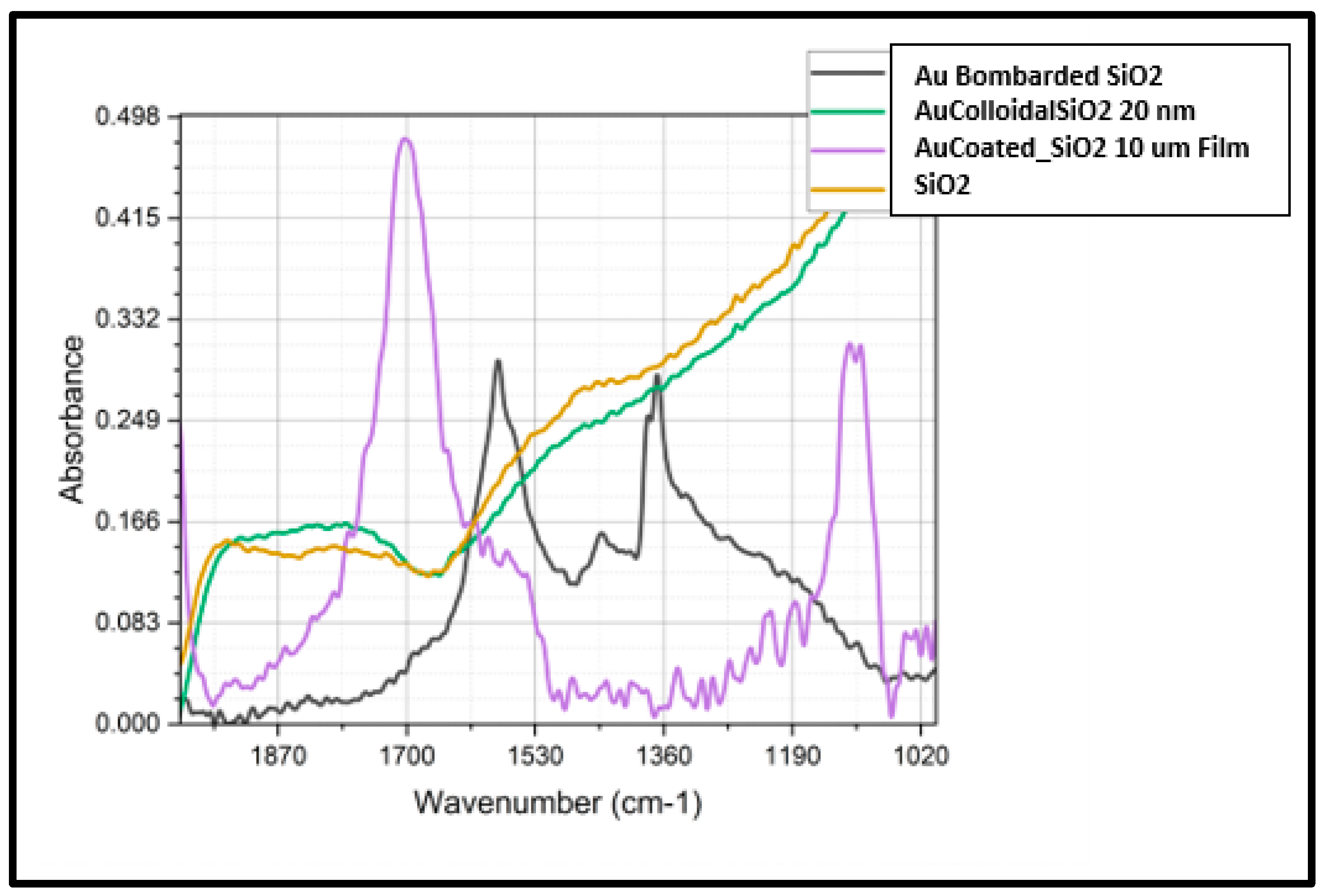
3.1. Elevated Baseline and Enhanced Fano Resonance in Ion-Bombarded Au Samples

The FTIR absorbance spectrum for the ion-bombarded Au sample shows a significantly elevated baseline. This elevation is attributed to the presence of a large number of nano-charge carriers within the nanoparticle cavities, which consistently increases the FTIR baseline [12,13]. The formation of nanocrystals and nanocavities due to ion bombardment leads to significant interactions between plasmonic and vibrational modes, resulting in Fano resonance in the strong coupling regime [14,15]. The closer spacing of modes observed in the ion-bombarded sample can be explained by the increased plasmonic coupling resulting from the formation of closely spaced nanocrystals and nanocavities. This enhanced coupling leads to stronger interactions between the plasmonic modes and the vibrational modes of the SiO2 substrate, producing the observed closer spacing of the modes [16]. Additionally, the asymmetric lineshape observed in the ion-bombarded sample is a characteristic feature of Fano resonance. This lineshape arises from the interference between the discrete plasmonic resonance of the gold nanocrystals and the continuum of vibrational states of the SiO2 substrate [12]. The strong coupling between these modes enhances the asymmetry of the lineshape, making it more pronounced in the ion-bombarded sample compared to the other samples [2].

3.2. Comparison with Other Samples

The Au-coated SiO2 sample in Figure 1, displays peaks similar to those seen in the ion-bombarded Au sample but at slightly different positions. This difference may result from variations in the method of Au deposition, affecting the local environment and the interaction strength of the modes [17]. The 20 nm Au colloidal sample in the UV-Vis spectra shows the largest and most saturated SPR peaks, indicating strong plasmonic activity. However, this strong plasmonic activity does not translate into the vibrational coupling observed in the FTIR spectrum. This suggests that while colloidal Au exhibits significant plasmonic resonance, it lacks the vibrational mode coupling seen in the ion-bombarded samples [16]. The SiO2 control sample confirms that the observed peaks in the other samples are due to the presence of Au and its interaction with the SiO2 substrate [18,19].

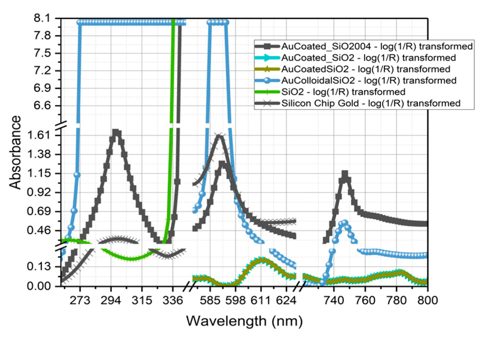
3.3. UV-Vis Spectra Analysis

The UV-Vis spectra of the samples provide additional insights into the SPR characteristics of gold nanoparticles under different preparation conditions. The SPR peaks observed in the UV-Vis spectra are highly sensitive to the local environment and preparation methods. The 20 nm Au colloidal sample shows the largest and most saturated SPR peaks, indicating significant plasmonic activity. This suggests that colloidal gold particles have a high density of free electrons, contributing to the strong plasmon resonance peaks observed in the UV-Vis spectra [4,5]. However, this strong plasmonic activity does not correlate with vibrational mode coupling in the FTIR spectrum, indicating that the plasmonic resonance in colloidal gold is not accompanied by significant vibrational interactions [12,13].

3.4. Mechanistic Insights and Theoretical Modeling

The pronounced Fano resonance and elevated baseline in the ion-bombarded Au sample Figure 2. can be attributed to the complex interplay between plasmonic and vibrational modes. When Au ions bombard the SiO2 substrate, they embed into the surface, creating nanocrystals and nanocavities that increase surface roughness and electron density. These regions act as plasmonic hotspots, where localized surface plasmons interact more strongly with the vibrational modes of the SiO2 matrix.The closer spacing of the modes in the ion-bombarded sample results from enhanced plasmonic coupling due to the formation of closely spaced nanocrystals and nanocavities. This leads to stronger hybridization of the plasmonic modes, reducing the energy separation between them. This hybridization effect aligns with the plasmon hybridization theory, which predicts the formation of new plasmonic modes with energies dependent on nanoparticle geometry and arrangement. The asymmetric lineshape characteristic of Fano resonance arises from the interaction of discrete plasmonic modes with a continuum of vibrational states. In the ion-bombarded samples, the discrete plasmonic mode provided by the gold nanocrystals interferes with the continuum of vibrational states in the SiO2 substrate. This interaction creates the distinctive Fano resonance lineshape, with an asymmetric profile that can be tuned by modifying the nanoparticle configuration [1,2]. Ion bombardment uniquely enhances plasmonic–vibrational coupling compared to other preparation methods like sputtering or colloidal deposition. Ion-bombarded samples exhibit closer mode spacing and elevated baseline absorbance, indicating higher charge carrier density and stronger plasmonic activity. In contrast, while colloidal Au shows significant plasmonic resonance, it lacks the vibrational mode coupling observed in ion-bombarded samples, demonstrating the superior interaction mechanism achieved through ion bombardment.

3.5. Implications for Nanophotonic Applications

The enhanced Fano resonance in ion-bombarded Au-SiO2 nanocomposites has profound implications for nanophotonic applications [20,21,22,23,24,25,26]. The strong coupling and characteristic asymmetric lineshape of Fano resonance can be exploited to develop high-sensitivity sensors that detect subtle changes in the local environment. These nanocomposites can be used in optical devices requiring precise light–matter interaction control, such as tunable filters and modulators. By adjusting the ion bombardment parameters, the optical properties of these nanocomposites can be tailored for specific applications [27,28].

4. Conclusions

The ion bombardment process dramatically transforms the structure of Au-SiO2 nanocomposites, leading to the formation of nanocrystals and nanocavities that generate plasmonic hotspots. These modifications foster strong coupling between localized surface plasmons and the vibrational modes of the SiO2 matrix, resulting in pronounced Fano resonance characterized by asymmetric lineshapes and closer mode spacing. Plasmon hybridization theory and Fano interference theory provide a solid theoretical basis for these observations, predicting the formation of new plasmonic modes that depend on nanoparticle geometry and arrangement. These findings underscore the critical role of structural engineering in achieving enhanced optical properties in nanocomposites and highlight ion bombardment as a powerful technique for fabricating materials with superior plasmonic–vibrational coupling. Future research should focus on scaling this method and exploring its applicability to other material systems. Investigating the long-term stability and performance of these nanocomposites in practical applications will be crucial for their deployment in high-performance nanophotonic devices and sensors based on Fano resonance [29,30].

Author Contributions

P.G. Management and coordination responsibility for the research activity planning and execution. S.P. Writing—Reviewing/Editing. A.D. Writing—Reviewing/Editing. J.L. Resources. S.B. Resources. C.V. Conceptualization, Visualization, Methodology, Data Curation, Management and Coordination. All authors have read and agreed to the published version of the manuscript.

Funding

This research was funded by NSF grant number 2331969.

Institutional Review Board Statement

Not applicable.

Informed Consent Statement

Not applicable.

Data Availability Statement

Data are contained within the article.

Conflicts of Interest

The authors declare no conflicts of interest.

References

  1. Limonov, M.F.; Rybin, M.V.; Poddubny, A.N.; Kivshar, Y.S. Fano Resonances in Photonics. Nat. Photonics 2017, 11, 543–554. [Google Scholar] [CrossRef]
  2. Miroshnichenko, A.E.; Flach, S.; Kivshar, Y.S. Fano Resonances in Nanoscale Structures. Rev. Mod. Phys. 2010, 82, 2257–2298. [Google Scholar] [CrossRef]
  3. Luk’yanchuk, B.; Zheludev, N.I.; Maier, S.A.; Halas, N.J.; Nordlander, P.; Giessen, H.; Chong, C.T. The Fano Resonance in Plasmonic Nanostructures and Metamaterials. Nat. Mater. 2010, 6, 707–715. [Google Scholar] [CrossRef]
  4. Willets, K.A.; Van Duyne, R.P. Localized Surface Plasmon Resonance Spectroscopy and Sensing. Annu. Rev. Phys. Chem. 2007, 58, 267–297. [Google Scholar] [CrossRef]
  5. Kelly, K.L.; Coronado, E.; Zhao, L.L.; Schatz, G.C. The Optical Properties of Metal Nanoparticles: The Influence of Size, Shape, and Dielectric Environment. J. Phys. Chem. B 2003, 107, 668–677. [Google Scholar] [CrossRef]
  6. Cottancin, E.; Celep, G.; Lerme, J.; Pellarin, M.; Huntzinger, J.-R.; Vialle, J.-L.; Broyer, M. Optical Properties of Noble Metal Clusters as a Function of the Size: Comparison between Experiments and a Semi-Quantal Theory. Theor. Chem. Acc. 2006, 116, 514–523. [Google Scholar] [CrossRef]
  7. Schmidt, M.K.; Esteban, R.; Gonzalez-Tudela, A.; Giedke, G.; Aizpurua, J. Quantum Mechanical Description of Raman Scattering from Molecules in Plasmonic Cavities. ACS Nano 2016, 10, 6291–6298. [Google Scholar] [CrossRef]
  8. Xiao, Y.; Du, J. Superparamagnetic Nanoparticles for Biomedical Applications. J. Mater. Chem. B 2020, 8, 354–367. [Google Scholar] [CrossRef]
  9. Wang, X.; Liu, G.; Xia, S.; Meng, H.; Shang, X.; He, P.; Zhai, X. Dynamically Tunable Fano Resonance Based on Graphene Metamaterials. IEEE Photonics Technol. Lett. 2018, 30, 2147–2150. [Google Scholar] [CrossRef]
  10. Güner, S.; Budak, S.; Amaral, R.; Minamisawa, C.; Muntele, C.; Ila, D. Thickness and MeV Si Ions Bombardment Effects on the Thermoelectric Properties of Ce3Sb10 Thin Films. Nucl. Instrum. Methods Phys. Res. Sect. B Beam Interact. Mater. At. 2008, 266, 1261–1264. [Google Scholar] [CrossRef]
  11. Hentschel, M.; Saliba, M.; Vogelgesang, R.; Giessen, H.; Alivisatos, A.P.; Liu, N. Fano Resonances in Plasmonic Nanoparticle Aggregates. J. Phys. Chem. C 2009, 113, 5540–5544. [Google Scholar] [CrossRef]
  12. Huck, C.; Vogt, J.; Sendner, M.; Hengstler, D.; Neubrech, F.; Pucci, A. Plasmonic Enhancement of Infrared Vibrational Signals: Nanoslits versus Nanorods. ACS Photonics 2015, 2, 1489–1497. [Google Scholar] [CrossRef]
  13. Hao, F.; Nehl, C.L.; Hafner, J.H.; Nordlander, P. Symmetry Breaking in the Hydrogenation of Gold Clusters. Nano Lett. 2004, 4, 327–330. [Google Scholar] [CrossRef]
  14. SPIE. Coupling Effects in Plasmonic Nanostructures and Metamaterials. Available online: https://spie.org/news/4948-coupling-effects-in-plasmonic-nanostructures-and-metamaterials (accessed on 17 May 2025).
  15. Kauranen, M.; Zayats, A.V. Nonlinear Plasmonics. Nat. Photonics 2012, 6, 737–748. [Google Scholar] [CrossRef]
  16. Steele, J.M.; Grady, N.K.; Nordlander, P.; Halas, N.J. Plasmon Hybridization in Complex Nanostructures. In Surface Plasmon Nanophotonics; Maier, S.A., Atwater, H.A., Eds.; Springer Series in Optical Sciences; Springer: Dordrecht, The Netherlands, 2007; Volume 131, pp. 183–196. [Google Scholar] [CrossRef]
  17. Bhat, S.A.; Rao, S.; Prasad, S.K.; Yelamaggad, C. Tuning the Optical Properties of Gold Nanoparticles via Photoactive Liquid Crystalline Azo Ligands. Nanoscale 2025, 17, 3123–3132. [Google Scholar] [CrossRef] [PubMed]
  18. Jain, P.K.; Lee, K.S.; El-Sayed, I.H.; El-Sayed, M.A. Calculated absorption and scattering properties of gold nanoparticles of different size, shape, and composition: Applications in biological imaging and biomedicine. J. Phys. Chem. B 2006, 110, 7238–7248. [Google Scholar] [CrossRef] [PubMed]
  19. Link, S.; El-Sayed, M.A. Optical properties and ultrafast dynamics of metallic nanocrystals. Annu. Rev. Phys. Chem. 2003, 54, 331–366. [Google Scholar] [CrossRef]
  20. Grimmer, M.; Tao, W.; Fleischer, M. Enhancing Fano Resonances through Coupling of Dark Modes in a Dual-Ring Nanostructure. Opt. Express 2024, 32, 1926–1940. [Google Scholar] [CrossRef]
  21. Lu, H.; Li, D.; Shi, S.; Li, Y.; Zhao, J. Exciton-Induced Fano Resonance in Metallic Nanocavity with Tungsten Disulfide Atomic Layer. Opt. Express 2023, 31, 20761–20768. [Google Scholar] [CrossRef]
  22. Ma, C.; Yan, J.; Huang, Y.; Yang, G. Directional Fano Resonance in an Individual GaAs Nanospheroid. Small 2019, 15, 1900546. [Google Scholar] [CrossRef]
  23. Gong, T.; Guan, F.; Wei, Z.; Huang, W.; Zhang, X. Tunable Magnetic Fano Resonances on Au Nanosphere Dimer–Dielectric–Gold Film Sandwiched Structure. Front. Phys. 2021, 9, 691027. [Google Scholar] [CrossRef]
  24. Grineviciute, L.; Nikitina, J.; Babayigit, C.; Staliūnas, K. Fano-like Resonances in Nanostructured Thin Films for Spatial Filtering. Appl. Phys. Lett. 2021, 118, 131114. [Google Scholar] [CrossRef]
  25. Petrosyan, P. Influence of Interaction Retardation and Radiation Reaction on the Fano Resonance Efficiency in the System of Nanoparticles. J. Contemp. Phys. 2018, 53, 324–331. [Google Scholar] [CrossRef]
  26. Ren, J.; Qiu, W. Generating and Tuning the Fano Resonance by Graphene Oligomers with Different Nanostructures. Proc. SPIE 2018, 10964, 109645I. [Google Scholar] [CrossRef]
  27. Chen, J.; Gan, F.; Wang, Y.; Li, G. Plasmonic Sensing and Modulation Based on Fano Resonances. Adv. Opt. Mater. 2018, 6, 1701152. [Google Scholar] [CrossRef]
  28. Zheng, C.; Jia, T.; Zhao, H.; Xia, Y.; Zhang, S.; Feng, D.; Sun, Z. Theoretical Study on Narrow Fano Resonance of Nanocrescent for the Label-Free Detection of Single Molecules and Single Nanoparticles. RSC Adv. 2018, 8, 3381–3391. [Google Scholar] [CrossRef]
  29. Shiryaev, A.; Ekimov, E.; Prokof’ev, V.; Kondrin, M. Temperature Dependence of the Fano Resonance in Nanodiamonds Synthesized at High Static Pressures. JETP Lett. 2022, 115, 651–656. [Google Scholar] [CrossRef]
  30. Kim, K.-H.; Ri, M. Metal–Dielectric Composite Nanostructures for Fano Resonance-Based Highly Sensitive SECARS from Visible to Deep-UV. J. Phys. Chem. C 2018, 122, 16281–16288. [Google Scholar] [CrossRef]
Figure 1. Comparison of ATR FTIR spectra of Au bombardment on SiO2, Au colloidal 20 nm on SiO2, 10 nm Au film on SiO2, and SiO2.
Coatings 15 00605 g001
Figure 2. Comparison of UV-Vis spectra of SiO2 and Au-SiO2 samples after different preparation techniques.
Coatings 15 00605 g002
Disclaimer/Publisher’s Note: The statements, opinions and data contained in all publications are solely those of the individual author(s) and contributor(s) and not of MDPI and/or the editor(s). MDPI and/or the editor(s) disclaim responsibility for any injury to people or property resulting from any ideas, methods, instructions or products referred to in the content.

