Article

Interfacial Adsorption Mechanisms of Arginine, Glutamic Acid, Aspartic Acid, and Valine on Magnesium and Magnesium Alloy Surfaces: A First-Principles Investigation

by
Zhe Fang
1,
Shuaiwei Xu
1,
Rui Cao
1,
Mingli Jiao
1,*,
Keyi Liu
2,
Quan Diao
1,
Shaokang Guan
3 and
Yu Jia
4,5,*
1
School of Materials Electronics and Energy Storage, Zhongyuan University of Technology, Zhengzhou 450007, China
2
Department of Physics, Imperial College London, South Kensington Campus, London SW7 2AZ, UK
3
School of Materials Science and Engineering, Zhengzhou University, Zhengzhou 450001, China
4
Key Laboratory for Special Functional Materials of Ministry of Education, School of Materials Science and Engineering, Henan University, Kaifeng 475004, China
5
International Laboratory for Quantum Functional Materials of Henan, School of Physics, Zhengzhou University, Zhengzhou 450001, China
*
Authors to whom correspondence should be addressed.
Coatings 2025, 15(5), 586; https://doi.org/10.3390/coatings15050586
Submission received: 18 April 2025 / Revised: 12 May 2025 / Accepted: 13 May 2025 / Published: 15 May 2025
(This article belongs to the Special Issue Advanced Alloy Degradation and Implants, 2nd Edition)

Abstract

:
Elucidating the interfacial interaction mechanisms between biomolecules and metal surfaces is crucial for designing functionalized biomedical materials. This study employs first-principles calculations based on density functional theory (DFT) to investigate the adsorption behaviors of arginine (Arg), glutamic acid (Glu), aspartic acid (Asp), and valine (Val) on magnesium (Mg) and Mg alloy surfaces. The adsorption behaviors of four kinds of amino acids on Mg and Mg alloy surfaces were analyzed through optimized adsorption configurations, adsorption energies (Eads), bond lengths, projected densities of states (PDOSs), and differential charge densities. The calculated results of Eads followed the order of Arg > Glu > Asp > Val, driven by functional group spatial configurations and electron transfer efficiency. Alloying elements facilitated charge redistribution on the Mg and Mg alloy surfaces, enhancing the interaction between amino acids and the alloy surfaces. Notably, the guanidino group of Arg exhibited exceptional adsorption stability and multi-dentate bonding, increasing electron donation to the Mg(0001) surface, achieving the highest Eads (−1.67 eV). This work provides insights into the structure–activity relationships between amino acids and Mg and Mg alloy surfaces, offering a foundation for designing biomolecule-derived functional coatings and strategies for improving the biocompatibility of Mg and Mg alloy implants.

1. Introduction

The interaction between metal surfaces and biomolecules has garnered considerable attention in the fields of materials science and biomedicine [1,2,3,4]. Amino acids, as the basic constituents of proteins, are pivotal in modulating adsorption behavior on metal surfaces, thereby influencing the performance and potential applications of the material [5,6]. Mg alloys, as the lightest structural metal, offer high specific strength, biodegradability, and exceptional biocompatibility, making them a promising material for biomedical applications such as orthopedic implants, cardiovascular stents, and drug delivery systems [7,8,9,10]. However, the spontaneous formation of a native oxide layer on Mg surfaces acts as a passive barrier, which hinders the effective interactions between Mg and biomolecules [11,12]. Consequently, elucidating the adsorption mechanisms of biomolecules on both pure Mg and Mg alloy surfaces is of paramount importance for optimizing the performance of Mg alloys in biomedical applications.
In recent years, first-principles calculations based on DFT have emerged as a powerful tool for studying interface interactions, particularly in the investigation of biomolecule adsorption mechanisms on metal surfaces [13,14,15]. Xia et al. [16] demonstrated, through ab initio molecular dynamics (MD) simulations, that amino acids tend to align parallel to the surface of Zn metal, thereby enhancing corrosion protection and inhibiting dendritic growth. Meng et al. [17] evaluated the corrosion inhibition performances of three amino acids on the 7150 Al alloy in saline solution using electrochemical methods, revealing a negative correlation between the Eads and corrosion inhibition efficiency. In the field of biomolecule adsorption on Mg and Mg alloy surfaces, the integration of experimental techniques with multiscale simulations has provided deeper insights into interfacial interaction mechanisms. Wang et al. [18] developed a fucoidan/collagen composite coating on the ZE21B Mg alloy surface, achieving synergistic improvements in both corrosion resistance and biocompatibility. Kasprzhitskii et al. [19] applied theoretical methods, including DFT and Monte Carlo (MC) simulation, to study the potential inhibition efficiency of sulfur-containing and aromatic amino acids on the Mg surface in Hanks’ solution. The results showed that tyrosine formed shorter Mg-O bonds and more negative Eads. Pei et al. [20] investigated the adhesion behavior of β-phenylethylamine (8-phe-4) derived from phenylalanine on Mg(0001) surfaces via MD simulations, finding stronger adhesion of 8-phe-4 compared to lactide co-glycolide (PLGA) on Mg surfaces. Ma et al. [21] used micro-arc oxidation technology to prepare coatings on Mg alloy surfaces, followed by stearic acid deposition via self-assembly. Their MD simulations indicated that the adsorption of stearic acid on the micro-arc oxidation coating was primarily chemical in nature. Xie et al. [22] explored the microstructural characteristics and corrosion behavior of Mg-Sc alloy, employing both DFT calculations and experimental tests to elucidate the corrosion mechanisms. Chen et al. [23] investigated the interactions and solid solution behaviors of 24 alloying elements in Mg alloys using first-principles and machine-learning methods, offering valuable insights into the design of high-performance multi-component Mg alloys.
While these studies have advanced the understanding of the interactions between biomolecules and Mg alloy surfaces from various perspectives, systematic investigations into the structure–activity relationships of amino acid side chains remain limited. The influence of different amino acid side-chain structures and the effect of alloying elements on the adsorption behaviors on Mg and Mg alloy surfaces is not yet fully understood. Notably, amino acids with distinct functional groups, such as Arg, Glu, Asp, and Val, exhibit differing adsorption characteristics due to variations in their side-chain structures. Despite the significance of these amino acids as fundamental protein building blocks, research on their adsorption characteristics on Mg and Mg alloy surfaces remains sparse. This is particularly crucial for understanding protein interactions with Mg alloys, which profoundly affect the performance of Mg alloys in practical applications. Building upon the aforementioned research, the Arg-Glu-Asp-Val (REDV) peptide sequence, a characteristic cell adhesion motif of fibronectin, plays a pivotal role in biochemical reactions. It facilitates cell adhesion and regulates proliferation by specifically recognizing the integrin α4β1 receptor. Through integrin binding, the REDV peptide activates intracellular signaling pathways, significantly promoting osteoblast differentiation and bone regeneration [24,25]. Despite progress in understanding REDV peptide-modified Mg alloys [26,27], theoretical investigations into its adsorption mechanisms and surface interactions remain limited. In particular, first-principles computational studies are urgently required to elucidate the nature of REDV peptide–metal interface interactions at the electronic structure level. This gap in knowledge hinders the understanding of fundamental interactions and restricts the rational design of coatings based on molecular simulations.
Furthermore, the adsorption kinetics of the characteristic functional groups of individual amino acids (such as the guanidino group of Arg and the carboxyl group of Glu/Asp/Val) on Mg and Mg alloy surfaces, as well as their synergistic effects, remain inadequately understood. The present study employs the first-principles calculation to systematically investigate the adsorption configurations and electronic structures of Arg, Glu, Asp, and Val on Mg and Mg alloy surfaces, offering valuable theoretical insights for the rational design of functional coatings on biomedical Mg alloys.

2. Computational Methods

In this study, the Vienna ab initio simulation package (VASP) software package [28,29] was employed to investigate the adsorption characteristics of amino acids on Mg and Mg alloy surfaces based on DFT [30,31]. The Projector Augmented Wave (PAW) method [32] was utilized to model electron–ion interactions, and the Perdew–Burke-Ernzerhof (PBE) functional [33] was selected for treating the exchange-correlation energy during the calculations. The Mg surface was modeled based on the Mg(0001) crystal plane of the hexagonal close-packed (HCP) structure, with a supercell of size 6 × 6 × 5 constructed. To mitigate the effects of periodic boundary conditions, a vacuum layer of 20 Å was introduced in the direction normal to the surface. During structural optimization, the energy convergence criterion was set to 1 × 10⁻⁵ eV, and the convergence threshold for the interatomic forces was set to 0.02 eV/Å. The optB86b van der Waals (vdW) correction [34] was applied to account for the weak vdW interactions of the small biomolecules and Mg and Mg alloy surfaces. A Gamma-centered k-point grid of 1 × 1 × 1 was used for sampling the Brillouin zone. The lowest two layers of Mg atoms were kept fixed, while the remaining Mg and alloy atoms and amino acid molecules were fully relaxed until the convergence criteria were obtained. The adsorption energy (Eads) was calculated as follows:
E a d s = E m o l + s u b E m o l E s u b
where Emol+sub, Emol, and Esub represent the total energy of the optimized stable adsorption systems, the total energy of the optimized isolated biomolecules, and the energy of the different substrates, respectively.

3. Results and Discussion

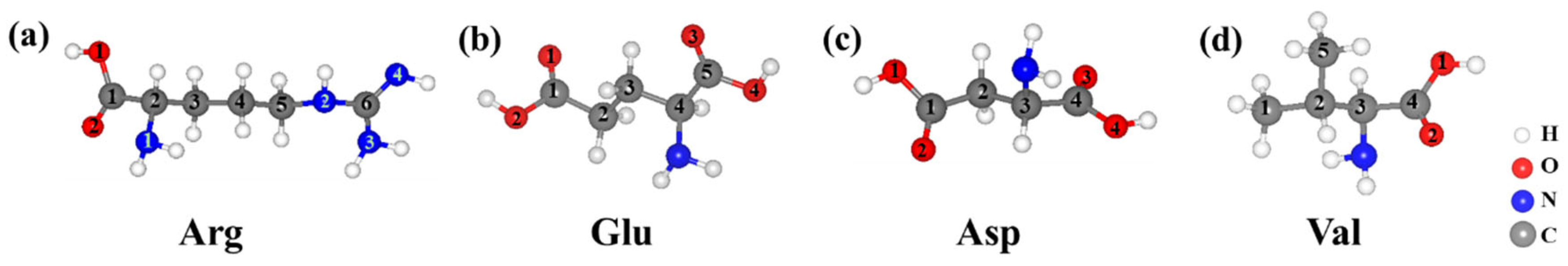
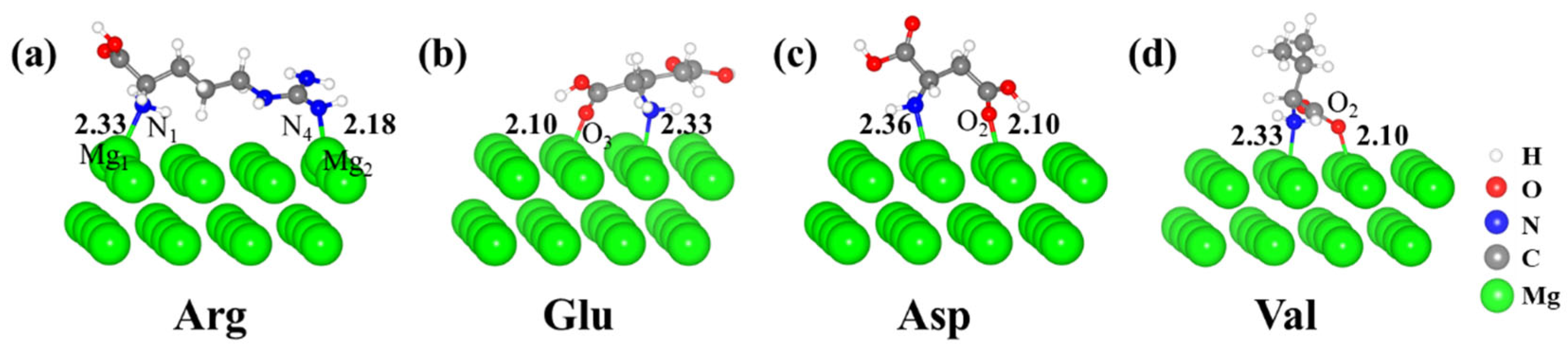
3.1. Adsorption Properties of Amino Acids on Mg(0001) Surfaces

The adsorption of molecules on metal surfaces is influenced by both the surface properties of the metal and the structural and chemical characteristics of the adsorbed species. This relationship plays a crucial role in modulating interface interactions, optimizing adsorption behavior, and designing high-performance functional materials [35,36,37]. Sudhakar et al. [38] employed periodic DFT calculations to investigate the adsorption behavior of L-cysteine on the Au(111) surface as a function of applied potential. The study highlighted the potential-dependent rearrangement of cysteine on the electrode surface, revealing the underlying mechanisms governing this process. Furthermore, the results demonstrated that the adsorption structure of zwitterionic cysteine was primarily dictated by the interactions between the cationic ammonium and anionic carboxylate groups. To improve the clarity of the findings, specific atoms within the various amino acid molecules were labeled. The optimized molecular structures of Arg, Glu, Asp, and Val are depicted in Figure 1. Additionally, the optimized lattice parameters of the bulk hcp Mg were a = b = 3.188 Å and c = 5.193 Å [39], which are in close agreement with the experimental values of a = b = 3.210 Å, c = 5.213 Å [40].
Following optimization calculations, the adsorption configurations of four amino acid molecules on the Mg(0001) surfaces were determined to reach their lowest energy states. The optimized adsorption configurations of Arg, Glu, Asp, and Val on the Mg(0001) surfaces are presented in Figure 2. Notable differences in the adsorption sites and modes were observed for the various amino acids. Specifically, Arg interacted strongly with the Mg surface through its amino and guanidino groups, whereas Glu, Asp, and Val formed bonds with the Mg surfaces primarily through their amino and carboxyl groups. The Eads, calculated using Equation (1), are shown in Table 1. The calculated Eads values, ranked by descending absolute magnitude, were as follows: Arg (−1.67 eV) > Glu (−1.31 eV) > Asp (−1.23 eV) > Val (−1.16 eV). These results indicate that Arg exhibited the strongest Eads on the Mg(0001) surface, suggesting that its adsorption behavior was the most stable among the four amino acids studied.
The bond length is a critical parameter for assessing the interaction strength between adsorbed molecules and surfaces, with changes in bond length reflecting the stability of interfacial chemical bonds. The bond lengths for the four kinds of amino acid molecules adsorbed on the Mg(0001) surfaces are summarized in Table 2. The theoretical covalent radii for N, O, and Mg atoms were 0.71 Å, 0.66 Å, and 1.41 Å, respectively. Additionally, the theoretical bond lengths for the N-Mg and O-Mg covalent bonds were 2.11 Å and 2.09 Å [41]. For the adsorption configurations of the different amino acids on the Mg(0001) surfaces, the calculated bond lengths for N-Mg and O-Mg ranged from 2.13 to 2.36 Å and from 1.94 to 2.28 Å, respectively. In the case of Arg, the guanidino group, containing three N atoms, formed a strong covalent bond with the Mg surface. The N-Mg bond directly interacting with the surface was 2.18 Å, indicating the formation of a robust covalent bond. The N-Mg bond in the amino group was relatively longer; however, it provided additional adsorption sites via weak electron sharing, thus enhancing the overall adsorption stability. For Glu, the Eads was primarily influenced by the strong chemisorption of the carboxyl group. The O-Mg bond length between the carboxyl O atom and the binding Mg atom on surface was 2.10 Å, indicating the formation of a strong covalent bond. Similarly, for Asp, the O-Mg bond length (2.10 Å) of the carboxyl group was close to the theoretical value, suggesting strong chemisorption. However, the N-Mg bond length (2.33 Å) was relatively long, resulting in weak electron transfer and insufficient enhancement of Eads. As a result, the adsorption of Asp was dominated by a single carboxyl group, lacking the multi-functional group synergy observed in Arg. For Val, despite the O-Mg (2.10 Å) and N-Mg (2.36 Å) bond lengths being close to the theoretical values, the hydrophobic isopropyl side chain hindered the synergistic adsorption between the amino and carboxyl groups, weakening its overall Eads. Consequently, Val exhibited the lowest Eads among the four amino acids.

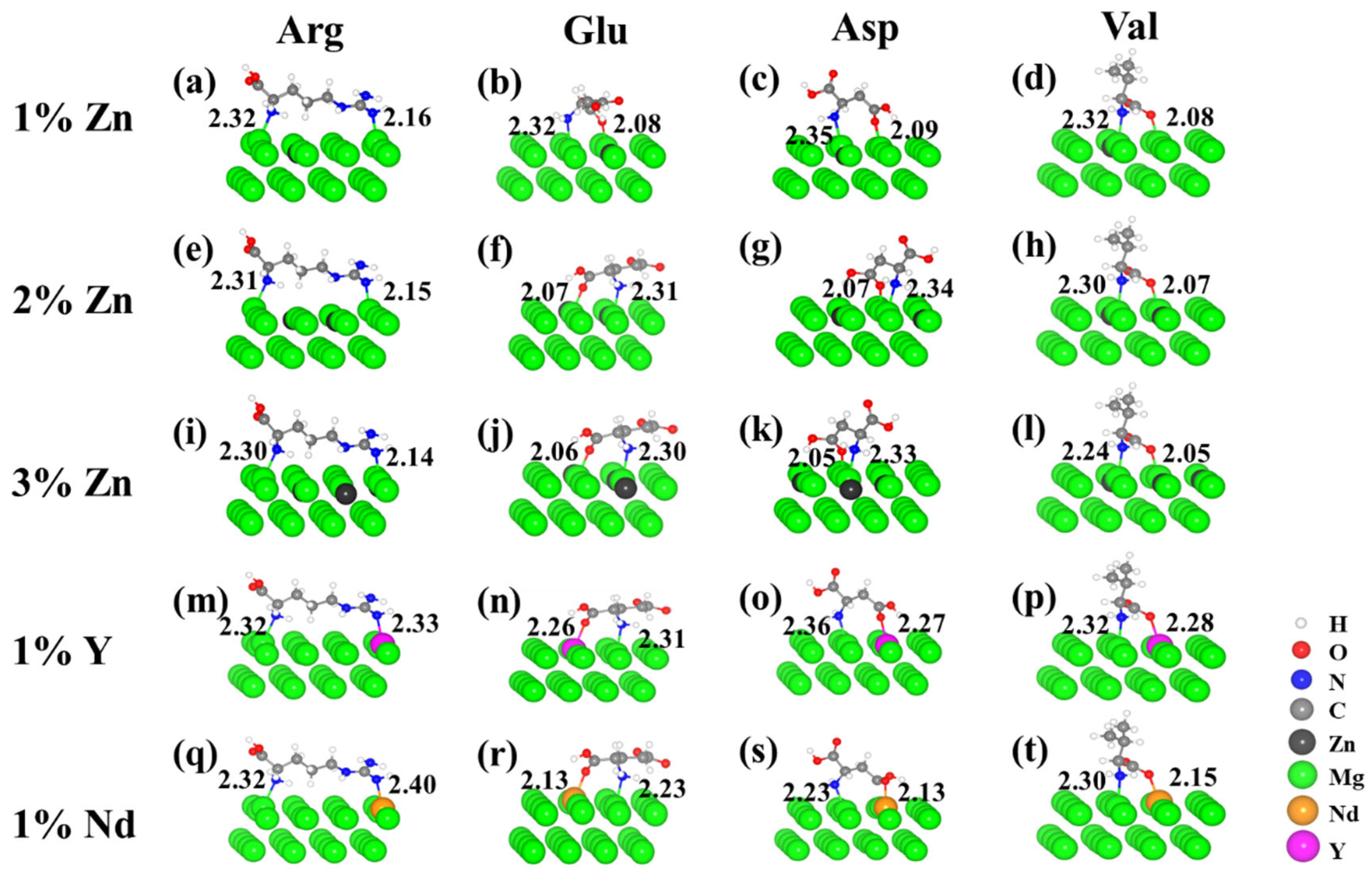
3.2. Effects of Alloying Elements on the Adsorption Properties

The incorporation of alloying elements not only influenced the physical and chemical properties of Mg alloys but also had a profound impact on the adsorption behavior of biomolecules on their surfaces. Among the common alloying elements, zinc (Zn), yttrium (Y), and neodymium (Nd) play distinct roles in modifying the characteristics of Mg alloys [42,43]. The effects of these elements on the adsorption of amino acids on the Mg alloy surfaces were investigated by incorporating Zn at concentrations of 1%, 2%, and 3%, alongside 1% Y and 1% Nd. The stable adsorption configurations of four kinds of amino acids on the Mg alloy surfaces with varying element concentrations are depicted in Figure 3. The Eads, calculated using Equation (1), are presented in Table 3.
Following the incorporation of alloying elements, the bond lengths of four kinds of amino acids adsorbed on the Mg alloy surfaces were summarized, as shown in Table 4. Specifically, the bond distances for N-Y, N-Nd, O-Y, and O-Nd on the surfaces of Mg-Y and Mg-Nd alloys were observed to range from 2.32 Å to 2.36 Å, 2.23 Å to 2.40 Å, 2.26 Å to 2.28 Å, and 2.13 Å to 2.15 Å, respectively. These optimized bond lengths closely align with the theoretical covalent bond lengths for N-Y, N-Nd, O-Y, and O-Nd (2.37 Å, 2.39 Å, 2.35 Å, and 2.37 Å, respectively) [40]. Further analysis of the effects of alloying elements revealed that the relatively high electronegativity of Zn contributed to a reduction in the electron density of adjacent Mg atoms. This, in turn, enhanced the propensity for N and O atoms in the amino acid molecules to share electrons with the neighboring Mg atoms. Consequently, the N-Mg bond length was reduced to 2.14–2.36 Å, and the O-Mg bond length decreased to 2.04–2.10 Å, suggesting a strengthened interaction and an improvement in Eads.
In contrast, for Mg-Y and Mg-Nd alloys, the Y and Nd elements exhibited a tendency to transfer electrons to the surfaces, facilitating a more direct interaction between the amino acids and the alloy elements. For instance, the N-Y bond length for Val was measured at 2.32 Å, which is in close agreement with the theoretical value of 2.37 Å. This indicates the formation of a stable covalent bond, further enhancing the Eads. These observations demonstrate that the electronic properties of the alloying elements effectively optimized the charge transfer pathways between the amino acids and the alloy surfaces by adjusting the bond lengths.

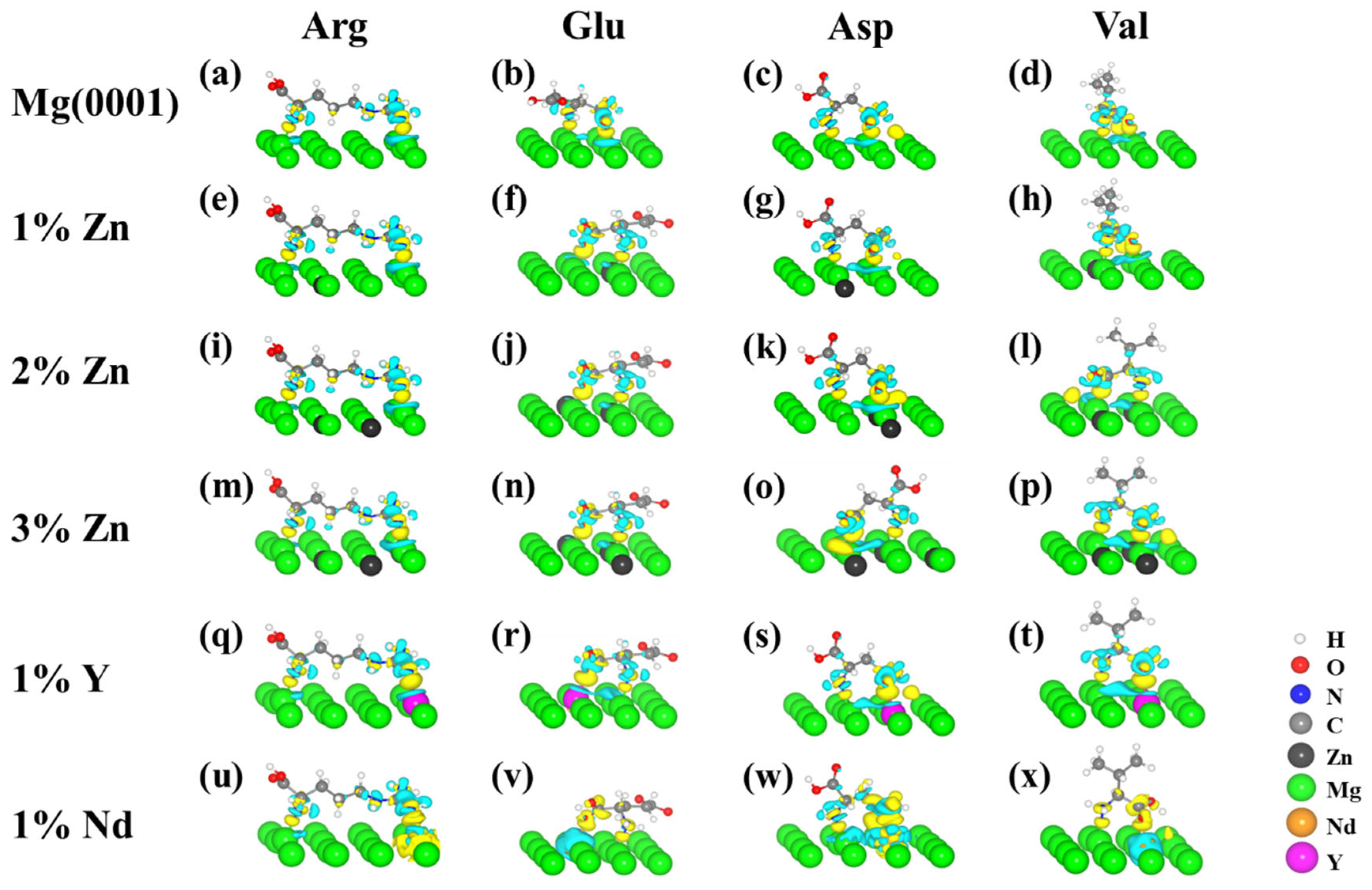
3.3. Electronic Structure Properties of Amino Acids on the Mg and Mg Alloy Surfaces

To explore the electronic structure properties of the molecules on the Mg(0001) surface, PDOS analyses was performed for Arg before and after its adsorption on the Mg(0001) surface. The results are presented in Figure 4.
Prior to adsorption, the amino N atom (N1) and the guanidino N atom (N4) of Arg exhibited localized electronic states near the Fermi level, with prominent peaks at approximately −2 eV and −6 eV, corresponding to the contributions from the p orbitals of the N atoms. The PDOS of the Mg surface displayed typical metallic characteristics near the Fermi level, with continuously distributed s and p electronic states. Upon adsorption, the PDOSs of both N1 and N4 broadened significantly within the −2 eV to −6 eV range, overlapping with the s and p states of the adjacent Mg atoms, indicating a strong electronic interaction between N and binding Mg atoms. Specifically, the p orbital of N4 and the s orbital of Mg formed a new hybridization peak within the −6 eV to 2 eV range. The emergence of this hybridization peak reflects the reconstruction of the electronic states at the Mg-Arg interface, providing an electronic structural foundation for the formation of covalent bonds between Arg and the Mg surface. Furthermore, after adsorption, new peaks appeared in the PDOS of the Mg atoms within the −2 eV to 2 eV range, which were unobservable in the PDOS of the Mg surface before adsorption. These new features are attributed to the charge transfer and electron sharing between the binding Mg atoms of the Mg(0001) and the N atoms of Arg, further supporting the formation of chemisorption. Coupled with the Eads data (Eads of Arg is −1.67 eV), the PDOS analysis suggests that the strong electronic coupling between N4 and the Mg surface plays a crucial role in the enhanced adsorption stability of Arg compared to other three amino acids. It is noteworthy that, although the PDOS of N1 also exhibited some overlap with the Mg surface, the coupling strength was weaker than that of N4. This observation is consistent with the longer bond length observed for N1 (the bond length of N1 and Mg is 2.33 Å) compared to N4 and Mg (2.18 Å). This implies that Arg achieves efficient electron transfer with the Mg surface primarily through the synergistic adsorption of the guanidino and amino groups, with the guanidino group, in particular, playing a key role.
To obtain deeper insights into the bonding mechanism and charge redistribution during the adsorption process between amino acids and Mg alloy surfaces, the charge density difference (∆ρ) of the adsorption system was calculated. The results are presented in Figure 5. A positive ∆ρ (>0) indicates an increase in charge density following the interaction, whereas a negative ∆ρ (<0) signifies a decrease in charge density post-interaction. The charge density difference (∆ρ) was computed using the following formula:
Δ ρ = ρ m o l + s u b ρ m o l ρ s u b
where ρmol+sub represents the charge density of the optimized adsorbate–substrate system; ρmol refers to the charge density of the adsorbed molecule in the absence of the surface; and ρsub denotes the charge density of the substrate surface, respectively.
The charge density difference analyses revealed that charge transfer occurred between the Mg surface and the amino acid molecules during adsorption. In the case of Arg, its notably high Eads was attributed to the strong electronic coupling between the guanidino functional group and the Mg surface. The differential charge density showed a significant electron accumulation (the yellow region) at the interface between the N4 atom of the guanidino group and the adjacent Mg atoms, suggesting that electrons were transferred from the Mg surface to the guanidino group, resulting in the formation of localized covalent bonds. Simultaneously, electron depletion (the light cyan region) was observed on the adjacent Mg surface, further supporting the directional charge transfer. This charge redistribution was in good agreement with the PDOS analysis, which showed strong hybridization between the N4 p orbital and the Mg s orbital near the Fermi level, indicating that the guanidino group achieved efficient electron sharing through the N-Mg bond, thereby significantly enhancing the adsorption stability.
The Eads of Glu was found to be intermediate between that of Arg and Asp, primarily due to the strong chemisorption effect of its carboxyl functional group. The differential charge density showed significant electron accumulation at the interface between the carboxyl O atom (O3) and the Mg surface, indicating a high density of electron sharing between O3 and the Mg atoms. This bonding strength was comparable to that of the carboxyl group of Asp. However, the N-Mg bond length in the amino group of Glu was shorter than that of Asp, leading to more significant electron transfer, which manifested as localized electron dissipation. This suggests that although the amino group of Glu did not form a strong covalent bond, its weak electron contribution worked synergistically with the carboxyl group, resulting in an increase in Eads, surpassing that of Asp, which relied solely on the carboxyl group.
In contrast to Arg and Glu, Asp exhibited a lower Eads, primarily due to the single bonding effect of its carboxyl functional group. The differential charge density showed considerable electron accumulation around the O atom (O2) of the carboxyl group, indicating a strong chemical bond formation between O2 and the Mg surface. However, the N-Mg bond in the amino functional group was relatively long, resulting in weak electron transfer and a limited range of charge redistribution at the interface. Additionally, Asp lacked the synergistic effect of multiple functional groups present in Arg, leading to an insufficient increase in the overall Eads. This result was consistent with the weaker adsorption stability and single bonding mechanism observed for Asp, which contributed to its relatively low Eads value among the four amino acids.
Val exhibited the lowest Eads. Although the O-Mg bond length between the O atom of the carboxyl group and the Mg surface was close to the theoretical value, and the N-Mg bond length in the amino group was similar to that of Asp, the hydrophobic isopropyl side chain significantly restricted the spatial interaction of the functional groups, as evidenced by the differential charge density. The electron accumulation at the interface was primarily concentrated around the O atom of the carboxyl group, while the electron transfer around the amino group was limited, with no noticeable charge redistribution in the side-chain region. This resulted in the adsorption of Val being predominantly dependent on the bonding of the isolated carboxyl and amino functional groups, lacking the multiple bonding interactions shown in Arg or the weak synergistic effect observed in Glu. The weak interaction mechanism of Val can, thus, be attributed to its simplified side-chain structure and the limited electron coupling pathways.

4. Conclusions

This study initiated from the atomic scale and systematically investigated the adsorption properties of Arg, Glu, Asp, and Val on Mg and Mg alloy surfaces through first-principles calculations based on DFT. The main conclusions are summarized as follows:
(1) The calculated order of Eads was Arg (−1.67 eV) > Glu (−1.31 eV) > Asp (−1.23 eV) > Val (−1.16 eV). This trend in adsorption stability was primarily attributed to the distinct spatial configurations and electronic properties of the amino acid side chains.
(2) The incorporation of alloying elements such as Zn, Y, and Nd influenced the electronic structure of the Mg surfaces, thereby modifying the adsorption behavior. Specifically, for the Mg-Zn alloy, the higher electronegativity of Zn reduced the electron density of the adjacent Mg atoms, leading to shorter bond lengths and enhanced electron sharing. In the case of Mg-Y/Nd alloys, the electron donation from Y and Nd atoms to the Mg surfaces facilitated more direct interactions between the amino acids and the alloying elements.
(3) The PDOSs of the guanidino and amino N atoms broadened and overlapped with those of the Mg atoms, forming hybridization peaks that confirmed the formation of covalent bonds between Arg and the Mg surface. For Val, the charge density analysis showed that due to the hydrophobic nature of its side chain, the electron transfer from the amino group was limited, resulting in weak hybridization and a relatively long N-Mg bond length. Overall, the charge density difference analysis indicated significant charge transfer and redistribution between the amino acids and the Mg and Mg alloy surfaces, with chemisorption being the dominant interaction mechanism.

Author Contributions

Conceptualization, Z.F., S.G. and Y.J.; Software, Y.J.; Validation, Q.D.; Formal analysis, S.X. and K.L.; Resources, S.G. and Y.J.; Data curation, R.C.; Writing—original draft, S.X.; Writing—review & editing, Z.F.; Visualization, Q.D.; Supervision, M.J.; Project administration, Z.F. and M.J.; Funding acquisition, M.J. All authors have read and agreed to the published version of the manuscript.

Funding

The authors are very grateful for the financial support from the Program for Innovative Research Team (in Science and Technology) in University of Henan Province (No. 23IRTSTHN019), the project of Young Backbone Teacher of Zhongyuan University of Technology (2023XQG10), and the project of Graduate Education Quality Improvement of Zhongyuan University of Technology (JD202506).

Institutional Review Board Statement

Not applicable.

Informed Consent Statement

Not applicable.

Data Availability Statement

There were no new data created.

Conflicts of Interest

The authors declare no conflicts of interest.

References

  1. Janisse, S.E.; Fernandez, R.L.; Heffern, M.C. Characterizing metal-biomolecule interactions by mass spectrometry. Trends Biochem. Sci. 2023, 48, 815–825. [Google Scholar] [CrossRef]
  2. Xu, W.J.; Lin, Z.X.; Kim, C.J.; Wang, Z.R.; Wang, T.Z.; Jugo, C.; Caruso, F. Assembly and biological functions of metal-biomolecule network nanoparticles formed by metal-phosphonate coordination. Sci. Adv. 2024, 10, eads9542. [Google Scholar] [CrossRef] [PubMed]
  3. Alvisi, N.; Vries, R. Biomedical applications of solid-binding peptides and proteins. Mater. Today Bio. 2023, 19, 100580. [Google Scholar] [CrossRef] [PubMed]
  4. Giovana, C.C.; Carlos, R.G.; Julietta, V.R. Comprehensive review of PEO coatings on titanium alloys for biomedical implants. J. Mater. Res. Technol. 2024, 31, 311–328. [Google Scholar]
  5. Nassar, M.F.; Taban, T.Z.; Obaid, R.F.; Shadhar, M.H.; Almashhadani, H.A.; Kadhim, A.M.; Liu, P. Study to amino acid-based inhibitors as an effective anti-corrosion material. J. Mol. Liq. 2022, 360, 119449. [Google Scholar] [CrossRef]
  6. Zhu, L.W.; Mori, Y.; Song, J.Q.; Kuroda, K.; Okido, M.; Peng, C. Surface modification by pre-adsorption of proteins and polypeptides on Ti substrate with controlled hydrophilicity to improve biocompatibility. Mater. Today Commun. 2023, 37, 107124. [Google Scholar] [CrossRef]
  7. Dubey, A.; Ghosh, S.; Jaiswal, S.; Roy, P.; Lahiri, D. Assessment of protein adhesion behaviour and biocompatibility of magnesium/Co-substituted HA-based composites for orthopaedic application. Int. J. Biol. Macromol. 2022, 208, 707–719. [Google Scholar] [CrossRef]
  8. Liu, K.P.; Cheng, A.Y.; You, J.L.; Chang, Y.H.; Tseng, C.C.; Ger, M.D. Biocompatibility and corrosion resistance of drug coatings with different polymers for magnesium alloy cardiovascular stents. Colloids Surf. B Biointerfaces 2025, 245, 114202. [Google Scholar] [CrossRef]
  9. Wang, J.; Sun, C.; Huang, Q.X.; Chi, Y.; Yan, J.H. Adsorption and thermal degradation of microplastics from aqueous solutions by Mg/Zn modified magnetic biochars. J. Hazard. Mater. 2021, 419, 126486. [Google Scholar] [CrossRef]
  10. Fang, Z.; Xu, S.; Wang, Z.; Sun, Y. Effect of Solution and Aging Treatment on the Microstructure and Properties of LAZ931 Mg-Li Alloy by Friction Stir Processing. Metals 2025, 15, 314. [Google Scholar] [CrossRef]
  11. Cazzola, R.; Della Porta, M.; Piuri, G.; Maier, J.A. Magnesium: A defense line to mitigate inflammation and oxidative stress in adipose tissue. Antioxidants 2024, 13, 893. [Google Scholar] [CrossRef]
  12. Fattah, A.; Chaharmahali, R.; Askari, A.; Alizad, S.; Kaseem, M. Unraveling the impact of purification and alloying elements on corrosion performance and passivation of magnesium alloys. J. Magnes. Alloys 2024, 12, 4808–4827. [Google Scholar] [CrossRef]
  13. Ventura, E.; Martinez, P.M.; Pérez, R.; Vilhena, J.G. Quantum mechanical derived (VdW-DFT)transferable Lennard–Jones and morse potentials to model cysteine and alkanethiol adsorption on Au(111). Adv. Mater. Interfaces 2024, 11, 30. [Google Scholar]
  14. Xu, M.L.; Liu, S.H.; Vijay, S.; Bligaard, T.; Kastlunger, G. Benchmarking water adsorption on metal surfaces with ab initio molecular dynamics. J. Chem. Phys. 2024, 160, 244707. [Google Scholar] [CrossRef]
  15. Weber, I.; Penschke, C.; Michaelides, A.; Morgenstern, K. Importance of ion size on the dominance of water-ion versus water-water interactions in Au-supported solvatomers. Nano Lett. 2025, 25, 6. [Google Scholar] [CrossRef]
  16. Xia, Y.F.; Hong, Z.J.; Zhou, L.M.; Chen, S.; Luo, Z.; Jin, S.T.; Huang, Y.H.; Jiang, Y.Z.; Wu, Y.J. Multiscale simulations of surface adsorption characteristics of amino acids on zinc metal anode. J. Energy Chem. 2023, 87, 153–161. [Google Scholar] [CrossRef]
  17. Meng, X.Z.; Zhang, Q.R.; Lv, X.T.; Chen, J.B.; Peng, Y.Z.; Dong, P.X.; Sun, Q.Q. Corrosion inhibition effect of several amino acids on an Al-Zn-Mg-Cu alloy: Electrochemical study, DFT and MD simulation. Mater. Today Commun. 2024, 41, 110529. [Google Scholar] [CrossRef]
  18. Wang, Y.H.; Zhao, Y.; Wang, X.Y.; Xie, Y.D.; Bai, L.C.; Guan, S.K. Fucoidan/collagen composite coating on magnesium alloy for better corrosion resistance and pro-endothelialization potential. Int. J. Biol. Macromol. 2024, 255, 128044. [Google Scholar] [CrossRef]
  19. Kasprzhitskii, A.; Lazorenko, G. Corrosion inhibition potential of sulfur-containing and aromatic amino acids on magnesium in Hank’s solution. Colloids Surf. A Physicochem. Eng. Asp. 2024, 703, 135267. [Google Scholar] [CrossRef]
  20. Pei, K.M.; Ying, Y.D.; Chu, C.C. Molecular dynamic simulations of a new family of synthetic biodegradable amino acid-based poly(ester amide) biomaterials: Glass transition temperature and adhesion behavior. Mater. Today Chem. 2017, 4, 90–96. [Google Scholar] [CrossRef]
  21. Ma, C.; Liu, J.Y.; Zhang, Z.Y.; Wu, F.; Wen, Y.Q.; Shang, W. Preparation and Properties of Micro-Arc Oxidation/Self-Assembly Coatings with Different Hydrophobicities on Magnesium Alloy. Adv. Eng. Mater. 2022, 24, 2200741. [Google Scholar] [CrossRef]
  22. Xie, T.; Zhao, P.Y.; Chen, Y.Y.; Zhang, M.Y.; Wang, Y.W.; Ying, T.; Zhu, H.; Zeng, X.Q. Investigation on the corrosion behavior of single-phase and binary-phase Mg-Sc alloys: An experimental and first-principles study. Mater. Charact. 2021, 179, 111294. [Google Scholar] [CrossRef]
  23. Chen, T.; Yuan, Y.; Mi, X.X.; Wu, J.J.; Tang, A.T.; Wang, J.F.; Moelans, N.; Pan, F.S. Interaction of elements in dilute Mg alloys: A DFT and machine learning study. J. Mater. Res. Technol. 2022, 21, 4512–4525. [Google Scholar] [CrossRef]
  24. Zhou, R.Y.; Wu, Y.M.; Chen, K.; Zhang, D.T.; Chen, Q.; Zhang, D.H.; She, Y.R.; Zhang, W.J.; Liu, L.Q.; Zhu, Y.Q.; et al. A polymeric strategy empowering vascular cell selectivity and potential application superior to extracellular matrix peptides. Adv. Mater. 2022, 34, 2200464. [Google Scholar] [CrossRef]
  25. Zhang, B.; Qin, Y.M.; Wang, Y.B. A nitric oxide-eluting and REDV peptide-conjugated coating promotes vascular healing. Biomaterials 2022, 284, 121478. [Google Scholar] [CrossRef]
  26. Bai, L.C.; Wang, Y.H.; Chen, L.; Wang, J.; Li, J.A.; Zhu, S.J.; Wang, L.G.; Guan, S.K. Preparation of functional coating on magnesium alloy with hydrophilic polymers and bioactive peptides for improved corrosion resistance and biocompatibility. J. Magnes. Alloys 2022, 10, 1957–1971. [Google Scholar] [CrossRef]
  27. Pan, C.J.; Liu, X.H.; Hong, Q.X.; Chen, J.; Cheng, Y.X.; Zhang, Q.Y.; Meng, L.J.; Dai, J.; Yang, Z.M.; Wang, L.R. Recent advances in surface endothelialization of the magnesium alloy stent materials. J. Magnes. Alloys 2023, 11, 48–77. [Google Scholar] [CrossRef]
  28. Kresse, G.; Furthmüller, J. Efficient iterative schemes for ab initio total-energy calculations using a plane-wave basis set. Phys. Rev. B 1996, 54, 11169–11186. [Google Scholar] [CrossRef]
  29. Kresse, G.; Hafner, J. Ab initio molecular dynamics for liquid metals. Phys. Rev. B 1993, 47, 558–561. [Google Scholar] [CrossRef]
  30. Hohenberg, P.; Kohn, W. Inhomogeneous electron gas. Phys. Rev. 1964, 136, B864–B871. [Google Scholar] [CrossRef]
  31. Kohn, W.; Sham, L.J. Self-Consistent equations including exchange and correlation effects. Phys. Rev. 1965, 140, A1133–A1138. [Google Scholar] [CrossRef]
  32. Kresse, G.; Joubert, D. From ultrasoft pseudopotentials to the projector augmented-wave method. Phys. Rev. B 1999, 59, 1758–1775. [Google Scholar] [CrossRef]
  33. Perdew, J.P.; Burke, K.; Ernzerhof, M. Generalized gradient approximation made simple. Phys. Rev. Lett. 1996, 77, 3865–3868. [Google Scholar] [CrossRef]
  34. Berland, K.; Cooper, V.R.; Lee, K.; Schröder, E.; Thonhauser, T.; Hyldgaard, P.; Lundqvist, B.I. van der Waals forces in density functional theory: A review of the vdW-DF method. Rep. Prog. Phys. 2015, 78, 066501. [Google Scholar] [CrossRef]
  35. Kennedy, P.S.B.; Emily, A.C.; Maria, A.P.; Erin, V.I. Extreme atomic-scale surface roughening: Amino acids on Ag on Au(111). J. Vac. Sci. Technol. A 2021, 39, 060404. [Google Scholar]
  36. Mosaddeghi Amini, P.; Subbotina, J.; Lobaskin, V. Milk Protein Adsorption on Metallic Iron Surfaces. Nanomaterials 2023, 13, 1857. [Google Scholar] [CrossRef]
  37. Merv, A.; Guglielmo, A.C.; Marie, V.G.; Tomohiko, Y.; Manuela, S.K.; Rob, L.; Erik, V.E.; Hans, P.S.; Annabel, B. Influence of polydopamine functionalization on the rapid protein immobilization by alternating current electrophoretic deposition. Surf. Interfaces 2022, 34, 102347. [Google Scholar]
  38. Sudhakar, H.R.; Renner, J.N.; Warburton, R.E. Interfacial Electric Fields Drive Rearrangement of Adsorbed Cysteine and Electrolyte Ions on Au Electrodes. J. Phys. Chem. C 2024, 128, 18063–18073. [Google Scholar] [CrossRef]
  39. Fang, Z.; Ma, B.W.; Liang, E.J.; Jia, Y.; Guan, S.K. Interaction Regularity of Biomolecules on Mg and Mg-Based Alloy Surfaces: A First-Principles Study. Coatings 2024, 14, 25. [Google Scholar] [CrossRef]
  40. Swanson, H.E.; McMurdie, H.F.; Morris, M.C.; Evans, E.H. Standard X-Ray Diffraction Powder Patterns. In National Bureau of Standards Monograph 25-Section 6; U.S. Department of Commerce, National Bureau of Standards: Gaithersburg, MD, USA, 1968. [Google Scholar]
  41. Sargent, W. Table of Periodic Properties of the Elements; Sargent-Welch Scientific: Skokie, IL, USA, 1980. [Google Scholar]
  42. Xi, T.F.; Wei, L.N.; Liu, J.; Liu, X.L.; Zhen, Z.; Zheng, Y.F. Research Progress in Bioresorbable Magnesium Scaffolds. Acta Metall. Sin. 2017, 53, 1153–1167. [Google Scholar]
  43. Yu, Y.; Zhu, S.J.; Dong, H.T.; Zhang, X.Q.; Li, J.A.; Guan, S.K. A novel MgF2/PDA/S-HA coating on the bio-degradable ZE21B alloy for better multi-functions on cardiovascular application. J. Magnes. Alloys 2023, 11, 480–492. [Google Scholar] [CrossRef]
Figure 1. Optimized molecular configurations of (a) Arg, (b) Glu, (c) Asp, and (d) Val.
Coatings 15 00586 g001
Figure 2. Optimized adsorption configurations of (a) Arg, (b) Glu, (c) Asp, and (d) Val on the Mg(0001) surfaces. The bond lengths (in Å) of optimized structures are also listed.
Coatings 15 00586 g002
Figure 3. Stable adsorption configurations of four kinds of amino acids on the Mg alloys surfaces. The bond lengths (in Å) of optimized structures are also listed.
Coatings 15 00586 g003
Figure 4. PDOS diagrams of Arg on the Mg(0001) surfaces before and after adsorption. The Fermi level was set to 0 eV.
Coatings 15 00586 g004
Figure 5. The charge density difference diagrams of four kinds of amino acids adsorbed on the Mg and Mg alloy surfaces. The value of the isosurface was ±0.003 e/Å3. Yellow and light cyan represent the accumulation and depletion of the charge density.
Coatings 15 00586 g005
Table 1. The Eads of four kinds of amino acids on Mg(0001) surfaces.
ArgGluAspVal
Eads (eV)−1.67−1.31−1.23−1.16
Table 2. The bond lengths of Arg, Glu, Asp, and Val adsorbed on the Mg(0001) surfaces.
Distance from N/O Atom to Nearest Mg Atom (Å)
ArgN12.33
N42.18
GluN2.33
O32.10
AspN2.36
O22.10
ValN2.33
O22.10
Table 3. The Eads of Arg, Glu, Asp, and Val on the Mg alloy surfaces.
Eads (eV)
1% Zn2% Zn3% Zn1% Y1% Nd
Arg−1.88−1.91−1.99−2.41−2.43
Glu−1.43−1.47−1.50−2.23−2.18
Asp−1.30−1.45−1.42−1.72−1.98
Val−1.21−1.28−1.33−1.79−1.85
Table 4. The bond lengths of the four kinds of amino acids adsorbed on the Mg alloy surfaces.
Distance from N or O Atom to Nearest Mg Atom (Å)
1% Zn2% Zn3% Zn1% Y1% Nd
ArgN12.322.312.302.322.32
N42.362.152.142.332.40
GluN2.322.312.302.312.23
O32.082.072.062.262.13
AspN2.352.342.332.362.23
O22.092.072.052.272.13
ValN2.322.302.242.322.30
O22.082.072.052.282.15
Disclaimer/Publisher’s Note: The statements, opinions and data contained in all publications are solely those of the individual author(s) and contributor(s) and not of MDPI and/or the editor(s). MDPI and/or the editor(s) disclaim responsibility for any injury to people or property resulting from any ideas, methods, instructions or products referred to in the content.

