Article

The Potential Application of AZ31-Mg(OH)2/CeO2 as Temporary Medical Implants: Evaluation of the Corrosion Resistance and Biocompatibility Properties

by
Edgar Onofre-Bustamante
1,
Rosa M. Lozano
2,
María L. Escudero
3,
Ana C. Espíndola-Flores
1 and
Sandra E. Benito-Santiago
1,4,*
1
Instituto Politécnico Nacional, Centro de Investigación en Ciencia Aplicada y Tecnología Avanzada, Unidad Altamira, Red de Medio Ambiente, Grupo BioReCon+Sustentabilidad, Km. 14.5 Carretera Tampico-Puerto Industrial Altamira, Altamira 89600, Tamaulipas, Mexico
2
Centro de Investigaciones Biológicas-Margarita Salas (CIB Margarita Salas), Consejo Superior de, Investigaciones Científicas (CSIC), C/Ramiro de Maeztu, 28040 Madrid, Spain
3
Centro Nacional de Investigaciones Metalúrgicas (CENIM), Consejo Superior de Investigaciones, Científicas (CSIC), Avenida Gregorio del Amo, 8, 28040 Madrid, Spain
4
Departamento de Ingeniería en Energía, Universidad Politécnica de Altamira, Nuevo Libramiento Altamira Km. 3, Santa Amalia, Altamira 89602, Tamaulipas, Mexico
*
Author to whom correspondence should be addressed.
Coatings 2025, 15(4), 450; https://doi.org/10.3390/coatings15040450
Submission received: 20 March 2025 / Revised: 5 April 2025 / Accepted: 8 April 2025 / Published: 10 April 2025
(This article belongs to the Special Issue Electrochemistry and Corrosion Science for Coatings)

Abstract

:
Magnesium-based alloys are considered to be promising materials for the fabrication of temporary bone repair medical implants. The AZ31 magnesium-based (AZ31-Mg) alloy contains 3% aluminum and 1% zinc in its microstructure, which gives it mechanical strength and corrosion resistance. Nonetheless, the corrosion rate is high, which can lead to implant failure due to rapid degradation, which triggers the release of harmful metal ions. In the present work, a passive layer was obtained on the AZ31-Mg alloy, and subsequently, a cerium oxide (CeO2) coating was deposited through a chemical conversion treatment using 0.01 M CeO2 as a precursor. Based on X-ray photoelectron spectroscopy, the calculated amount of Ce(IV) and Ce(III) present in AZ31-Mg(OH)2/CeO2 was 93.6% and 6.4%, respectively. AZ31-Mg(OH)2/CeO2 showed improved corrosion resistance compared with the bare sample. The in vitro assessment of MC3T3-E1 pre-osteoblast cell viability showed that AZ31-Mg(OH)2/CeO2 was biocompatible after incubation for 24 and 72 h. The results revealed that the CeO2 coating confers greater electrochemical stability and biocompatibility properties, mostly due to the presence of Ce4+ ions.

1. Introduction

Magnesium (Mg) and its alloys have been considered candidate materials for manufacturing biodegradable implants for medical use [1,2,3]. Mg differs from other biomaterials in that it has similar mechanical and physical properties to human bone due to a similar density and modulus of elasticity [4]. Therefore, elastic mismatches between implants and bone are minimized [5,6]. In addition, 50%–60% of Mg is naturally present in the body as part of the bone, and it is one of the metals necessary for energy metabolism, RNA and DNA synthesis, protein synthesis, and maintenance of nervous tissue and cell membranes [7,8,9,10]. However, Mg has a high negative standard potential (−2.37 V at 25 °C) and, therefore, corrodes relatively faster than other metallic materials, especially in an aqueous environment containing chloride ions (Cl) [5]. Nevertheless, the advantage of Mg-based implants lies in the fact that they can be completely degraded under physiological conditions when dissolved, absorbed, and excreted by the body, thus preventing the need for a second surgical intervention to remove the device after repair of the damaged bone [11]. In clinical studies, their potential use as biomaterials has been analyzed because they have a specific density (1.74–2 g/cm3) and Young’s modulus (40–45 GPa) closer to that of bone in the human body (1.8–2.1 g/cm3 and 3–20 GPa, respectively). The high strength-to-weight ratio of Mg makes them attractive for applications in the biomedical field [12]. Based on recent research, Mg and its alloys have demonstrated good osseointegration, leading to an increase in bone mass around the alloys, possibly due to the activation of bone cells by Mg [13].
Several studies have shown that the corrosion behavior of Mg alloys is generally related to the alloying elements, microstructure, and phases of intermetallic elements [14,15]. Moreover, as a consequence of the interaction with the proteins present in the biological fluids in the body, deposits can occur on the surface of the implant. These deposits can trigger a negative immune response, causing biochemical reactions that can adversely affect the functionality of the device [16,17]. It has been reported that in the presence of proteins, Mg corrosion increases by 4–7 times [18]. Hence, it is necessary to monitor the interactions between biomedical implants and their surrounding biological environment. In addition, before using Mg-based materials as biodegradable metal implants, it is necessary to improve the rate of Mg degradation by alloying it with other elements and surface engineering [19]. Surface modification of Mg is a strategy that is mainly proposed to control the degradation process and to prevent or minimize the adverse physiological response that occurs due to exposure to the implanted material. According to recent studies, there are several strategies to control the corrosion rate of Mg alloys, including anodizing, electrodeposition, chemical bath, chemical conversion treatments, dip coatings, painting, sol-gel conversion, chemical vapor deposition, plasma electrolytic oxidation, and incorporating metal or ceramic oxides [20]. Cerium, a rare-earth element, has had important applications in biology and medicine due to its +4 and +3 oxidation states, which allow it to act as an electron acceptor, facilitating electron transfer between Ce4+ and Ce3+ [21]. The properties of CeO2 nanoparticles allow it to act as a cellular antioxidant, protecting against reactive oxygen species (ROS) through its redox centers [22]. Therefore, its use as a coating has been studied due to its corrosion resistance and biomedical benefits [23,24].
The aim of this work was to identify the structural characteristics of AZ31-magnesium hydroxide (Mg(OH)2) coated with cerium oxide (CeO2)—AZ31-Mg(OH)2/CeO2— and to evaluate the corrosion rate in simulated physiological media (Hank’s Balanced Salt Solution [HBSS]) and its biocompatibility with MC3T3-E1 cells, a pre-osteoblast cell line, for its potential applications in biomedical implants.

2. Materials and Methods

2.1. Sample Preparation

The samples used in this work were obtained from a bar of the AZ31-Mg alloy (ELEKTRON, Hadco Metal Trading Co., LLC, Bensalem, PA, USA), which was cut into discs (13 mm in diameter and 3 mm thick). The nominal composition (% by weight) is shown in Table 1. Subsequently, all samples were polished with 1200-grit SiC paper using ethanol as a polishing solution, and then acetone and distilled water were used to clean the surface, and it was air-dried at room temperature.

2.2. CeO2 Coating

To obtain a stable electrochemical surface, the specimens were anodized using 1 M NaOH (analytical grade reagent; Fermont, Canada). The CeO2 coating was deposited through chemical conversion treatment (CCT) by means of 0.01 M CeCl3·7H2O (particle size 25 nm, Sigma-Aldrich, St. Louis, MO, USA). A solution of 3% H2O2 (Sigma-Aldrich, St. Louis, MO, USA) was used as an oxidant agent. Finally, to establish electric contact, all discs were fixed with a copper wire.

2.3. Characterization Techniques

2.3.1. Structural Characterization

To identify the oxidation states of modified samples, anodized (AZ31-Mg(OH)2) and CeO2-coated (AZ31-Mg(OH)2/CeO2) samples were analyzed with X-ray photoelectron spectroscopy (XPS) using a Fisons MT500 spectrometer (Loughborough, UK) equipped with a hemispherical electron analyzer (CLAM2) and a non-monochromatic Mg Kα X-ray source operated at 300 W. High-resolution spectra were collected at a pass energy of 20 eV. The intensities of each peak were determined by calculating its area after subtraction of the S-shaped background and fitting the curve by using a combination of Lorentzian and Gaussian lines.

2.3.2. Roughness

The surface roughness of the bare (AZ31-Mg), anodized (AZ31-Mg(OH)2), and CeO2-coated (AZ31-Mg(OH)2/CeO2) samples was characterized by atomic force microscopy with a table-top microscope (AFM Workshop, Hilton Head Island, South Carolina, USA) in contact mode using 40 N/m Si tips with a resonance frequency of ca. 190 KHz.

2.3.3. Electrochemical Testing

Electrochemical testing was performed using a potentiostat–galvanostat instrument (BioLogic SP150, Seyssinet-Pariset, France) at room temperature and HBSS, which is a simulated physiological solution. The HBSS chemical composition is shown in Table 2. A conventional three-electrode cell was used, with the AZ31-Mg, AZ31-Mg(OH)2, and AZ31-Mg(OH)2/CeO2 samples as the working electrode; a graphite bar as the counter electrode; and Ag/AgCl (saturated 3.5 M KCl) as the reference electrode. The corrosion potential (Ecorr) was monitored as a function of time for 10 min. Subsequently, electrochemical impedance spectroscopy (EIS) was performed for a frequency range from 100,000 to 0.1 Hz with 10 points per frequency decade with an amplitude of 10 mV. The polarization curves were generated by applying a potential scan of –100 to +700 mV and a scan rate of 0.5 mV/s.

2.3.4. In Vitro Cell Culture Assays

MC3T3-E1 cells, a commonly used in vitro bone cell model system, were provided by DSMZ Human and Animal Cell Bank (DSMZ, Braunschweig, Germany). Cells were cultured in Dulbecco’s Modified Eagle Medium (DMEM, 21063, Gibco Thermo Fisher Scientific, Waltham, MA, USA) supplemented with sodium pyruvate, 10% heat-inactivated fetal bovine serum (FBS, Gibco Thermo Fisher Scientific, Waltham, MA, USA), and a mixture of antibiotics (penicillin at 100 units/mL and streptomycin at 100 μg/mL, Gibco, BRL), hereafter referred to as complete cell culture medium. To evaluate the effect of AZ31-Mg, AZ31-Mg(OH)2, and AZ31-Mg(OH)2/CeO2 on osteoblast biocompatibility, suspensions of 200,000 and 125,000 cell/cm2 were seeded on sterilized metallic surfaces in 24-well culture plates and cultured for 24 and 72 h, respectively, at 37 °C in 5% CO2. Controls without the alloy were carried out in parallel.

2.3.5. Cell Proliferation Assay

The reduction of the 4-[3-4-iodophenyl)-2-(4-nitro-phenyl)-2H-5-tetrazolio]-1,3-benzene disulfonate (WST-1) reagent (Roche Diagnostics GmbH, Mannheim, Germany) to formazan by cellular enzymes was used to evaluate, by spectrophotometric quantification, the effect of modified surfaces on cell proliferation and viability. MC3T-E1 cells were exposed to materials for 24 and 72 h. After cultures, cell culture medium was recovered from each well and stored at 4 °C to evaluate lactate dehydrogenase (LDH) activity and replaced with 1 mL of fresh complete cell culture medium with 100 µL of the WST-1 reagent. After 4 h of incubation with the WST-1 reagent, the difference in absorbance at 415 and 655 nm was measured with an iMark microplate absorbance reader (Bio-Rad, Richmond, CA, USA). The absorbance was corrected by the absorbance of the blank (complete cell culture medium for the assay in the absence of any material and the respective material assayed in the absence of cells to correct for the interference of the material with the WST-1 reagent). All experiments were carried out independently and in triplicate. The data are expressed as the means and standard deviation.

2.3.6. LDH Assay

The effect of the tested materials on cell death and cell lysis was assessed by measuring LDH activity in the supernatants of cell cultures using the Cytotoxicity Detection Kit PLUS enzymatic assay (Roche Diagnostics GmbH, Mannheim, Germany). LDH activity was measured based on the difference in absorbance at 490 and 655 nm measured with an iMark microplate absorbance reader (Bio-Rad). Cell culture medium without cells was used as the blank. Cell proliferation was assessed by measuring the total LDH content. In brief, the cells were lysed with the lysis solution to measure LDH from dead cells plus LDH released from live cells that were lysed. Cell culture medium with the assayed materials but without cells was used as the blank. The assays were carried out independently and in triplicate. The data are expressed as the means and standard deviation.

2.3.7. Live/Dead Assay

Cytotoxicity was assessed after culturing MC3T3-E1 for 24 and 72 h. When cultured for 24 h, 133,000 MC3T3-E1 cells were seeded on AZ31-Mg and AZ31-Mg(OH)2 discs in a volume of 200 µL of complete cell culture medium (a density of 100,000 cells/cm2) and incubated for 30 min to allow cell attachment. When cultured for 72 h, the experimental procedure was the same except for the number of cells seeded on AZ31-Mg, AZ31-Mg(OH)2, and AZ31-Mg(OH)2/CeO2 was 83,125 cells/cm2 to keep the cell density constant (62,500 cells/cm2).
Cell viability was evaluated using calcein-AM/Hoechst double staining, as described previously [25]. In the presence of the calcein-AM dye (excitation/emission = 494/515 nm, C3100MP, Invitrogen), intact and viable cells fluoresce green. 2,7-Hoechst 33258 (excitation/emission = 350/461 nm, Sigma Aldrich, St. Louis, MO, USA) is a fluorescent dye for labeling DNA. It is used to stain the nuclei of dead cells because its permeability in live cells is very low. For this assay, samples were incubated with 1 mL of a mixture of dyes (0.5 mM of each) in DMEM without FBS for 20 min, protected from the light in a cell incubator. Then, the cells were washed with phosphate-buffered saline twice and observed with a fluorescence microscope (DMIL LED Inverted Routine Fluorescence Microscope with a 3-plate stage, Leica, Germany).

3. Results and Discussion

3.1. Structural Characterization

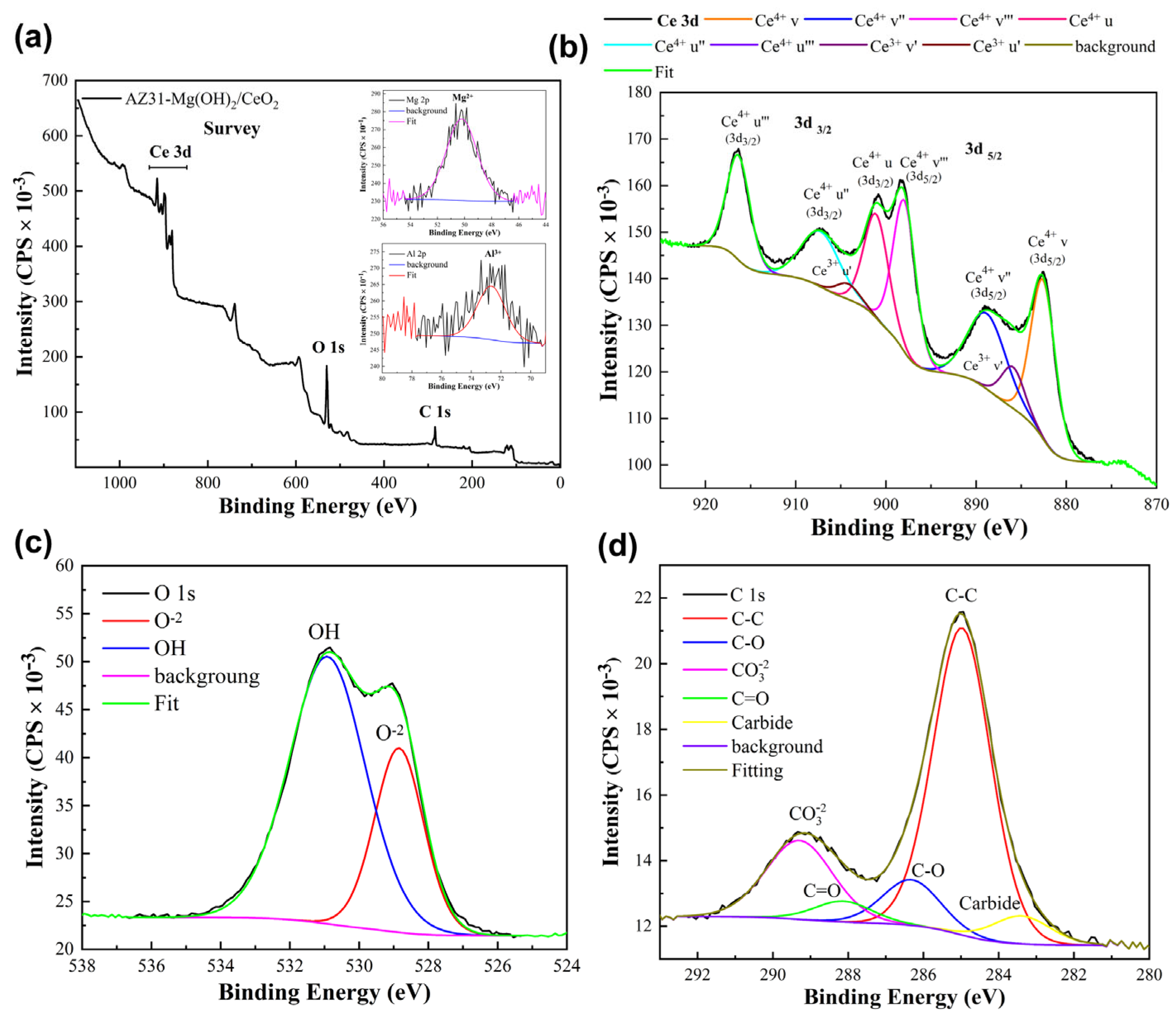
X-Ray Photoelectron Spectroscopy (XPS) Analysis

Figure 1 shows the XPS spectra of AZ31-Mg(OH)2/CeO2. The two most common oxidation states of Ce are Ce3+ and Ce4+, both of which were observed in the Ce 3d XPS spectrum. Figure 1a shows the general spectrum where the presence of O1s and C1s was detected, located at 531 and 285 eV, respectively. Additionally, Mg 2p and Al 2p were found, whose binding energies were 49.8 and 72.3 eV, respectively, associated with the Mg base substrate and the aluminum (Al) alloying element. Both Ce(III) and Ce(IV) appeared in the Ce 3d region (Figure 1b). For the Ce4+ peaks, the highest energy levels correspond to the u‴/v‴ doublet at ~916.40 and ~898.25 eV, which are assigned to the final state of Ce4+3d94f0 O2p6. The u″/v″ doublets at ~907.40 and ~888.95 eV are attributed to the hybridization of the Ce4+3d94f1 O2p5 state, and the u/v doublet region at ~900.98 and ~882.58 eV corresponds to the Ce4+3d94f2 O2p4 state [26]. The Ce(IV) content was calculated using the method proposed by Shyu et al. [27]. According to this method, in the case of pure Ce(IV) species, the contribution of the u‴ satellite (see the spectrum) was approximately 14% of the total Ce 3d peak area. Therefore, the percentage of Ce(IV) species can be calculated as
Ce(IV)% = u‴ % /14 × 100%
where u‴ is the percentage of the peak area of the satellite u‴ with respect to the total area of Ce 3d. With this method, the calculation error is < 10%. Based on the calculation, the sample contained 93.36% Ce4+ and 6.64% Ce3+.
The XPS spectra of the CeO2 coating on AZ31OH show the corresponding Ce 3d and O 1s core level spectra. According to the notation introduced by Borroughs et al. [28], for a deconvolution region, there are four spin-orbit doublets (3d5/2 and 3d3/2), which are labeled v–v‴ and u–u‴. For Ce4+, there are u/v, u″/v″, and u‴/v″, and for Ce3+, there is u′/v′. In the XPS spectrum of O 1 s (Figure 1c), the satellite peak is broad and asymmetric, which could indicate the presence of more than one OCe-O chemical state, as well as OH.

3.2. Roughness

Atomic Force Microscopy

The surface of the AZ31-Mg(OH)2/CeO2 sample was evaluated with atomic force microscopy. Figure 2a–c show images of surface roughness for the AZ31-Mg, AZ31-Mg(OH)2, and AZ31-Mg(OH)2/CeO2 samples, respectively. Figure 2a–b show the topography of the AZ31 surface; it had a root mean square roughness (Rq) of 132.9 nm. The anodized AZ31-Mg(OH)2 sample had a roughness of 128.3 nm, indicating a small decrease with respect to the bare sample as a consequence of the passive layer of oxides/hydroxides formed on the surface, which is attributed to the sanding lines. On the other hand, the AZ31-Mg(OH)2/CeO2 sample (Figure 2c) had an Rq of 7.54 μm. This change is probably due to the accelerated growth of the CeO2 coating due to the immediate precipitation of Ce2+ ions in the synthesis process, as observed by scanning electron microscopy in our previous work [29]. Figure 2c(i,ii) show the growth of the coating which follows the sanding lines of the substrate surface, as can be seen in the surface roughness in Figure 2c(iii). Microcracks are created as a consequence of drying, and there are also non-adherent areas of the CeO2 coating. These non-adherent zones could create defects that facilitate the access of aggressive ions. However, due to its self-healing and inhibitory characteristics, CeO2 is easily changed to Ce2O3, which dissolves in the corrosive medium and produces Ce3+. These ions migrate to the substrate surface and become Ce4+, forming a film that increases the corrosion resistance of the AZ31 substrate [30]. This is consistent with the results found in XPS, indicating that the formed film is mainly composed of Ce4+ with a 93.36% purity. The mechanism has been proposed by Gobara M. et al., in which the Ce3+ oxidation state reacts to form a hydroxide layer of Ce3+. Therefore, by oxygen reduction, H2O2 is formed to promote Ce4+ precipitation at cathodic sites, and more Ce4+ ions form a thicker layer of cerium that protects the metal surface [31].

3.3. Electrochemical Results

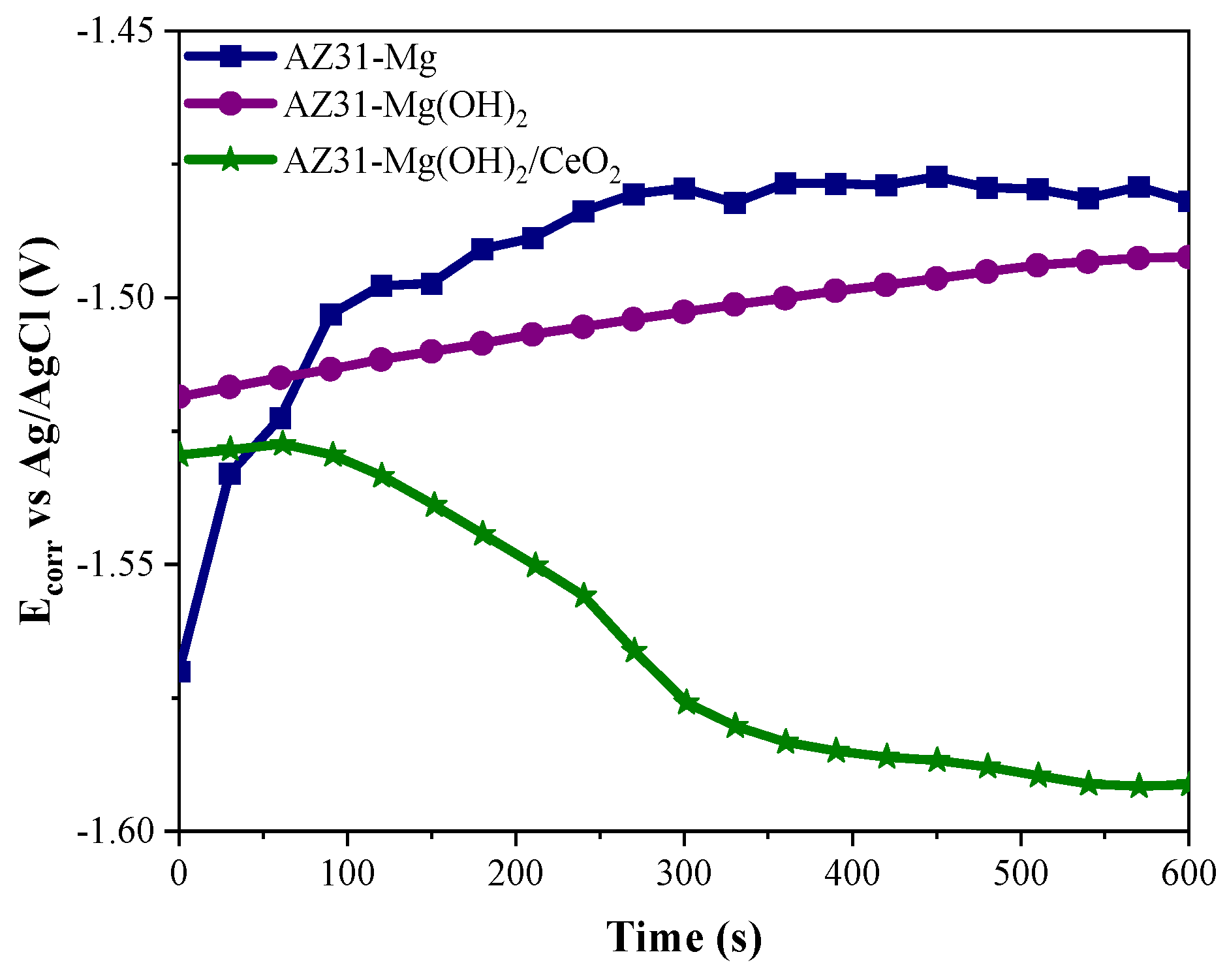
3.3.1. Open Circuit Potential (OCP)
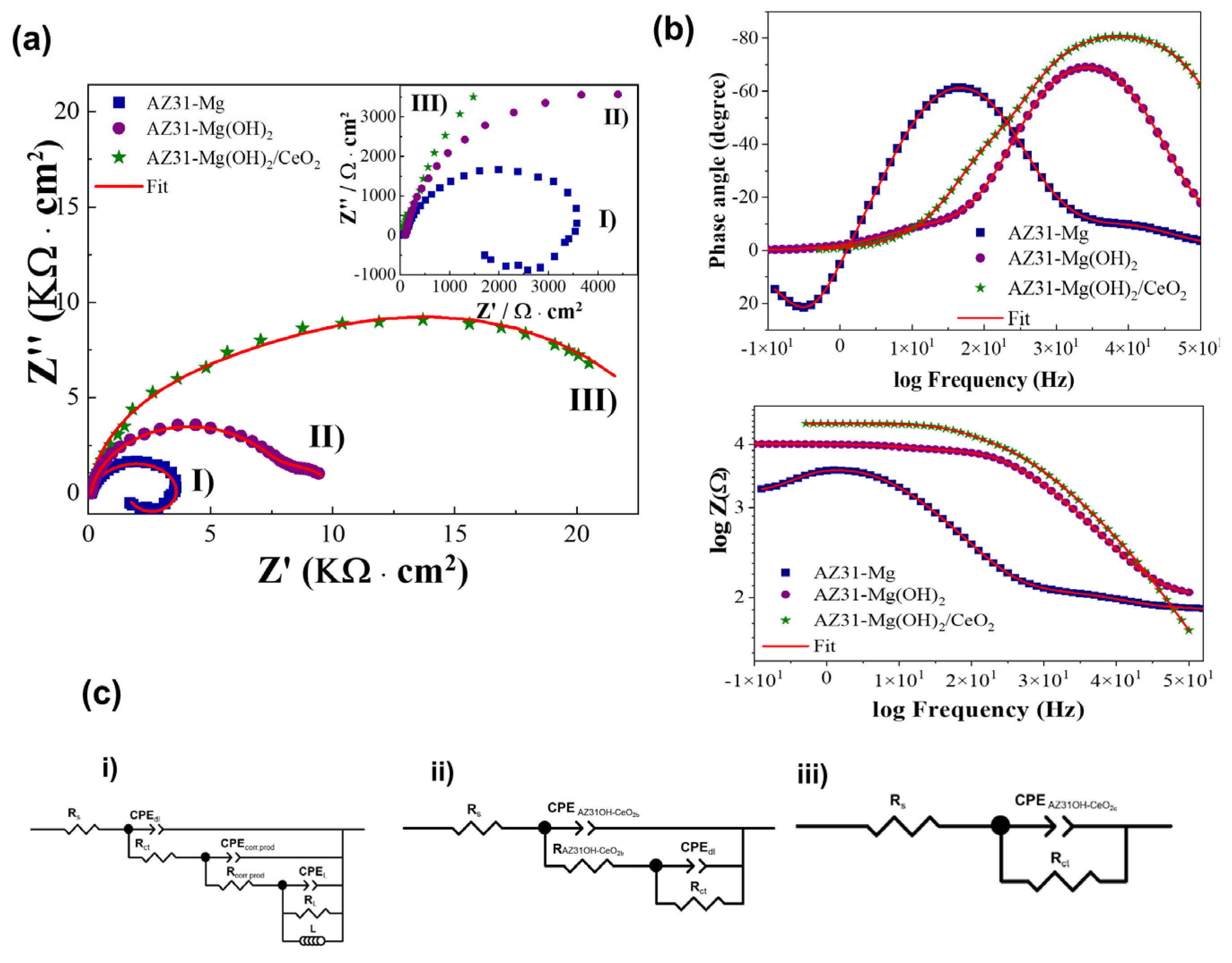
All electrochemical tests were performed using HBSS. The Ecorr was monitored for 10 min (Figure 3).
Based on OCP, the Ecorr ranged from -1.45 to 1.60 V. However, the bare AZ31 sample showed the most positive and stable Ecorr after evaluation for 10 min (−1.47 V). Nevertheless, this sample initially displayed the most negative Ecorr (−1.57 V), characteristic of Mg-based alloys, due to its high surface reactivity [32,33]. This behavior can be justified considering that initially, the surface of the bare AZ31 sample is highly active, and when it comes into contact with an electrolyte (in this study, HBSS), it immediately begins to oxidize, forming corrosion products that temporarily stabilize the surface of the bare AZ31 and shifting the Ecorr to become more positive [34]. Nevertheless, the corrosion products that commonly form as a result of the accelerated oxidation of a bare substrate are a combination of Mg oxy-hydroxides, which are unstable and porous [35]. Therefore, this oxide film has poor adherence to the substrate. Hence, electrolyte and aggressive ions move in through the pores, allowing oxidation–reduction reactions to re-establish, and the oxide film detaches from the substrate. This behavior could occur cyclically.
On the other hand, the AZ31-Mg(OH)2 sample displayed a slightly more active Ecorr than the bare AZ31-Mg sample. However, its behavior was more stable as a function of time because the potential difference during the OCP was approximately 25 mV. This behavior is principally due to anodization, which generates a homogeneous, porous, and passive Mg(OH)2 film that stabilizes the AZ31 surface [36], allowing the application of the Ce base film.
At the beginning of the OCP test, the AZ31-Mg(OH)2/CeO2 sample showed an Ecorr of approximately 1.53 V; subsequently, it decreased to -1.59 V, the most negative Ecorr of the evaluated samples. It is important to mention that the CeO2 coating has been reported as a cathodic corrosion inhibitor [37]. Therefore, the presence of CeO2 shifted the potential to more active (negative) values compared with AZ31-Mg and the anodized sample AZ31-Mg(OH)2, at least 100 and 90mV, respectively.

3.3.2. EIS

The EIS results for the bare AZ31-Mg, AZ31-Mg(OH)2, and AZ31-Mg(OH)2-CeO2 samples are shown in Figure 4. At low frequencies, the Nyquist and Bode plots corresponding to the AZ31-Mg sample display an inductive loop characteristic of Mg-based alloys, (Figure 4a). This behavior is associated with the high surface reactivity of the substrate [38]. It has been reported that the most common corrosion mechanism is based on a strong dissolution of AZ31 due to the oxidation reaction. Because of the reduction reaction, there is an intense evolution of hydrogen [39,40,41,42]. The presence of this inductive loop at a low frequency has also been associated with pitting corrosion and has been related to the adsorption/desorption on the surface electrode of ions such as Cl or intermediate ions [43,44], as well as accelerated anodic dissolution [45,46]. In this case, the presence of Cl (≈8.5 g/L) in HBSS favored the pitting of AZ31-Mg. Additionally, a greater concentration of Cl could increase their adsorption/desorption on the surface of the bare substrate, resulting in the accelerated dissolution of AZ31-Mg and, therefore, a total impedance of approximately 1.2 KΩ·cm−2.
For the AZ31-Mg(OH)2 sample, there are two semicircles (time constants) in the Nyquist plot. One is well defined at high frequencies, which is associated with the passive film obtained by anodizing (AZ31Mg(OH)2), showing a total impedance of 8 KΩ/cm−2, which is higher than the impedance for the AZ31-Mg sample, and increasing its corrosion resistance by at least 6 KΩ·cm2 (Figure 4b). Even so, the film that forms on the surface of the bare substrate, composed mainly of Mg(OH)2, is porous. This allows aggressive ions and water to pass through the pores and reach the substrate, allowing the oxidation–reduction reactions to occur and thus increasing the total impedance of the system, albeit only temporarily. Consequently, the impedance signal does not cross the real axis (Z’), forming a new semicircle that is not well defined and appears at low frequencies; it is associated with double-layer electrochemical resistance (charge transfer resistance), showing a total impedance of approximately 10 KΩ·cm−2. However, by eliminating the impedance associated with the anodizing film and considering only the response associated with the formation of the electrochemical double layer and/or the resistance to charge transfer, the total impedance is reduced by approximately 2 KΩ·cm−2.
The Nyquist plot for the AZ31-Mg(OH)2/CeO2 sample shows a single semicircle (a single time constant) with a phase angle close to −80° and n = 0.94 according to the simulation results (Figure 4b). This response is associated with the presence of the cerium-sealed Mg(OH)2 film (CeO2), which gives AZ31 corrosion resistance and a higher total impedance of around 30 KΩ·cm−2. In other words, it increases the corrosion resistance of bare Mg more than 20-fold. This behavior is associated with the presence of the CeO2 film, which seals the anodized pores, stabilizing the surface and decreasing the susceptibility of bare AZ31 to pitting corrosion, as has been reported in the literature [47,48,49].
Finally, to corroborate the corrosion behavior of the samples evaluated in HBSS, the Nyquist and Bode plots were analyzed using EC-Lab software (version V11.36) and fitted using an equivalent circuit (Figure 4c(i–iii)), and the resistance of the corrosion environment Rs (electrolyte), the resistance of the passive film (RP-F), the resistance of the cerium coating (RCeC), and the resistance of charge transfer (RCT) were estimated. Additionally, AZ31 was thought to exhibit inductance (L) associated with the redox reaction owing to the high reactivity of Mg. The constant phase element (CPE) was used instead of a capacitive element (ΥCPE (ω) = 1/ZCPE = Q(ϳω)n), where ω is the angular frequency, Q is the modulus, and n is the phase deviation from the ideal electrical component behavior that provides information about surface inhomogeneity. Figure 4c(i) corresponds to the equivalent circuit for sample AZ31-Mg. Figure 4c(ii) corresponds to the behavior of sample AZ31-Mg(OH)2. Finally, Figure 4c(iii) shows the electrical behavior of sample AZ31-Mg(OH)2-CeO2. Table 3 lists the fitting parameters.

3.3.3. Polarization Curves

The polarization curves were generated by applying a potential scan from −100 to +700 mV at a scan rate of 0.5 mV/s. The AZ31-Mg(OH)2/CeO2 polarization curve revealed that the layer of Mg(OH)2 sealed with CeO2 formed on the AZ31 surface strongly increased corrosion resistance. Note that CeO2 shifted the corrosion potential in the cathodic direction. This behavior is associated with the cathodic character of the CeO2 coating as a corrosion inhibitor [50]. Nevertheless, the key contribution of the CeO2 coating was reflected in a greater amplitude of the passive zone (ΔEpass) and a displacement of the pitting potential (Epitt) toward more positive values, as presented in Figure 5a, which shows a summary of the behavior of samples treated with CeO2.
The increase in the passive zone of the CeO2 coating can be attributed to its self-healing mechanism [30]. In this mechanism, CeO2 is reduced to Ce2O3 by releasing Ce3+. Subsequently, these ions diffuse through the Mg(OH)2 layer to the AZ31 substrate, whose surface allows the oxidation of Ce3+ to Ce4+, which is attributed to the passive layer (Mg(OH)2/CeO2) formed on the substrate [51]. Clearly, AZ31-Mg(OH)2/CeO2 exhibited the most positive pitting potential (−1.1 V) as well as a larger passivation zone (0.35 V) compared with the anodized sample (0.20 V) and the reference (0.06 V).
Finally, the larger passivation zone is associated with the presence of more stable cerium oxides, such as CeO2, whereas a noble or positive pitting potential indicates that the passive film is more resistant to pitting corrosion [52]. The polarization curves are in good agreement with the EIS and OCP results. Figure 5b shows photographs of the different surfaces after being electrochemically evaluated. It can be observed that the bare sample presents pits with an average diameter of 7.33 mm. The AZ31-Mg(OH)2 sample presents pits with a diameter of 4.53 mm, while the sample with a cerium oxide coating shows a pit size of 1.70 mm. These results suggest improved corrosion resistance using a CeO2 coating that minimizes pitting damage to the sample surface. As observed in Table 4, the current density and resistance values of the coating are in the order of magnitude of other reported materials/systems.

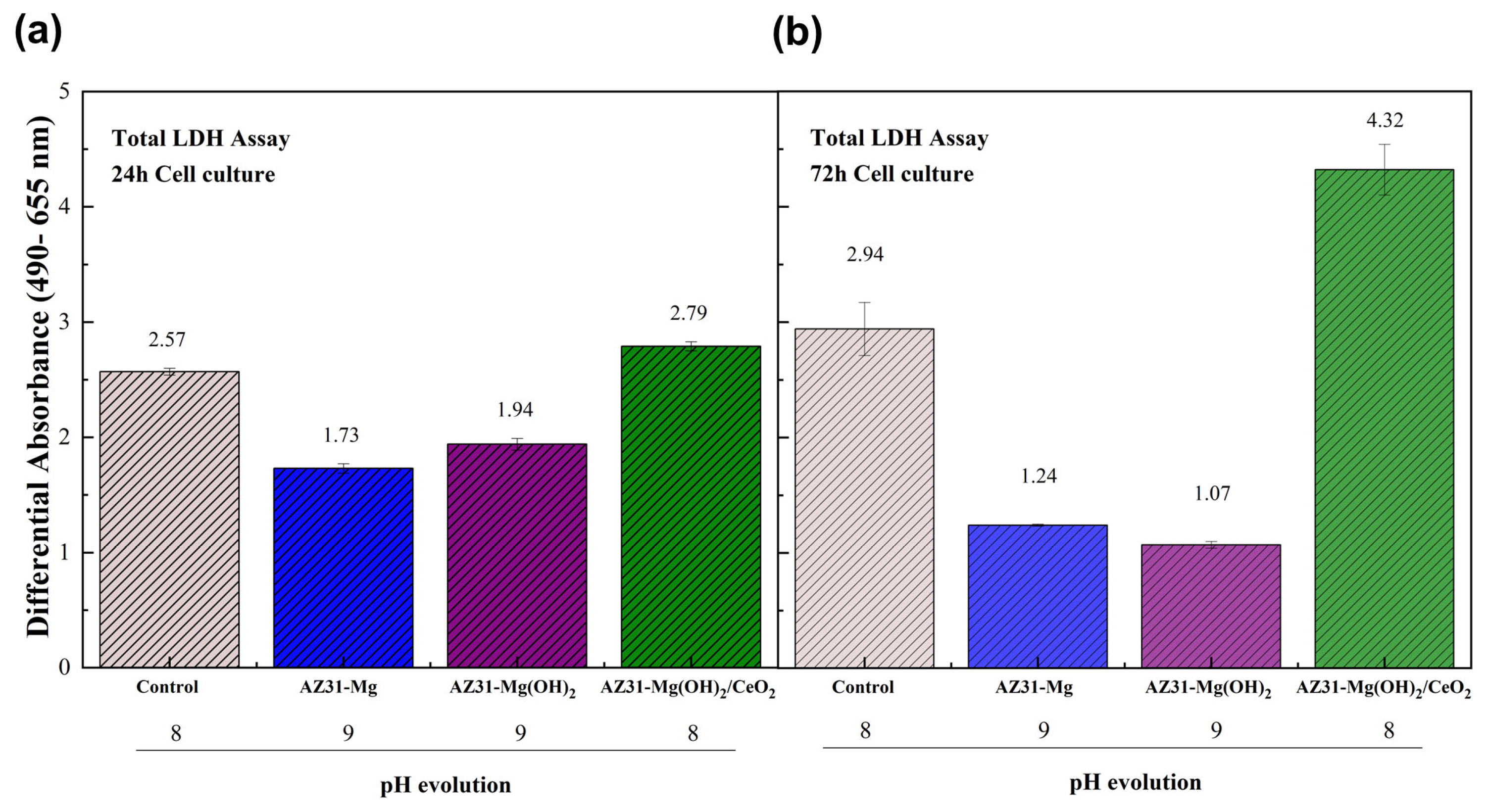
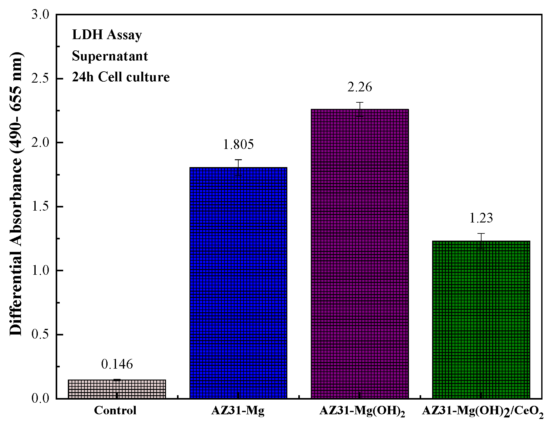
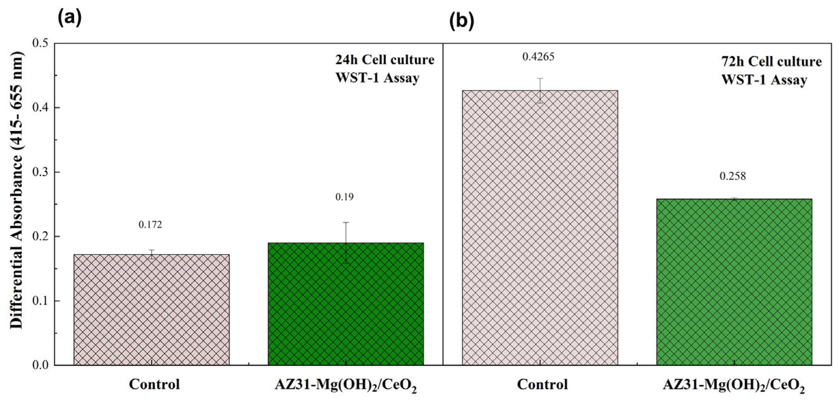
3.4. Biocompatibility

MC3T3-E1 cells were grown on AZ31, AZ31-Mg(OH)2, and AZ31-Mg(OH)2/CeO2 discs, and their viability and morphology were compared.
Cell viability was evaluated by staining the cultured cells with calcein-AM and Hoechst (Figure 6), which stain live and dead cells, respectively. Calcein-AM dye stains intact and viable cells as intracellular esterases remove the ester groups from the dye molecule and convert them into a green fluorescent compound. Hoechst is a fluorescent dye that labels DNA and is used to stain the nuclei of dead cells, given that its ability to permeate live cells is very low [57]. Live and dead cells were evaluated after incubation for 24 and 72 h. MC3T3-E1 cells cultured in the absence of any material were used as a control. The control MC3T3-E1 cells showed almost no dead cells (Hoechst-stained cells, which appear blue). The control cells showed the characteristic morphology of pre-osteoblasts, with an elongated shape and filopodia, which are typical extensions of the cytoplasm (Figure 6a,e) [58]. Exposure of the MC3T3-E1 cells to AZ31 killed all cells (Figure 6b,f). The few cells that were on the metal surface appeared to form clusters of dead cells. This is probably due to the high reactivity of Mg with the cell culture medium, which gives rise to degradation products such as CaCO3, MgCl2, Mg(CO3)2, and Mg3(PO4)2 and the release of H2 gas, corrosion products that affect cell viability due to pH alkalinization and corrosion [59,60]. The anodization of AZ31 (AZ31-Mg(OH)2) produced a clear change in the cell distribution on the metal surface (Figure 6c,g). The MC3T3-E1 cells cultured on this surface exhibited a more uniform and dispersed distribution on the metal surface compared with AZ31. This behavior on anodized metal suggests an improvement with surface passivation, probably due to the enhancement of the hydrophilic character that may affect cell adhesion to the metal surface [61]. However, only dead cells were detected on AZ31-Mg(OH)2, indicating the lack of biocompatibility of this material. There were markedly different results for MC3T3-E1 cells cultured on AZ31-Mg(OH)2/CeO2. Almost all of the cells appeared to be living, denoted by the high green fluorescence, and there were only a few dead cells (Figure 6d,h). Overall, the MC3T3-E1 cells cultured on AZ31-Mg(OH)2/CeO2 were similar to the control cells, confirming the good biocompatibility of this surface.
Total LDH activity was determined as an indirect measurement of the number of cells to evaluate the effect of the different surfaces on MC3T3-E1 proliferation. Of note, proliferation is estimated by an experimental procedure that does not use trypsin to remove cells from materials, as some cells change their adhesion properties upon material interaction, as has been reported for Mg [62]. To avoid the effect of metallic material on cell adhesion, total LDH, which is from live and dead cells, was measured as an estimate of the total number of cells after culture in the presence of the different materials (Figure 7). After incubation for 24 and 72 h, MC3T3-E1 cells grown on AZ31-Mg and AZ31-Mg(OH)2 showed lower total LDH values compared with the control cells, while MC3T3-E1 cells grown on AZ31-Mg(OH)2/CeO2 showed a higher total LDH value. Thus, the CeO2 film on anodized AZ31-Mg favors cell proliferation, a finding consistent with the biocompatibility of this material. According to the scanning electron microscopy/energy-dispersive X-ray spectroscopy results from our previous work [29], the CeO2 coating shields the AZ31-Mg(OH)2 surface, so Mg corrosion products do not adversely affect the cultured cells. In addition, the pH of this material is maintained at around 8 (similar to the control well, in the absence of any material) for the entire culture period, which is beneficial and critical for cell growth. The LDH values were similar for the control cells and the cells grown on AZ31-Mg(OH)2/CeO2 at 24 h, although there was a higher value for the cells grown on AZ31-Mg(OH)2/CeO2 for 72 h. Given that the density (cells/cm2) was similar, but the surface area for the AZ31-Mg(OH)2/CeO2 sample was different, the correction factor necessary to normalize the data for surface area could be responsible for a certain overestimation of the total LDH content. Hence, there may not have been a difference in the proliferation of the control cells and the cells grown on AZ31-Mg(OH)2/CeO2. The differences in cell proliferation of the CeO2 sample with respect to AZ31 and AZ31-Mg(OH)2 were larger at the longer incubation time. When cells are exposed to corrosion products of AZ31 and AZ31-Mg(OH)2 for a longer time, there are more pronounced, deleterious effects on cell proliferation, as denoted by a decrease in total LDH for the 72 h culture compared with the 24 h culture. The AZ31-Mg(OH)2/CeO2 sample did not show this time-dependent behavior, as cell proliferation was comparable to the control (the absence of material), and the pH did not change during the assay, further confirming the good biocompatibility between pre-osteoblasts and AZ31-Mg(OH)2/CeO2.
The toxicity of the materials to MC3T3-E1 cells was assessed by measuring LDH in the supernatants. LDH is a stable cytoplasmic enzyme present in all cells and rapidly released into the cell culture supernatant when the plasma membrane is damaged, which is a sign of cell death [63]. Figure 8 shows LDH release after incubation for 24 h. In the presence of AZ31-Mg and AZ31-Mg(OH)2, there was greater LDH release compared with the control cells and the cells grown on AZ31-Mg(OH)2/CeO2. These data indicate that AZ31-Mg and AZ31-Mg(OH)2 damage the MC3T3-E1 cell membranes. This damage could be attributed to oxidative stress due to the high reactivity of AZ31-Mg and AZ31-Mg(OH)2 and to the alkalinization of the medium due to the presence of these materials. The decreased LDH release from MC3T3-E1 cells cultured on AZ31-Mg(OH)2/CeO2 demonstrates the beneficial effect of these surface modifications in decreasing toxicity and reactivity and preventing the alkalinization of the culture medium.
Finally, to assess the biocompatibility of AZ31-Mg(OH)2/CeO2, the number of metabolically active cells was determined with the WST-1 assay, where the amount of formazan dye is related to cell proliferation [64]. Figure 9 shows the mitochondrial activity of control MC3T3-E1 cells and MC3T3-E1 cells grown on AZ31-Mg(OH)2/CeO2 discs for 24 and 72 h. After 24 h, the mitochondrial activity of the control cells and cells grown on AZ31-Mg(OH)2/CeO2 was comparable. After 72 h, however, it was lower for the cells grown on AZ31-Mg(OH)2/CeO2 compared with the control cells.
This finding was expected because of the interaction with a material that usually impairs cell growth. It is remarkable that mitochondrial activity was not enhanced in the cells grown on AZ31-Mg(OH)2/CeO2; the lack of an increase indicates that this surface does not induce oxidative stress. Many studies reported cytotoxic effects under certain conditions, leading to cell damage [65,66]. Due to the crystal structure, oxygen vacancy defects in CeO2, and the oxidation states determined in the XPS results, cerium’s mechanism of action is hypothesized to involve its ability to mimic superoxide dismutase and catalase activity, as reported by various authors [67,68]. Cerium exists in two oxidation states, attributed to its 4f1 5d1 6s2 electron configuration, with an unpaired electron in the 4f orbital. The Ce4+ ions act as electron acceptors, while the Ce3+ ions act as electron donors. Ce3+ is converted to Ce4+ and vice versa, resulting in a change in vacancies of oxygen. Then, superoxide is converted to H2O2. Finally, Ce4+ is reduced to Ce3+ through catalase mimetic activity (H2O2 is converted to O2 + 4H+) [69]. Therefore, in the presence of ROS, they can capture free electrons through a redox reaction, in which the Ce3+ ions donate electrons to the ROS, effectively neutralizing them [70].

4. Conclusions

The electrochemical results confirmed that the surface activity of the bare AZ31-Mg sample is reduced by depositing a layer of Mg(OH)2 by anodization, and it also promotes the formation of a CeO2 coating. The XPS analysis of the AZ31-Mg(OH)2/CeO2 sample confirmed that the cerium is mostly present as Ce4+; according to the literature, this valence state produces the least damage to cells. Additionally, AZ31-Mg(OH)2/CeO2 showed good biocompatibility with MC3T3-E1 pre-osteoblasts after incubation for 24 and 72 h. The cerium coating that appears on the surface of AZ31-Mg(OH)2 seems to act as a barrier, as it is the first coating that interacts with the surrounding medium, which is aggressive for the material. Cerium can mimic the activity of superoxide dismutase and catalase, reducing reactive oxygen species based on its structural configuration (Ce3+/Ce4+). In this way, it could contribute to improving the reactive environment in which the cerium-modified anodized AZ31-Mg sample is found, decreasing reactive oxygen species and thus increasing osteoblast biocompatibility, which is required for bone repair applications.

Author Contributions

Conceptualization, E.O.-B. and S.E.B.-S.; methodology, E.O.-B., S.E.B.-S. and R.M.L.; investigation, E.O.-B. and A.C.E.-F.; data curation, E.O.-B. and R.M.L.; XPS analysis, M.L.E.; Resources, E.O.-B. and R.M.L.; Writing—original draft preparation, E.O.-B.; Writing—review and editing, R.M.L. and S.E.B.-S. All authors have read and agreed to the published version of the manuscript.

Funding

This research was supported by Secretaría de Ciencia, Humanindades, Tecnología e Innovación (SECIHTI) Science grant, (SIP project 20253511 and 20250589) through Instituto Politécnico Nacional. Financial support was also obtained from ICOOP 2021 (Ref COOPA20479) of Consejo Superior de Investigaciones Científicas (CSIC, Spain) and by RTI2018-101506-B-C33 from the Ministerio de Ciencia, Innovación y Universidades (MICIU/FEDER) in Spain.

Data Availability Statement

Data is contained within the article.

Acknowledgments

Instituto Politécnico Nacional; Secretaría de Ciencia, Humanidades, Tecnología e Innovación (SECIHTI); Consejo Superior de Investigaciones Científicas (CSIC) and the Spanish Ministry of Science, Innovation and Universities is gratefully acknowledged.

Conflicts of Interest

The authors declare no conflicts of interest.

References

  1. Seelig, M.G. A study of magnesium wire as an absorbable suture and ligature material. Arch. Surg. 1924, 8, 669–680. [Google Scholar] [CrossRef]
  2. Zheng, Y.F.; Gu, X.N.; Witte, F. Biodegradable metals. Mater. Sci. Eng. R Rep. 2014, 77, 1–34. [Google Scholar] [CrossRef]
  3. Badkoobeh, F.; Mostaan, H.; Rafiei, M.; Bakhsheshi-Rad, H.R.; RamaKrishna, S.; Chen, X. Additive manufacturing of biodegradable magnesium-based materials: Design strategies, properties, and biomedical applications. J. Magnes. Alloys 2023, 11, 801–839. [Google Scholar] [CrossRef]
  4. Thanigaivel, S.; Priya, A.K.; Balakrishnan, D.; Dutta, K.; Rajendran, S.; Soto-Moscoso, M. Insight on recent development in metallic biomaterials: Strategies involving synthesis, types and surface modification for advanced therapeutic and biomedical applications. Biochem. Eng. J. 2022, 187, 108522. [Google Scholar] [CrossRef]
  5. Ding, Y.; Wen, C.; Hodgson, P.; Li, Y. Effects of alloying elements on the corrosion behavior and biocompatibility of biodegradable magnesium alloys: A review. J. Mater. Chem. B 2014, 2, 1912–1933. [Google Scholar] [CrossRef]
  6. Wu, G.; Ibrahim, J.M.; Chu, P.K. Surface design of biodegradable magnesium alloys—A review. Surf. Coat. Technol. 2013, 233, 2–12. [Google Scholar] [CrossRef]
  7. Yamagami, R.; Sieg, J.P.; Bevilacqua, P.C. Functional Roles of Chelated Magnesium Ions in RNA Folding and Function. Biochemistry 2021, 60, 2374–2386. [Google Scholar] [CrossRef]
  8. Gray-Munro, J.E.; Seguin, C.; Strong, M. Influence of surface modification on the in vitro corrosion rate of magnesium alloy AZ31. J. Biomed. Mater. Res. Part A 2009, 91 Pt A, 221–230. [Google Scholar] [CrossRef]
  9. Pierson, D.; Edick, J.; Tauscher, A.; Pokorney, E.; Bowen, P.; Gelbaugh, J.; Stinson, J.; Getty, H.; Lee, C.H.; Drelich, J.; et al. A simplified in vivo approach for evaluating the bioabsorbable behavior of candidate stent materials. J. Biomed. Mater. Res. Part B Appl. Biomater. 2012, 100 Pt B, 58–67. [Google Scholar] [CrossRef]
  10. Abraham, A.M.; Subramani, V. Effect of Magnesium as Biomaterial in Biodegrdation. Mater. Today Proc. 2023. [Google Scholar] [CrossRef]
  11. Bowen, P.K.; Drelich, J.; Buxbaum, R.E.; Rajachar, R.M.; Goldman, J. New approaches in evaluating metallic candidates for bioabsorbable stents. Emerg. Mater. Res. 2012, 1, 237–255. [Google Scholar] [CrossRef]
  12. Liu, F.; Chen, C.; Niu, J.; Pei, J.; Zhang, H.; Huang, H.; Yuan, G. The processing of Mg alloy micro-tubes for biodegradable vascular stents. Mater. Sci. Eng. C 2015, 48, 400–407. [Google Scholar] [CrossRef] [PubMed]
  13. Lu, Y.; Deshmukh, S.; Jones, I.; Chiu, Y.-L. Biodegradable magnesium alloys for orthopaedic applications. BMT 2021, 2, 214–235. [Google Scholar] [CrossRef]
  14. Zhou, W.; Xue, F.; Li, M. Corrosion Behavior of Al-Mg Alloys with Different Alloying Element Contents in 3.5% NaCl Solution. Metals 2025, 15, 327. [Google Scholar] [CrossRef]
  15. Venkateswarlu, B.; Sunil, B.R.; Kumar, R.S. Microstructure, mechanical properties and corrosion behavior of Rare Earths (RE) containing Mg-Zn alloy for biomedical applications. Mater. Today Proc. 2023, 102, 6–10. [Google Scholar] [CrossRef]
  16. Kim, J.; Pan, H. Effects of magnesium alloy corrosion on biological response—Perspectives of metal-cell interaction. Prog. Mater. Sci. 2023, 133, 101039. [Google Scholar] [CrossRef]
  17. Kumar, S.; Katyal, P.; Chaudhary, R.N.; Singh, V. Assessment of factors influencing bio-corrosion of magnesium based alloy implants: A review. Mater. Today Proc. 2022, 56, 2680–2689. [Google Scholar] [CrossRef]
  18. Gonzalez, J.; Hou, R.Q.; Nidadavolu, E.P.; Willumeit-Römer, R.; Feyerabend, F. Magnesium degradation under physiological conditions—Best practice. Bioact. Mater. 2018, 3, 174–185. [Google Scholar] [CrossRef]
  19. Púa, L.D.C.G.; Montenegro, J.C.R.; Reyes, A.M.F.; Rodríguez, H.Z.; Méndez, V.N.P. Biomaterials for orthopedic applications and techniques to improve corrosion resistance and mechanical properties for magnesium alloy: A review. J. Mater. Sci. 2023, 58, 3879–3908. [Google Scholar] [CrossRef]
  20. Wei, F.; Zhang, W.; Zhang, T.; Wang, F. Effect of variations of Al content on microstructure and corrosion resistance of PEO coatings on Mg Al alloys. J. Alloys Compd. 2017, 690, 195–205. [Google Scholar] [CrossRef]
  21. Jahan, U.M.; Blevins, B.; Minko, S.; Reukov, V.V. Advancing biomedical applications: Antioxidant and biocompatible cerium oxide nanoparticle-integrated poly-ε-caprolactone fibers. Biomed. Phys. Eng. Express 2025, 11, 015020. [Google Scholar] [CrossRef] [PubMed]
  22. Nosrati, H.; Heydari, M.; Khodaei, M. Cerium oxide nanoparticles: Synthesis methods and applications in wound healing. Mater. Today Bio 2023, 23, 100823. [Google Scholar] [CrossRef]
  23. Chen, D.; Mei, D.; Li, Y.; Chen, L.; Wang, H.; Huang, W.; Wang, L.; Zhu, S.; Guan, S. Protective nature of cerium-based oxides coating against Mg corrosion in Hanks’ balanced salt solution. Corros. Sci. 2023, 219, 111255. [Google Scholar] [CrossRef]
  24. Petrova, V.A.; Poshina, D.N.; Golovkin, A.S.; Mishanin, A.I.; Zhuravskii, S.G.; Yukina, G.Y.; Naumenko, M.Y.; Sukhorukova, E.G.; Savin, N.A.; Erofeev, A.S.; et al. Electrospun Composites of Chitosan with Cerium Oxide Nanoparticles for Wound Healing Applications: Characterization and Biocompatibility Evaluation In Vitro and In Vivo. Polymers 2024, 16, 1787. [Google Scholar] [CrossRef]
  25. Fagali, N.S.; Madrid, M.A.; Maceda, B.T.P.; Fernández, M.E.L.; Puerto, R.M.L.; de Mele, M.F.L. Effect of degradation products of iron-bioresorbable implants on the physiological behavior of macrophages in vitro. Metallomics 2020, 12, 1841–1850. [Google Scholar] [CrossRef] [PubMed]
  26. Inoue, T.; Nakata, Y.; Shida, S. Electron beam induced orientation selective epitaxial growth of CeO2(100) layers on Si(100) substrates by dc reactive sputtering. J. Phys. Conf. Ser. 2008, 100, 082014. [Google Scholar] [CrossRef]
  27. Shyu, J.Z.; Otto, K.; Watkins, W.L.H.; Graham, G.W.; Belitz, R.K.; Gandhi, H.S. Characterization of Pd/γ-alumina catalysts containing ceria. J. Catal. 1988, 114, 23–33. [Google Scholar] [CrossRef]
  28. Allahgholi, A.; Flege, J.I.; Thieß, S.; Drube, W.; Falta, J. Oxidation-State Analysis of Ceria by X-Ray Photoelectron Spectroscopy. Available online: www.chemphyschem.org (accessed on 15 November 2024).
  29. Benito-Santiago, S.E.; Onofre-Bustamante, E.; Lozano-Puerto, R.M. Synthesis and Characterisation of CeO2 Coatings on the AZ31 Alloy for Corrosion Protection and In Vitro Biocompatibility of MC3T3-E1 Pre-Osteoblasts. Metals 2023, 13, 653. [Google Scholar] [CrossRef]
  30. Anjum, M.J.; Zhao, J.; Ali, H.; Tabish, M.; Murtaza, H.; Yasin, G.; Malik, M.U.; Khan, W.Q. A Review on Self-Healing Coatings Applied to Mg Alloys and Their Electrochemical Evaluation Techniques. Int. J. Electrochem. Sci. 2020, 15, 3040–3053. [Google Scholar] [CrossRef]
  31. Gobara, M.; Baraka, A.; Akid, R.; Zorainy, M. Corrosion protection mechanism of Ce4+/organic inhibitor for AA2024 in 3.5% NaCl. RSC Adv. 2020, 10, 2227–2240. [Google Scholar] [CrossRef]
  32. Yu, J.; Lv, Y.; Duan, T.; Meng, F.; Cheng, Q.; Xu, Y.; Zhou, K.; Xue, L.; Leng, Z. Discharge and Corrosion Behaviors of Mg-Li and Mg-Li-La Alloys as Anodes for Seawater Battery. Int. J. Electrochem. Sci. 2020, 15, 10922–10935. [Google Scholar] [CrossRef]
  33. Singh, I.B.; Singh, M.; Das, S. A comparative corrosion behavior of Mg, AZ31 and AZ91 alloys in 3.5% NaCl solution. J. Magnes. Alloys 2015, 3, 142–148. [Google Scholar] [CrossRef]
  34. Kumar, R.; Katyal, P.; Gupta, M.; Singh, V. Electrochemical Corrosion Behaviour Analysis of Mg-Alloys Used for Orthopaedics and Vascular Implants. IOP Conf. Ser. Mater. Sci. Eng. 2022, 1225, 012063. [Google Scholar] [CrossRef]
  35. Luo, Y.; Sun, Y.; Lv, J.; Wang, X.; Li, J.; Wang, F. Transition of interface oxide layer from porous Mg(OH)2 to dense MgO induced by polyaniline and corrosion resistance of Mg alloy therefrom. Appl. Surf. Sci. 2015, 328, 247–254. [Google Scholar] [CrossRef]
  36. Luo, Y.; Wang, X.; Guo, W.; Rohwerder, M. Growth Behavior of Initial Product Layer Formed on Mg Alloy Surface Induced by Polyaniline. J. Electrochem. Soc. 2015, 162, C294–C301. [Google Scholar] [CrossRef]
  37. Brito-Franco, A.; Vazquez-Velez, E.; Lopez-Sesenes, R.; Larios-Gálvez, A.K.; Ramirez-Arteaga, A.M.; Gonzalez-Rodriguez, J.G. Use of CeO2 nanoparticles as CO2-corrosion inhibitors of a duplex stainless steel. Green Chem. Lett. Rev. 2024, 17, 2360497. [Google Scholar] [CrossRef]
  38. Feliu, S., Jr. Electrochemical Impedance Spectroscopy for the Measurement of the Corrosion Rate of Magnesium Alloys: Brief Review and Challenges. Metals 2020, 10, 775. [Google Scholar] [CrossRef]
  39. Song, G. Recent Progress in Corrosion and Protection of Magnesium Alloys. Adv. Eng. Mater. 2005, 7, 563–586. [Google Scholar] [CrossRef]
  40. Cao, F.; Song, G.-L.; Atrens, A. Corrosion and passivation of magnesium alloys. Corros. Sci. 2016, 111, 835–845. [Google Scholar] [CrossRef]
  41. Jamali, S.S.; Moulton, S.E.; Tallman, D.E.; Forsyth, M.; Weber, J.; Wallace, G.G. Evaluating the corrosion behaviour of Magnesium alloy in simulated biological fluid by using SECM to detect hydrogen evolution. Electrochimica Acta 2015, 152, 294–301. [Google Scholar] [CrossRef]
  42. Atrens, A.; Chen, X.; Shi, Z. Mg Corrosion—Recent Progress. Corros. Mater. Degrad. 2022, 3, 566–597. [Google Scholar] [CrossRef]
  43. Benbouzid, A.Z.; Gomes, M.P.; Costa, I.; Gharbi, O.; Pébère, N.; Rossi, J.L.; Tran, M.T.; Tribollet, B.; Turmine, M.; Vivier, V. A new look on the corrosion mechanism of magnesium: An EIS investigation at different pH. Corros. Sci. 2022, 205, 110463. [Google Scholar] [CrossRef]
  44. Gomes, M.P.; Costa, I.; Pébère, N.; Rossi, J.L.; Tribollet, B.; Vivier, V. On the corrosion mechanism of Mg investigated by electrochemical impedance spectroscopy. Electrochimica Acta 2019, 306, 61–70. [Google Scholar] [CrossRef]
  45. Cao, F.; Shi, Z.; Song, G.-L.; Liu, M.; Dargusch, M.S.; Atrens, A. Influence of casting porosity on the corrosion behaviour of Mg0.1Si. Corros. Sci. 2015, 94, 255–269. [Google Scholar] [CrossRef]
  46. Li, J.; Zhang, B.; Wei, Q.; Wang, N.; Hou, B. Electrochemical behavior of Mg-Al-Zn-In alloy as anode materials in 3.5 wt.% NaCl solution. Electrochim. Acta 2017, 238, 156–167. [Google Scholar] [CrossRef]
  47. Dong, K.; Song, Y.; Shan, D.; Han, E.-H. Formation mechanism of a self-sealing pore micro-arc oxidation film on AM60 magnesium alloy. Surf. Coat. Technol. 2015, 266, 188–196. [Google Scholar] [CrossRef]
  48. Jiang, D.; Zhou, H.; Wan, S.; Cai, G.-Y.; Dong, Z.-H. Fabrication of superhydrophobic coating on magnesium alloy with improved corrosion resistance by combining micro-arc oxidation and cyclic assembly. Surf. Coat. Technol. 2018, 339, 155–166. [Google Scholar] [CrossRef]
  49. Guo, L.; Zhang, F.; Song, L.; Zeng, R.-C.; Li, S.-Q.; Han, E.-H. Corrosion resistance of ceria/polymethyltrimethoxysilane modified magnesium hydroxide coating on AZ31 magnesium alloy. Surf. Coat. Technol. 2017, 328, 121–133. [Google Scholar] [CrossRef]
  50. Chaudry, U.M.; Farooq, A.; bin Tayyab, K.; Malik, A.; Kamran, M.; Kim, J.-G.; Li, C.; Hamad, K.; Jun, T.-S. Corrosion behavior of AZ31 magnesium alloy with calcium addition. Corros. Sci. 2022, 199, 110205. [Google Scholar] [CrossRef]
  51. Zhao, Y.; Zhang, Z.; Shi, L.; Zhang, F.; Li, S.; Zeng, R. Corrosion resistance of a self-healing multilayer film based on SiO2 and CeO2 nanoparticles layer-by-layer assembly on Mg alloys. Mater. Lett. 2019, 237, 14–18. [Google Scholar] [CrossRef]
  52. Poinern, G.E.J.; Brundavanam, S.; Fawcett, D. Biomedical Magnesium Alloys: A Review of Material Properties, Surface Modifications and Potential as a Biodegradable Orthopaedic Implant. Am. J. Biomed. Eng. 2013, 2, 218–240. [Google Scholar] [CrossRef]
  53. Ouyang, Y.; Chen, Z.; Jiang, C.; Yang, W.; Chen, Y.; Yin, X.; Liu, Y. Design of the double-layer biocompatible coating on AZ31 magnesium alloy for highly effective corrosion resistance. Surf. Coat. Technol. 2021, 428, 127897. [Google Scholar] [CrossRef]
  54. Zhang, J.; Hou, S.; Zhang, M.; Zhang, S.; Li, W. Corrosion resistance and biocompatibility of silica coatings on AZ31 magnesium alloy via magnetron sputtering. Mater. Today Commun. 2024, 41, 110890. [Google Scholar] [CrossRef]
  55. Tiyyagura, H.R.; Puliyalil, H.; Filipič, G.; Kumar, K.C.; Pottathara, Y.B.; Rudolf, R.; Fuchs-Godec, R.; Mohan, M.K.; Cvelbar, U. Corrosion studies of plasma modified magnesium alloy in simulated body fluid (SBF) solutions. Surf. Coat. Technol. 2020, 385, 125434. [Google Scholar] [CrossRef]
  56. Wang, J.; Deng, S.; Meng, M.; Tu, W.; Ou, J. Improved corrosion resistance and biocompatibility of AZ31 alloy by acid pickling pretreatment and (H+) hydroxyapatite/chitosan composite coating. Surf. Coat. Technol. 2023, 454, 129157. [Google Scholar] [CrossRef]
  57. Carter, S.-S.D.; Barbe, L.; Tenje, M.; Mestres, G. Exploring microfluidics as a tool to evaluate the biological properties of a titanium alloy under dynamic conditions. Biomater. Sci. 2020, 8, 6309–6321. [Google Scholar] [CrossRef]
  58. Sammons, R. Biological responses to hydroxyapatite. In Hydroxyapatite (Hap) for Biomedical Applications; Elsevier: Amsterdam, The Netherlands, 2015; pp. 53–83. [Google Scholar] [CrossRef]
  59. Peron, M.; Skaret, P.C.; Fabrizi, A.; Varone, A.; Montanari, R.; Roven, H.J.; Ferro, P.; Berto, F.; Torgersen, J. The effect of Equal Channel Angular Pressing on the stress corrosion cracking susceptibility of AZ31 alloy in simulated body fluid. J. Mech. Behav. Biomed. Mater. 2020, 106, 103724. [Google Scholar] [CrossRef]
  60. Gonzalez, J.; Lamaka, S.V.; Mei, D.; Scharnagl, N.; Feyerabend, F.; Zheludkevich, M.L.; Willumeit-Römer, R. Mg Biodegradation Mechanism Deduced from the Local Surface Environment under Simulated Physiological Conditions. Adv. Healthc. Mater. 2021, 10, 2100053. [Google Scholar] [CrossRef]
  61. Hsu, C.; Nazari, M.H.; Li, Q.; Shi, X. Enhancing degradation and corrosion resistance of AZ31 magnesium alloy through hydrophobic coating. Mater. Chem. Phys. 2019, 225, 426–432. [Google Scholar] [CrossRef]
  62. Alvarez, F.; Puerto, R.M.L.; Pérez-Maceda, B.; Grillo, C.A.; de Mele, M.F.L. Time-Lapse Evaluation of Interactions Between Biodegradable Mg Particles and Cells. Microsc. Microanal. 2016, 22, 1–12. [Google Scholar] [CrossRef]
  63. Kumar, P.; Nagarajan, A.; Uchil, P.D. Analysis of Cell Viability by the Lactate Dehydrogenase Assay. Cold Spring Harb. Protoc. 2018, 2018, 465. [Google Scholar] [CrossRef] [PubMed]
  64. Koyanagi, M.; Kawakabe, S.; Arimura, Y. A comparative study of colorimetric cell proliferation assays in immune cells. Cytotechnology 2016, 68, 1489–1498. [Google Scholar] [CrossRef] [PubMed]
  65. Soltani, M.; Soltani, M.; Karami-Mohajeri, S.; Mohadesi, A.; Ranjbar, M.; Oghabian, Z.; Mehrpour, O.; Khosravi, F. An interdisciplinary approach to assessing the toxicity reduction of cerium oxide nanoparticles coated with polyethylene glycol and polyvinylpyrrolidone polymers: An in vitro study. Toxicol. In Vitro 2025, 105, 106022. [Google Scholar] [CrossRef]
  66. Filippova, A.D.; Sozarukova, M.M.; Baranchikov, A.E.; Kottsov, S.Y.; Cherednichenko, K.A.; Ivanov, V.K. Peroxidase-like Activity of CeO2 Nanozymes: Particle Size and Chemical Environment Matter. Molecules 2023, 28, 3811. [Google Scholar] [CrossRef]
  67. Zhang, S.; Ruan, H.; Xin, Q.; Mu, X.; Wang, H.; Zhang, X.-D. Modulation of the biocatalytic activity and selectivity of CeO2 nanozymes via atomic doping engineering. Nanoscale 2023, 15, 4408–4419. [Google Scholar] [CrossRef]
  68. Mukherjee, A.; Ashrafi, A.M.; Bytesnikova, Z.; Svec, P.; Richtera, L.; Adam, V. An investigation on the multiple roles of CeO2 nanoparticle in electrochemical sensing: Biomimetic activity and electron acceptor. J. Electroanal. Chem. 2023, 935, 117301. [Google Scholar] [CrossRef]
  69. Rzigalinski, B.A.; Carfagna, C.S.; Ehrich, M. Cerium oxide nanoparticles in neuroprotection and considerations for efficacy and safety. WIREs Nanomed. Nanobiotechnol. 2017, 9, e1444. [Google Scholar] [CrossRef]
  70. Pota, G.; Silvestri, B.; Vitiello, G.; Gallucci, N.; Di Girolamo, R.; Scialla, S.; Raucci, M.G.; Ambrosio, L.; Di Napoli, M.; Zanfardino, A.; et al. Towards nanostructured red-ox active bio-interfaces: Bioinspired antibacterial hybrid melanin-CeO2 nanoparticles for radical homeostasis. Biomater. Biomater. Advances 2023, 153, 213558. [Google Scholar] [CrossRef]
Figure 1. The XPS spectra of AZ31-Mg(OH)2/CeO2: (a) general, (b) Ce region, (c) O1s region, and (d) C1s region.
Coatings 15 00450 g001
Figure 2. Atomic force micrographs of the (a) AZ31-Mg, (b) AZ31-Mg(OH)2, and (c) AZ31-Mg(OH)2/CeO2 (i) topography, (ii) AFM height, and (iii) roughness (Rq) samples.
Coatings 15 00450 g002
Figure 3. OCP behavior as a function of time for the different AZ31 samples.
Coatings 15 00450 g003
Figure 4. The EIS results based on the (a) Nyquist and (b) Bode plots and (c) an equivalent circuit model.
Coatings 15 00450 g004
Figure 5. (a) Polarization curves and (b) parameters obtained from the polarization curves and degradation observed after the test.
Coatings 15 00450 g005
Figure 6. Fluorescence of the MC3T3-E1 cells, showing the presence of live/dead cells after incubation for 24 and 72 h in ((a) and (e)) the absence of material (control), ((b) and (f)) on an AZ31-Mg disc, ((c) and (g)) on an AZ31-Mg(OH)2 disc, and ((d) and (h)) on an AZ31-Mg(OH)2/CeO2 disc.
Coatings 15 00450 g006
Figure 7. Estimation of the total number of cells cultured in the presence of different materials (based on the total LDH measurement) and the effect on the pH of the AZ31 discs and the coatings after incubation for (a) 24 and (b) 72 h.
Coatings 15 00450 g007
Figure 8. Damage to the cell membrane of cells cultured in the presence of different materials (based on LDH released into the supernatant) after incubation for 24 h.
Coatings 15 00450 g008
Figure 9. The number of metabolically active cells in culture (based on the WST-1 assay) in the absence of any material (control) and in the presence of an AZ31-Mg(OH)2/CeO2 disc after incubation for (a) 24 and (b) 72 h.
Coatings 15 00450 g009
Table 1. The nominal composition of the AZ31-Mg alloy.
AlloyChemical Composition (wt. %)
Zn%Ca%Al%Si%Cu%Mn%Fe%Ni%Total OthersMg% Balance
AZ31-Mg1.140.003.10.0010.000.200.0040.0004˂0.3Remainder
Table 2. Chemical composition of HBSS at pH 7.4.
HBSS Solution Composition (g/L)
NaCl8
KCl0.4
CaCl20.14
MgSO4·7H2O0.1
MgCl2·6H2O0.1
Na2HPO4·2H2O0.06
KH2PO40.06
D-glucose1
NaHCO30.35
Table 3. The parameters obtained from the EIS simulation.
SampleRs
( Ω · cm2)
CPEAZ31OH
( Ω −1 sn/cm2)
ηCPEcorr.prod
( Ω −1 sn/cm2)
ηCPEAZ31OH-CeO2
( Ω −1 sn/cm2)
ηRcorr.prod
( Ω · cm2)
CPEdl
( Ω −1sn/cm2)
ηRct
( Ω · cm2)
CPEL
( Ω −1 sn/cm2)
ηRL
( Ω · cm2)
L
(H cm2)
AZ31-Mg782 × 10−60.75401 × 10−60.983690−5 × 10−60.4−20000.05
AZ31O-Mg(OH)21008 × 10−70.8510,0002 × 10−70.9320,000
AZ31O-Mg(OH)2/CeO2201.7 × 10−70.9652001.3 × 10−70.9324,500
Table 4. Comparison of electrochemical results with other systems of protection.
Materials/SystemEcorr (V)Icorr (A·cm2)R (Ω·m2)Reference
PA@PCL@Ce-HA−1.344.56 × 10−77000[53]
AZ31/Si-3−1.596.56 × 10−62947[54]
AZ1(G)−1.4702.07 × 10−330[55]
AZ31-(H+)HAp−1.306.26 × 10−62.75 × 10⁴[56]
AZ31-Mg(OH)2/CeO2−1.592.37 × 10−62.45 × 10⁴This work
Disclaimer/Publisher’s Note: The statements, opinions and data contained in all publications are solely those of the individual author(s) and contributor(s) and not of MDPI and/or the editor(s). MDPI and/or the editor(s) disclaim responsibility for any injury to people or property resulting from any ideas, methods, instructions or products referred to in the content.

