Article

Effect of Different Surface Treatments and Toothbrushing Durations on Surface Roughness and Color Stability of CAD/CAM Interim Crown Material †

by
Onur Şahin
1,
Ayşegül Köroğlu
2,*,
Doğu Ömür Dede
3,
Hüsniye Yıldırım
4,
Ünsun Yağcı
1 and
Selda Gökçe Erdal
2
1
Land of Smile Clinic, 07230 Antalya, Türkiye
2
Department of Prosthodontics, Faculty of Dentistry, Zonguldak Bülent Ecevit University, 67600 Zonguldak, Türkiye
3
Department of Prosthodontics, Faculty of Dentistry, Ordu University, 52200 Ordu, Türkiye
4
Nimet Bayraktar Oral and Dental Health Hospital, 38050 Kayseri, Türkiye
*
Author to whom correspondence should be addressed.
This study was previously presented as an oral presentation at the 48th Annual Conference of the European Prosthodontic Association (EPA), Cappadocia, Türkiye, 11–13 September 2025.
Coatings 2025, 15(12), 1377; https://doi.org/10.3390/coatings15121377
Submission received: 24 October 2025 / Revised: 19 November 2025 / Accepted: 21 November 2025 / Published: 25 November 2025
(This article belongs to the Section Surface Characterization, Deposition and Modification)

Abstract

The clinical performance of interim restorations, particularly in the anterior region, largely depends on surface roughness (Ra) and color stability. This study investigated the influence of different toothbrushing durations on the surface roughness and color stability of CAD/CAM interim restorative materials subjected to varying polishing protocols. A total of 140 rectangular specimens (15 × 9 × 2 mm) were fabricated from highly cross-linked PMMA blocks (Telio-CAD, Ivoclar Vivadent, Schaan, Liechtenstein) and allocated to two surface treatment groups: conventional polishing and surface coating with Optiglaze Color (GC Corp, Tokyo, Japan). Each group was further divided into seven subgroups (n = 10), including a control (no brushing) and groups that performed simulated brushing (DentArge TB-6.1, Analitik Medikal, Gaziantep, Turkiye) with distilled water or toothpaste (Colgate Total; Colgate-Palmolive, New York, NY, USA), for 2 weeks, 3 months, or 1 year. Ra values were recorded before (Ra0) and after brushing (Ra1), and color changes (ΔE00) following immersion in coffee solution were calculated. Statistical analysis was performed using a three-way ANOVA and Tukey’s HSD test (α = 0.05). Specimens coated with Optiglaze Color exhibited significantly lower Ra values compared with conventionally polished specimens (p < 0.05). The Con_Tp_1Yr group demonstrated the highest Ra value (0.53 ± 0.08 µm) compared to all other specimen groups (p < 0.05). A one-year brushing duration markedly increased ΔE00 values in both surface treatment groups regardless of brushing medium (p < 0.05). While surface coating was more effective than conventional polishing in obtaining smoother surfaces at all brushing durations, prolonged brushing with toothpaste produced a progressive increase in surface roughness in both treatments. Ra values increased consistently over time, with the most pronounced changes observed after one year of brushing. Within the limitations of using a single CAD/CAM material, it may be concluded that surface coating improves the initial smoothness of interim crowns; however, extended brushing and different brushing media can intensify color changes, indicating that the long-term stability of surface-coated interim restorations may be compromised under abrasive conditions.

1. Introduction

Interim restorative materials are an indispensable component of fixed crown and bridge procedures that are used to maintain function, esthetics, periodontal health, and the relationship between abutments during the period from tooth preparation to the placement of the definitive prosthesis; they allow the esthetic and functional suitability of the final prosthesis to be evaluated [1,2]. Interim restorations help to preserve the positions of prepared teeth and the adjacent/opposing teeth in the antagonistic arch. They support the maintenance of marginal periodontal health and allow plaque accumulation and soft tissue responses to be monitored over time, thereby facilitating the assessment of oral hygiene. Additionally, they help establish a balanced and functional occlusal relationship in the patient [3,4]. Poor marginal fit has been associated with plaque accumulation, periodontal inflammation, secondary caries, and pulpal irritation due to microleakage. Therefore, achieving and maintaining an ideal marginal seal in provisional restorations is essential for successful prosthodontic treatment [5]. In addition, a well-adapted provisional restoration protects the prepared tooth by covering exposed dentin; reducing thermal, chemical, and microbial irritation; and helping preserve pulpal health and soft tissue stability [6].
Color stability is one of the key criteria in selecting interim materials, particularly those used in esthetically critical areas [2]. Discoloration of interim crowns and bridges in the esthetic zone during the treatment period may negatively affect patient satisfaction and increase overall treatment costs because the restoration needs to be remade [7]. Interim restorations may also be used for periods of up to 3 to 6 months in cases of occlusal vertical dimension reestablishment requirements, as well as in the management of functional problems such as bruxism or temporomandibular joint disorders [8]. The duration of use for interim restorations may be considerably extended due to orthodontic, periodontal, or endodontic treatments, as well as procedures such as implant placement or grafting [9]. In long-term clinical applications, both the mechanical strength and color stability of the restoration are crucial for ensuring functional success. Several factors may influence color change, including surface roughness (Ra), occlusal wear, water sorption, chemical composition, insufficient polymerization, dietary habits, and oral hygiene of the individual [2]. Toothbrushing is one of the primary causes of surface wear that may occur on dental materials over time. Although therapeutic agents in toothpastes contribute to oral health, abrasive particles can lead to increased Ra on both dental enamel and restorative materials [10].
Various provisional materials (such as autopolymerizing PMMA, bis-acryl composites, and light-cured resins) and fabrication procedures (direct chair-side procedures, indirect laboratory procedures, and CAD/CAM methods) are currently available, allowing the clinician to select the most appropriate option according to the patient’s clinical situation and treatment timeline [11]. These materials have been evaluated in various clinical scenarios, considering the polymerization technique, type of surface treatment, brushing resistance, color stability, and marginal adaptation/biofilm accumulation. Milled PMMA materials generally show better surface stability, color resistance, and mechanical strength than 3D-printed PMMA due to their higher polymer density [10,12,13]. In contrast, 3D-printed PMMA often presents lower hardness and greater discoloration because of increased porosity and residual monomer content, although some new-generation printed resins have demonstrated performance comparable to CAD-CAM PMMA after brushing [14,15]. In addition to comparisons made based on manufacturing techniques, several studies have also evaluated different interim restorative materials. While several investigations suggest that PMMA materials demonstrate smoother surfaces and better color stability than bis-acryl [16,17], other studies indicate that conventional or PMMA-based materials may be rougher or less stable than bis-acryl under certain conditions [18,19]. The literature indicates that the performance of interim materials can vary substantially depending on the conditions under which they are tested.
Computer-aided design and manufacturing (CAD-CAM) technology has become integral to many dental disciplines, especially prosthetic applications, with PMMA-based materials showing superior mechanical performance compared with conventionally produced ones [20]. PMMA is a widely preferred polymer in dentistry due to its low density, esthetic appearance, cost-effectiveness, and adaptable mechanical properties [21]. However, the main limitation of such materials is their tendency to lose mechanical integrity and esthetic quality over time within the oral cavity [22].
Resin composition and particle morphology directly influence discoloration resulting from external stimuli. Intrinsic causes of color change include incomplete polymerization, initiator type, matrix structure, particle size, hardness, water absorption, hydrolysis, and oxidation of residual carbon double bonds [23,24]. Color alteration in interim restorations is governed not only by surface parameters—such as polishability and presence of a sealant or glaze—but also by environmental variables, including temperature fluctuations, humidity, exposure duration, and intake of staining beverages [25]. Various finishing procedures, including fine pumice, polishing pastes or liquids, silicone burs, or surface coating agents, can be employed to produce a smooth surface on interim restorations [26]. However, although the surface may appear smooth following initial polishing, wearing out over time in the oral environment can lead to increased Ra [18]. It has been demonstrated that the critical Ra threshold for bacterial adhesion is 0.20 μm on average [27].
Earlier investigations have shown that applying surface sealants or glaze layers on restorations can effectively decrease Ra values and enhance surface smoothness [15,16,18,28,29]. Related studies have recommended applying surface coating agents onto interim restorations to reduce surface porosities and irregularities while enhancing wear resistance and limiting discoloration [10,30]. However, other studies have found that surface coating procedures may lead to certain drawbacks, such as low wear resistance, poor adhesion to the underlying material, and irregular distribution due to high viscosity [31,32]. In this context, further studies are needed to investigate their effectiveness in terms of both color stability and Ra against intraoral factors causing erosion.
This in vitro study aimed to examine how varying toothbrushing durations and toothbrushes/toothpastes affect the surface roughness (Ra) and color stability of CAD-CAM interim crown materials finished using distinct polishing methods. The study’s null hypothesis was that Optiglaze Color-treated specimens would not exhibit statistically significant differences in Ra or color stability compared to conventionally polished specimens following brushing.

2. Materials and Methods

2.1. Specimen Fabrication

A total of 140 rectangular specimens (15 × 9 × 2 mm) were prepared from crosslinked PMMA blocks (Telio CAD; Ivoclar Vivadent, Schaan, Liechtenstein) using a precision cutting device (Mecatone T180; Presi, Eybens, France) with continuous water cooling. To standardize the initial surface textures, the specimens were pre-polished with a sanding device (Phoenix Beta; Buehler, Leinfelden-Echterdingen, Germany) at 100 rpm for 15 s with 400-grit silicon carbide abrasive paper (Atlas Waterproof Sheet; Saint Gobain, Gebze, Türkiye).

2.2. Surface Treatment and Finishing Procedures

All specimens were randomly allocated into two primary experimental groups. Specimen surfaces were sequentially polished with 600-, 800-, and 1000-grit silicon carbide abrasive papers (Atlas Waterproof Sheet; Saint Gobain, Türkiye) for 15 s each under continuous water irrigation. In the Surface Sealant Agent Treatment Group (Og), the surfaces of the specimens were coated with a surface coating agent (Optiglaze Color; GC, Tokyo, Japan) using a bristle brush, and an LED polymerization device (Labolight Duo; GC, Leuven, Belgium) was used. The device operates with a broad-spectrum LED light source (wavelength range: 380–780 nm) and delivers an output intensity of approximately 1000–1400 mW/cm2. Each surface treatment group was further divided into three subgroups using a simple randomization process: a control (Cnt) group (no brushing), a group that underwent simulated toothbrushing with distilled water (Dw), and a group that underwent simulated toothbrushing with toothpaste (Tp). The toothbrushing simulator used in this study (DentArge TB-6.1, Analitik Medikal, Gaziantep, Turkiye) is shown in Figure 1.
Two subgroups underwent simulated toothbrushing using a mechanical brushing device (DentArge TB-6.1; Analitik Medikal, Gaziantep, Türkiye) and were further subdivided according to time, representing different brushing durations of 2 weeks (2 Wk) (400 cycles), 3 months (3 Mn) (2500 cycles), and 1 (1 Yr) year (10,000 cycles), resulting in a total of seven subgroups with n = 10 specimens in each [33,34]. The slurry mixture was created using a 1:1 ratio of distilled water and toothpaste (Colgate Total; Colgate-Palmolive, New York, NY, USA), and a new toothbrush and new mixture were used for each specimen. This toothpaste has a Relative Dentin Abrasivity (RDA) value of approximately 70–80, placing it within the medium abrasive category. Its formulation contains hydrated silica as the primary abrasive agent, sodium fluoride (1450 ppm F) as the anticaries component, and additional antimicrobial agents. Brushing was conducted at 25 °C in a reciprocating motion, standardized by applying a 350 g vertical load, a 10 mm stroke length, and a 40 mm/s cycle rate.

2.3. Surface Roughness Measurement

Ra values of the specimens were measured with a contact profilometer (Perthometer M2; Mahr, Göttingen, Germany). The instrument was set to a 0.01 mm resolution, a 0.8 mm cutting length, a 5.5 mm scan length, and a 1 mm/s probe speed. Each specimen was measured with a diamond-tipped probe (NHT-6) under constant pressure, and the average Ra values were recorded in µm. Measurements were taken at three separate surface points, and the mean Ra0 value was calculated.

2.4. Scanning Electron Microscopy (SEM)

Scanning electron microscopy (SEM) was performed using a high-resolution field emission microscope (Quanta 450 FEG; FEI Company, Hillsboro, OR, USA) operated at 1000× magnification, 20 kV accelerating voltage, and a 5.0 spot size. Representative micrographs were obtained from each group after sputter-coating with gold.

2.5. Color Measurements

Initial color values were determined three times for each specimen using a digital spectrophotometer (VITA Easyshade; Vita Zahnfabrik, Bad Sackingen, Germany), and the average L0*, a0*, and b0* readings were recorded. Each specimen was immersed in a 304 stainless-steel container (Ersoy Dental, ODC, Samsun, Turkiye) filled with a staining solution made by dissolving 7.5 g of coffee (Nescafé Classic; Nestlé, Türkiye) in 500 mL of boiled distilled water. To simulate the intraoral conditions, the specimens were stored in this solution in a dark environment at 37 °C for 14 days, and the solution was changed every 24 h throughout the test. Following the staining procedure, each specimen was washed under running water and spray-dried with air, and color measurements were repeated [35,36]. The data were recorded as L1*, a1*, and b1*. Color change values were calculated using the CIEDE2000 (ΔE00) color difference formula [10,37]:
ΔE00 = [(ΔL′/KL SL)2 + (ΔC′/KC SC)2 + (ΔH′/KH SH)2 + R T (ΔC′/KC SC) (ΔH′/KH SH)]½
Thresholds for perceptible and clinically acceptable color differences were defined as 0.80 and 1.8, respectively [38].

2.6. Statistical Analysis

The power analysis was performed using the G*Power 3.1.9 software. The analysis indicated that, for a large effect size (d = 0.58) with 95% power and a 5% Type I error probability, 140 specimens were required. Statistical analyses of Ra and ΔE00 data were conducted using SPSS software (version 23.0; IBM Corp). Data distribution was evaluated using the Kolmogorov–Smirnov test, and normal distribution was determined (p > 0.05). Levene’s test was applied to assess the homogeneity of variances, and two-way ANOVA tests were independently performed for Ra and ΔE00 values to obtain descriptive statistics. The means were also compared using Tukey’s HSD post hoc test, and statistical significance was set at p < 0.05.
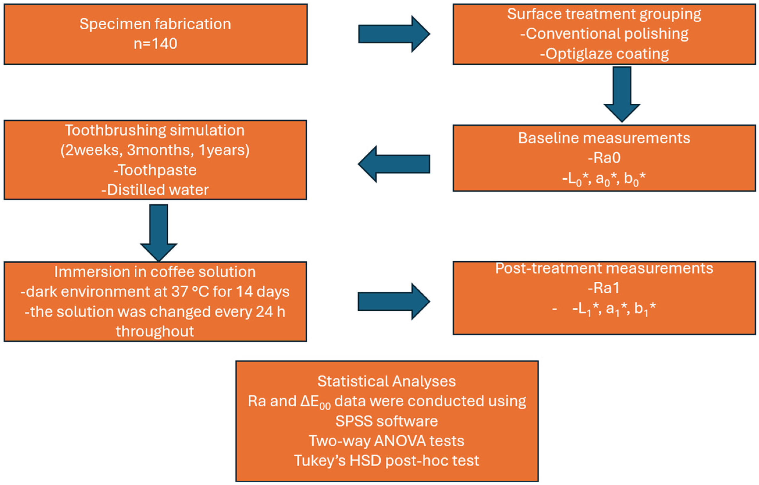
The workflow diagram summarizing the experimental procedures is presented in Figure 2.

3. Results

The results of the variance analysis regarding the effects of surface treatment, brushing agent, and duration on Ra are presented in Table 1. Statistical analysis revealed that surface treatment, brushing agent, and duration had a significant independent effect on Ra. Surface coating showed the strongest influence (p < 0.001, η2 = 0.779), followed by brushing agent and duration (both p < 0.001). Among interactions, only the combination of surface treatment and duration was statistically significant (p < 0.001) (Table 1).
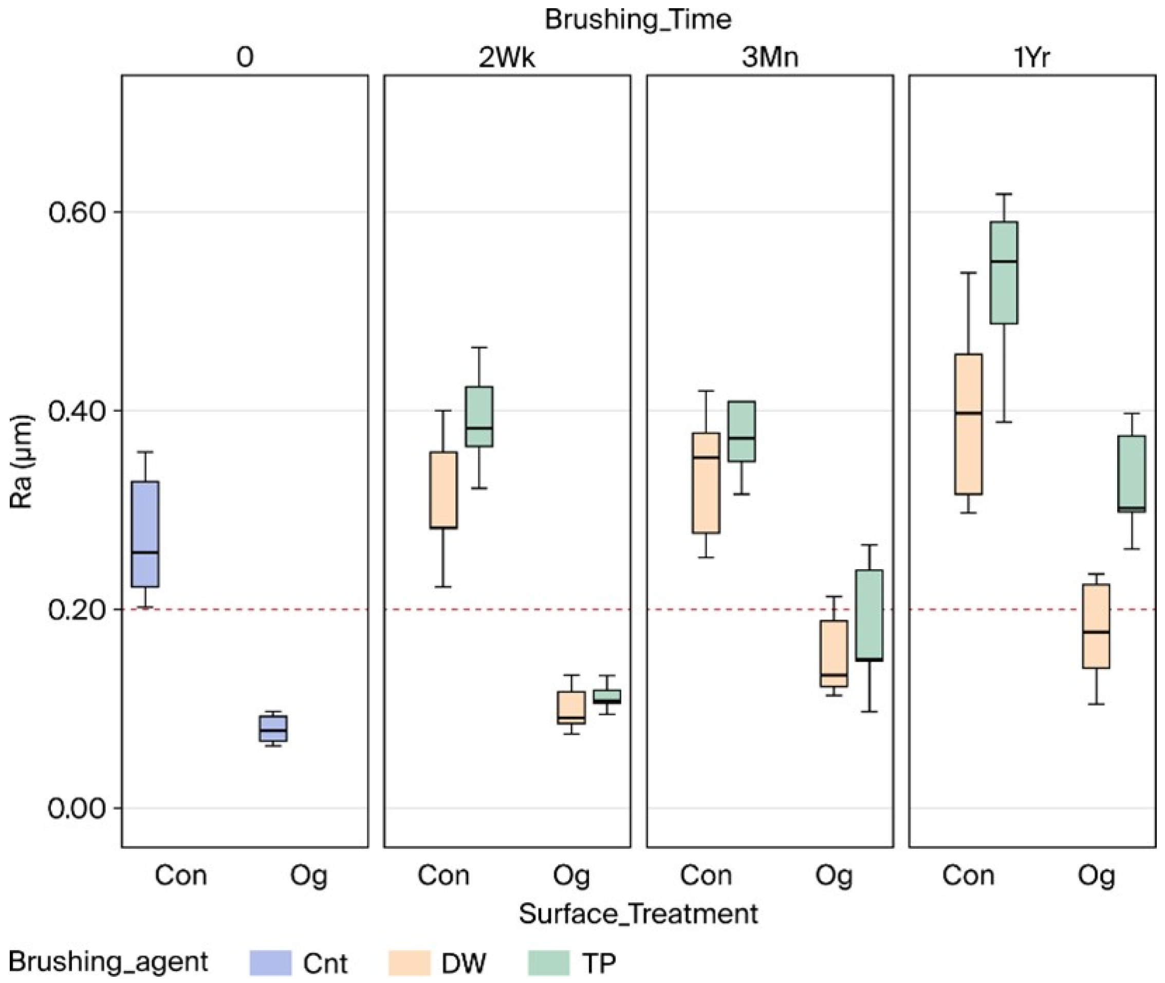
Table 2 presents the mean Ra values measured at baseline (unbrushed) and after 2 weeks, 3 months, and 1 year of brushing across various surface treatments (ST) and brushing agents (BA). The Con_Tp_1Yr group demonstrated the highest Ra value (0.53 ± 0.08), whereas the Og_Cnt group showed the lowest (0.08 ± 0.01). In the Con test groups, the Cnt value (Ra0) significantly increased from 0.28 µm to 0.40 µm after 1 year of brushing with distilled water, while brushing with toothpaste caused statistically significant changes, showing mean values of 0.39 ± 0.05 µm for Con_Tp_2Wk, 0.40 ± 0.09 µm for Con_Tp_3Mn, and 0.53 ± 0.08 µm for Con_Tp_1Yr (p < 0.05). The results of all test groups that underwent conventional polishing were higher than the plaque accumulation threshold (0.20 µm) (Figure 3).
Og_Cnt specimens were found to have the lowest Ra value (0.08 ± 0.01 µm) among all experimental groups. This value significantly increased to 0.17 ± 0.05 µm after 1 year of brushing with distilled water, while after 3 months and 1 year of brushing with toothpaste, it increased to 0.17 ± 0.06 m and 0.33 ± 0.05 µm, respectively (p < 0.05). The mean Ra values for all Og groups were lower than the reported plaque accumulation threshold (0.20 ± 0.10 µm), except for the 1 Yr Og_Tp specimens (Figure 3). When the pooled Ra values were evaluated, we found that TP agent application caused significantly higher Ra values than Dw, and 1 Yr of brushing application had significantly higher Ra values than the 2 Wk and 3 Mn durations for both surface treatment procedures (p < 0.05). Additionally, SEM micrographs supported the profilometric findings, showing smoother and more homogeneous surfaces for the coated groups, while prolonged brushing led to evident surface irregularities and micro-abrasions (Figure 4).
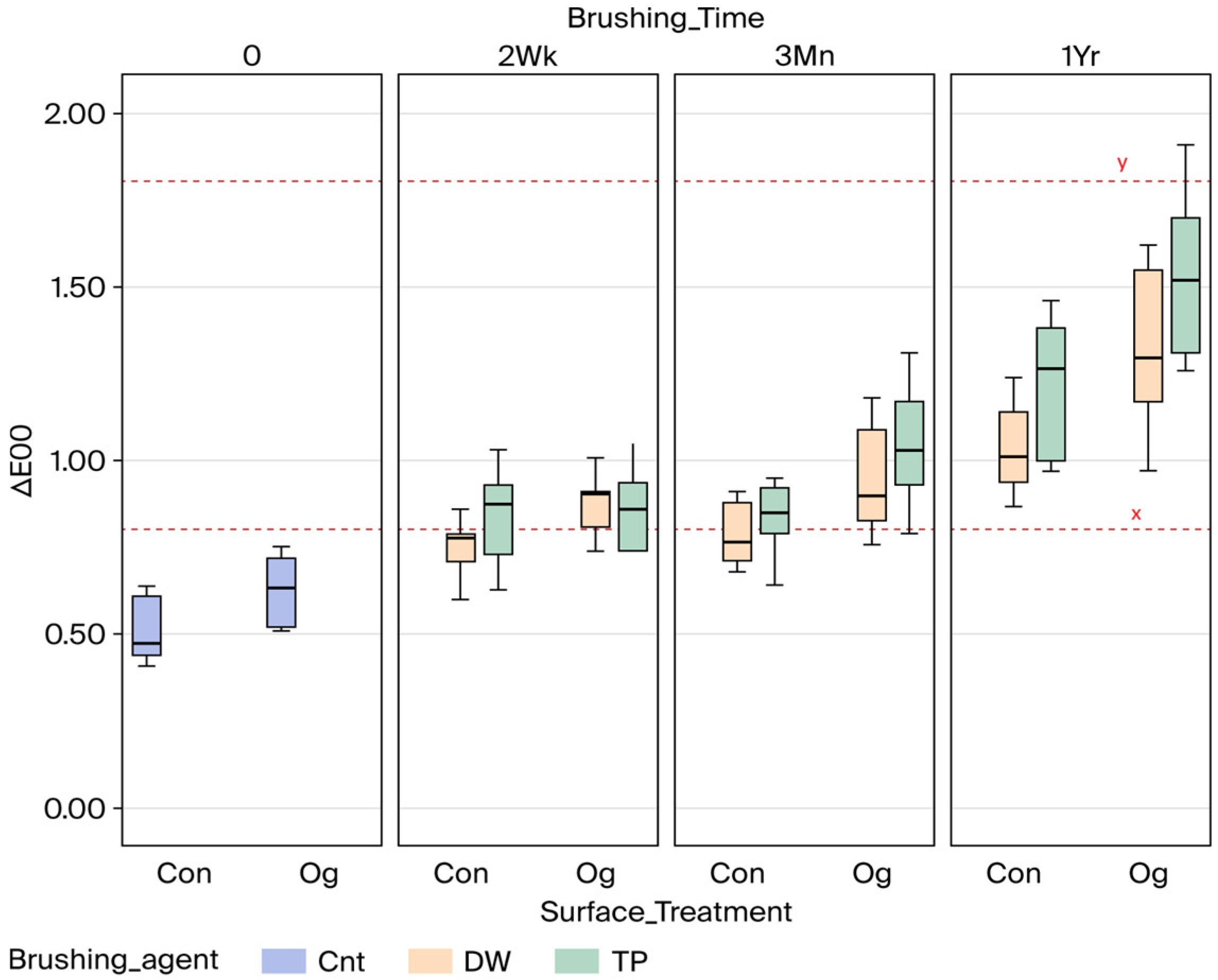
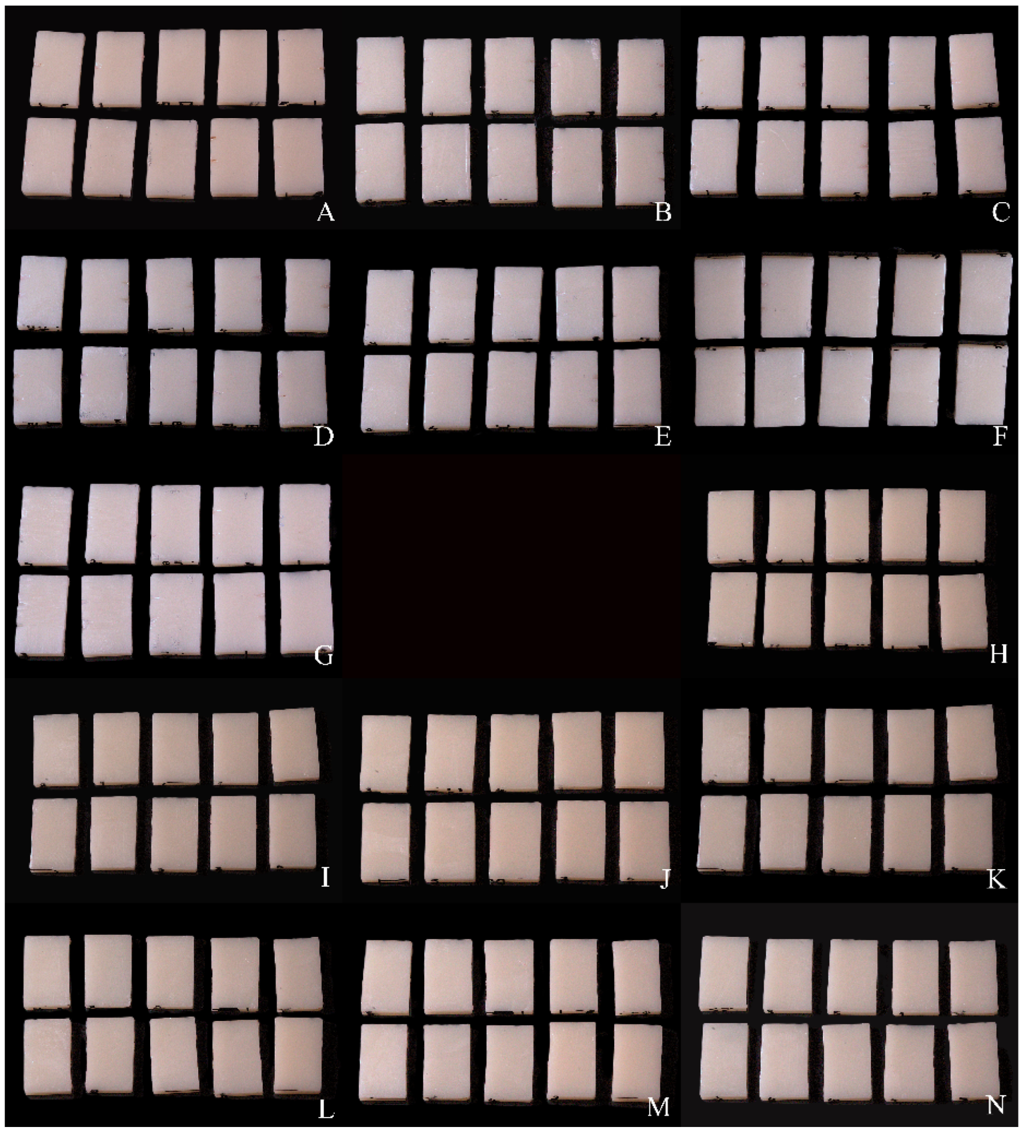
The two-way ANOVA revealed that each principal factor significantly influenced ΔE00 values (p < 0.001). Surface treatment (A) showed statistical significance (F = 42.547, p < 0.001) and exhibited a moderate effect size (Partial η2 = 0.252). This finding indicates that conventional polishing and Optiglaze Color application have different influences on color stability. Similarly, the brushing agent factor (B) was also significant (F = 15.442, p < 0.001; partial η2 = 0.109). Specimens brushed with toothpaste exhibited greater color change compared to those brushed with distilled water. This suggests that the abrasive effect of toothpaste negatively impacts color stability. The strongest effect, however, was observed for brushing duration (C) (F = 104.625, p < 0.001; partial η2 = 0.624). ΔE00 values increased significantly over time, demonstrating that prolonged brushing simulation has a pronounced adverse effect on color stability (Table 3). A significant A × C interaction (F = 5.820, p = 0.004; partial η2 = 0.085) indicated that the influence of surface treatment on color variation depended on brushing duration. The B × C interaction was also significant (F = 3.563, p = 0.031; partial η2 = 0.054) and showed that the time-dependent abrasive effect of toothpaste was more pronounced than that of distilled water. Mean ΔE00 values, standard deviations, and the multiple comparison results of the test groups are presented in Table 4. According to Table 4, the highest ∆E00 value was observed for the Og_Tp_1Yr group (1.53 ± 0.22), while the lowest was observed for the Con_Cnt group (0.51 ± 0.09) (p < 0.05). After the toothbrushing procedure, the ΔE00 values of both surface treatment groups increased, and statistically significant differences were detected between 2 Wk, 3 Mn, and 1 year of brushing (p < 0.05). Furthermore, specimens brushed with toothpaste (Tp) showed significantly greater color change compared to those brushed with distilled water (Dw) in both surface treatment groups (p < 0.05). Overall, the Optiglaze group exhibited slightly greater color changes than the control group, with statistically significant differences observed at all time points. As ΔE00 values of the Con_Cnt, Con_Dw_2Wk, Con_Dw_3 Mn, and Og_Cnt test groups were below the perceptibility threshold (∆E00 < 0.80); all other test groups were above the perceptibility threshold but still within clinically acceptable limits (0.80 < ∆E00 < 1.80) (Figure 5). The images of the specimens before and after the surface treatments are presented in Figure 6.

4. Discussion

The null hypothesis predicting no significant difference in surface roughness (Ra) or color stability (ΔE00) was rejected. The Optiglaze Color-coated groups exhibited significantly lower Ra values at all time points compared to conventionally polished groups (p < 0.05), while ΔE00 values were significantly higher after prolonged brushing, particularly with toothpaste, indicating a trade-off between surface smoothness and color stability (p < 0.05).
CAD-CAM-manufactured interim restorations provide notable clinical benefits arising from enhanced mechanical, physical, and esthetic characteristics [20,39]. Production under controlled industrial conditions increases the mechanical strength, marginal adaptation, resistance to bacterial adhesion, and overall reliability of these materials in the oral environment [8,9,10,20]. In this context, a CAD-CAM PMMA-based interim material (Teliocad) was evaluated in the current study due to its documented adequate color stability and surface resistance for long-term temporary use for esthetics and functionality. The manufacturing process itself—whether subtractive (milling) or additive (3D printing)—plays a critical role in determining surface texture and uniformity. Milling typically yields more consistent polymer density and fewer surface irregularities, which enhances smoothness and reduces plaque retention, whereas layer-by-layer 3D printing may introduce micro-irregularities and higher roughness, thereby potentially compromising surface quality over time [40]. This is due to the optimal curing conditions and highly cross-linked methacrylic acid ester-based polymer in milled PMMA, which increases its mechanical properties [41]. Wechkunanukul et al. reported that conventionally fabricated specimens exhibited significantly higher surface hardness compared with those produced using 3D printing while showing similar hardness values to the milled group [40]. In contrast, the lowest hardness was observed in the 3D-printed specimens, a finding attributed by Souza et al. [42] to the higher residual monomer content and increased porosity characteristic of 3D-printed PMMA. Additionally, Farina et al. [43] demonstrated that homogeneous heat polymerization enhances monomer conversion, reduces the plasticizing effect of residual monomers, and consequently increases surface hardness.
A variety of materials can be used to fabricate provisional restorations, including autopolymerizing polymethyl methacrylate (PMMA), polyvinyl methacrylate, urethane methacrylate, polyethylene methacrylate, micro-filled resins, and bis-acryl composites. Among these, polyethylene methacrylate is less frequently selected for clinical use due to its inferior esthetic properties and limited wear resistance, whereas PMMA-based and bis-acryl resin materials are generally favored for their broader clinical applicability [4]. Furthermore, in agreement with previous research, PMMA-based provisional materials demonstrated superior color stability compared to bis-acryl resins under staining conditions, which may be attributed to their more homogeneous polymeric structure and lower water sorption capacity [44].
Surface roughness is a critical factor that negatively impacts the biocompatibility and esthetics of restorative materials. Rough surfaces can facilitate plaque accumulation, jeopardize periodontal health, and compromise esthetics by causing discoloration [17,27]. Therefore, proper finishing and polishing of interim restorations are crucial for maintaining the materials’ clinical performance. Typical polishing approaches involve using burs, abrasive stones, fine pumice, silicone points, or polishing pastes; surface coating agents have also been shown to effectively increase surface smoothness [26]. In this context, the present study aimed to compare the effects of both the conventional polishing technique and the application of the Optiglaze Color surface coating material to compare their effects on surface roughness after simulated toothbrushing.
Alternative finishing materials have been introduced, incorporating resins such as Bis-GMA (bisphenol A-glycidyl methacrylate), TEGDMA (Triethylene glycol dimethacrylate), THFMA (Tetrahydrofurfuryl methacrylate), and UDMA (Urethane dimethacrylate), to replace traditional polishing techniques [45]. Surface coating agents can offer several advantages, including reducing surface irregularities, limiting plaque accumulation, and improving the esthetic appearance of interim restorations [18,46]. Optiglaze Color was chosen as the surface coating agent in the present study due to its favorable properties. It resulted in Ra values significantly below the reported plaque accumulation threshold of 0.2 µm compared to the conventionally polished group and maintained lower roughness levels, although its protective effect slightly decreased with prolonged brushing. The present results regarding surface roughness are supported by several studies in the literature [15,16,28,29,47].
In esthetically critical areas, the initial color match of interim restorations and the maintenance of their esthetic appearance and color stability throughout their service life are essential. Noticeable color changes may compromise the clinical acceptability of such restorations, especially with long-term use. In the present study, statistical analysis revealed that the brushing procedure had the most significant influence on ΔE00 values. Considering the perceptibility threshold of ΔE00 = 0.8 and the acceptability threshold of ΔE00 = 1.8, the results obtained using the CIEDE2000 formula [15] showed that, after one year of simulated brushing, ΔE00 values exceeded the perceptibility threshold. However, all test groups remained within the clinically acceptable range (ΔE00 < 1.8).
Restorative materials’ discoloration may result from the chemical composition of the material, water sorption, surface roughness, incomplete polymerization, and environmental factors such as temperature fluctuations, humidity, and the consumption of staining beverages [2,24]. These variables can significantly impact the esthetic success of the restoration over time. In the present study, Optiglaze Color, which contributed to smoother surfaces, was found to negatively affect color stability. The color change in the Optiglaze Color-treated group approached the acceptability threshold of 1.8, especially after the one-year brushing cycle with toothpaste [38]. The positive correlation between Ra and ΔE00 values observed by Radwan et al. [48] further supports our conclusion that increased roughness is often accompanied by greater color change. However, in the present study, color change after brushing was unexpectedly observed more often in the specimens treated with surface coating agents. This suggests that despite their ability to reduce roughness, surface coatings may negatively affect color stability over time. Therefore, reducing surface roughness alone may not be sufficient to prevent discoloration, and the long-term durability of coating agents should be carefully considered. Discrepancies between this study’s results and prior research could stem from variations in coating chemistry, application techniques, or polymerization methods. Furthermore, differences with other studies may be due to the duration and intensity of simulated brushing, microcracks that form on the surface after brushing, and the resulting gradual wear of the applied thin coating layer over time, resulting in an inadequate barrier effect. These factors highlight the need for standardized protocols in future studies to better understand the long-term behavior of surface coatings under dynamic oral conditions.
Brushing with abrasive toothpaste may cause micro-abrasions on restorative materials, progressively raising surface roughness and diminishing color stability [18]. In this study, specimens brushed for 2 weeks, 3 months, and 1 year were immersed in a coffee solution for 14 days after the simulations. The specimens that underwent brushing for only 2 weeks exceeded the perceptibility threshold for color change (ΔE00 > 0.8), which indicates that short-term brushing combined with coffee exposure can lead to clinically noticeable discoloration. In the current study, extended brushing caused greater color changes in all groups, particularly in those using toothpaste, where ΔE00 values gradually approached the clinically acceptable limit. These results suggest that brushing—particularly with abrasive agents—can cause microscopic surface alterations that negatively affect the color stability of interim restorations. Additionally, the mechanical action of brushing may gradually degrade the integrity of surface coating agents, reducing their protective capacity and allowing greater water sorption and pigment penetration, especially over longer periods such as the one-year cycle evaluated in this study. Similarly, several studies in the literature have also emphasized that toothbrushing can significantly affect the color stability of dental materials and may lead to discoloration over time [10,24,49].
This in vitro research presents certain limitations, as the brushing simulation could not entirely reproduce intraoral variables like temperature, saliva presence, or masticatory forces. Factors such as toothbrush hardness, toothpaste abrasively, and brushing motion may have influenced the results. Additionally, variations in intraoral pH and the frequent intake of acidic beverages—both of which are known to accelerate surface degradation and discoloration—were not evaluated in the present study. Flat specimens do not completely represent the complex surface morphology encountered clinically. Artificial saliva, smoking, inadequate oral hygiene, and different staining solutions were not considered, and additional aging protocols such as thermal cycling and chewing simulation were not applied. Therefore, future studies should aim to simulate more realistic intraoral conditions and evaluate both short- and long-term effects to enhance the clinical relevance and reliability of the findings.

5. Conclusions

Findings from this in vitro investigation indicate that applying a surface coating agent (Optiglaze Color) to CAD-CAM PMMA interim crowns effectively decreased initial surface roughness. Nevertheless, extended brushing duration and the toothpaste used led to greater surface roughness and color variation. Notably, Optiglaze-coated surfaces exhibited more pronounced discoloration after brushing compared to conventionally polished surfaces. This study also revealed that brushing duration was a strong determining factor for both color stability and surface roughness. These results highlight the critical importance of surface treatment and brushing conditions for ensuring the long-term esthetic and biological success of interim restorations. These observations should be interpreted within the scope of the specific materials and in vitro conditions tested.

Author Contributions

O.Ş.: conceptualization, supervision, and writing—review and editing. A.K.: methodology, evaluation, and writing—review and editing. D.Ö.D.: investigation, methodology, and data validation. H.Y.: data acquisition, sample preparation, and investigation. Ü.Y.: resources, data acquisition, and visualization. S.G.E.: Writing—original draft, data acquisition, and sample preparation. All authors have read and agreed to the published version of the manuscript.

Funding

This research received no external funding.

Institutional Review Board Statement

Not applicable.

Informed Consent Statement

Not applicable.

Data Availability Statement

The data that support the findings of this study are available from the corresponding author upon reasonable request.

Acknowledgments

The authors have reviewed and edited the output and take full responsibility for the content of this publication. This article is a revised and expanded version of a paper [50], This study was previously presented as an oral presentation at the 48th Annual Conference of the European Prosthodontic Association (EPA), 11–13 September 2025, Cappadocia, Türkiye.

Conflicts of Interest

Authors Onur Şahin and Ünsun Yağcı were employed by Land of Smile Clinic. However, the clinic had no role in funding, supporting, or influencing the study in any way. Therefore, these affiliations do not constitute commercial involvement in the research, and no commercial or financial relationship exists that could be interpreted as a potential conflict of interest. The remaining authors also declare that the research was conducted independently, without any commercial or financial support.

Abbreviations

CAD-CAMComputer-aided design and computer-aided manufacturing
RaSurface roughness
PMMAPolymethyl methacrylate
ΔE00Color changes
Bis-GMABisphenol A-glycidyl methacrylate
TEGDMATriethylene glycol dimethacrylate
THFMATetrahydrofurfuryl methacrylate
UDMAUrethane dimethacrylate

References

  1. Sadek, H.A.; Wahsh, M.; Saeed, M. Surface Roughness and Color stability of Provisional Restorative Materials Fabricated Using Different Methods After Immersion in Various Storage Media. Egypt. Dent. J. 2023, 69, 683–695. [Google Scholar] [CrossRef]
  2. Song, S.Y.; Shin, Y.H.; Lee, J.Y.; Shin, S.W. Color stability of provisional restorative materials with different fabrication methods. J. Adv. Prosthodont. 2020, 12, 259. [Google Scholar] [CrossRef]
  3. Fox, C.W.; Abrams, B.L.; Doukoudakis, A. Provisional restorations for altered occlusions. J. Prosthet. Dent. 1984, 52, 567–572. [Google Scholar] [CrossRef] [PubMed]
  4. Almohareb, T.; Alkatheeri, M.S.; Vohra, F.; Alrahlah, A. Influence of experimental staining on the color stability of indirect computer-aided design/computer-aided manufacturing dental provisional materials. Eur. J. Dent. 2018, 12, 269–274. [Google Scholar] [CrossRef]
  5. Mohsen AlMutairi, R. In-Vitro Marginal Fit of 3D-Printed vs Milled Provisional Crowns. J. Pioneer. Med. Sci. 2025, 14 (Suppl. S2), 176–180. [Google Scholar] [CrossRef]
  6. Igreț, A.; Rotar, R.N.; Ille, C.; Topală, F.; Jivănescu, A. Marginal fit of milled versus different 3D-printed materials for provisional fixed dental prostheses: An in vitro comparative study. Med. Pharm. Rep. 2023, 96, 298–304. [Google Scholar] [CrossRef]
  7. Sham, A.S.K.; Chu, F.C.S.; Chai, J.; Chow, T.W. Color stability of provisional prosthodontic materials. J. Prosthet. Dent. 2004, 91, 447–452. [Google Scholar] [CrossRef]
  8. Knobloch, L.A.; Kerby, R.E.; Pulido, T.; Johnston, W.M. Relative fracture toughness of bis-acryl interim resin materials. J. Prosthet. Dent. 2011, 106, 118–125. [Google Scholar] [CrossRef] [PubMed]
  9. Lieu, C.; Nguyen, T.M.; Payant, L. In vitro comparison of peak polymerization temperatures of 5 provisional restoration resins. J. Can. Dent. Assoc. 2001, 67, 36–39. [Google Scholar] [PubMed]
  10. Köroğlu, A.; Şahin, O.; Küçükekenci, A.S.; Dede, D.Ö.; Yıldırım, H.; Yilmaz, B. Influences of Toothbrushing and Different Toothpastes on the Surface Roughness and Color Stability of Interim Prosthodontic Materials. Materials 2022, 15, 5831. [Google Scholar] [CrossRef]
  11. Jehan, A.; Chidambaranathan, A.S.; Balasubramanium, M. Application and trends in provisional dental restorative materials for fixed partial denture. J. Oral. Res. Rev. 2023, 15, 65–71. [Google Scholar] [CrossRef]
  12. Rayyan, M.M.; Aboushelib, M.; Sayed, N.M.; Ibrahim, A.; Jimbo, R. Comparison of interim restorations fabricated by CAD/CAM with those fabricated manually. J. Prosthet. Dent. 2015, 114, 414–419. [Google Scholar] [CrossRef] [PubMed]
  13. Shenoy, A.; Rajaraman, V.; Maiti, S. Comparative analysis of various temporary computer-aided design/computer-aided manufacturing polymethyl methacrylate crown materials based on color stability, flexural strength, and surface roughness. J. Adv. Pharm. Technol. Res. 2022, 13 (Suppl. S1), S130–S135. [Google Scholar] [CrossRef]
  14. Nam, N.E.; Shin, S.H.; Lim, J.H.; Shim, J.S.; Kim, J.E. Effects of Artificial Tooth Brushing and Hydrothermal Aging on the Mechanical Properties and Color Stability of Dental 3D Printed and CAD/CAM Materials. Materials 2021, 14, 6207. [Google Scholar] [CrossRef] [PubMed]
  15. Bozoğulları, H.N.; Köroğlu, A.; Şahin, O.; İzzetağa, B. The Effect of the Toothbrushing and Surface Treatments on the Surface Roughness of Interim Crown Materials Used for Conventional, Subtractive, and Additive Manufacturing Techniques: An in Vitro Study. Selcuk. Dent. J. 2023, 10, 554–559. [Google Scholar] [CrossRef]
  16. Şen, D.; Göller, G.; İşsever, H. The effect of two polishing pastes on the surface roughness of bis-acryl composite and methacrylate-based resins. J. Prosthet. Dent. 2002, 88, 527–532. [Google Scholar] [CrossRef]
  17. Yildiz, P.; Güneş Ünlü, D. Farkli Yöntemler İle Kullanilan Geçici Materyallerinin Renk Stabilitesine Polisaj Patlarinin Etkisi. Selcuk. Dent. J. 2021, 8, 420–426. [Google Scholar] [CrossRef]
  18. Sattawatthamrong, S.; Kamonkhantikul, K.; Homsiang, W.; Arksornnukit, M. Effect of toothbrushing on surface roughness and gloss of CAD-CAM versus conventional interim materials with different surface treatments. J. Prosthet. Dent. 2025, 133, 282.e1–282.e12. [Google Scholar] [CrossRef] [PubMed]
  19. Gantz, L.; Fauxpoint, G.; Arntz, Y.; Pelletier, H.; Etienne, O. In vitro comparison of the surface roughness of polymethyl methacrylate and bis-acrylic resins for interim restorations before and after polishing. J. Prosthet. Dent. 2021, 125, 833.e1–833.e10. [Google Scholar] [CrossRef] [PubMed]
  20. Al-Dwairi, Z.N.; Tahboub, K.Y.; Baba, N.Z.; Goodacre, C.J. A Comparison of the Flexural and Impact Strengths and Flexural Modulus of CAD/CAM and Conventional Heat-Cured Polymethyl Methacrylate (PMMA). J. Prosthodont. 2020, 29, 341–349. [Google Scholar] [CrossRef]
  21. Al-Thobity, A.M. The Impact of Polymerization Technique and Glass-Fiber Reinforcement on the Flexural Properties of Denture Base Resin Material. Eur. J. Dent. 2020, 14, 092–099. [Google Scholar] [CrossRef] [PubMed]
  22. Aysan, İ.; Uçar, Y.; Üşümez, A. Üç Farkli Kaide Materyalinin Farkli Solüsyonlardaki Renk Stabilitesinin Karşilaştirilmasi. Atatürk Üniv. Diş Hek. Fak. Derg. 2011, 21, 219–225. [Google Scholar]
  23. Lepri, C.P.; Palma-Dibb, R.G. Surface roughness and color change of a composite: Influence of beverages and brushing. Dent. Mater. J. 2012, 31, 689–696. [Google Scholar] [CrossRef] [PubMed]
  24. Peñate, L.; Mercade, M.; Arregui, M.; Roig, M.; Basilio, J.; Cedeño, R. Color Stability of CAD/CAM Interim Material for Long-Term Fixed Dental Prostheses vs. Conventional Materials after Immersion in Different Staining Solutions. J. Compos. Sci. 2021, 5, 106. [Google Scholar] [CrossRef]
  25. Yao, Q.; Morton, D.; Eckert, G.J.; Lin, W.S. The effect of surface treatments on the color stability of CAD-CAM interim fixed dental prostheses. J. Prosthet. Dent. 2021, 126, 248–253. [Google Scholar] [CrossRef]
  26. Haselton, D.R.; Diaz-Arnold, A.M.; Dawson, D.V. Effect of Storage Solution on Surface Roughness of Provisional Crown and Fixed Partial Denture Materials. J. Prosthodont. 2004, 13, 227–232. [Google Scholar] [CrossRef]
  27. Bollenl, C.M.L.; Lambrechts, P.; Quirynen, M. Comparison of surface roughness of oral hard materials to the threshold surface roughness for bacterial plaque retention: A review of the literature. Dent. Mater. 1997, 13, 258–269. [Google Scholar] [CrossRef]
  28. Santos, M.; Soo, S.; Petridis, H. The effect of Parylene coating on the surface roughness of PMMA after brushing. J. Dent. 2013, 41, 802–808. [Google Scholar] [CrossRef]
  29. Rao, D.; Kalavathy, N.; Mohammad, H.; Hariprasad, A.; Kumar, C.R. Evaluation of the surface roughness of three heat-cured acrylic denture base resins with different conventional lathe polishing techniques: A comparative study. J. Indian Prosthodont. Soc. 2015, 15, 374. [Google Scholar] [CrossRef]
  30. Potluri, N.; Savitha, P.N. Comparison of two surface sealants on surface roughness and color stability of a commercially available denture base material—An in vitro study. IP Ann. Prosthodont. Restor. Dent. 2023, 9, 22–26. [Google Scholar]
  31. Takeuchi, C.Y.G.; Orbegoso Flores, V.H.; Palma Dibb, R.G.; Panzeri, H.; Lara, E.H.G.; Dinelli, W. Assessing the surface roughness of a posterior resin composite: Effect of surface sealing. Oper. Dent. 2003, 28, 281–286. [Google Scholar] [PubMed]
  32. Korkut, B.; Bud, M.; Kukey, P.; Sancakli, H. Effect of surface sealants on color stability of different resin composites. Med. Pharm. Rep. 2022, 95, 71–79. [Google Scholar] [CrossRef] [PubMed]
  33. Yuan, J.C.C.; Barão, V.A.R.; Wee, A.G.; Alfaro, M.F.; Afshari, F.S.; Sukotjo, C. Effect of brushing and thermocycling on the shade and surface roughness of CAD-CAM ceramic restorations. J. Prosthet. Dent. 2018, 119, 1000–1006. [Google Scholar] [CrossRef]
  34. Goldstein, G.R.; Lerner, T. The effect of toothbrushing on a hybrid composite resin. J. Prosthet. Dent. 1991, 66, 498–500. [Google Scholar] [CrossRef]
  35. Aydın, N.; Karaoglanoglu, S.; Oktay, E.A. Investigation the effects of whitening toothpastes on color change of resin-based CAD/CAM blocks. J. Esthet. Restor. Dent. 2021, 33, 884–890. [Google Scholar] [CrossRef]
  36. Zhang, L.; Yu, Y.; Li, S.; Yang, F.; Liang, S.; Xing, W. Effect of staining solutions on color and translucency stability of resin-composite computer-aided design and computer-aided manufacturing blocks. J. Am. Dent. Assoc. 2024, 155, 1012–1021. [Google Scholar] [CrossRef]
  37. Ghinea, R.; Pérez, M.M.; Herrera, L.J.; Rivas, M.J.; Yebra, A.; Paravina, R.D. Color difference thresholds in dental ceramics. J. Dent. 2010, 38, e57–e64. [Google Scholar] [CrossRef]
  38. Paravina, R.D.; Pérez, M.M.; Ghinea, R. Acceptability and perceptibility thresholds in dentistry: A comprehensive review of clinical and research applications. J. Esthet. Restor. Dent. 2019, 31, 103–112. [Google Scholar] [CrossRef] [PubMed]
  39. Mayer, J.; Stawarczyk, B.; Vogt, K.; Hickel, R.; Edelhoff, D.; Reymus, M. Influence of cleaning methods after 3D printing on two-body wear and fracture load of resin-based temporary crown and bridge material. Clin. Oral. Investig. 2021, 25, 5987–5996. [Google Scholar] [CrossRef]
  40. Wechkunanukul, N.; Klomjit, K.; Kumtun, T.; Jaikumpun, P.; Kengtanyakich, S.; Katheng, A. Comparison of Mechanical and Surface Properties between Conventional and CAD/CAM Provisional Restorations. Eur. J. Dent. 2025, 19, 697–703. [Google Scholar] [CrossRef]
  41. Alt, V.; Hannig, M.; Wöstmann, B.; Balkenhol, M. Fracture strength of temporary fixed partial dentures: CAD/CAM versus directly fabricated restorations. Dent. Mater. 2011, 27, 339–347. [Google Scholar] [CrossRef]
  42. Souza, A.L.C.; de Oliveira Cruvinel Filho, J.L.; da Rocha, S.S. Flexural strength and Vickers hardness of milled and 3D-printed resins for provisional dental restorations. Braz. J. Oral. Sci. 2023, 22, e238439. [Google Scholar] [CrossRef]
  43. Farina, A.P.; Cecchin, D.; Soares, R.G.; Botelho, A.L.; Takahashi, J.M.F.K.; Mazzetto, M.O.; Mesquita, M.F. Evaluation of Vickers hardness of different types of acrylic denture base resins with and without glass fibre reinforcement. Gerodontology 2012, 29, e155–e160. [Google Scholar] [CrossRef]
  44. Rençber Kizilkaya, A.; Dursun, M.N. The Effect of Beverages and Polishing on Color Change of Different Temporary Prosthetic Materials: In Vitro Study. Selcuk. Dent. J. 2023, 10, 560–564. [Google Scholar] [CrossRef]
  45. Eslemez Topcu, E.; Şahin, O.; Köroğlu, A.; Cömert, F.; Yilmaz, B. Surface roughness and Streptococcus mutans adhesion on surface sealant agent coupled interim crown materials after dynamic loading. BMC Oral Health 2022, 22, 299. [Google Scholar] [CrossRef]
  46. Rizzante, F.A.; Bombonatti, J.S.; Vasconcelos, L.; Porto, T.S.; Teich, S.; Mondelli, R.F. Influence of resin-coating agents on the roughness and color of composite resins. J. Prosthet. Dent. 2019, 122, 332.e1–332.e5. [Google Scholar] [CrossRef]
  47. Mahrous, A.A.; Alhammad, A.; Alqahtani, F.; Aljar, Y.; Alkadi, A.; Taymour, N.; Alotaibi, A.; Akhtar, S.; Gad, M.M. The Toothbrushing Effects on Surface Properties and Color Stability of CAD/CAM and Pressable Ceramic Fixed Restorations—An In Vitro Study. Materials 2023, 16, 2950. [Google Scholar] [CrossRef] [PubMed]
  48. Radwan, H.; Elnaggar, G.; El Deen, I.S. Surface roughness and color stability of 3D printed temporary crown material in different oral media (In vitro study). Int. J. Appl. Dent. Sci. 2021, 7, 327–334. [Google Scholar] [CrossRef]
  49. Bezgin, T.; Özer, L.; Tulga Öz, F.; Özkan, P. Effect of Toothbrushing on Color Changes of Esthetic Restorative Materials. J. Esthet. Restor. Dent. 2015, 27 (Suppl. S1), S65–S73. [Google Scholar] [CrossRef] [PubMed]
  50. Şahin, O.; Köroğlu, A.; Dede, D.Ö.; Yıldırım, H.; Yağcı, Ü.; Erdal, S.G. Influence of Toothbrushing and Toothpaste on Surface Roughness and Color Stability of CAD/CAM Interim Crown Material. In Proceedings of the 48th EPA Annual Conference, Cappadocia, Türkiye, 11–13 September 2025; Abstract no. SS215. p. 102. [Google Scholar]
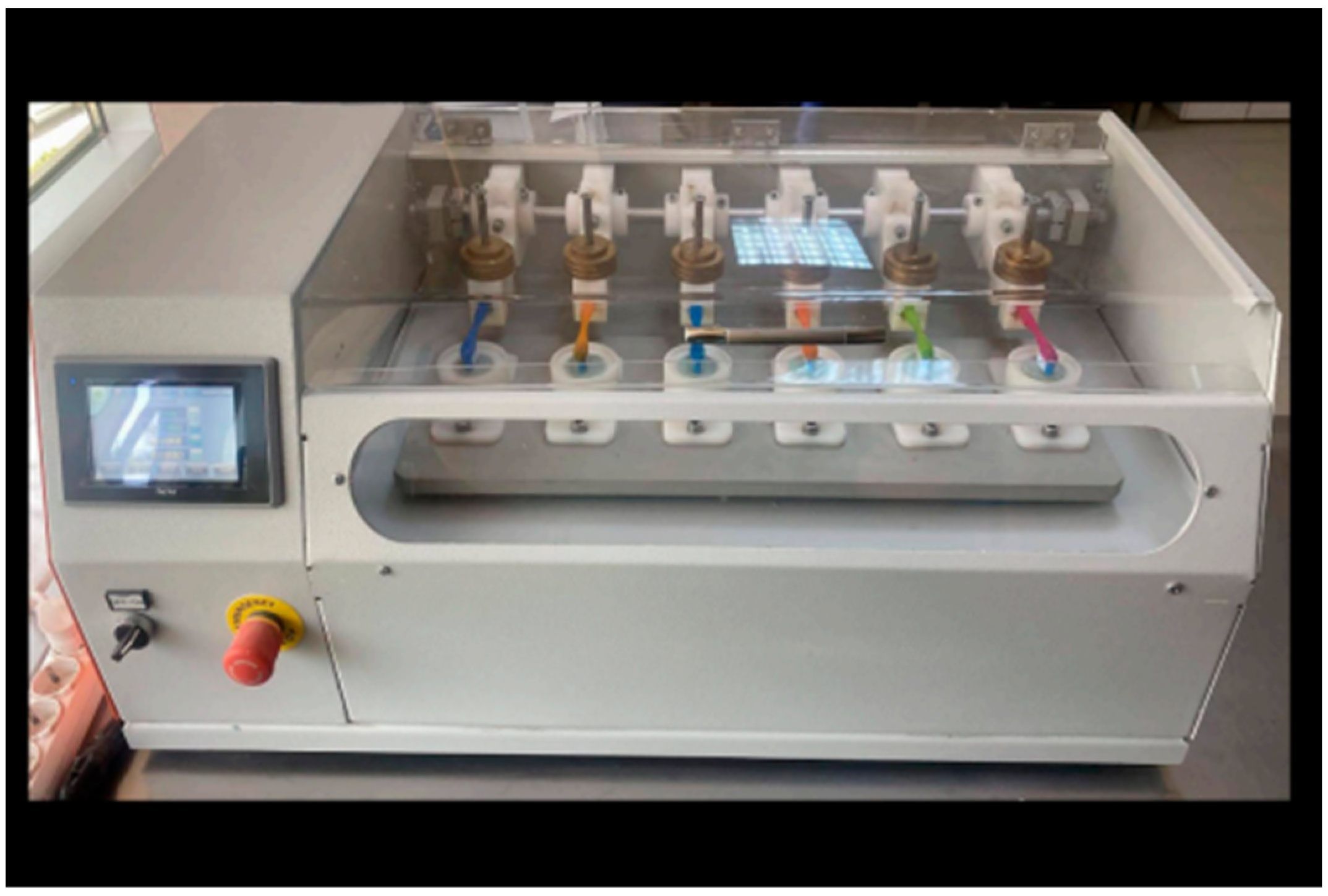
Figure 1. The toothbrushing simulator used in the study (DentArge TB-6.1, Analitik Medikal, Gaziantep, Turkiye) [10].
Coatings 15 01377 g001
Figure 2. A schematic representation of the experimental protocol, including specimen preparation, surface treatments, baseline measurements, toothbrushing simulation, post-brushing analyses, and statistical evaluation steps.
Coatings 15 01377 g002
Figure 3. Mean Ra (±SD) values of the test groups. The plaque accumulation threshold (Ra = 0.2 mm) is indicated on the x-axis.
Coatings 15 01377 g003
Figure 4. Scanning electron microscope (SEM) images of the TelioCAD specimens obtained before and after the surface treatment procedures to visualize surface morphology and treatment-related alterations. Og_Cnt (1), Og_DW_2Wk (2), Og_DW_3 Mn (3), Og_DW_1 Yr (4), Og _TP_2Wk (5), Og_TP_3 Mn (6), Og_TP_1 Yr (7), Con_Cnt (8), Con_DW_2Wk (9), Con_DW_3 Mn (10), Con_DW_1 Yr (11), Con _TP_2Wk (12), Con_TP_3 Mn (13), Con_TP_1 Yr (14). Scale bar: 100 μm.
Coatings 15 01377 g004
Figure 5. Mean (±SD) ∆E00 values of the test groups. The perceptibility threshold of the color differences (∆E00 = 0.8) is indicated on the x-axis, and the acceptability threshold (∆E00 = 1.8) on the y-axis.
Coatings 15 01377 g005
Figure 6. The test specimens of group Con_Cnt (A), Con_DW_2Wk (B), Con_DW_3 Mn (C), Con_DW_1 Yr (D), Con _TP_2Wk (E), Con_TP_3 Mn (F), Con_TP_1 Yr (G), Og_Cnt (H), Og_DW_2Wk (I), Og_DW_3 Mn (J), Og_DW_1 Yr (K), Og _TP_2Wk (L), Og_TP_3 Mn (M), Og_TP_1 Yr (N).
Coatings 15 01377 g006
Table 1. Results of variance analysis for the effects of surface treatment, brushing agent, and duration on surface roughness (Ra).
Tests of Between-Subject Effects
Dependent Variable: Ra
SourceType III Sum of SquaresdfMean SquareFpPartial Eta Squared
Surface_treatment (A)1.47911.479443.273<0.0010.779
Brushing_agent (B)0.19110.19157.311<0.0010.313
Brushing_time (C)0.35320.17652.875<0.0010.456
A × B0.00910.0092.6500.1060.021
A × C0.00620.0030.9680.3830.015
B × C0.06220.0319.254<0.0010.128
A × B × C0.01220.0061.8490.1620.029
Error0.4201260.003
Total12.879140
R Squared = 0.855 (Adjusted R Squared = 0.840); df = degree of freedom; F = mean square of factor.
Table 2. Mean Ra values (µm) and standard deviations (SD) of test groups.
Time0 (Ra0)2 Wk (Ra1)3 Mn (Ra1)1 Yr (Ra1)Pooled
STBA
ConCnt0.28 ± 0.06 C
Dw 0.30 ± 0.06 C0.33 ± 0.06 CD0.40 ± 0.08 D0.34 ± 0.08 a
Tp0.39 ± 0.05 D0.40 ± 0.09 D0.53 ± 0.08 F0.44 ± 0.10 b
Pooled 0.35 ± 0.07 #0.37 ± 0.08 #0.46 ± 0.1 ^
OgCnt0.08 ± 0.01 A
Dw 0.10 ± 0.02 A0.15 ± 0.04 AB0.17 ± 0.05 B0.14 ± 0.0 a
Tp0.11 ± 0.01 A0.17 ± 0.06 B0.33 ± 0.05 C0.20 ± 0.10 b
Pooled 0.11 ± 0.02 #0.16 ± 0.05 #0.25 ± 0.09 ^
ST, surface treatment; BA, brushing agent; Cnt, none; Dw, distilled water; Tp, toothpaste. The multiple comparison results of test groups are shown as capital letters (A–D,F) and the pooled results comparisons are shown as lowercase letters (a,b) and symbols (#,^).
Table 3. Two-way ANOVA results for ΔE00 values.
Tests of Between-Subject Effects
Dependent Variable: ΔE00
SourceType III Sum of SquaresdfMean SquareFpPartial Eta Squared
Surface_treatment (A)0.90210.90242.547<0.0010.252
Brushing_agent (B)0.32710.32715.442<0.0010.109
Brushing_time (C)4.43722.218104.625<0.0010.624
A × B0.00010.0000.0090.9240.000
A × C0.24720.1235.8200.0040.085
B × C0.15120.0763.5630.0310.054
A × B × C0.03320.0170.7870.4570.012
Error2.6711260.021
Total137.326140
R Squared = 0.782 (Adjusted R Squared = 0.760); df = degrees of freedom; F = mean square of factor.
Table 4. Mean ΔE00 values and standard deviations of test groups.
Time02 Wk3 Mn1 YrPooled
STBA
ConCnt0.51 ± 0.09 A
Dw 0.77 ± 0.15 B0.79 ± 0.09 B1.04 ± 0.12 C0.87 ± 0.17 a
Tp0.85 ± 0.13 B0.83 ± 0.11 B1.22 ± 0.18 C0.97 ± 0.23 b
Pooled 0.81 ± 0.14 #0.81 ± 0.10 #1.13 ± 0.18 ^
OgCnt0.63 ± 0.10 A
Dw 0.88 ± 0.08 B0.94 ± 0.15 BC1.31 ± 0.22 D1.05 ± 0.25 a
Tp0.87 ± 0.12 B1.05 ± 0.17 C1.53 ± 0.22 D1.15 ± 0.33 b
Pooled 0.88 ± 0.10 #1.00 ± 0.16 #1.42 ± 0.24 ^
ST, surface treatment; BA, brushing agent; Cnt, none; Dw, distilled water; Tp, toothpaste. The multiple comparison results of tests groups are shown as capital letters (A–D) and the pooled result comparisons are shown as lowercase letters (a,b) and symbols (#,^).
Disclaimer/Publisher’s Note: The statements, opinions and data contained in all publications are solely those of the individual author(s) and contributor(s) and not of MDPI and/or the editor(s). MDPI and/or the editor(s) disclaim responsibility for any injury to people or property resulting from any ideas, methods, instructions or products referred to in the content.