Article

Effect of Hydroxyapatite Post-Treatment on the Corrosion Resistance, Cytocompatibility and Antibacterial Properties of Copper-Containing Micro Arc Oxidation Coatings on Mg Alloy as Oral GBR Membrane Application

1
Suzhou Stomatological Hospital, Gusu District, Suzhou 215000, China
2
Key Laboratory of Materials Surface Science and Technology, Jiangsu Province Higher Education Institutes (Changzhou University), Changzhou 213164, China
3
Mechanical Engineering Department, School of Engineering and Technology, Federal Polytechnic, Offa P.M.B 420, Nigeria
4
Metallurgical Engineering Department, NED University of Engineering and Technology, University Road, Karachi 75270, Pakistan
5
School of Mechanical Engineering, Yangzhou University, Yangzhou 225127, China
*
Author to whom correspondence should be addressed.
These authors contributed equally to this work.
Coatings 2025, 15(11), 1344; https://doi.org/10.3390/coatings15111344
Submission received: 20 October 2025 / Revised: 3 November 2025 / Accepted: 13 November 2025 / Published: 19 November 2025
(This article belongs to the Special Issue Advanced Coatings for Alloy Protection and Performance Enhancement)

Abstract

Biodegradable magnesium (Mg) alloys hold promising application prospects in the field of guided bone regeneration (GBR) membranes, particularly for oral and maxillofacial applications. However, their corrosion resistance requires further improvement. Additionally, Mg alloys are susceptible to bacterial infection upon implantation, while copper (Cu) is known for its excellent antibacterial properties. Introducing Cu into the micro-arc oxidation (MAO) coating can enhance both the corrosion resistance and antibacterial performance of Mg alloys. However, the sealing effect of such coatings remains suboptimal. Hydroxyapatite (HA), which possesses outstanding bioactivity, is a promising bone substitute material. This study investigates the influence of HA content on the microstructure, corrosion resistance, cytotoxicity, and antibacterial properties of Cu-containing MAO coatings. The results demonstrate that as the HA concentration increases, the corrosion resistance of the composite coating is significantly enhanced. The corrosion rate decreased from 0.32 mm/y for the untreated MAO coating to 0.27 mm/y and 0.23 mm/y for the HA-treated samples with EDTA–Ca concentrations of 125 mmol/L and 175 mmol/L, respectively. Cytotoxicity assessment indicates that the incorporation of an HA layer significantly improves cell compatibility compared to the bare MAO coating. However, the enhanced corrosion resistance provided by the denser HA layer (at 175 mmol/L EDTA–Ca) unfortunately acts as a barrier, limiting the release of antibacterial Cu2+. Among the coatings tested, the one with 125 mmol/L EDTA–Ca exhibited the best overall performance, demonstrating good corrosion resistance, cytocompatibility, and effective antibacterial properties.

1. Introduction

Guided Bone Regeneration (GBR) technology is a core therapeutic approach in modern oral implantology for addressing insufficient alveolar bone volume [1]. Its fundamental principle involves implanting a barrier membrane to prevent the premature invasion of epithelial cells and fibrous connective tissue into the bone defect area, thereby creating a relatively independent space that prioritizes the growth of bone tissue cells. The most significant limitation of traditional titanium alloy membranes is their non-biodegradability, which necessitates a second surgery for removal, thereby increasing patient discomfort. On the other hand, absorbable polymer membranes often fail to provide adequate mechanical support due to their relatively poor mechanical properties. In contrast, biodegradable magnesium alloys, owing to their excellent osteogenic properties and good biocompatibility, hold promising application prospects in the field of guided bone regeneration membranes. However, the poor corrosion resistance of magnesium alloys, coupled with excessively rapid degradation rates that lead to hydrogen gas accumulation around implants [2], necessitates further improvements in their corrosion resistance. Surface modification is widely employed to enhance the corrosion resistance of magnesium alloys [3]. Micro-arc oxidation (MAO), an eco-friendly surface modification technique, not only improves corrosion resistance [4] but also fosters an osteoconductive and cell-friendly microenvironment on the alloy surface [5]. Although MAO coatings can improve corrosion resistance to some extent, their porous structure allows corrosive agents to penetrate the micropores, leading to galvanic corrosion between the coating and the substrate. Additionally, magnesium alloys are prone to infection during implantation, which may result in surgical failure. Copper (Cu) exhibits broad-spectrum bactericidal properties, and numerous studies have fabricated Cu/MAO composite coatings on titanium alloy surfaces [6,7,8]. These results consistently demonstrate that Cu enhances osteogenesis and antibacterial efficacy. Furthermore, studies have indicated that Mg-Cu alloys show promising potential for treating osteomyelitis [9]. However, in such alloys, galvanic corrosion between secondary phases and the matrix accelerates their degradation. To address this, one study incorporated Cu compounds into coatings via micro-arc oxidation, which mitigated galvanic corrosion between the substrate and Cu, albeit at the cost of enlarged pore diameters in the MAO coating [10]. Alternatively, Zhang et al. [11] developed a Cu-doped Al2O3 coating with a dense outer layer, which, compared to undoped Al2O3 coatings, significantly reduced adherent cell counts while exhibiting superior antibacterial capability.
Incorporating copper into MAO coatings not only enhances the corrosion resistance of magnesium alloys but also imparts robust antibacterial properties. However, the release of Cu2+ can compromise cell viability [12], while excessive Cu2+ content or a rapid release rate may adversely affect osteogenic capacity. Hydroxyapatite (HA), characterized by stable chemical properties and a composition/structure similar to human bone, stimulates or induces bone regeneration and exhibits excellent bioactivity [13]. HA possesses exceptional ion-exchange and adsorption capabilities, effectively neutralizing the detrimental cellular effects of Cu2+ [14,15]. Qin et al. [16] fabricated an HA coating doped with an optimal Mg content on a titanium alloy surface, significantly enhancing the coating’s performance and bioactivity. One study has indicated [17] that MAO and MAO-HA coatings show no statistically significant difference in average pore size, and their cytocompatibility remains suboptimal. Tang et al. [18] refined the fabrication method by preparing MAO/HA composite coatings on magnesium alloy substrates via the sol–gel technique. Results revealed that the MAO/HA composite coating heat-treated at 400 °C exhibited the lowest corrosion current density (4.52 × 10⁻⁷ A/cm2). The HA sol–gel layer acts as a barrier layer, sealing the pores of the MAO coating and thereby improving both corrosion resistance and bioactivity. In a recent study [19], researchers employed the sol–gel method to dope metallic copper into caprine tooth-derived HA, demonstrating exceptionally high antibacterial efficacy.
Compared to the copper-containing micro-arc oxidation coatings on magnesium alloys, the HA coating seals the micropores of the MAO coating. This simultaneously enhances the corrosion resistance and osteogenic properties of the MAO coating, while also enabling a controlled release of copper ions from it, resulting in effective antibacterial performance. Building upon these research foundations, this study systematically investigates the effects of HA post-treatment on the physicochemical and biological properties of copper-containing micro-arc oxidation (Cu-MAO) coatings for use as GBR membranes. The objective is to develop a novel composite coating that yields a magnesium implant surface combining exceptional antibacterial performance with enhanced osseointegration capability in the context of GBR.

2. Experimental Details

2.1. Coating Preparation

The substrate used was an extruded Mg-2Zn-1Gd-0.5Zr alloy, which was sectioned into disk specimens (Φ15 mm × 1 mm) via wire electrical discharge machining. Following polishing with 2000-grit SiC paper, the specimens were ultrasonically cleaned sequentially in deionized water and anhydrous ethanol, dried, and finally vacuum-sealed. The coatings were subsequently fabricated using a 2000 W micro-arc oxidation (MAO) system. The electrolyte for the copper-containing MAO (Cu-MAO) coatings was composed of 1.2 g/L Ca(OH)2, 8 g/L KF, 4 g/L sodium hexametaphosphate ((NaPO3)6), and 3 g/L CuO. The MAO process was carried out under a constant voltage of 360 V, at a frequency of 1000 Hz with a 40% duty cycle, for a duration of 5 min. The inter-electrode distance was set at 10 cm, with the oxidation process conducted at room temperature. Throughout the process, air was introduced to agitate the electrolyte.
Following the MAO treatment, the samples were rinsed with distilled water and dried under a stream of cold air. Hydrothermal solutions were prepared by dissolving Ethylenediaminetetraacetic Acid Calcium Disodium Salt (EDTA-Ca) and KH2PO4 in distilled water at molar ratios of 75:45, 125:75, and 175:105 mmol/L (maintaining a constant Ca/P ratio of 5:3). The solutions were freshly prepared. The pH of each solution was adjusted to 9–10 using a NaOH solution. The MAO-treated samples were then immersed in the solutions and subjected to a hydrothermal treatment at 80 °C for 4 h. Subsequently, the samples were retrieved, rinsed with ethanol, dried, and stored for further analysis. The resulting specimens were designated as follows: the untreated Cu-MAO coating was labeled H0, while the post-hydrothermal treatment samples were labeled H75, H125, and H175 according to their respective EDTA-Ca concentrations.

2.2. Microstructural Characterization

The surface morphology of the MAO/HA coatings was examined using scanning electron microscopy (SEM, JSM-6510). The JSM-6510 scanning electron microscope is manufactured by JEOL Ltd. in Akishima, Tokyo, Japan. The chemical composition of the coatings was characterized by energy-dispersive spectroscopy (EDS). The phase composition was determined via X-ray diffraction (XRD, D/MAX2500) using Cu-Kα radiation. The D/MAX2500 X-ray diffractometer (XRD) is manufactured by Rigaku Corporation in Akishima, Tokyo, Japan.

2.3. Electrochemical Testing

Tests were conducted using a standard three-electrode configuration. The specimen served as the working electrode, with a saturated calomel electrode (SCE) as the reference electrode and a platinum sheet as the counter electrode. Experiments were performed in Hank’s solution (composition detailed in Table 1) maintained at 37 °C. To ensure system stabilization, open circuit potential (OCP) was monitored for 30 min. Potentiodynamic polarization curves were subsequently recorded with a potential sweep from −0.25 V to +0.35 V relative to OCP at a scan rate of 0.5 mV/s. All experiments were performed in triplicate.

2.4. Immersion Test

The disk specimens (Φ15 mm × 1 mm) were immersed in Hank’s solution at a specimen-to-solution ratio of 1.25 cm2/mL for 14 days. The solution was refreshed daily, and the pH value was measured on days 1, 3, 5, 7, 9, and 14. The corrosion rate was determined after 7 and 14 days of immersion. Following the immersion test, the corrosion products were removed by immersing the samples in a chromic acid solution (200 g/L CrO3 + 10 g/L AgNO3) for 20 min. The corrosion rate (Pi) was then calculated using Equation (1):
P i =   ( K   ×   W ) / ( A   ×   T   ×   D )
where Pi denotes corrosion rate (mm/y), K is a constant (8.76 × 10⁴), W represents mass loss (g), A is the specimen surface area (cm2), T is immersion duration (h), and D is alloy density (g/cm3). Final microstructural analysis was conducted using SEM, EDS, and XRD. All experiments were performed in quintuplicate.

2.5. Cytotoxicity Testing

The cytotoxicity of the samples with varying H contents was evaluated using mouse osteoprecursor cells (MC3T3-E1) and human osteosarcoma cells (MG63). The experimental setup included the test groups, a negative control (phosphate-buffered saline, PBS), and a positive control (5% DMSO solution). Cells were harvested into single-cell suspensions and seeded into 96-well plates. After a 24 h pre-culture, the medium was replaced with sample extracts or control solutions (fresh medium as a negative control and 5% DMSO as a positive control). Cell proliferation was assessed on days 1, 3, and 5 using a CCK-8 assay. Briefly, the medium in each well was replaced with 200 μL of CCK-8 solution, followed by a 4 h incubation in a humidified incubator (37 °C, 5% CO2). The absorbance at 450 nm was then measured using a μQuant Microplate Spectrophotometer. To enhance measurement reliability, a dual-wavelength detection was employed, using test wavelengths of 450–490 nm and a reference wavelength of 600–650 nm. The cell proliferation rate, expressed as the Relative Growth Rate (RGR), was subsequently calculated using Equation (2):
RGR = OD sample OD blank OD control OD blank × 100 %
ODblank refers to the optical density value of wells containing culture medium and CCK-8 solution without cells. All experiments were performed in triplicate. For the ICP (inductively coupled plasma) analysis, the samples were immersed in Hank’s solution with an immersion ratio of 1.25 cm2/mL for 24 h.

2.6. ALP Test

Cells were seeded in 24-well plates at a density of 1 × 10⁴ cells/mL and pre-cultured for 24 h to allow adhesion. The culture medium was then replaced with the immersion extracts, and the cells were further cultured for 1 or 2 weeks. After the incubation, the extracts were aspirated, and the cells were gently washed three times with PBS. Subsequently, the cells were lysed using 0.2% Triton X-100 for 5 h. The resulting lysates were centrifuged at 10,000 r/min for 10 min to collect the supernatant for subsequent biochemical analyses. Alkaline phosphatase (ALP) activity was determined by incubating the supernatant with ALP reagent at 37 °C for 15 min and measuring the absorbance at 520 nm. The total protein content in the supernatant was quantified using a BCA Protein Assay Kit.

2.7. Antibacterial Testing

The antibacterial properties of the coatings were evaluated against Escherichia coli (ATCC 25922). Bacterial suspensions were adjusted to a concentration of 105 CFU/mL. For the test groups, 80 μL of the suspension was inoculated directly onto the surface of the triplicate samples. In parallel, 80 μL of the same suspension was incubated in PBS as blank and control groups. All groups were then cultured at 37 °C under high relative humidity (>90%) for 24 h. After incubation, the viable bacteria from each group were collected by dilution with PBS and enumerated using the plate count method. Additionally, the concentration of Cu2+ ions released from the samples into Hank’s solution over 24 h of immersion was quantified by inductively coupled plasma (ICP) analysis. All experiments were performed in triplicate.

3. Results

3.1. Microstructural Analysis

Figure 1 displays the cross-sectional microstructures of the H0, H75, H125, and H175 samples. The thickness of the hydroxyapatite (HA) coating ranged from approximately 2.5 to 5.5 μm and exhibited a positive correlation with the Ca-P content. Specifically, the H175 sample possessed the maximum thickness, ranging from 5.0 to 5.5 μm. The interface between the MAO layer and the HA coating was free of discernible defects, suggesting strong adhesion. Furthermore, while some cracks and defects were observed in the untreated H0 coating, they were effectively sealed after the hydrothermal treatment. This indicates that HA particles permeated and filled these micro-defects during the process.
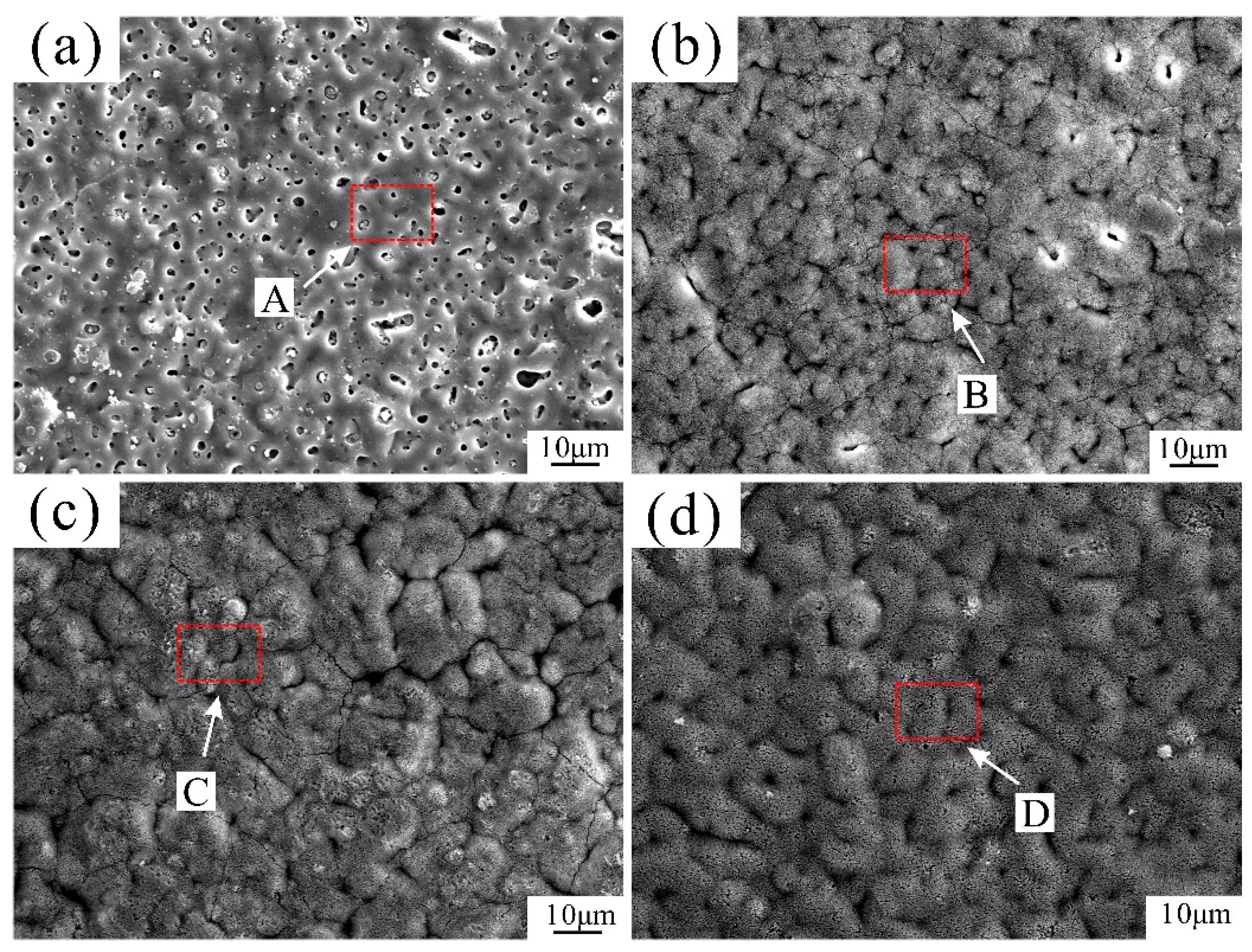
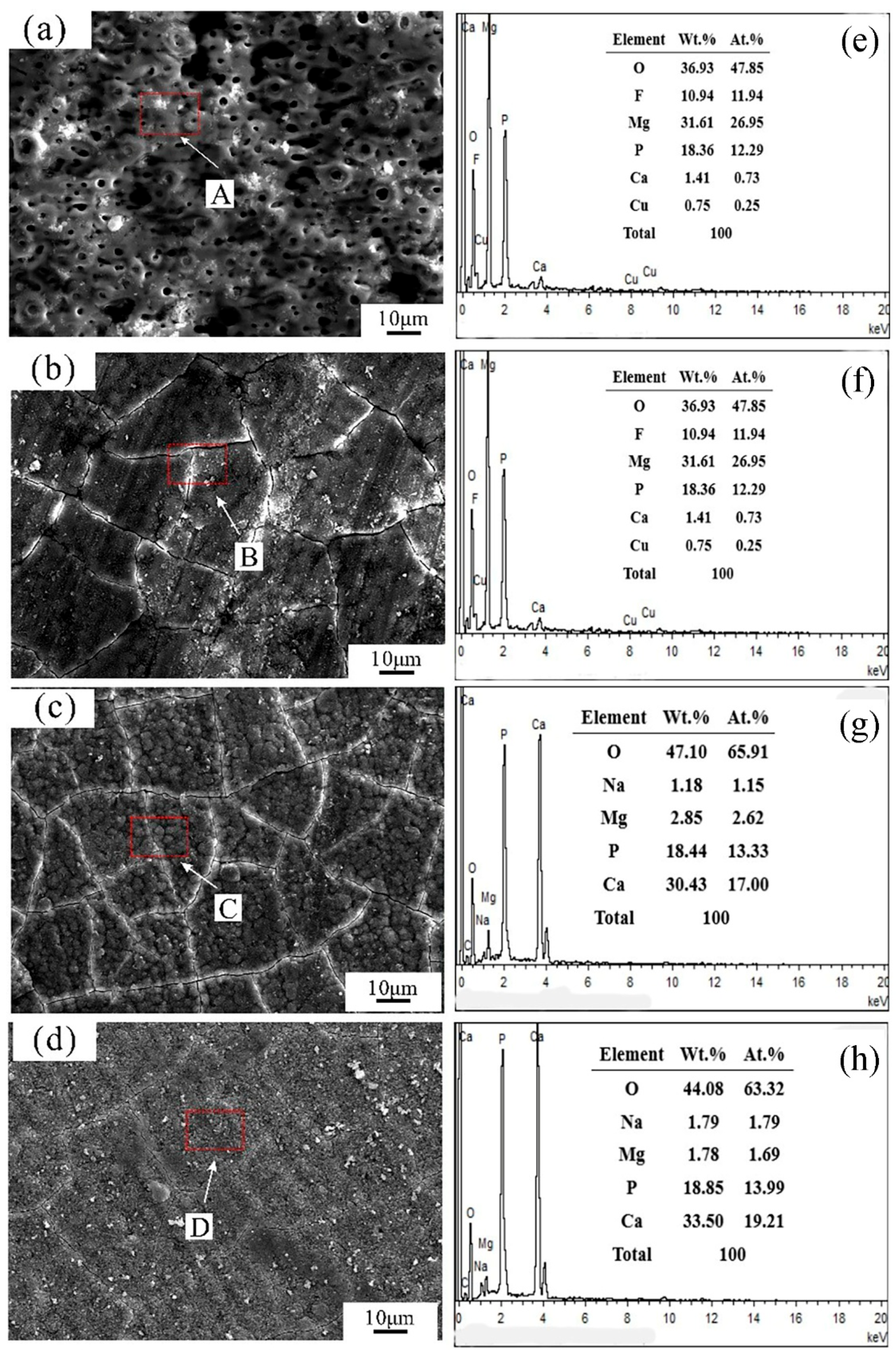
The surface morphologies of the samples are presented in Figure 2. Sample H0 featured a typical MAO structure with numerous micropores. In contrast, sample H75 showed a significant reduction in micropore density, with only a few pores remaining visible. The characteristic micropores of the MAO coating were entirely obscured in the H125 sample, as its surface was covered by a new HA layer, although some cracks and pits persisted. Finally, the H175 sample exhibited a dense and uniform HA film that completely covered the underlying MAO structure, providing a high degree of substrate protection.
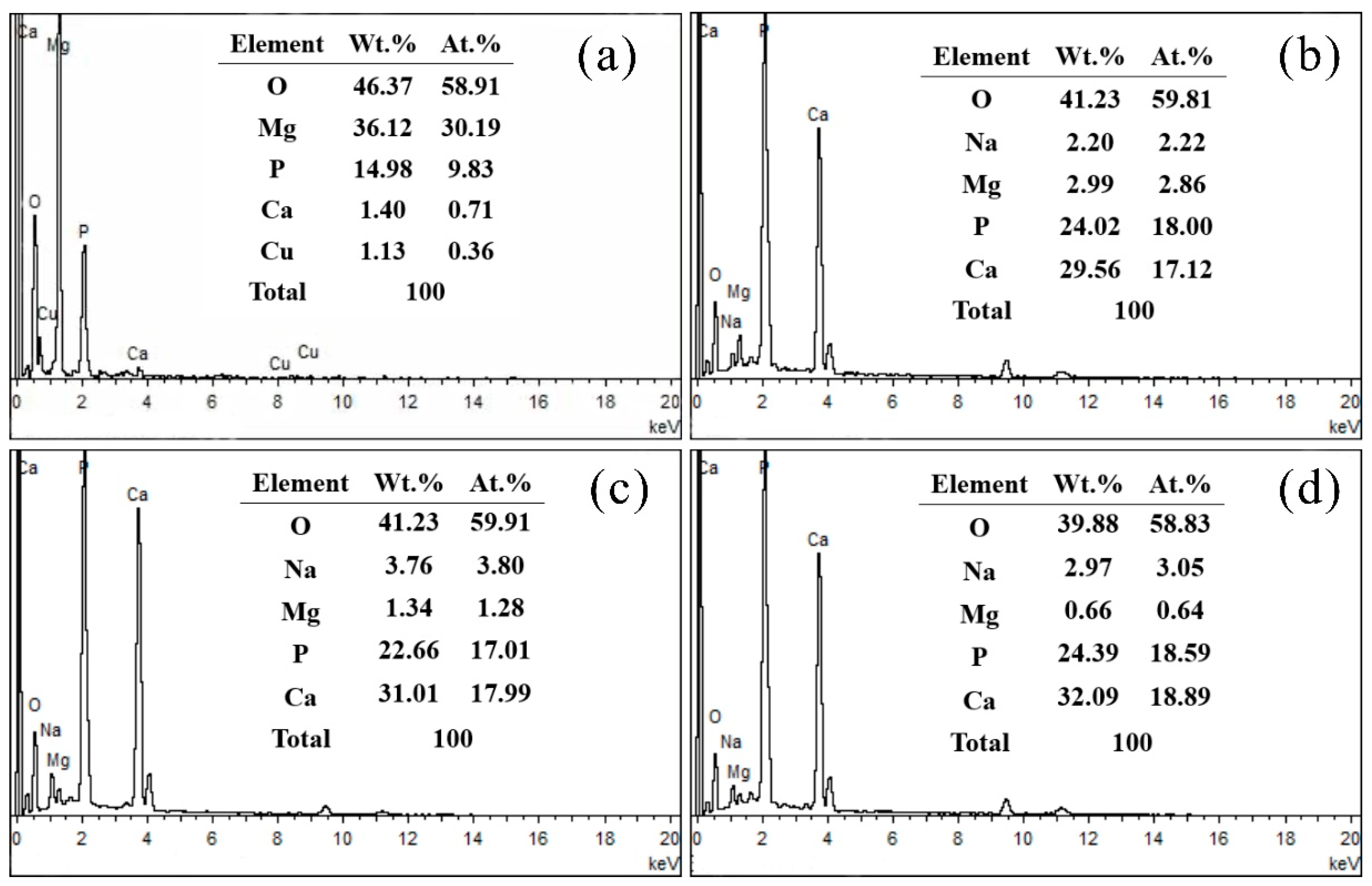
Figure 3 presents the EDS analysis of the surface areas indicated by arrows in Figure 2. The spectra were predominantly composed of Mg, O, Ca, and P. After the hydrothermal treatment, the surface Mg content decreased significantly, while the intensities of Ca and P increased markedly, confirming that the HA coating effectively sealed the porous MAO layer. The average Ca/P ratio for the three HA-coated samples (H75, H125, H175) was calculated to be 1.24 ± 0.09. It should be noted that the intrinsic presence of Ca and P in the underlying MAO coating precludes the definitive identification of surface compounds based solely on the Ca/P ratio. Therefore, phase identification was further conducted by XRD.
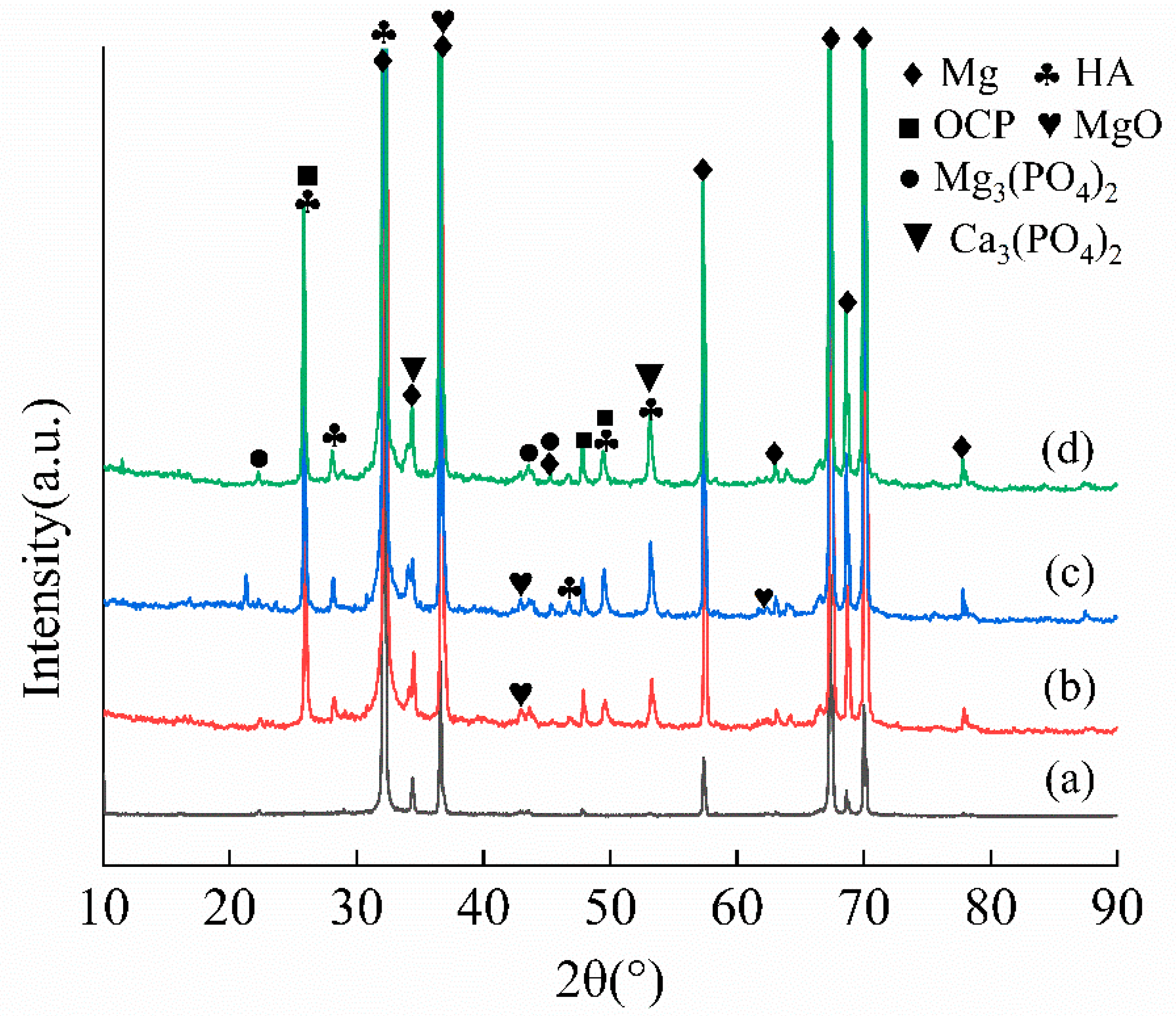
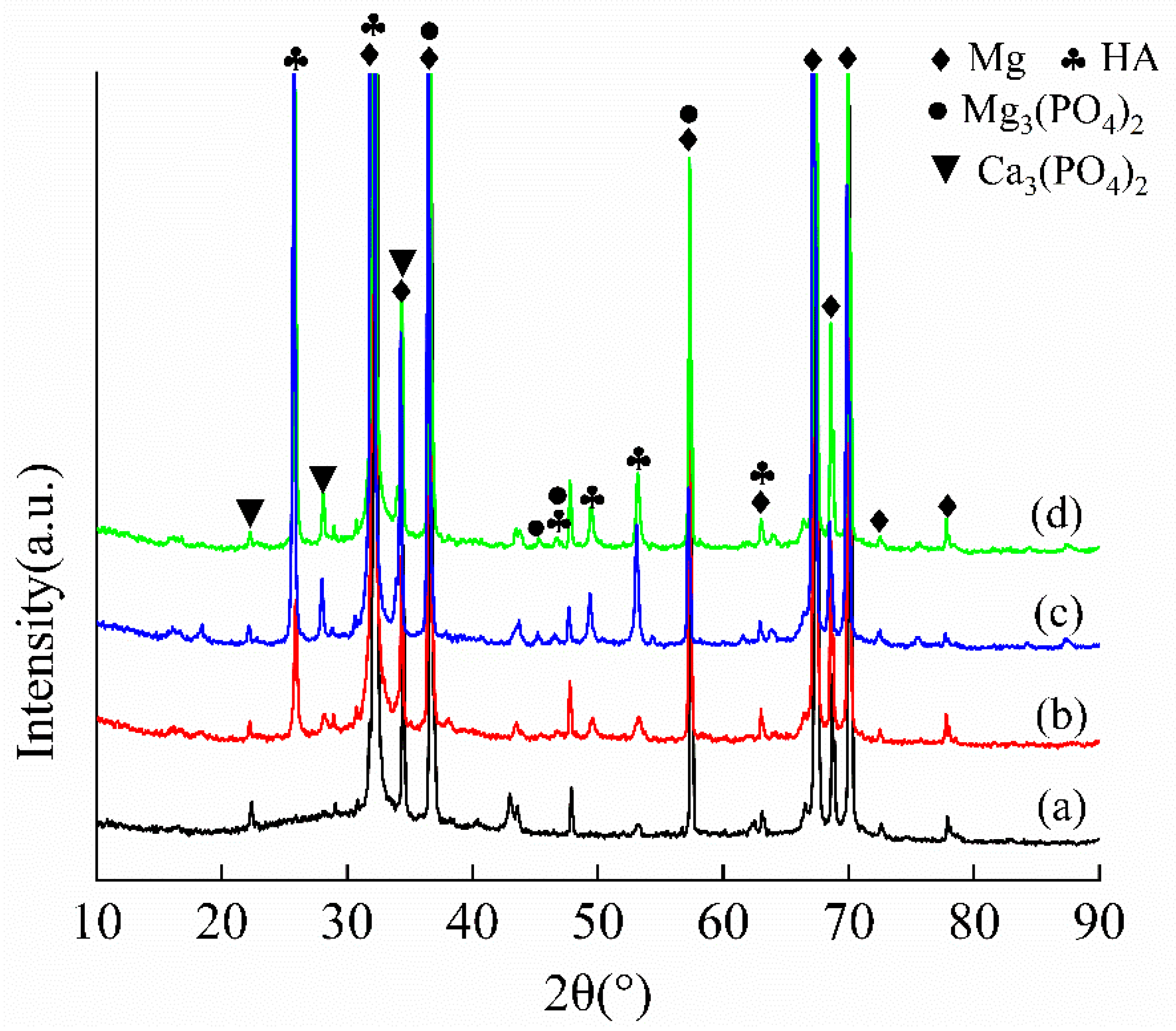
The XRD patterns in Figure 4 confirmed the formation of hydroxyapatite (HA) on all composite coatings. Distinct diffraction peaks were observed at 2θ = 25.8°, corresponding to the (002) crystallographic plane of HA (JCPDS #09-0432 [20]). Additional characteristic peaks for HA were identified at 28.1°, 32.1°, 46.7°, 49.4°, and 53.1°, which are indexed to the (210), (211), (222), (213), and (004) planes, respectively [21]. Additional HA, the coatings also contained tricalcium phosphate (Ca3(PO4)2), octacalcium phosphate (Ca8H2(PO4)6, OCP)—which is a known precursor to HA—and trimagnesium phosphate (Mg3(PO4)2).
During the hydrothermal synthesis, the Ca2+ concentration at the substrate surface significantly exceeded that of Mg2+. This concentration gradient was established and maintained by the continuous supply of Ca2+ from the thermal dissociation of Ca-EDTA complexes in the solution. In contrast, Mg2+ release was confined to the initial stage of the reaction, originating solely from the pre-existing MAO coating. The resultant ionic environment favored the preferential co-deposition of calcium and phosphate ions, leading to the initial formation of OCP on the MAO surface. Subsequently, a portion of this OCP precursor underwent a phase transformation into the more stable hydroxyapatite (Ca5(PO4)3OH). Concurrently, the Mg2+ released into the solution precipitated as magnesium phosphate compounds.

3.2. Electrochemical Analysis

Figure 5 displays the potentiodynamic polarization curves of the four samples tested in Hank’s solution, with the corresponding electrochemical parameters—corrosion current density (icorr), corrosion potential (Ecorr), and calculated corrosion rates—detailed in Table 2. All MAO/HA composite coatings demonstrated superior corrosion resistance compared to the untreated H0 sample, as evidenced by their nobler Eicorr and significantly lower icorr values. This enhancement is attributed to the microstructural evolution from a porous MAO coating to a sealed composite surface. Specifically, the dense HA top layer in the composite coatings acts as an effective barrier, impeding the permeation of corrosive ions to the underlying magnesium substrate.
Among the coated samples, the corrosion resistance improved progressively with increasing Ca-P content. Sample H175 exhibited the optimal performance, with the most positive Ecorr (−1.42 V), the lowest icorr (0.21 μA/cm2), and the minimal corrosion rate (0.107 mm/y). This represents a positive shift of 54 mV in Ecorr and a 72.1% reduction in icorr compared to H0. In contrast, H75 demonstrated the poorest performance among the coated groups. The H125 sample showed a marked improvement over H75, with a substantially decreased corrosion current density, effectively more than doubling the corrosion resistance.

3.3. Immersion Analysis

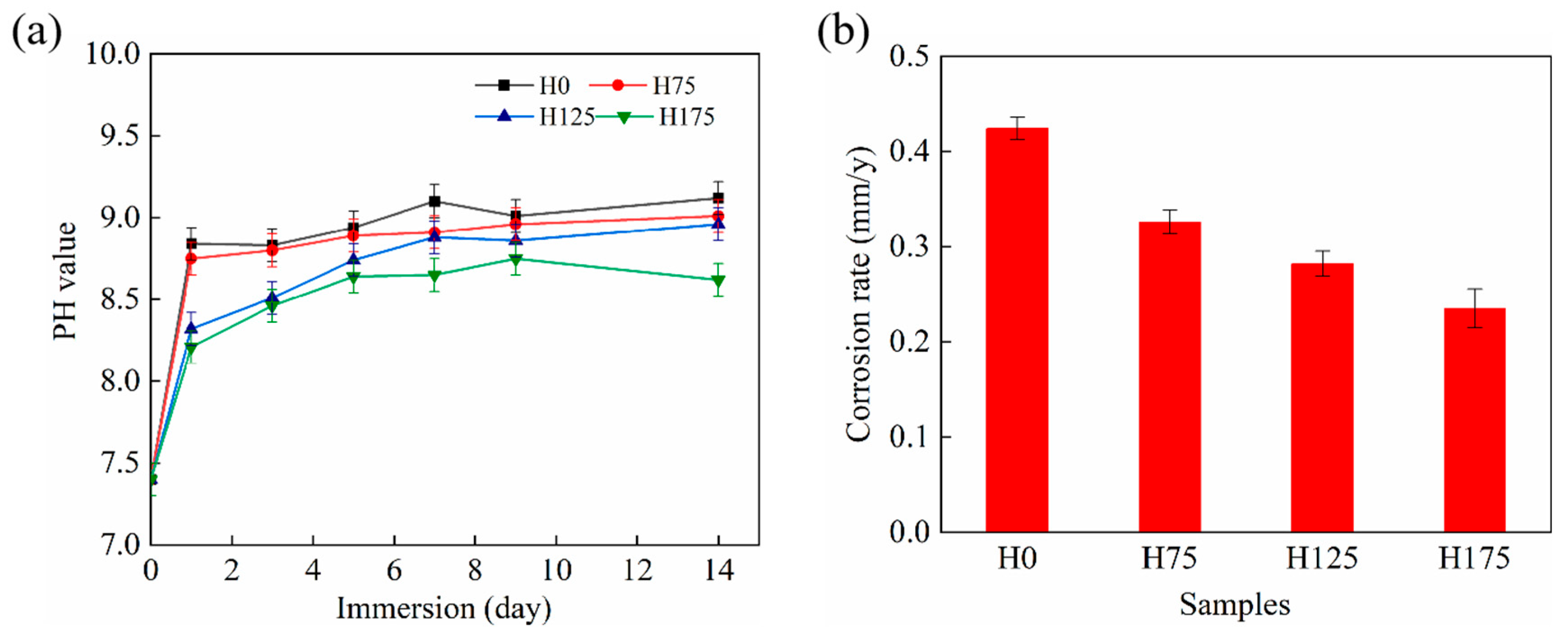
Immersion tests in Hank’s solution were conducted to further evaluate the long-term corrosion behavior. Figure 6a shows the pH variation in the solution during the immersion period. Upon initial immersion, all samples triggered a sharp pH increase due to the rapid corrosion reaction and alkalization. As insoluble corrosion products accumulated and formed a barrier layer, the pH trends stabilized after 7 days. Throughout the test, the MAO/HA composite coatings consistently maintained a lower solution pH than the H0 sample, following the order: H0 > H75 > H125 > H175. A lower final pH is indicative of a slower corrosion reaction and thus superior resistance.
This trend was corroborated by the corrosion rates calculated from mass loss after 14 days (Figure 6b). The corrosion rates ascended in the sequence: H175 (0.23 ± 0.002 mm/y) < H125 (0.27 ± 0.002 mm/y) < H75 (0.32 ± 0.010 mm/y) < H0, demonstrating that the hydrothermal treatment significantly enhanced corrosion protection. This enhancement is attributed to the HA layer effectively sealing the intrinsic micropores of the MAO coating and increasing the overall thickness of the protective layer. Among the composites, H175’s optimal performance—characterized by the greatest coating thickness and minimal microcracking—resulted in the most effective barrier against corrosive attack, even under more aggressive acidic conditions reflected by the lower environmental pH.
Figure 7 presents the surface morphologies and corresponding EDS analyses of the corrosion products formed on the samples after the 14-day immersion. The uncoated H0 sample suffered from severe corrosion, manifesting as numerous corrosion pits. In contrast, the MAO/HA composite coatings demonstrated remarkable integrity, with their original surface features largely preserved. Specifically, sample H75 showed limited corrosion with only a few isolated pits. The extent of pitting decreased significantly with increasing Ca-P concentration, as evidenced by the H125 sample, which exhibited only minor pitting. The H175 coating displayed the best performance, maintaining a crack-free surface with minimal signs of degradation. EDS analysis confirmed that the surfaces of the corroded MAO/HA samples were primarily composed of Ca, P, and O.
The XRD patterns in Figure 8 provided further insight into the phase transformations after corrosion. A notable increase in the intensity of tricalcium phosphate (Ca3(PO4)2) peaks was observed, while the characteristic peaks of octacalcium phosphate (OCP) completely disappeared. Moreover, the average Ca/P ratio of the MAO/HA coatings after corrosion was measured at 0.93 ± 0.06, which was statistically indistinguishable from their pre-immersion values. This consistency indicates the exceptional structural stability and preservation of the composite coatings in a corrosive environment.

3.4. Cytotoxicity Testing Analysis

Figure 9 illustrates the cytocompatibility and osteogenic activity of the samples. The cell proliferation rates, expressed as the Relative Growth Rate (RGR), for both MC3T3-E1 and MG63 cell lines increased over the 5-day culture period with sample extracts. Notably, the H125 and H175 groups significantly promoted cell proliferation compared to the H0 and H75 groups, with no statistically significant difference observed between H125 and H175 themselves. Additionally, MG63 cells generally exhibited slightly lower RGR values than MC3T3-E1 cells across all groups.
A consistent trend was observed in the osteogenic differentiation assay. The Alkaline Phosphatase (ALP) activity of cells cultured for 7 and 14 days closely paralleled the proliferation results. Specifically, the H125 and H175 samples again demonstrated a significant enhancement in ALP activity, confirming their superior ability to promote osteogenic differentiation compared to the other groups.

3.5. Antibacterial Testing Analysis

Antibacterial efficacy against E. coli after 24 h of co-culture is presented in Figure 10. Samples H0, H75, and H125 exhibited outstanding antibacterial performance, with no bacterial colonies observed on the agar plates, corresponding to a 100% antibacterial rate. In contrast, sample H175 demonstrated substantially reduced efficacy, as evidenced by the abundant bacterial growth on its plate. The control group, which contained no Cu2+, showed the lowest level of antibacterial activity. The variation in antibacterial performance is directly explained by the Cu2+ release profiles measured after 24 h of immersion (Figure 10f). The concentrations of released Cu2+ from H0, H75, H125, and H175 were 35 ppb, 32 ppb, 29 ppb, and 15 ppb, respectively. This data indicates a clear inverse correlation between the HA coating concentration and Cu2+ release, which in turn accounts for the diminished antibacterial activity of the H175 sample.

4. Discussion

4.1. Effect of Hydroxyapatite Post-Treatment on the Corrosion Resistance of MAO Coating

Magnesium alloys hold significant promise for guided bone regeneration (GBR) membranes owing to their biocompatibility and bioactivity; however, their rapid corrosion remains a critical limitation. While micro-arc oxidation coatings can enhance the corrosion resistance of biodegradable Mg alloys and have demonstrated good bioactivity in bone formation [22,23], the inherent micropores in these coatings restrict further improvement [24]. The introduction of additional elements, such as copper, can alter this porous microstructure. For instance, Chen et al. [10], reported that Cu-containing MAO coatings exhibited larger micropores (average diameter ~5 μm) compared to Cu-free MAO coatings (~2 μm). Shan et al. [25] evaluated an MAO-treated pure Mg membrane for GBR applications, finding superior early-stage bioactivity to a pure Ti membrane; however, after 8 weeks of implantation, the bioactivity declined due to coating fracture.
Hydroxyapatite coating, renowned for its excellent bioactivity and clinical applicability, presents an effective strategy for modification. Numerous studies have incorporated HA into Mg alloys and coatings to improve both corrosion resistance and bioactivity [26]. In this study, the post-treatment of MAO coatings with HA significantly enhanced corrosion resistance, as evidenced by the systematically decreasing corrosion rates in Hank’s solution. With increasing concentrations of HA precursors, the resulting coating became progressively thicker and more compact. This microstructural evolution directly contributes to a superior barrier effect, reducing the corrosion rate from 0.32 mm/y for the bare MAO coating (H0) to 0.27 mm/y and 0.23 mm/y for the HA-treated samples. This enhancement is attributed to the effective sealing of the intrinsic MAO pores by the HA layer, which impedes the penetration of corrosive ions and provides robust protection to the substrate. The denser HA layer formed at higher concentrations offers a more formidable physical shield, thereby systematically improving the long-term durability of the implant material in physiological environments.
Based on the experimental findings, the corrosion resistance mechanism of the MAO/HA composite coatings was elucidated. The corrosion resistance of a standalone MAO coating is governed by its porosity, thickness, and chemical stability. Upon exposure to a corrosive medium, the electrolyte penetrates through the inherent micropores, initiating rapid corrosion of the underlying magnesium substrate. In contrast, within the MAO/HA composites, the hydroxyapatite layer effectively seals these micropores and repairs structural defects, thereby significantly delaying the contact between the electrolyte and the substrate. Furthermore, HA enhances the overall chemical inertness of the surface [27]. During the initial stage of immersion, limited electrolyte infiltration through microcracks leads to the generation of hydrogen gas (H2) and hydroxide ions (OH) at the magnesium interface. This localized alkaline microenvironment facilitates the continuous redeposition of HA onto the coating surface. The disappearance of OCP after immersion, confirmed by XRD, can be attributed to two primary factors: (1) the elevated pH promotes the direct nucleation of HA, often via the transformation of the metastable OCP precursor; and (2) the dissolution of OCP releases Ca2+ and PO43−, which subsequently reprecipitate as the more stable HA [28]. Consequently, the HA coating effectively suppresses the release of Mg2+ and establishes a surface chemistry dominated by Ca2+ over Mg2+. As the MAO/HA coatings release Ca2+ and PO43− during immersion, the supersaturation of the solution with respect to apatite increases. This enables apatite nuclei to spontaneously adsorb these ions, resulting in the rapid reconstruction of a HA layer—a clear demonstration of its bioactive potential. Ultimately, the corrosion resistance improves substantially with increasing HA concentration, as a denser and thicker HA layer provides a more effective and durable barrier.

4.2. Effect of Hydroxyapatite Post-Treatment on the Cytotoxicity and Antibacterial Performance of MAO Coating

Copper is widely incorporated into magnesium and titanium alloys to enhance their mechanical and antibacterial properties [29,30]. Shan et al. [31] developed an Mg-Ca/Mg-Cu bilayer GBR membrane that demonstrated excellent biocompatibility, osteogenic performance, and a 100% antibacterial rate against S. aureus and E. coli within 24 h. In the present work, an HA coating was applied to a copper-containing MAO coating primarily to improve corrosion resistance. However, this modification also significantly influenced the coating’s cytocompatibility and antibacterial performance. Cytotoxicity assessment revealed that the HA layer markedly improved cell compatibility compared to the bare MAO coating in a dose-dependent manner. Samples with higher HA concentrations (H125 and H175) provided a more favorable environment for cell proliferation, consistent with the well-known biocompatibility of hydroxyapatite [32]. Conversely, the antibacterial efficacy against E. coli, which is primarily governed by the release of Cu2+ ions from the underlying MAO layer, exhibited an inverse relationship with the HA concentration. ICP measurements confirmed a substantial decrease in Cu2+ release from 35 ppb for the uncoated sample (H0) to 15 ppb for the sample with the highest HA content (H175). According to previous studies, a Cu2+ concentration exceeding approximately 18–40 ppb in material extracts is required for effective antibacterial activity [24,33], The dense HA layer, while excellent for corrosion protection, acts as a barrier that impedes the release of Cu2+. Consequently, although the H75 and H125 samples maintained strong antibacterial efficacy, the H175 sample exhibited a significant decline. Therefore, the coating with a medium HA concentration (H125, 125:75 mmol/L) is identified as the optimal formulation. It successfully strikes a critical balance, offering significantly enhanced corrosion resistance, excellent cytocompatibility, and uncompromised antibacterial functionality. In this study, only E.coli was used as a model system to evaluate antibacterial activity. It should be mentioned that the results may differ between different bacterial strains, and that no Gram-positive strains were evaluated.

5. Conclusions

This study fabricated MAO/HA composite coatings on Mg-2Zn-1Gd-0.5Zr alloy substrates and investigated their microstructural characteristics, elemental composition, in vitro corrosion resistance, and biocompatibility for potential use as a GBR membrane. The main findings are as follows:
(1)
Among MAO/HA coatings, H175 exhibited maximum thickness (5.0–5.5 μm) with minimal microcracking and the lowest corrosion rate.
(2)
The cytotoxicity assessment indicates that the composite coatings significantly improves cell compatibility compared to the bare MAO coating. Moreover, with increase in the HA concentration, the cell viability increased.
(3)
When the HA concentration was high, the denser HA layer acted as a barrier to retard the release of antibacterial Cu2+, leading to the decrease in the antibacterial property. The composite coating with 125 mmol/L EDTA–Ca exhibited good corrosion resistance, cytocompatibility and effective antibacterial property.
When the hydroxyapatite and copper-containing micro-arc oxidation composite coating is applied in guided bone regeneration membranes, it can significantly enhance the corrosion resistance, osteogenic properties, and antibacterial performance of the alloy membrane. The hydroxyapatite coating prepared under the “optimal condition” (125 mmol/L EDTA-Ca) holds significant importance for designing magnesium alloy guided bone regeneration membranes.

Author Contributions

Writing—original draft, formal analysis, T.M. and Q.C.; software, Q.C. and Q.Z.; data curation, Q.Z. and Y.X.; investigation, T.M. and S.K.K.; resources, Q.C. and Y.X.; methodology, M.A.S.; validation, Y.X. and H.C.; visualization, S.K.K. and M.A.S.; writing—review and editing, supervision, J.C. All authors have read and agreed to the published version of this manuscript.

Funding

This research was funded by the Suzhou Science and Technology Innovation Program (Healthcare Innovation) (Project No. SYW2025082).

Institutional Review Board Statement

Not applicable.

Informed Consent Statement

Not applicable.

Data Availability Statement

The original contributions presented in this study are included in this article. Further inquiries can be directed to the corresponding author.

Conflicts of Interest

Authors Tingting Ma, Qiang Chen and Qian Zhang were employed by Suzhou Stomatological Hospital. The remaining authors declare that the research was conducted in the absence of any commercial or financial relationships that could be construed as potential conflicts of interest.

References

  1. Taghizadeh, E.; Negargar, S.; Nuroozi Larki, K.; Saberi Haghighi, R.; Shahoon, H. The Role of Guided Bone Regeneration in Enhancing Dental Implant Success. Galen Med. J. 2024, 13, e3681. [Google Scholar] [CrossRef]
  2. Kabir, H.; Munir, K.; Wen, C.; Li, Y. Recent Research and Progress of Biodegradable Zinc Alloys and Composites for Biomedical Applications: Biomechanical and Biocorrosion Perspectives. Bioact. Mater. 2021, 6, 836–879. [Google Scholar] [CrossRef]
  3. Zou, Y.; Zhang, Z.; Liu, S.; Chen, D.; Wang, G.; Wang, Y.; Zhang, M.; Chen, Y. Ultrasonic-Assisted Electroless Ni-P Plating on Dual Phase Mg-Li Alloy. J. Electrochem. Soc. 2014, 162, C64. [Google Scholar] [CrossRef]
  4. Liu, S.; Li, G.; Qi, Y.; Peng, Z.; Ye, Y.; Liang, J. Corrosion and Tribocorrosion Resistance of MAO-Based Composite Coating on AZ31 Magnesium Alloy. J. Magnes. Alloys 2022, 10, 3406–3417. [Google Scholar] [CrossRef]
  5. Li, C.-Y.; Gao, L.; Fan, X.-L.; Zeng, R.-C.; Chen, D.-C.; Zhi, K.-Q. In Vitro Degradation and Cytocompatibility of a Low Temperature In-Situ Grown Self-Healing Mg-Al LDH Coating on MAO-Coated Magnesium Alloy AZ31. Bioact. Mater. 2020, 5, 364–376. [Google Scholar] [CrossRef]
  6. Huang, Q.; Li, X.; Elkhooly, T.A.; Liu, X.; Zhang, R.; Wu, H.; Feng, Q.; Liu, Y. The Cu-Containing TiO2 Coatings with Modulatory Effects on Macrophage Polarization and Bactericidal Capacity Prepared by Micro-Arc Oxidation on Titanium Substrates. Colloids Surf. B Biointerfaces 2018, 170, 242–250. [Google Scholar] [CrossRef] [PubMed]
  7. Wen, X.; Liu, Y.; Xi, F.; Zhang, X.; Kang, Y. Micro-Arc Oxidation (MAO) and Its Potential for Improving the Performance of Titanium Implants in Biomedical Applications. Front. Bioeng. Biotechnol. 2023, 11, 1282590. [Google Scholar] [CrossRef]
  8. Wang, Z.; Lan, N.; Zhang, Y.; Deng, W. Microstructure and Properties of MAO-Cu/Cu-(HEA)N Composite Coatings on Titanium Alloy. Coatings 2022, 12, 1877. [Google Scholar] [CrossRef]
  9. Li, Y.; Liu, L.; Wan, P.; Zhai, Z.; Mao, Z.; Ouyang, Z.; Yu, D.; Sun, Q.; Tan, L.; Ren, L.; et al. Biodegradable Mg-Cu Alloy Implants with Antibacterial Activity for the Treatment of Osteomyelitis: In Vitro and in Vivo Evaluations. Biomaterials 2016, 106, 250–263. [Google Scholar] [CrossRef] [PubMed]
  10. Chen, J.; Zhang, Y.; Ibrahim, M.; Etim, I.P.; Tan, L.; Yang, K. In Vitro Degradation and Antibacterial Property of a Copper-Containing Micro-Arc Oxidation Coating on Mg-2Zn-1Gd-0.5Zr Alloy. Colloids Surf. B Biointerfaces 2019, 179, 77–86. [Google Scholar] [CrossRef]
  11. Zhang, X.; Wu, Y.; Wang, J.; Xia, X.; Lv, Y.; Cai, G.; Liu, H.; Xiao, J.; Liu, B.; Dong, Z. Microstructure, Formation Mechanism and Antifouling Property of Multi-Layered Cu-Incorporated Al2O3 Coating Fabricated through Plasma Electrolytic Oxidation. Ceram. Int. 2020, 46, 2901–2909. [Google Scholar] [CrossRef]
  12. Zhang, C.; Li, X.; Xiao, D.; Zhao, Q.; Chen, S.; Yang, F.; Liu, J.; Duan, K. Cu2+ Release from Polylactic Acid Coating on Titanium Reduces Bone Implant-Related Infection. J. Funct. Biomater. 2022, 13, 78. [Google Scholar] [CrossRef] [PubMed]
  13. Zhang, Z.-Y.; Huang, T.-Y.; Zhai, D.-J.; Wang, H.-B.; Feng, K.-Q.; Xiang, L. Study on Strontium Doped Bioactive Coatings on Titanium Alloys Surfaces by Micro-Arc Oxidation. Surf. Coat. Technol. 2022, 451, 129045. [Google Scholar] [CrossRef]
  14. Wang, H.; Yan, K.; Xing, H.; Chen, J.; Lu, R. Effective Removal of Cu2+ from Aqueous Solution by Synthetic Abalone Shell Hydroxyapatite Microspheres Adsorbent. Environ. Technol. Innov. 2021, 23, 101663. [Google Scholar] [CrossRef]
  15. Ma, J.; Xia, M.; Zhu, S.; Wang, F. A New Alendronate Doped HAP Nanomaterial for Pb2+, Cu2+ and Cd2+ Effect Absorption. J. Hazard. Mater. 2020, 400, 123143. [Google Scholar] [CrossRef] [PubMed]
  16. Qin, J.; Chen, Y.; Chen, C.; Zhong, S.; Yan, Z.; Liu, W.; Wang, Y.; Lai, X.; Zhao, Y.; Zhao, R.; et al. Preparation of HA-Containing Coating by One-Step MAO on Titanium Alloys through Synergistic Effect of Calcium Gluconate and Calcium Glycerophosphate. Surf. Coat. Technol. 2023, 466, 129655. [Google Scholar] [CrossRef]
  17. Ma, X.; Zhu, S.; Wang, L.; Ji, C.; Ren, C.; Guan, S. Synthesis and Properties of a Bio-Composite Coating Formed on Magnesium Alloy by One-Step Method of Micro-Arc Oxidation. J. Alloys Compd. 2014, 590, 247–253. [Google Scholar] [CrossRef]
  18. Tang, H.; Tao, W.; Wang, C.; Yu, H. Fabrication of Hydroxyapatite Coatings on AZ31 Mg Alloy by Micro-Arc Oxidation Coupled with Sol–Gel Treatment. RSC Adv. 2018, 8, 12368–12375. [Google Scholar] [CrossRef]
  19. Abbasi, M.; Rashnavadi, M.; Gholami, M.; Molaei, S. Antibacterial Property of Hydroxyapatite Extracted from Biological Sources and Doped with Cu2+ and Ag+ by Sol-Gels Method. Sci. Rep. 2025, 15, 12101. [Google Scholar] [CrossRef]
  20. Li, H.; Qin, Z.; Ouyang, Y.; Zheng, B.; Wei, H.; Ou, J.; Shen, C. Hydroxyapatite/Chitosan-Metformin Composite Coating Enhances the Biocompatibility and Osteogenic Activity of AZ31 Magnesium Alloy. J. Alloys Compd. 2022, 909, 164694. [Google Scholar] [CrossRef]
  21. Shen, S.; Cai, S.; Li, Y.; Ling, R.; Zhang, F.; Xu, G.; Wang, F. Microwave Aqueous Synthesis of Hydroxyapatite Bilayer Coating on Magnesium Alloy for Orthopedic Application. Chem. Eng. J. 2017, 309, 278–287. [Google Scholar] [CrossRef]
  22. He, W.; Shao, Z.; He, J.; Zhang, Y.; Sun, M.; Jiang, Y.; Wen, Z.; Chen, F. Enhanced Anticorrosive, Antimicrobial and Biocompatible Properties of AZ91D Magnesium Alloy by MAO-Polycaprolactone-Modified ZnO Composite Coating. Surf. Coat. Technol. 2024, 494, 131484. [Google Scholar] [CrossRef]
  23. Feng, J.; Yuan, S.; Shi, Z.; Li, X.; Zhao, Y.; Atrens, A.; Zhao, M.-C. Micro-Arc Oxidation (MAO) Synthesized MgAl2O4 Spinel-Integrated Coatings on Mg Alloys: Multidimensional Exploration of Biodegradation, Biocompatibility, and Osseointegration. J. Mater. Res. Technol. 2025, 36, 6486–6498. [Google Scholar] [CrossRef]
  24. Shen, Y.; Shan, X.; Etim, I.P.; Siddiqui, M.A.; Yang, Y.; Shi, Z.; Su, X.; Chen, J. Comparative Study of the Effects of Nano ZnO and CuO on the Biodegradation, Biocompatibility, and Antibacterial Properties of Micro-Arc Oxidation Coating of Magnesium Alloy. Acta Metall. Sin. Engl. Lett. 2024, 37, 242–254. [Google Scholar] [CrossRef]
  25. Shan, X.; Xu, Y.; Kolawole, S.K.; Wen, L.; Qi, Z.; Xu, W.; Chen, J. Degradable Pure Magnesium Used as a Barrier Film for Oral Bone Regeneration. J. Funct. Biomater. 2022, 13, 298. [Google Scholar] [CrossRef]
  26. Zheng, Z.; Zhao, M.C.; Tan, L.; Zhao, Y.C.; Xie, B.; Huang, L.; Yin, D.F.; Yang, K.; Atrens, A. Biodegradation Behaviour of Hydroxyapatite-Containing Self-Sealing Micro-Arc-Oxidation Coating on Pure Mg. Surf. Eng. 2021, 37, 942–952. [Google Scholar] [CrossRef]
  27. Liu, C.; Zhang, W.; Xu, T.; Li, H.; Jiang, B.; Miao, X. Preparation and Corrosion Resistance of a Self-Sealing Hydroxyapatite- MgO Coating on Magnesium Alloy by Microarc Oxidation. Ceram. Int. 2022, 48, 13676–13683. [Google Scholar] [CrossRef]
  28. Wang, B.; Huang, P.; Ou, C.; Li, K.; Yan, B.; Lu, W. In Vitro Corrosion and Cytocompatibility of ZK60 Magnesium Alloy Coated with Hydroxyapatite by a Simple Chemical Conversion Process for Orthopedic Applications. Int. J. Mol. Sci. 2013, 14, 23614–23628. [Google Scholar] [CrossRef] [PubMed]
  29. Zhao, M.-C.; Shi, Z.; Li, X.; Zhao, C.; Wang, W.; Yin, D.; Atrens, A. Insights into Nano-ZrO2 Reinforced Self-Antibacterial Ti–3Cu Composites via Laser Metal Deposition: Content-Optimized Bioactive Nano-ZrO2 Integrated for Wear Resistance, in Vitro Antibacterial and Biological Properties. J. Mater. Chem. B 2025, 13, 4353–4373. [Google Scholar] [CrossRef] [PubMed]
  30. Yan, X.; Wan, P.; Tan, L.; Zhao, M.; Qin, L.; Yang, K. Corrosion and Biological Performance of Biodegradable Magnesium Alloys Mediated by Low Copper Addition and Processing. Mater. Sci. Eng. C 2018, 93, 565–581. [Google Scholar] [CrossRef]
  31. Shan, Y.; Qiao, B.; Ouyang, S.; Du, C.; Zhao, L.; Wang, G.; Ye, J.; Xiong, Y.; Wei, Y.; Song, J.; et al. Biodegradable Mg-Ca/Mg-Cu Bilayer Membranes with Enhanced Mechanical, Osteogenesis and Antibacterial Performances for GBR Applications. J. Magnes. Alloys 2025, 13, 792–809. [Google Scholar] [CrossRef]
  32. Gao, H.; Chen, Y.; Chen, X.; Huang, L.; Yao, H.; Zhu, X.; Tang, M.; Wang, Y.; Li, X.; Xie, L. In Vitro and in Vivo Studies on Bioactive Hydroxyapatite-Coated Magnesium for Glaucoma Drainage Implant. J. Magnes. Alloys 2025, 13, 442–455. [Google Scholar] [CrossRef]
  33. Zhang, X.; Zhao, J.; Yang, C.; Yang, K. Release-Type Bacteriostasis of Cu-Bearing Stainless Steel against Planktonic Bacteria Served in Liquid System. Mater. Chem. Phys. 2023, 295, 127083. [Google Scholar] [CrossRef]
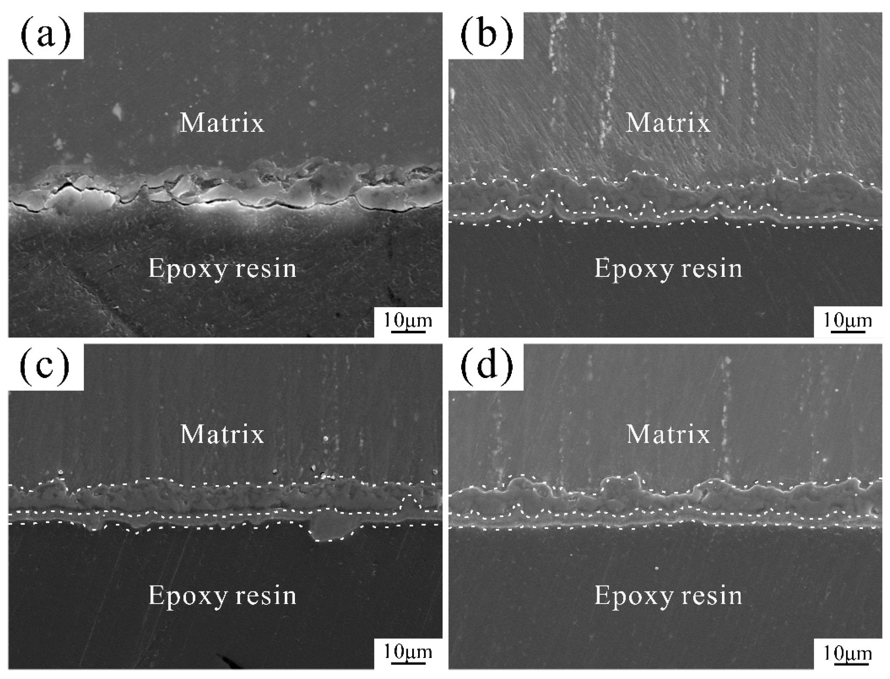
Figure 1. Cross-sectional microstructures of coatings (a) H0; (b) H75; (c) H125; (d) H175.
Coatings 15 01344 g001
Figure 2. Surface morphologies of coatings: (a) H0; (b) H75; (c) H125; (d) H175.
Coatings 15 01344 g002
Figure 3. EDS spectra of labeled regions in Figure 2: (a) Region A; (b) Region B; (c) Region C; (d) Region D.
Coatings 15 01344 g003
Figure 4. XRD patterns of coatings (a) H0; (b) H75; (c) H125; (d) H175.
Coatings 15 01344 g004
Figure 5. Potentiodynamic polarization curves of four samples in Hank’s solution.
Coatings 15 01344 g005
Figure 6. (a) pH evolution of four coatings during immersion in Hank’s solution; (b) Corrosion rates after 14-day immersion.
Coatings 15 01344 g006
Figure 7. Micro morphologies and EDS analysis of labeled regions after 14-day immersion: (a) H0, (b) H75, (c) H125, and (d) H175; (eh) EDS results of areas of A, B, C, and D.
Coatings 15 01344 g007
Figure 8. XRD patterns of post-immersion coatings (a) H0; (b) H75; (c) H125; (d) H175.
Coatings 15 01344 g008
Figure 9. The relative growth rates (RGR) of cells co-cultured with extracts for 1/3/5 days (* p < 0.05). (a,b) depict the RGR values of MC3T3-E1 and MG63 cells, respectively. (c,d) depict the ALP values of MC3T3-E1 and MG63 cells, respectively.
Coatings 15 01344 g009
Figure 10. Antibacterial efficacy against E. coli after 24 h co-culture: (a) H0; (b) H75; (c) H125; (d) H175; (e) blank control; (f) the concentration of released copper ions.
Coatings 15 01344 g010
Table 1. Composition of Hank’s Solution (g/L).
CompositionNaClKClKH2PO4MgSO4·7H2ONaHCO3CaCl2Na2HPO4
Content80.40.060.20.350.140.13
Table 2. Fitted parameters from potentiodynamic polarization curves in Hank’s solution.
Samplesicorr
(μA/cm2)
Ecorr
(V vs. SCE)
Corrosion Rate Pi (mm/y)
H00.75±0.03−1.49 ± 0.010.384 ± 0.01
H750.59±0.002−1.47 ± 0.020.301 ± 0.02
H1250.31±0.001−1.45 ± 0.010.152 ± 0.01
H1750.21±0.002−1.42 ± 0.020.107 ± 0.01
Disclaimer/Publisher’s Note: The statements, opinions and data contained in all publications are solely those of the individual author(s) and contributor(s) and not of MDPI and/or the editor(s). MDPI and/or the editor(s) disclaim responsibility for any injury to people or property resulting from any ideas, methods, instructions or products referred to in the content.