Article

Characteristics of the Gold-Decorated Wooden Sculptures of Qing Dynasty Collected in Qianjiang Cultural Administration Institute, Chongqing, China

1
History and Social Work College, Chongqing Normal University, Chongqing 401331, China
2
Institute of Cultural Heritage, Shandong University, Qingdao 266237, China
3
Department of Cultural Relics Conservation and Archaeology, Chongqing China Three Gorges Museum, Chongqing 400015, China
4
Qianjiang Cultural Administration Institute, Chongqing 409000, China
*
Author to whom correspondence should be addressed.
Coatings 2025, 15(10), 1163; https://doi.org/10.3390/coatings15101163
Submission received: 28 August 2025 / Revised: 17 September 2025 / Accepted: 22 September 2025 / Published: 5 October 2025
(This article belongs to the Section Environmental Aspects in Colloid and Interface Science)

Abstract

Two gold-decorated wooden sculptures of Qing Dynasty collected in Qianjiang Cultural Administration Institute, Chongqing, China, holds significant cultural value. Although in appearance they were preserved completely, the wooden bodies exhibited a certain degree of decay with severe peeling of the surface painted layer and gold lacquer layer. In this study, the samples from the sculptures were characterized by microscopy, SED–EDS (Scanning Electron Microscopy and Energy Dispersive Spectrometer) analysis, and Raman spectroscopy, while the preservation state of wooden core was assessed through the fluorescence microscopy and NREL (National Renewable Energy Laboratory) chemical analysis methods. Findings reveal that the raw material for wooden sculpture is cypress, and holocellulose content of wooden core is as low as 32%. The raw materials for red pigment include cinnabar (HgS) and hematite (Fe2O3). There are multiple layers of lacquer and gold can be observed. There is a layer made of clay, gypsum, or brick ash beneath the lacquer layer and colored layer. The gold layer on the surface adopted traditional Chinese gilding technique which is called sticking gold. This study provides insights into the material properties and technological features of these wooden sculptures, offering a reference for future protection of similar sculptures.

1. Introduction

Lacquer and wooden cultural relics, as a significant branch of China’s cultural heritage, carried unique historical, artistic, and scientific value. These lacquer and wooden cultural relics can be primarily categorized into two types: those unearthed from archaeological sites and those passed down through generations. Extensive research on the excavated lacquer and wooden cultural relics has significantly advanced our understanding of the deterioration mechanisms and improved the dehydration and consolidation techniques for waterlogged lacquerware artifacts [1,2,3,4,5,6,7,8,9,10]. In contrast, studies focusing on lacquer and wooden cultural relics passed down through generations remained relatively limited. While existing research on such surviving wood carvings often emphasize their historical and cultural significance, especially artistic form and aesthetic appreciation [11,12,13,14,15,16,17], and manufacturing techniques and decorative processes by traditional methods such as direct observation and literature review for the purpose of restoration [18,19,20,21,22,23,24]. However, the application of modern scientific analytical methods in preliminary studies of these relics has been relatively limited. For example, X-ray radiography was employed to determine whether the wood carving technique was integral carving or mortise and tenon splicing in the Shenzhen Museum. Multiple techniques such as microscopy, Raman spectroscopy, X-ray diffraction, fluorescence spectroscopy, and infrared spectroscopy were utilized to identify the painting, lacquer layer composition, and manufacturing process of the woodcarving cultural relics in the Museum. Additionally, observing section’s cell tissue to identify species of the wood was utilized [25,26]. Similarly, X-ray imaging, cross-sectional microscopic analysis, X-ray fluorescence surface scanning, Raman spectroscopy, and pyrolysis–gas chromatography/mass spectrometry were applied to scientifically analyze a polychrome wooden sculpture of a Bodhisattva from the Liao Dynasty by the Palace Museum. The painting materials used were identified, and the processes of restoration and reworking were traced. This provided valuable evidence for studying both the craftsmanship and historical context of the artifact [19,27].
Located in southwestern China within the upper Yangtze River basin, Chongqing experiences a subtropical monsoon climate characterized by mild seasons and abundant forestry resources [28]. According to statistics, the current collection of registered wooden cultural relics in Chongqing is about 2621 pieces (sets). Due to the summer heat and fall cool in Chongqing, the humid and hot climate for wooden cultural relics is extremely unfavorable. Coupled with the long-term usage, circulation, and collection of the process of man-made damage and stress erosion and other uncertainties, the degree of disease to cultural relics would be aggravate. Moreover, current protection of wooden cultural relics is not enough [29], the relevant scientific and technological analysis of the research case is relatively small. There is an urgent need to establish methodological frameworks that can guide future scientific analysis and conservation of similar artifacts.
The two gold-decorated wooden sculptures examined in this study are preserved in the Qianjiang Cultural Administration Institute in Chongqing. They are relatively well-preserved in appearance, finely carved, and both decorated with gold leaf and polychromy. However, numerous forms of deterioration can be observed, including decay and cracking of the wooden body, as well as flaking of the paint and lacquer layers. These relics hold significant material value for the study of folk religious beliefs in Qing Dynasty in Chongqing and the craftsmanship of lacquered wooden objects. In this study, multiple modern analytical techniques were employed to characterize the physical and chemical properties of the relics. This approach enabled a comprehensive scientific characterization of this group of relics.

2. Materials and Methods

2.1. Cultural Relics Information

The photos of the two wooden sculptures were shown in Figure 1.
Gold-decorated wooden statue 1: Qing Dynasty. This one has a total height of 88 cm, a width of 35 cm, a thickness of 26 cm, and weighs 20.8 kg. The figure stands on a square platform approximately 5 cm thick. It depicts a shaven-headed monk with a serene expression, dressed in a right-lapel cross-collar robe and a kasaya draped over his left shoulder. The loose sleeves hang down to the knees, and both hands are clasped together in front of his chest.
Gold-decorated wooden statue 2: Qing Dynasty. This one has a total height of 75.5 cm, a width of 24 cm, a thickness of 21 cm, and weighs 10.6 kg. The statue leans slightly to the right due to uneven deterioration of its base. It stands on a platform approximately 5 cm thick. The figure is depicted as a monk with half-closed eyes and a slightly raised mouth, conveying a quiet and peaceful expression. His hands are clasped together in front of the chest. He wears a right-lapel, cross-collar robe with wide sleeves extending to the knees, a kasaya draped over the left shoulder and a kasaya ring hook visible at the front. The overall posture is similar to that of the gold-decorated wooden statue 1.
Different degrees of damage existed in both the two cultural relics, such as decay of the body, peeling of the painted surfaces, gold layers, and lacquer layers. The condition map is shown below (Figure 2).

2.2. Sample Information

A total of 8 samples were used in this study and the details are shown in Table 1.

2.3. Microscopy Analysis

Microscopic observations of the samples were conducted using a 3D digital microscope (VHX-5000, Keyence Corporation, Osaka, Japan) equipped with a super-depth field zoom lens ranging from 20× to 200× magnification.

2.4. SEM-EDS Analysis

A desktop scanning electron microscope (TM4000PlusII, Hitachi Ltd., Tokyo, Japan) coupled with an energy dispersive spectrometer (AZtecOne, Oxford Ltd., Oxford, UK) was used to show the morphology in detail and perform elemental analysis. The test conditions are as follows: high vacuum mode, acceleration voltage 15 Kv. The samples were examined without any conductive coating. They were affixed to sample holders with conductive tape.
S1, Mag = X500, WD = 14.0 mm
S3, Mag = X180, WD = 14.9 mm
S4, Mag = X200, WD = 12.1 mm

2.5. Raman Spectroscopy Analysis

The pigment compositions were analyzed using a Renishaw in Via confocal micro-Raman spectrometer (Renishaw, London, UK). Detection conditions are as follows: wave number range (100–2000 cm−1); objective lens: 50L×; spot size: 1 μm; laser wavelength (532 nm), power, time, and cumulative number of times were adjusted at any time with different samples.

2.6. Slice Analysis

Using manual techniques or a sliding microtome (TU-213, Yamato Koki Kogyo KK, Yamato-shi, Japan) prepared approximately 20 μm think sections of archaeological wood in the three sectional planes (transverse, radial, and tangential). The sections were then embedded in a water-soluble gum as the embedding medium. The embedded samples were placed in a drying oven at 60 °C for continuous drying for 72 h. Finally, the sections were observed using an optical microscope.

2.7. Wood Apecies Identification

The small archaeological wood samples were embedded in Epon812 resin. Sections of 1–1.5 μm thickness were cut using a Leica RM2265 rotary microtome (Leica Microsystems, Wetzlar, Germany). The sections were observed and imaged using an Olympus CX33 optical microscope (Olympus Corporation, Tokyo, Japan) and Olympus BX51 fluorescence microscope (Olympus Corporation, Tokyo, Japan).

2.8. Wood Composition Analysis

Chemical composition analysis was conducted using an Alliance e2695 high-performance liquid chromatography (HPLC) system (Waters Corporation, Milford, MA, USA) and an L6 UV-Vis spectrophotometer (Shanghai Youke Instrument Co., Ltd., Shanghai, China).
The relative contents of cellulose, hemicellulose, and lignin in the wood samples from the wood statue were tested in accordance with the standard procedure [30] established by the U.S. National Renewable Energy Laboratory (NREL). The extractive content and ash content were tested in accordance with Chinese national standards GB/T 35816-2018 [31] and GB/T 36057-2018 [32], respectively.
After the sample is sieved through a 60-mesh sieve, it is extracted with a Benzene-alcohol solution for 8 h to remove lipids. The lipid-free sample is then hydrolyzed with sulfuric acid. After the hydrolysis reaction, the reaction mixture and residue are filtered through a dried G4 sintered glass funnel. The filter residue is used to calculate the acid-insoluble lignin content, while the filtrate is analyzed for acid-soluble lignin and sugar content using a UV-Vis spectrophotometer and ion chromatography, respectively.

3. Results

3.1. Microscopy Observation Results

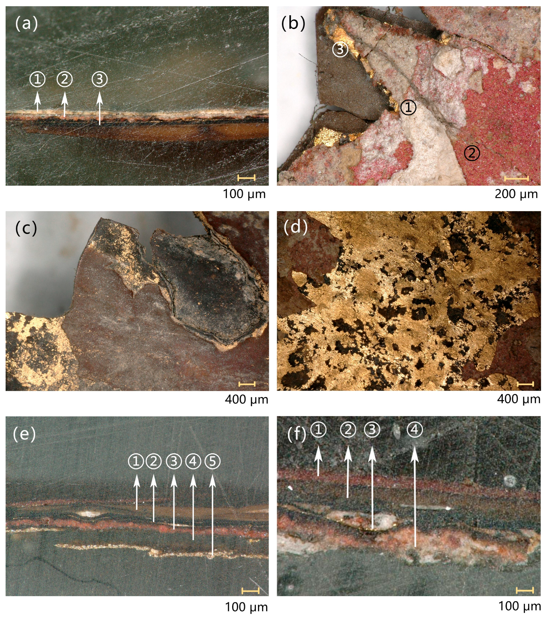
The micrographs of the surface and cross-section of S1 (sample from statue 1) were presented in Figure 3a,b. Based on color differences of the layers in the cross-section of the micrographs (Figure 3a), it was revealed that the decorative layers of the gold-decorated wooden statue 1 primarily consist of three distinct parts, from inside to outside: ① a white ground layer directly contacted with the wood, ② a red painted layer, and ③ a gold lacquer layer. The gold lacquer layer, applied during a later restoration process, covered the underlying painted layer. Additionally, the gold lacquer layer was complex, having been applied more than once. The painted layer was tightly bonded to the white ground layer. White granular crystals were observed within the red painted layer (Figure 3b), which may have functioned as a binding medium.
The surface micrographs of S2 (sample from statue 2) were shown in Figure 3c,d. There were multiple layers of repeated lacquer layers that can be observed from detached part, and some of the lacquer layers were pasted with gold foil (Figure 3c). The color of outer lacquer layer was reddish brown, while the inner layer was blackish, which may be attributed to the type or purity of the pigments added to the layer. From the micrograph of the gold foil layer in Figure 3d, the gold foil was bright with obvious wrinkles and scratches. The micrographs of the cross-section of S3 (sample from statue 2) and S4 (sample from statue 2) were shown in Figure 3e,f. Based on the color differences of each layer in the sample cross-section, the layer sequence of S3 from outside to inside was as follows ① brown lacquer layer, ② black lacquer layer, ③ red pigment layer, ④ black lacquer layer, and ⑤ yellowish-white layer. The layer sequence of S4 from outside to inside presented as ① red lacquer layer, ② brown layer, ③ black layer, and ④ painted layer and ash layer.

3.2. Raman Spectroscopy Analysis Results

To identify the pigments non-destructively, S1 and S5 from gold-decorated wooden statue 1 were tested by Raman. The Raman spectrum of S5 was shown in Figure 4a. The result showed that the Raman peak at 253 cm−1 was consistent with the characteristic peak of cinnabar (HgS) [33]. The Raman spectroscopy analysis result of S1 was the same as that of S5; the red pigment was cinnabar too.
The red layer and yellow layer of S3 and red pigment of S6 from gold-decorated wooden statue 2 were tested by Raman. The Raman spectrum of them were shown in Figure 4b–d. The result showed that the Raman peaks at 223 cm−1, 291 cm−1, and 1316 cm−1 (Figure 3b) were consistent with the characteristic peaks of hematite (Fe2O3) [33], the Raman peaks at 152 cm−1, 200 cm−1, 308 cm−1, 353 cm−1, and 381 cm−1 (Figure 3c) were consistent with the characteristic peaks of orpiment (As2S3) [34], the Raman peaks at 253 cm−1 (Figure 3d) were consistent with the characteristic peaks of cinnabar (HgS). Thus, it was indicated that the red layer of S3 contained hematite, the yellow layer contained orpiment, and the red pigment of S6 was cinnabar. In addition, carbon black was detected in the S6 particles (Figure 4e).
The major color used for the two sculptures was red. Though the two sculptures were both decorated with red pigment, their raw material was different according to the results of Raman spectroscopy analysis. In the gold-decorated wooden statue 1, the red pigment sample was detected as cinnabar. However, in the gold-decorated wooden statue 2, not only cinnabar but also hematite was detected.

3.3. SEM–EDS Analysis Results

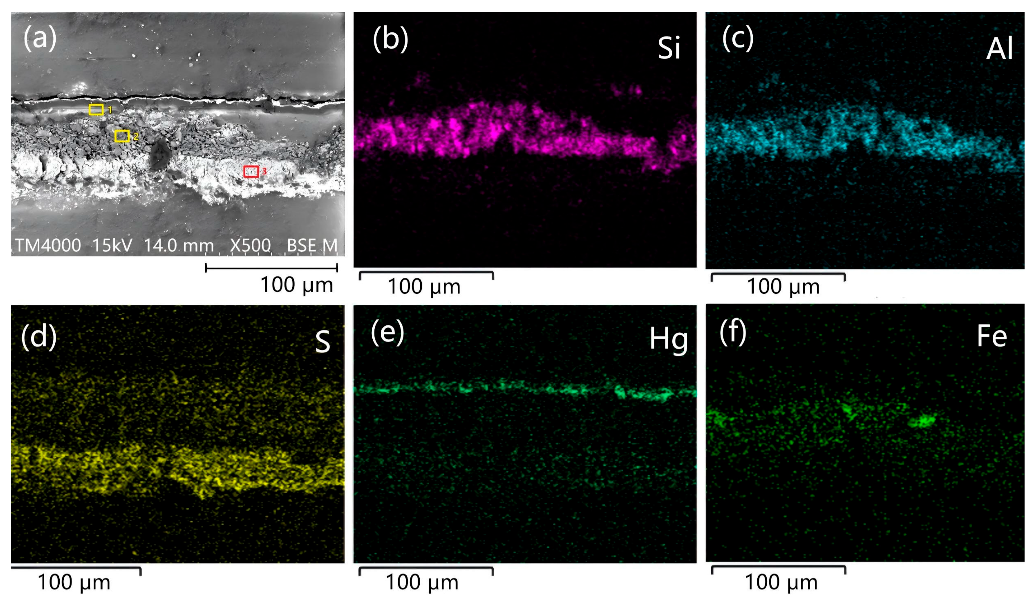
The SEM–EDS results of S1 (sample from statue 1) by cross-section were shown in Figure 5 and Table 2. The element distribution map of S1 (Figure 5) showed that there was an enriched area of Si and Al, which indicated that it may be a lacquer ash layer containing feldspar and quartz [35,36]. In addition, there were also sporadic distributions of Fe in the layer, which may be used as a color-developing element to make the area slightly red. In the upper red part, the element distribution map of Hg showed enrichment, and combined with the Raman spectrum diagram of S1 red pigment layer in the previous text, it can be determined that the red pigment in this layer was cinnabar. The S distribution map and elemental analysis (spot-3, Table 2) revealed S and Ca as dominant element in the light-yellow bottom layer, indicating gypsum (CaSO4·2H2O) as a probable constituent. To verify the inference and receive more information, another powder sample was taken and tested from the light-yellow bottom layer of S1, and the results were shown in Table 3. The EDS results showed that the main elements were Si, Al, Ca, and S. It can be inferred that its priming layer was made by mixing clay-like substances and gypsum-like substances. By comparing the carbon content in black and brown lacquer areas, it can be inferred that the color difference in the lacquer layer may be attributed to variations in carbon content.
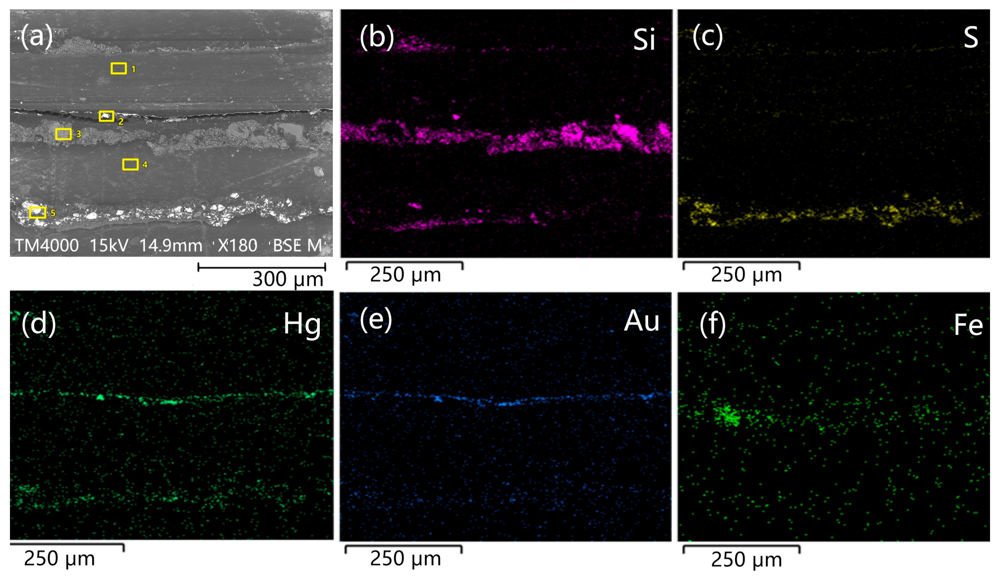
The SEM–EDS result of S3 (sample from statue 2) by cross-section was shown in Figure 6 and Table 4. The cross-section image showed that the structure of S3 can be divided into 5 layers: the element content of spot-5 showed that the main elements of ⑤ yellow-white layer were S (36.98% at spot-5) and As (56.27% at spot-5), and combined with the results of Raman spectroscopy, it can be conformed that the use of orpiment (As2S3). The element content of spot-1 and 4 showed that the main element of layer ① ② ④ was C, which might be the black primer layer made of charcoal ash. The EDS result of layer ③ suggested a high level of Fe (91.5% at spot-3), combined with the results of the Raman spectroscopy; it can be conformed that the pigment used here is iron-red. In the element distribution map of S3, a gold (Au) strip was detected with a high-level content of Au (96.16% at spot-2), suggesting that there was a gold layer in the middle. Combining the layering of gold leaf on the surface observed by microscope, it was inferred that the wooden sculpture decorated by sticking-gold technique.
The SEM–EDS result of S4 (sample from statue 2) by cross-section is shown in Figure 7 and Table 5. According to the element distribution map of Hg and S and EDS result of layer ①, the main elements in the red lacquer layer were Hg (33.68% at spot-1) and S (16.79% at spot-1). This indicated that cinnabar was used in the lacquer layer. The EDS result of layer ② showed that the main elements of the brown lacquer layer were C, suggesting that carbon black was used in the lacquer layer. A gold (Au) strip was detected with 88.37% Au concentration (spot-3), confirming a gold layer embedded within the lacquer layer. The EDS result of spot-5 showed that the main elements of the bottom layer were Si, Al, Ca, etc., and it was presumed that the layer may be a white ash layer made of a mixture of clay and gypsum.

3.4. Wood Analysis Results

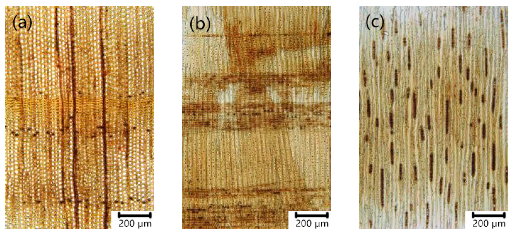
As shown in Figure 8 and Figure 9, the wood microstructure of S7 (sample from statue 1) and S8 (sample from statue 2) exhibited distinct growth rings with sharp transitions between earlywood and latewood. Wood rays were predominantly uniseriate and occasionally biseriate. Ray density ranged from 4 to 7 rays per millimeter. Ray height varied from 1 to 20 cells, with most rays measuring 2 to 15 cells in height. Ray tracheids displayed smooth inner walls. The cross-field pitting between ray parenchyma cells and earlywood tracheids was of the cupressoid type. Resin canals were not observed. Based on these microscopic anatomical characteristics, the wooden cores of both gold-decorated wooden statue 1 and gold-decorated wooden statue 2 were identified as wood from the genus Cupressus within the cypress family CUPRESSACEAE [37].
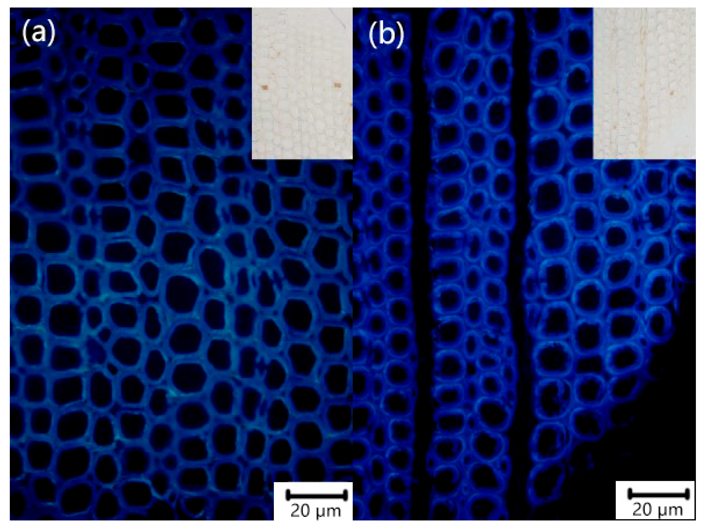
Lignin within the wood cell walls exhibits autofluorescence when excited by ultraviolet (UV) light. This phenomenon enables the observation of lignin distribution patterns across the wood cell wall and facilitates the comparison of relative lignin content differences among various cell types. In fresh wood, the composite intercellular layer (a collective term for the intercellular layer and the primary wall), exhibits a higher relative lignin content. Consequently, under UV excitation, this region displays stronger autofluorescence intensity than the secondary cell walls of the wood.
As shown in Figure 10, the overall fluorescence intensity of this group of wood samples was weaker than that of modern healthy wood, and the distribution of lignin in the whole cell wall tended to be uniform and consistent, and it was not possible to distinguish the difference between composite intercellular layer and secondary wall, and even the composite intercellular layer was weaker than the secondary wall. This was due to the lignin degradation in the cell wall of the wood statue samples, and the lignin structure changes. Due to the degradation of the cell wall, the difference in autofluorescence was relatively weaker.
The test results showed that the holocellulose (the total amount of cellulose and hemicellulose) of the two gold-decorated wooden sculpture samples were around 32%, whereas the holocellulose of modern healthy coniferous timber was usually 60–79% (Table 6), which suggested that a large amount of degradation of the main chemical components in the ancient timber took place, and that the wood deterioration degree was high.

4. Discussion

4.1. Assessment of the Preservation Status of Wood

This study employed fluorescence microscopy and NREL chemical analysis methods to examine wooden cores of the cultural relics. The results revealed that the holocellulose content of these cultural relics was approximately 32%, significantly lower than that of sound wood, indicating severe degradation of cellular components and advanced lignification of the cell structure. These findings provide quantitative data supporting the assessment of deterioration in the wooden cores.

4.2. Manufacturing Techniques and Decorative Processes

Analysis indicated that these two wooden sculptures were crafted from Cupressus wood and decorated with red-colored paint layers and gold lacquer layers, with clear evidence of restoration traces. Based on preliminary examination and literature search, the production steps of the two sculptures may be as follows:
(1)
Forming the shape: the wooden core was carved from Cupressus wood to form the basic shape.
(2)
Decoration and processing: First, a putty made of a mixture clay and gypsum was used for priming, and then decorated by pigments. The main pigment found in these two wooden sculptures was red, composed of cinnabar and hematite.
(3)
Lacquer putty application: a lacquer putty, composed of finely ground and sieved brick dust mixed with raw lacquer, was applied to the wood sculptures.
(4)
Lacquer coating: multiple layers of lacquer were applied over the putty, including a black base lacquer (pigmented with carbon black) and a red lacquer layer (pigmented with cinnabar).
(5)
Gilding: gold leaf was applied onto the lacquer layer using traditional Chinese gilding technique called sticking gold.
Current research indicated that mineral pigments such as cinnabar (HgS) and hematite (Fe2O3) were predominantly employed for the decoration of woodcarvings from the Chongqing region, with no plant-based dyes documented to have been used. In contrast, madder (Rubia tinctorum) has been identified as a botanical colorant in woodcarvings from other regions. Regarding timber selection, Chongqing artisans appeared to choose locally available Cupressus, whereas Cinnamomum camphora (camphor wood) and Cunninghamia lanceolata (China fir) were typically utilized by Guangdong-style gilded lacquer woodcarvings [25,26].

4.3. Evidence of Restoration

The two wooden cultural relics discussed in this study showed traces of later redecoration, which revealed more information about the relics.
Previous microscope observation and SED analysis revealed a complex hierarchical structure in the sample. The red pigment layer was mostly covered beneath the gold lacquer layer, indicating that subsequent redecoration has obscured the original color. Additionally, gold (Au) strips were discovered sandwiched between lacquer layers in structures S3 and S4, which also indicates the existence of restoration traces. Under normal circumstances, it is impossible to apply gold foil to the middle layer. This can only be explained by the possibility that the artifact underwent multiple instances of gold lacquer overpainting by later generations. What made them special was that instead of using painted pigments or colored lacquer directly for decoration during reshaping, the technique of sticking gold leaf over a lacquer layer was adopted. This was consistent with the “gold-ground lacquer” technique mentioned in another two studies on wooden statues and decorative Kanfang in the Chongqing area [38,39]. Wooden Buddhist statues from the moment they are created have the function of being enshrined and worshipped by people. Therefore, during their circulation in later generations, these statues inevitably undergo intentional restoration as a sign of respect. A Liao Dynasty wooden standing Bodhisattva statue collected by the Palace Museum [27] and seven painted woodcarving works in the Museum of Oriental Art in Turin, Italy [40], all have a history of complex restoration and redecoration.

Author Contributions

Methodology, Y.A. and X.F.; formal analysis, Y.A. and X.F.; investigation, Y.A.; resources, K.F. and M.P.; data curation, Y.A. and K.F.; writing—original draft preparation, Y.A.; writing—review and editing, K.F., M.P. and X.F.; visualization, Y.A.; supervision, K.F., M.P. and X.F.; project administration, X.F. All authors have read and agreed to the published version of the manuscript.

Funding

This research was funded by the Foundation of Chongqing Normal University for support, grant number 20xwB005.

Institutional Review Board Statement

Not applicable.

Informed Consent Statement

Not applicable.

Data Availability Statement

The data used during the study are available from the corresponding author on reasonable request.

Acknowledgments

The authors would like to thank the Chongqing China Three Gorges Museum for providing the experimental instruments. We also greatly thank Qianjiang Cultural Administration Institute for supplying the samples.

Conflicts of Interest

The authors declare no conflicts of interest.

References

  1. Zhou, S. Analysis of Corrosion Deformation Types and Mechanisms of Water-Saturated Wooden Cultural Relics. Jianghan Archaeol. 2014, S1, 51–56. [Google Scholar]
  2. Li, M. Research on the Degradation Mechanism and Chemical Protection of Rotten Silk Fabric Cultural Relics and Water-Saturated Bamboo and Wooden Cultural Relics. Ph.D. Thesis, Wuhan University, Wuhan, China, 2014. [Google Scholar]
  3. Chen, G.; Lu, Y.; Zhao, Y. Analysis of Corrosion Diseases and Mechanisms of Wooden Artifacts Unearthed from Mojuizi in Wuwei. Sci. Conserv. Archaeol. 2006, 2, 28–33. [Google Scholar] [CrossRef]
  4. Chen, G.; Lu, Y. Research on the Environmental Corrosion Effect and Mechanism of Rotten Wooden Artifacts Unearthed in Gansu Province. Sci. Conserv. Archaeol. 2009, 21, 67–73. [Google Scholar] [CrossRef]
  5. Chen, Z.; Wei, S.; Fu, Y.; Fang, Q. Analysis of Lacquer from the Zeng Cemetery (1046–771 BCE) at Guojiamiao. Coatings 2024, 14, 1559. [Google Scholar] [CrossRef]
  6. Chen, Z.; Wei, S.; Fu, Y. Pre-protection analysis of unearthed water-saturated lacquered woodware: With lacquered woodware unearthed from the Zeng Guo Cemetery of Guojiamiao, Hubei Province as examples. Res. Sq. 2021. preprint. [Google Scholar] [CrossRef]
  7. Jensen, P.; Jensen, J. Dynamic model for vacuum freeze-drying of waterlogged archaeological wooden artefacts. J. Cult. Heritage 2006, 7, 156–165. [Google Scholar] [CrossRef]
  8. Han, L.; Guo, J.; Tian, X.; Jiang, X.; Yin, Y. Evaluation of PEG and sugars consolidated fragile waterlogged archaeological wood using nanoindentation and ATR-FTIR imaging. Int. Biodeterior. Biodegrad. 2022, 170, 105390. [Google Scholar] [CrossRef]
  9. Rowell, R. Structure and Degradation Process for Waterlogged Archaeological Wood; American Chemical Society: Washington, DC, USA, 1990; pp. 35–50. [Google Scholar] [CrossRef]
  10. Liu, L.; Zhang, L.; Zhang, B.; Hu, Y. A comparative study of reinforcement materials for waterlogged wood relics in laboratory. J. Cult. Heritage 2019, 36, 94–102. [Google Scholar] [CrossRef]
  11. Lu, Z.; Wang, Y. A polychrome wooden sculpture with figure patterns of Liao Dynasty collected in Liaoning Provincial Museum. Steppe Cult. Relics 2017, 2, 98–107. [Google Scholar] [CrossRef]
  12. Dong, S. A polychrome wooden sculpture excavated from the White Elephant Pagoda of the Northern Song Dynasty. Cult. Relics East 2011, 3, 5–11. [Google Scholar]
  13. Cover Introduction: A seated statue of Guanyin Bodhisattva with painted wood carvings from the Song Dynasty. Collector 2019, 9, 1.
  14. Mo, L. On the Aesthetic Value of Chaozhou Wood Carvings in Guangdong Museum. Master’s Thesis, Guangdong University of Technology, Guangzhou, China, 2019. [Google Scholar] [CrossRef]
  15. Painted wooden carvings of seated Bodhisattvas. J. Natl. Mus. China 2018, 3, 161.
  16. Zhen, X. A Brief Analysis of the Characteristics of the polychrome wood Carvings in Shuanglin Temple. Appreciation 2017, 9, 289. [Google Scholar]
  17. Wu, H. An Analysis of the Wood Carvings of Figures from the Huaxi Pavilion in Bozhou during the Qing Dynasty. Tomorrow’s Fash. 2017, 5, 292. [Google Scholar]
  18. Huang, Q. Research on the Restoration of Small Box Buddha Wood Carvings of Standing Bodhisattvas in the Qing Dynasty. Art Mus. Mag. 2024, 4, 28–33. [Google Scholar]
  19. Xie, Y. Restoration Research on Standing Wooden Statues of Bodhisattvas from the Liao Dynasty collected by the Palace Museum. Cult. Relics World 2021, 12, 108–116. [Google Scholar]
  20. Xie, Y. Restoration Research on the Kuilong Pattern Luohan Bed Carved from Purple Sandalwood collected by the Palace Museum. China Cult. Herit. Sci. Res. 2021, 3, 65–71. [Google Scholar]
  21. Wang, Y. The Conservation and restoration of Chaozhou wood Carvings in the collection: Taking the through-carving, lacquered and gilded flower-and-bird pattern Board as an Example. Cult. Relics World 2016, 12, 46–48. [Google Scholar]
  22. Wang, J. The protection and restoration of the wooden Buddha statues unearthed from the Xiude Temple Pagoda in Quyang. In Proceedings of the 9th Academic Annual Conference of the China Association for Conservation Technology of Cultural Heritage, Hebei Provincial Cultural Relics Protection Center, Chongqing, China, 22–24 November 2016. [Google Scholar]
  23. Zhao, Y. The restoration of the Song Dynasty wooden standing Maitreya statue in Longxing Temple. Stories Relics 2002, 6, 61–65. [Google Scholar]
  24. Li, J. Replication and restoration of the lion head knob of the decorative piece “Purple Sandalwood Palace Fan Stand”. J. Jilin Univ. Arts 2010, 2, 25–28. [Google Scholar] [CrossRef]
  25. Lu, Y. Preliminary research on the Protection of Ming Dynasty Polychromoe Woodcarving Seated Statues in the collection. Sci. Conserv. Archaeol. 2014, 26, 57–68. [Google Scholar] [CrossRef]
  26. Lu, Y. The protection research and restoration of a Qing Dynasty lacquered and gilded wooden statue of Guanyin Bodhisattva. Sci. Conserv. Archaeol. 2016, 28, 38–46. [Google Scholar] [CrossRef]
  27. Li, G.; Xie, Y.; Zhang, X.; Wang, N.; Qu, F.; Lei, Y. Scientific analysis of a Polychrome Wooden Bodhisattva Statue in Standing Posture. Collect. Stud. Archaeol. 2020, 91, 108273. [Google Scholar]
  28. Zheng, L.; Xi, Z.; Wu, X.; Wang, Y. Investigation and Conservation of Diseases in Wooden Cultural Relics Collected in Chongqing Region. J. Chongqing Norm. Univ. 2008, 6, 80–86. [Google Scholar]
  29. Guo, Y. Yubei Collection of Cultural Relics Protection and Restoration of Wood. Master’s Thesis, Chongqing Normal University, Chongqing, China, 2016. [Google Scholar]
  30. Sluiter, A.; Hames, B.; Ruiz, R.; Scarlata, C.; Sluiter, J.; Templeton, D.; Crocker, D. Determination of structural carbohydrates and lignin in biomass, Laboratory analytical procedure. Lab. Anal. Proced. 2008, 1617, 1–16. [Google Scholar]
  31. GB/T 35816-2018; Standard Method for Analysis of Forestry Biomass—Determination of Extractives Content. Standardization Administration: Beijing, China, 2018.
  32. GB/T 36057-2018; Method for Analysis of Forestry Biomass—Determination of Ash Content. Standardization Administration: Beijing, China, 2018.
  33. Bell, I.M.; Clark, R.J.; Gibbs, P.J. Raman spectroscopic library of natural and synthetic pigments (pre- ≈ 1850 AD). Spectrochim. Acta Part A Mol. Biomol. Spectrosc. 1997, 53, 2159–2179. [Google Scholar] [CrossRef] [PubMed]
  34. Sun, D.; Li, H.; Zhang, G.; Yin, Y.; Su, M.; Bai, X.; Sikorski, M.; Zhang, D. Exploring the viability of combined laser-induced breakdown spectroscopy and Raman spectroscopy for stratigraphic analysis of murals containing isomeric pigments: A case study on realgar and orpiment. Heritage Sci. 2024, 12, 254. [Google Scholar] [CrossRef]
  35. Jin, P.; Xie, Y.; Li, N. Discussion on the Lacquer Coating Technique of Two Wooden Lacquerware from the Han Dynasty Tomb in Dongyang, Xuyi. Sci. Conserv. Archaeol. 2009, 21, 53–58. [Google Scholar] [CrossRef]
  36. Wu, S.; Cheng, P.; Li, T.; Yang, Y.; Tie, F.; Jin, P. Multispectral Imaging Study of Lacquerware Boxes Excavated from the Haiqu Han Dynasty Tombs, Shandong Province. Spectrosc. Spectr. Anal. 2022, 42, 1150–1155. [Google Scholar]
  37. Chen, J. Chinese Woody Flora: Records of Principal Timber Species; China Forestry Publishing House: Beijing, China, 1992; pp. 26–35. ISBN 9787503803925. [Google Scholar]
  38. Fan, J. Preliminary Study on Restoration and Protection of Gold-Decorated Wood Bodhisattva Statues in Qing Dynasty. Master’s Thesis, Chongqing Normal University, Chongqing, China, 2023. [Google Scholar]
  39. Li, L. Study on the Protection and Restoration of the Qing Dynasty Golded Lacquer Wood Carving Kanfang in the Cultural Heritage Protection Center of Yubei District, Chongqing. Master’s Thesis, Chongqing Normal University, Chongqing, China, 2023. [Google Scholar]
  40. Ricci, C.; Buscaglia, P.; Angelici, D.; Piccirillo, A.; Matteucci, E.; Demonte, D.; Tasso, V.; Sanna, N.; Zenucchini, F.; Croci, S.; et al. A Technical Study of Chinese Buddhist Sculptures: First Insights into a Complex History of Transformation through Analysis of the Polychrome Decoration. Coatings 2024, 14, 344. [Google Scholar] [CrossRef]
Figure 1. Photos of the two wooden sculptures and sampling areas ((a) gold-decorated wooden statue 1, (b) gold-decorated wooden statue 2).
Coatings 15 01163 g001
Figure 2. The condition map of the two wooden sculptures.
Coatings 15 01163 g002
Figure 3. The microscopic images of samples: (a) S1 cross-section, (b) S1 surface view, (c) S2 stratified gold lacquer layer, (d) S2 gold foil, (e) S3 cross-section, (f) S4 cross-section.
Coatings 15 01163 g003
Figure 4. Raman spectrum diagram of samples ((a) red pigment of S5, (b) red layer of S3. (c) yellow layer of S3, (d) red pigment of S6, and (e) black substances mixed in red pigment of S6).
Coatings 15 01163 g004
Figure 5. Element distribution map of the inlaid sample S1 ((a) SEM image, the detection position of EDS analysis is marked in box. (bf) element distribution map of Si, Al, S, Hg and Fe).
Coatings 15 01163 g005
Figure 6. Element distribution map of the inlaid sample S3 ((a) SEM image, the detection position of EDS analysis is marked in box. (bf) element distribution map of Si, S, Hg, Au and Fe).
Coatings 15 01163 g006
Figure 7. Element distribution map of the inlaid sample S4 ((a) SEM image, the detection position of EDS analysis is marked in box. (bf) element distribution map of Si, Al, Hg, Au and S).
Coatings 15 01163 g007
Figure 8. Microscopic view of three sections of S8 wood. (a) transection, (b) radial section, (c) tangential section.
Coatings 15 01163 g008
Figure 9. Microscopic view of three sections of S9 wood. (a) transection, (b) radial section, (c) tangential section.
Coatings 15 01163 g009
Figure 10. Micrograph of the cross-section of the sample under ultraviolet light excitation ((a) S8, (b) S9).
Coatings 15 01163 g010
Table 1. The information on samples.
Sample NumberSample StateSample SourceAnalysis
S1Fragment of the detached decorative layerGold-decorated wooden statue 1Ultra-depth-of-field microscopic observation (surface and cross-section); SEM-EDS and Raman spectroscopy analysis.
S2Fragment of the detached decorative layerGold-decorated wooden statue 2Surface ultra-depth-of field microscopic observation.
S3Fragment of the detached decorative layerGold-decorated wooden statue 2Observe the cross-section after the embedding process. SEM-EDS and Raman spectroscopy analysis.
S4Fragment of the detached decorative layerGold-decorated wooden statue 2Observe the cross-section after the embedding process and SEM-EDS analysis.
S5Sample of red pigment particlesGold-decorated wooden statue 1Raman spectroscopy analysis.
S6Sample of red pigment particlesGold-decorated wooden statue 2Raman spectroscopy analysis.
S7Wood fragmentGold-decorated wooden statue 1Slice analysis, fluorescence microscopy observation, and NREL component analysis.
S8Wood fragmentGold-decorated wooden statue 2Slice analysis, fluorescence microscopy observation, and NREL component analysis.
Table 2. Chemical composition of S1 measured by SEM–EDS (Scanning Electron Microscopy and Energy Dispersive Spectrometer, point scanning).
SpotElement Content (wt%)
CCuAlAuHgSiSCaFePAs
1 black lacquer area96.760.000.16/0.000.750.091.520.72//
2 brown lacquer area65.000.003.58/0.0025.690.430.205.10//
* 3 yellowish-white part/0.007.6421.320.009.6231.8913.489.732.373.95
The parts marked with * are the results after decarbonization and normalization treatment.
Table 3. Chemical composition of gold-decorated wooden statue 1’s white ash layer powder measured by SEM–EDS (point scanning).
SpotElement Content (wt%)
CuAlAuHgSiSCaFePAs
10.0023.058.590.0053.962.090.0010.500.521.29
20.003.506.490.006.0536.4244.762.170.590.03
30.0026.154.220.2551.013.570.2412.824.221.74
Table 4. Chemical composition of S3 measured by SEM-EDS (point scanning).
SpotElement Content (wt%)
CCuAlAuHgSiSCaFePAs
1 brown area98.610.000.010.370.000.290.530.110.08//
* 2 golden yellow area/0.000.1696.160.000.461.190.761.020.000.26
* 3 red area/0.001.980.080.003.161.010.5591.500.061.66
4 black-gray area99.170.000.220.000.130.370.040.040.04//
* 5 yellowish-white area/0.001.031.890.002.7436.980.390.110.5756.27
The parts marked with * are the results after decarbonization and normalization treatment.
Table 5. Chemical composition of S4 measured by SEM-EDS (point scanning).
SpotElement Content (wt%)
CCuAlAuHgSiSCaFePAs
* 1 red paint layer/0.000.9342.4733.681.4416.792.000.141.471.08
2 brown paint layers89.220.000.158.440.420.380.520.560.31//
* 3 golden areas/0.000.8088.373.182.922.090.701.880.060.00
* 4 pale yellow ash layers/0.001.830.210.0080.870.070.001.470.210.41
* 5 pale yellow ash layers/0.0021.5816.810.0040.131.6115.820.180.793.08
The parts marked with * are the results after decarbonization and normalization treatment.
Table 6. Analysis results of chemical components of wooden sculpture samples.
SampleExtract/%Lignin/%Cellulose/%Hemicellulose/%Ash Content/%
S814.38 (±0.55)28.13 (±2.38)28.38 (±1.06)9.52 (±0.71)6.56 (±2.04)
S915.16 (±0.35)32.54 (±1.70)19.75 (±0.20)11.5 (±0.28)7.47 (±1.14)
Disclaimer/Publisher’s Note: The statements, opinions and data contained in all publications are solely those of the individual author(s) and contributor(s) and not of MDPI and/or the editor(s). MDPI and/or the editor(s) disclaim responsibility for any injury to people or property resulting from any ideas, methods, instructions or products referred to in the content.