Article

First-Principles Study on the Electronic Structure and Optical Properties of BiOIO3 Doped with As, Se, and Te

1
Xinjiang Laboratory of Phase Transitions and Microstructures in Condensed Matters, College of Physics and Technology, Yili Normal University, Yining 835000, China
2
School of Information Science and Engineering, Xinjiang University of Science and Technology, Korla 841000, China
3
National Laboratory of Solid State Microstructures, School of Physics, Nanjing University, Nanjing 210093, China
4
Jiangsu Key Laboratory of Artificial Functional Materials, Department of Materials Science and Engineering, College of Engineering and Applied Sciences, Nanjing University, Nanjing 210093, China
5
Department of Physics, Changji University, Changji 831100, China
*
Authors to whom correspondence should be addressed.
Coatings 2025, 15(1), 111; https://doi.org/10.3390/coatings15010111
Submission received: 7 December 2024 / Revised: 2 January 2025 / Accepted: 17 January 2025 / Published: 20 January 2025
(This article belongs to the Special Issue Coatings as Key Materials in Catalytic Applications)

Abstract

:
This study calculates the electronic structure and optical properties of intrinsic BiOIO3 and X-BiOIO3 (X = As, Se, or Te) using PBE (Perdew–Burke–Ernzerhof) and MBJ (Modified Becke–Johnson) functionals based on density functional theory, with MBJ showing better correlation with experimental values. The X-BiOIO3 systems exhibit relative stability under MBJ potential and show crystal lattice distortion compared to intrinsic BiOIO3, creating localized potential differences that enhance polarization and adjust the bandgap. Doping reduces the bandwidth and increases energy level density, promoting electron transitions. Consequently, based on the computational results presented in this paper, it can be inferred that both BiOIO3 and X-BiOIO3 facilitate water hydrolysis and oxygen generation due to their favorable energy band positions. Notably, Se-BiOIO3 exhibits the highest visible light absorption capacity, which may enhance photocatalytic efficiency by strengthening the built-in electric field and promoting charge carrier generation.

1. Introduction

Under specific lighting conditions, TiO2 exhibits photocatalytic properties that enable water splitting for the production of clean and sustainable hydrogen energy, thereby driving traditional photocatalysis research in the scientific community [1]. Subsequent studies have revealed that conventional photocatalysts, such as TiO2, ZnO, and SiO2, possess the capability to not only facilitate water decomposition but also effectively degrade various pollutants [2]. However, the pronounced recombination rate of photo-generated electron–hole pairs in conventional photocatalytic materials significantly hampers their practical implementation in industrial production [3]. In recent years, the quest for novel photocatalytic materials [4] has emerged as a prominent trend in scientific research, garnering significant attention from numerous scholars.
As early as 2004, Grosso et al. [5] proposed the concept of utilizing ferroelectrics in the realm of photocatalysis, subsequently leading to the identification of several ferroelectric materials exhibiting exceptional photocatalytic activity [6,7]. Bismuth-based material BiOIO3, as a representative ferroelectric catalyst, achieved removal efficiencies of 100%, 85.2%, and 65.2% for methyl orange, rhodamine B, and methylene blue, respectively, after 16 min of ultraviolet light exposure. Compared to commercial TiO2 and other bismuth oxides, BiOIO3 nanosheets exhibited significantly enhanced photocatalytic activity, with the apparent reaction rate constant for methyl orange being 10.26 times higher than that of TiO2 [8]. The exceptional photocatalytic ability of BiOIO3 can be attributed to its unique layered structure [9], comprising nested [Bi2O2]2+ and [IO3] units. Notably, the latter exhibits a triangular pyramidal configuration, as illustrated in Figure 1. Due to the asymmetric distribution of its atoms, the separation of positive and negative charge centers gives rise to a dipole moment in the [IO3] triangular pyramidal system, with a flow of negative charge to positive charge. The polarization directions of [IO3] groups are systematically aligned along the C-axis, resulting in a pronounced macroscopic polarization effect [9] in the BiOIO3, as well as the establishment of an inherent electric field due to spontaneous polarization. The intrinsic electric field [10] plays a pivotal role in facilitating rapid carrier migration and suppressing electron–hole recombination, thereby significantly enhancing the photocatalytic efficiency of BiOIO3 [11,12,13]. As a result of these unique properties, BiOIO3 holds great potential for broad applications in the field of photocatalysis.
Although the presence of an internal electric field in BiOIO3 enhances its photocatalytic performance [11], its larger band gap width of 3.3 eV hinders further improvement in its photocatalytic ability [14]. Existing research has demonstrated that doping modification could enhance the photocatalytic performance of BiOIO3. In their experimental studies, Huang et al. [15] employed BiOIO3 and N-BiOIO3 as photocatalysts for the degradation of various pollutants including Rhodamine B, bisphenol A, and tetracycline. Their findings revealed that N-BiOIO3 demonstrated superior photocatalytic degradation efficiency compared to intrinsic BiOIO3. Liu et al. [16] incorporated sulfur atoms into the oxygen sites of BiOIO3, resulting in a downward shift of the conduction band edge and a reduction in the bandgap. Huang et al. [17] accomplished a visible-light photocatalytic performance in the layered bismuth-based photocatalyst BiOIO3 through iodine ion doping. In contrast to pure BiOIO3, the photoresponse range of iodine-doped BiOIO3 was significantly expanded from ultraviolet light to visible light, while attaining a tunable bandgap. The aforementioned modifications result in a reduction in the energy required for electron transition and facilitate the process, thereby significantly enhancing its photocatalytic efficacy. The enhancement achieved through non-metal doping demonstrates the remarkable potential of this modification strategy to improve the photocatalytic performance of BiOIO3. In order to evaluate the photocatalytic performance of BiOIO3 substitutional doped non-metallic elements, this study employs first principles to calculate the electronic structure and optical properties of BiOIO3-doped X. The rationale for selecting As, Se, and Te as doping elements lies in their classification within similar groups (Group VA and Group VIA) of the periodic table. These elements exhibit analogous chemical and physical properties, enabling them to form substitutional or interstitial dopants within the same host material matrix. Consequently, this facilitates the effective modification of the material’s electronic properties. The incorporation of arsenic, selenium, and tellurium can modulate the energy band structure of materials, particularly semiconductors, thereby enhancing light absorption capabilities and improving the separation efficiency of photo-generated carriers, which ultimately boosts photocatalytic activity. Additionally, the introduction of As, Se, and Te atoms significantly alters the carrier concentration of the material, thereby influencing its charge transport properties and reactivity. The obtained calculation results will offer theoretical guidance for the synthesis of BiOIO3 catalysts that are modified using the doping method.

2. Materials and Methods

In this study, the crystal structure of BiOIO3 is orthorhombic, exhibiting a space group of Pca21 (No. 29) [11]. This non-centrosymmetric arrangement (NCS) consists of interleaved [Bi2O2]2+ and [IO3] units. The model of doped BiOIO3, as depicted in Figure 2, demonstrates the utilization of one X atom being substituted for one I atom. Hence, the doping concentration of X-BiOIO3 is precisely 6.25%. The replaced position of atom I must be excluded outside the boundaries in order to mitigate boundary effects. Moreover, within the unit cell, any position of atom I is symmetrical and satisfies the requirements of symmetry. Hence, the replacement position of atom I can be chosen arbitrarily within the unit cell without impacting the final calculation results. In this study, we ultimately selected the coordinate position of replaced atom I as (0.748, 0.366, 0.335). The electronic configurations of the elements discussed in this article are as follows, Bi: 6s2 6p3; I: 5s2 5p5; O: 2s2 2p4; As: 4s2 4p3; Se: 4s2 4p4; Te: 5s2 5p4.
The calculations are predominantly performed using the Vienna Ab-initio Simulation Package (VASP 6) [18], specifically employing the generalized gradient approximation (GGA) within density functional theory (DFT) and utilizing the MBJ semi-local exchange-correlation potential [19] for material characterization. The cut-off energy for the BiOIO3 was set at 550 eV. The K-point grid was determined according to Monkhorst’s scheme [20], employing a 4 × 4 × 4 configuration. The convergence criterion for internal stress was established to be no greater than 0.02 GPa, while the energy convergence threshold was defined as being within 2 × 10⁻⁶ eV/atom. Additionally, the self-consistency iteration convergence accuracy was specified to be 2 × 10⁻⁶ eV/atom, and the MBJ parameter c value was determined to be 1.34. Due to the discrepancy between results obtained from calculations using monoclinic cells and actual conditions, an expanded cell model was employed to optimize computational resources while ensuring accuracy. Ultimately, a 2 × 2 × 1 BiOIO3 supercell model was selected.

3. Results and Discussion

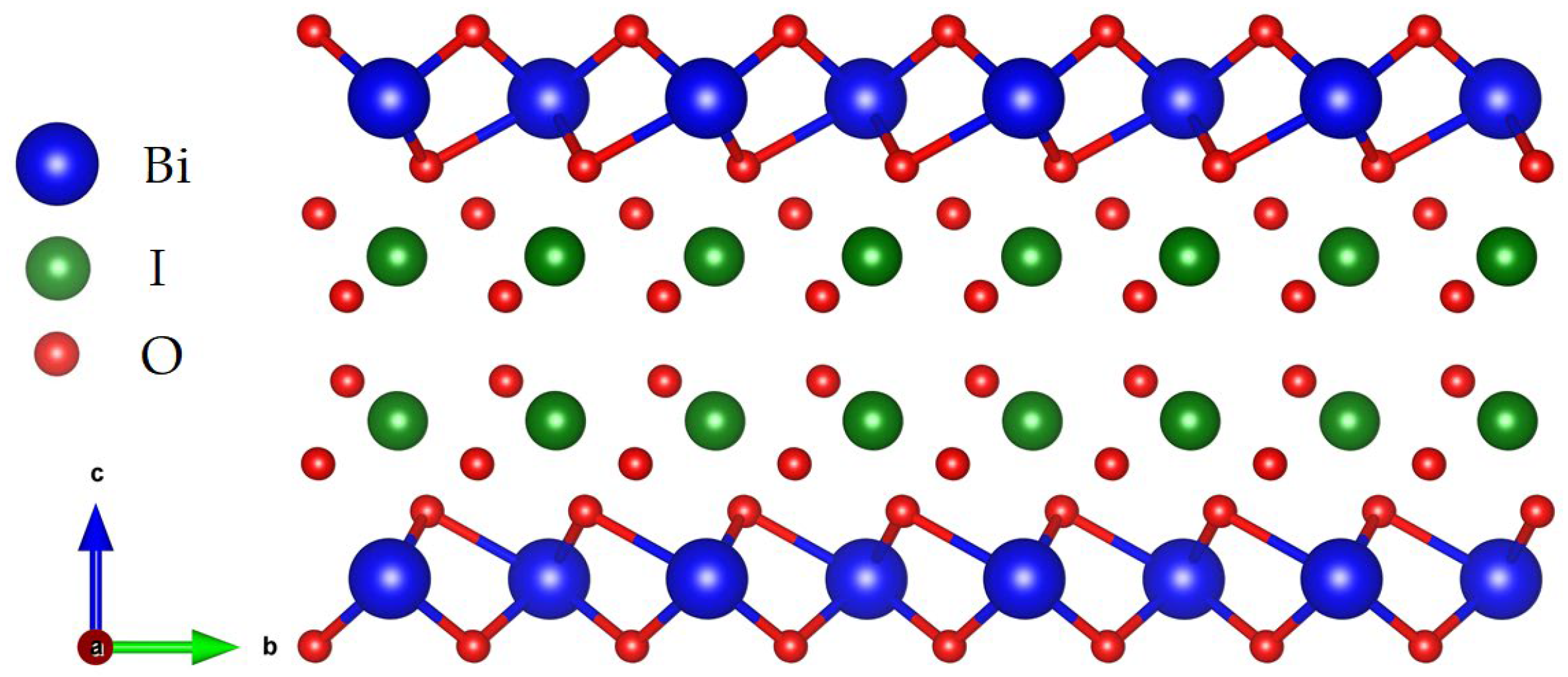
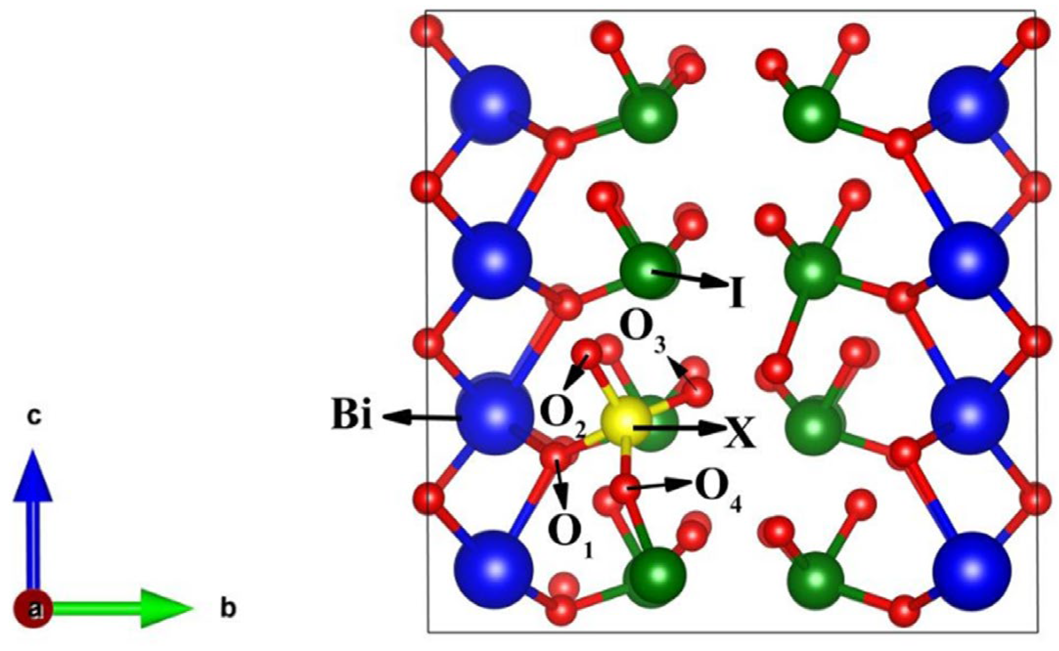
3.1. Geometric Optimization Results

To improve the precision of the computational results, this study conducts geometric optimization on the systems, with the outcomes presented in Table 1. After optimization, it is observed that the lattice constant ratio (c/a) for intrinsic BiOIO3 is 1.013, exhibiting a negligible deviation of only 0.2% from the experimental value of 1.0159. This observation suggests that the computational parameters employed in this study exhibit reliability and generate precise outcomes. The volume of each doped system changes, indicating that these systems experience lattice distortion relative to the calculated values of intrinsic BiOIO3. Considering the inherent non-centrosymmetric (NCS) nature of BiOIO3, it naturally induces a polarization effect. The doping further enhances this phenomenon, facilitating the generation of localized potential differences within the systems and augmenting the migration rate of photo-generated electron–hole pairs.

3.2. Structural Stability of BiOIO3 and X-BiOIO3

To conduct a detailed investigation of the stability of the four systems, the formation energy and binding energy are calculated for each system. The binding energy [22] and formation energy [23] serve to characterize the stability of the system and the ease of doping, respectively. A lower formation energy indicates a greater ease of doping, while a lower binding energy correlates with the enhanced stability of the system. Additionally, the doping formation energy (Ef) [24] and binding energy (Eb) [25] for different systems are determined using Equations (1) and (2).
E f = E X B I O E B I O m μ x + n μ I
E b = 1 / N ( E t o t E B I O )
In the equation, Etot is the total energy of all the atoms of the system in their free state. EXBIO represents the total energy of the system after doping. EBIO denotes the total energy of the BiOIO3. μx and μI denote the chemical formulas of the dopant atom X and the host atom I, respectively. m and n denote the quantities of dopant atoms and substituted atoms, respectively (in this study, m = n = 1). N denotes the total number of atoms present in the system. All the calculated results are listed in Table 2. The binding energy and formation energy of all doped systems exhibit negative values, implying that the formation of these systems is straightforward. The As-doped BiOIO3 demonstrates optimal stability and facile formation, as evidenced by its significantly lower binding energy and formation energy compared to the other systems observed.

3.3. Electronic Structure of BiOIO3 and X-BiOIO3

To further investigate the variations in electronic structure among different systems, this study computes the energy band and electron state density for each system. The high symmetry points in the Brillouin region of the BiOIO3 cells are denoted by Γ (0, 0, 0), M (0.5, 0, 0), K (0.5, 0.5, 0), Z (0, 0.5, 0), and Γ (0, 0, 0), as shown in Figure 3. The line with zero energy is designated as the Fermi level. The density of states for intrinsic BiOIO3 is presented in Figure 3a,b, calculated using the PBE functional [26] and MBJ functional, respectively. The results indicate that the bandgap value obtained from the PBE calculation is 2.043 eV, which deviates by approximately 38% from the experimental value (3.3 eV) [14]. This underestimation occurs because when solving the Cohen–Shen equation, PBE does not consider the excited states of the system, thereby leading to the underestimation of the band gap. To solve this problem, the kinetic energy density term is added to the MBJ function on the basis of GGA-PBE in order to improve the accuracy of energy and band structure calculations. Therefore, the MBJ potential achieves a significantly higher accuracy in energy and band structure calculations [27]. To enhance the accuracy of the bandgap, we employ the MBJ functional to perform a recalculation of the band structure of materials. The recalculated result of 3.308 eV exhibits a deviation of approximately 0.24% from the experimental value, thereby substantiating its reliability. Consequently, we will employ the MBJ functional approach for subsequent sections to conduct further investigations into the electronic structure.
The band structure of non-metallic doped materials is depicted in Figure 4. From the figure, it can be observed that the bandgap values of As-BiOIO3, Se-BiOIO3, and Te-BiOIO3 are calculated as 2.590 eV, 3.171 eV, and 2.867 eV, respectively. These values are found to be smaller than the intrinsic bandgap of BiOIO3 by 21.7%, 4.1%, and 13.3%, respectively, facilitating an easier transition of valence band electrons to the conduction band. Consequently, this enhances the probability of electron transition and further modulates the optical performance of the systems. Among them, As-BiOIO3 exhibits the smallest bandgap, accompanied by an impurity level at 2.072 eV that facilitates electron transitions and enhances the probability of such transitions. Accordingly, doping leads to a denser energy band structure, thereby promoting electron migration.
To further investigate the impact of doping on the electronic structure of the system, we present herein the total and partial density of states prior to and subsequent to doping, as visually depicted in Figure 5. Figure 5a presents the electron density of states for the intrinsic BiOIO3. The valence band maximum (VBM) primarily originates from the 2p orbitals of oxygen and the 6s orbitals of Bi, while the conduction band minimum (CBM) predominantly comprises the 5p orbitals of I, along with the 6p orbitals of Bi and the 2p orbitals of O. Figure 5b presents the electron density of states for the As-doped BiOIO3. The VBM primarily originates from the O-2p, Bi-6s, I-5p, and I-5s orbitals, with a minor contribution from the As-4p orbital. In contrast, the CBM is predominantly composed of the I-5p, O-2s, Bi-6p, and O-2p orbitals, as well as including contributions from the As-4s and As-4p orbitals. Furthermore, the introduction of an impurity element leads to the emergence of a defect energy level, thereby contributing to a reduction in the bandgap width within the system. The presence of this impurity level facilitates electron transitions by serving as a “bridge”, thereby augmenting the likelihood of such transitions and enhancing the optical performance of the system. Figure 5c illustrates the electron density of states for Se-doped BiOIO3, demonstrating that the contributions of Bi, O, and I to the system are largely consistent with those in intrinsic BiOIO3. However, the incorporation of Se leads to an upward shift in the valence band, causing the Fermi level to traverse through it and exhibit typical characteristics of a p-type semiconductor. Additionally, this upward shift of the valence band also leads to a slight reduction in the bandgap. The electron density of states for Te-doped BiOIO3, as shown in Figure 5d, exhibits a striking resemblance to that of Se-doped BiOIO3. The incorporation of Te induces an upward shift in the valence band, thereby conferring p-type semiconductor characteristics upon the system. The conduction band also undergoes a downward shift, resulting in a significant reduction in the width of the bandgap due to the influence exerted by the Te-5s orbital. Consequently, the introduction of As, Se, and Te as dopants in BiOIO3 leads to a reduction in the energy required for electron transitions, thereby indicating an augmentation in the optical performance of the systems.

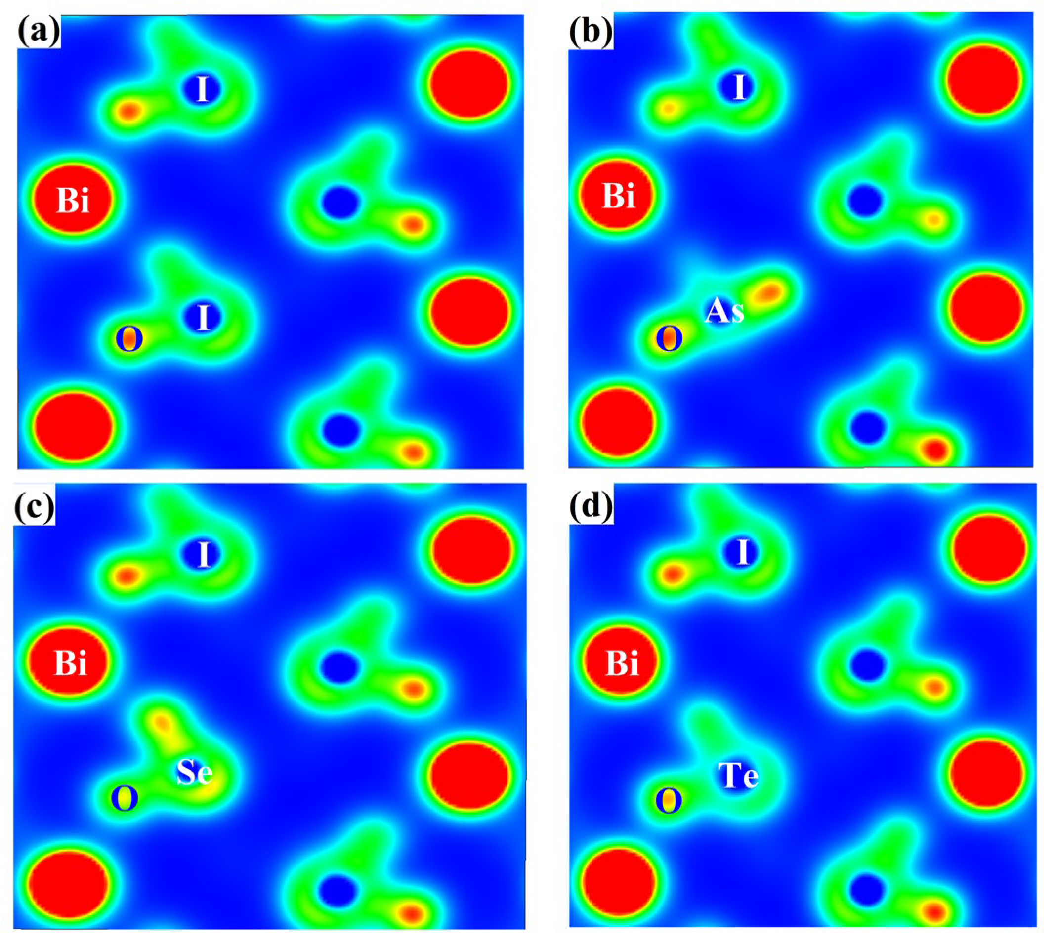
3.4. The Two-Dimensional Electron Density and Bader Charges of BiOIO3 and X-BiOIO3

The electron density diagrams on the (100) plane of BiOIO3 before and after doping are presented in Figure 6. It is evident that doping induces alterations in the distribution of electron clouds within the systems, implying a modification in atomic bonding. The introduction of an As atom into BiOIO3 leads to a shift in the bonding between the As and O atoms due to the deviation in atomic positions, resulting in enhanced charge delocalization near the As atoms, as depicted in Figure 6b. Meanwhile, no significant alteration in the electron cloud distribution is observed for the Se-BiOIO3 and Te-BiOIO3 systems, as depicted in Figure 6c,d. However, when compared to intrinsic BiOIO3, the Se atom demonstrates a higher degree of delocalization while enhancing the localization of the adjacent O atom. This suggests a transfer of electrons from the vicinity of the Se atom to the O atom. When one Te atom replaces one I atom, the degree of electron cloud overlap with the neighboring O atoms decreases, indicating a weakening in the bonding interaction between Te and its adjacent O atoms. Consequently, upon the substitution of I atoms by X atoms, the electron cloud undergoes varying degrees of changes, indicating that doping modifies the internal electron arrangement of the system and facilitates electron transfer [22]. The observed phenomenon can potentially be attributed to the disparity in non-metallic properties among the four atoms. In accordance with Pauling electronegativity, iodine (I) exhibits a higher electronegativity value of 2.66 compared to arsenic (As) at 2.18, selenium (Se) at 2.55, and tellurium (Te) at 2.1. Consequently, the non-metallic characteristics of these atoms follow the order I > Se > As > Te. Thus, upon the substitution of iodine atoms with X atoms, there is a reduction in electron cloud density near this position.
To further investigate the charge transfer dynamics in each system following doping, we employed the Bader charge method to analyze the charges of the oxygen atoms adjacent to the doping sites. The obtained results are presented in Table 3, with atomic numbers cross-referenced in Figure 1.
The table demonstrates a high degree of consistency in the charges of the four nearest oxygen atoms surrounding the I atom in intrinsic BiOIO3, implying an even distribution of electrons among these oxygen atoms and a lack of significant distortion. Upon doping with the non-metal X, a pronounced distortion in the charge distribution of oxygen is observed. In comparison to the intrinsic BiOIO3, the charges of the O atoms adjacent to the doping sites in the doped system are all elevated due to the higher electronegativity of I compared to that of the X atoms. This observation is consistent with electron density analysis findings. The table further demonstrates that variations in the doping element exert a significant influence on the dynamics of charge transfer. In As-BiOIO3, electron transfer predominantly occurs towards the O4 direction, whereas in Se-BiOIO3, there is a relatively higher electron flux towards the O2 direction; similarly, in Te-BiOIO3, there is also a pronounced movement of electrons towards the O2 direction. The charge transfer direction in the doped system has shifted compared to intrinsic BiOIO3, owing to an increased degree of distortion induced by doping. This alteration in the internal charge distribution aligns with previous analytical findings. Hence, it is evident that non-metal X doping can effectively modify the electronic structure and enhance the distortion degree of the system, resulting in a greater deviation between positive and negative charges. Consequently, this strengthens the polarization field and modulates its optical properties [28].

3.5. Optical Properties of BiOIO3 and X-BiOIO3

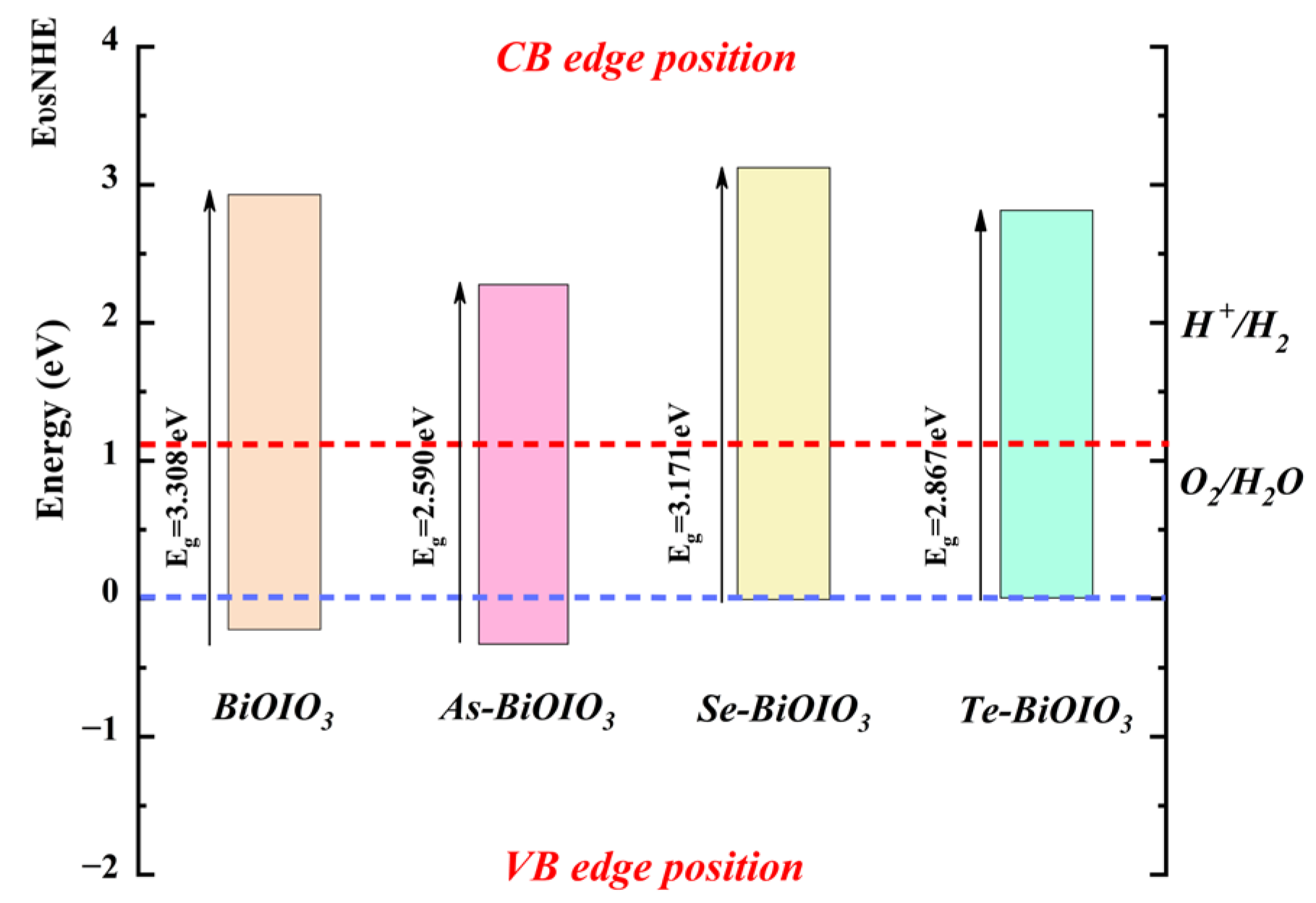
3.5.1. Alignment with Band Structure Edges

The photocatalyst is evaluated in accordance with the relative position of its semiconductor conduction band maximum (CBM) and valence band (VBM) values in relation to the standard hydrogen electrode minimum (NHE). The determination of the band edge potential on the NHE scale typically relies on Equations (3) and (4) [29]. In both equations, X represents the absolute electronegativity of the material. The energy level of the free electron on the NHE scale, approximately 4.5 eV, is denoted as Eelec. Instead, Eg refers to the band gap of a semiconductor.
E V B M = X E e l e c + 0.5 E g
E C B M = E V B M E g
The valence band edge potential of BiOIO3 and X-BiOIO3 at the NHE scale is depicted in Figure 7. The CBM of BiOIO3 and X-BiOIO3 lies above the H+/H2 potential, while the VBM is beneath the O2/H2O potential, suggesting that BiOIO3 and X-BiOIO3 possess the capability of water hydrolysis and oxygen generation. Nevertheless, due to the significant reduction in the bandgap width when Se is doped into BiOIO3, the energy required for electron transition becomes smaller. Hence, it can be inferred that Se-BiOIO3 has a superior water splitting photocatalytic activity among the four systems.

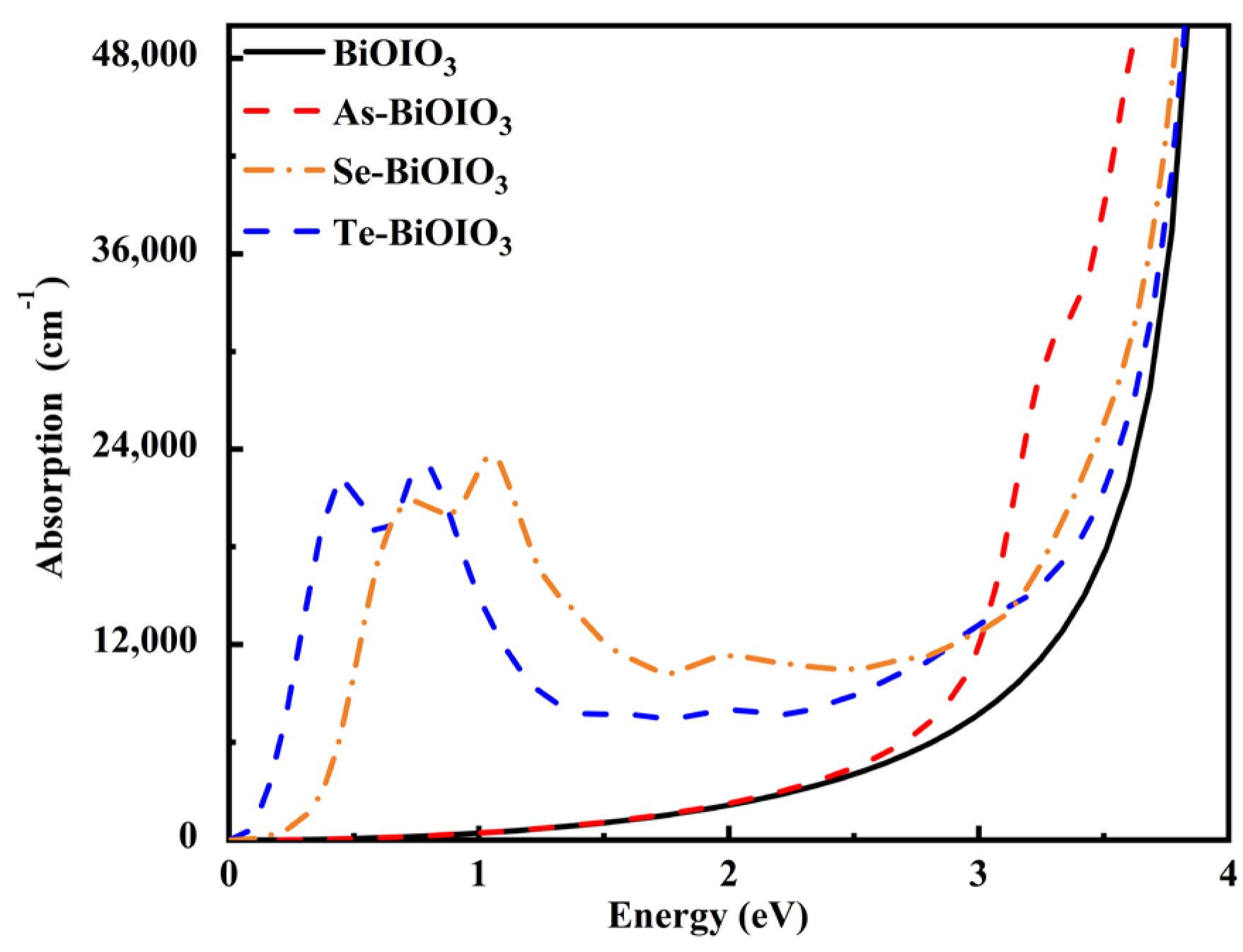
3.5.2. Absorption Spectrum

Doping significantly alters the optical properties of a crystal by introducing impurities that modify the electronic states within the system, thereby enhancing its light responsiveness and influencing its optical characteristics. In this study, we analyze optical absorption graphs and dielectric diagrams to investigate the changes in the optical characteristics of the doped systems.
The optical absorption coefficient α(ω) of a crystal can be determined by utilizing the real part εr(ω) and the imaginary part εi(ω) of the dielectric function, as demonstrated in Equations (5) and (6) [30].
ε ( ω ) = ε r ( ω ) + i ε i ( ω )
α ( ω ) = 2 ( ω ) ε r 2 ( ω ) + ε i 2 ( ω ) ε r ( ω ) 1 2
Theoretically, the optical absorption coefficient of X-BiOIO3 systems exhibits a remarkable enhancement within the visible light range, as illustrated in Figure 8. In comparison to intrinsic BiOIO3, X-BiOIO3 systems demonstrate a significant red shift towards lower energy levels, indicating an augmented light-responsive capacity. These findings suggest that the introduction of As, Se, and Te can significantly enhance the optical absorption capacity of this system. The comparative analysis reveals that among all doping systems, Se-BiOIO3 exhibits the highest light absorption capacity, thereby indicating its superior photocatalytic potential.

3.5.3. Dielectric Function

The dielectric constant characterizes the response of materials to external energy. Figure 9a illustrates the calculated real part of the dielectric function for the four systems both before and after doping, highlighting the trend in the dielectric constant as it varies with incident light energy. The greater the dielectric constant, the stronger the system’s polarization capability and internal electric field strength. When the incident light energy approaches zero, the corresponding value on the vertical axis represents the static dielectric constant. In the figure, the static dielectric constant of the intrinsic BiOIO3 is calculated at 4.929, whereas the static dielectric constants for the X-BiOIO3 systems are recorded as 4.8798, 7.567, and 5.938, respectively. In comparison to intrinsic BiOIO3, the dielectric constants of the doped systems exhibit notable variations. Specifically, the static dielectric constant of As-BiOIO3 is lower than that of intrinsic BiOIO3, suggesting that the incorporation of As diminishes the polarization capability of the system. However, this reduction amounts to only 1.20% relative to intrinsic BiOIO3 and can be considered negligible. Additionally, the dielectric constants of Se-BiOIO3 and Te-BiOIO3 exhibit increases of 53.51% and 20.47%, respectively, compared to the intrinsic BiOIO3. This suggests that doping with Se and Te alters the polarization capability of BiOIO3, leading to an enhancement in internal electric field strength and the accelerated movement of charge carriers within the doped systems, thereby improving charge binding capacity, which aligns with the previous analysis.
Figure 9b illustrates the calculated imaginary part of the dielectric function before and after doping, revealing significant variations among the three X-BiOIO3 systems within the 0–4 eV range. The imaginary part of the dielectric function represents the energy dissipated due to the induction of a large number of electric dipoles within the system in response to external energy stimulation. This value quantitatively reflects the degree of electronic transition, and its peak corresponds to the strengthened inter-band electronic transition of the system under the action of an external electric field. In addition, this value is also related to the probability of the system absorbing external energy [31]. Notably, Se-BiOIO3 exhibits a small peak at 0.370 eV, reaching a maximum value of 2.5236 eV, attributed to an electron transition between the 4p state of Se and the 2p state of O. At 0.592 eV, a new peak is observed in the Te-BiOIO3 system, attributed to the introduction of impurities that induce orbital hybridization and coupling at this energy level. In conclusion, the peak positions of the Se-BiOIO3 and Te-BiOIO3 systems following doping exhibit a tendency to shift towards the low-energy region. This observation aligns with the conclusions derived from band structure analysis and is also associated with the impurity levels introduced by doping. The doping of Se effectively reduces the bandgap of intrinsic BiOIO3, enhances its light absorption coefficient, and strengthens the polarization electric field within the system. Consequently, it can be inferred that this system exhibits superior optical performance.

4. Conclusions

The energy band of intrinsic BiOIO3 is calculated in this study using the PBE functional and MBJ functional based on the first principles of density functional theory. The results obtained under the MBJ functional exhibit superior agreement with experimental values. The X-BiOIO3 systems demonstrate relative stability when calculated using the MBJ potential. In comparison to intrinsic BiOIO3, the X-BiOIO3 systems exhibit crystal lattice distortion, thereby facilitating the generation of localized potential differences within the system. The intensification of polarization promotes the adjustment of the bandgap. The bandwidth of X-BiOIO3 reduces and the energy band distribution becomes denser. The diminished bandwidth and the denser energy levels of X-BiOIO3 suggest that doping enhances the probability of electron transitions. The CBM of BiOIO3 and X-BiOIO3 lies above the H+/H2 potential, while the VBM is beneath the O2/H2O potential, suggesting that BiOIO3 and X-BiOIO3 possess the capability of water hydrolysis and oxygen generation. The expansion of the light absorption range significantly enhances the optical performance of the system. In contrast, the Se-BiOIO3 system exhibits the highest absorption coefficient within the visible light region, followed by that of the Te-BiOIO3 system. Furthermore, both Se and Te doping can modify the polarization capability of BiOIO3. Based on the aforementioned conclusions, it can be deduced that doping with the non-metallic element Se enhances the internal electric field strength, accelerates charge carrier generation within the system, improves charge binding capability, increases electron mobility, and ultimately elevates the photocatalytic efficiency of BiOIO3-based materials, thereby paving a new pathway for the development of photocatalytic materials.

Author Contributions

Conceptualization: F.L., J.L., Y.H., L.Z. and B.Z.; data curation: F.L. and L.Z.; formal analysis: F.L., X.W. and L.Z.; funding acquisition: F.L. and L.Z.; investigation: F.L. and X.W.; methodology: F.L., J.L., L.Z., H.L. and K.J.; project administration: L.Z.; supervision: L.Z.; validation: H.L. and B.Z.; visualization: X.W. and K.J.; writing—original draft: F.L. and J.L.; writing—review and editing: Y.H., L.Z. and B.Z. All authors have read and agreed to the published version of the manuscript.

Funding

This research was funded by the Open Project of Xinjiang Condensed Matter Phase Transformation and Microstructure Laboratory (grant number XJDX0912Z2404), the Science and Technology Plan Project of Yili Kazakh Autonomous Prefecture (grant number YZ2022B021), the Xinjiang Province Graduate Research and Innovation Project (grant number XJ2024G260), and the Innovation and Entrepreneurship Training Project of Yili Normal University Students (grant number S202310764004).

Institutional Review Board Statement

Not applicable.

Informed Consent Statement

Not applicable.

Data Availability Statement

The original contributions presented in the study are included in the article; further inquiries can be directed to the corresponding authors.

Acknowledgments

We gratefully acknowledge HZWTECH for providing computation facilities.

Conflicts of Interest

The authors declare no conflicts of interest.

References

  1. Fujishima, A.; Honda, K. Electrochemical Photolysis of Water at a Semiconductor Electrode. Nature 1972, 238, 37–38. [Google Scholar] [CrossRef] [PubMed]
  2. Chatterjee, D.; Dasgupta, S. Visible light induced photocatalytic degradation of organic pollutants. J. Photochem. Photobiol. C Photochem. Rev. 2005, 6, 186–205. [Google Scholar] [CrossRef]
  3. Ma, S.; Yu, X.; Li, W.; Kong, J.; Long, D.; Bai, X. Bismuth-based photocatalysts for pollutant degradation and bacterial disinfection in sewage system: Classification, modification and mechanism. Environ. Res. 2024, 264, 120297. [Google Scholar] [CrossRef]
  4. El-Bahy, S.M.; Arshad, J.; Munir, S.; Chaudhary, K.; Alhashmialameer, D.; Eddy, D.R.; Warsi, M.F.; Shahid, M. Improved photocatalytic performance of a new silver doped BiSbO4 photocatalyst. Ceram. Int. 2022, 48, 23915. [Google Scholar] [CrossRef]
  5. Grosso, D.; Boissière, C.; Smarsly, B.; Brezesinski, T.; Pinna, N.; Albouy, P.A.; Amenitsch, H.; Antonietti, M.; Sanchez, C. Periodically ordered nanoscale islands and mesoporous films composed of nanocrystalline multimetallic oxides. Nat. Mater. 2004, 3, 787–792. [Google Scholar] [CrossRef] [PubMed]
  6. Mohan, S.; Subramanian, B. A strategy to fabricate bismuth ferrite (BiFeO3) nanotubes from electrospun nanofibers and their Solar light-driven photocatalytic properties. RSC Adv. 2013, 3, 23737–23744. [Google Scholar] [CrossRef]
  7. Cui, D.H.; Zheng, Y.F.; Song, X.C. Hydrothermal synthesis, characterisation and photocatalytic properties of BiOIO3 nanoplatelets. J. Exp. Nanosci. 2016, 11, 1000–1010. [Google Scholar] [CrossRef]
  8. Li, J.; Xie, J.; Zhang, X.; Lu, E.; Cao, Y. The Solid-State Synthesis of BiOIO3 Nanoplates with Boosted Photocatalytic Degradation Ability for Organic Contaminants. Molecules 2023, 28, 3681. [Google Scholar] [CrossRef] [PubMed]
  9. Zhi, Z.; Chen, T.Z.; Shui, J.Y.; Zhi, Y.Y.; Yun, Y. Ferroelectric polarization effect promoting the bulk charge separation for enhance the efficiency of photocatalytic degradation. Chem. Eng. J. 2021, 410, 128430. [Google Scholar]
  10. Huang, H.; Chen, F.; Reshak, A.H.; Auluck, S.; Zhang, Y. Insight into crystal-structure dependent charge separation and photo-redox catalysis: A combined experimental and theoretical study on Bi(IO3)3 and BiOIO3. Appl. Surf. Sci. 2018, 458, 129–138. [Google Scholar] [CrossRef]
  11. Dong, X.D.; Yao, G.Y.; Liu, Q.L.; Zhao, Q.M.; Zhao, Z.Y. Spontaneous polarization effect and photocatalytic activity of layered compound of BiOIO3. Inorg. Chem. 2019, 58, 2–9. [Google Scholar] [CrossRef] [PubMed]
  12. Patiphatpanya, P.; Phuruangrat, A.; Thongtem, S.; Kungwankunakorn, S.; Thongtem, T. Effect of microwave power on phase, morphology, and photocatalytic properties of BiOIO3 nanostructure. J. Aust. Ceram. Soc. 2019, 55, 501–506. [Google Scholar] [CrossRef]
  13. Sun, Y.; Xiong, T.; Dong, F.; Huang, H.; Cen, W. Interlayer-I-doped BiOIO3 nanoplates with an optimized electronic structure for efficient visible light photocatalysis. Chem. Commun. 2016, 52, 8243–8246. [Google Scholar] [CrossRef]
  14. Nguyen, S.D.; Yeon, J.; Kim, S.; Halasyamani, P.S. BiO(IO3): A New Polar Iodate that Exhibits an Aurivillius-Type (Bi2O2)2+ Layer and a Large Shg Response. J. Am. Chem. Soc. 2011, 133, 12422–12425. [Google Scholar] [CrossRef] [PubMed]
  15. Huang, L.; Wang, Y.; Li, Y.; Huang, S.; Xu, Y.; Xu, H.; Li, H. Calcination synthesis of N-doped BiOIO3 with high Led-light-driven photocatalytic activity. Mater. Lett. 2019, 246, 219–222. [Google Scholar] [CrossRef]
  16. Liu, Y.L.; Zhu, Z.R.; Liu, Y.Q.; Jiang, W.; Yang, L.; Zi, J.X.; Shuai, Q.; Yu, B.W.Y.; Meng, J.B. First principles insight on enhanced photocatalytic performance of sulfur-doped bismuth oxide iodate. Mater. Sci. Semicond. Process 2023, 165, 2–7. [Google Scholar] [CrossRef]
  17. Huang, H.; Ou, H.; Feng, J.; Du, X.; Zhang, Y. Achieving highly promoted visible-light sensitive photocatalytic activity on BiOIO3 via facile iodine doping. Colloids Surf. A Physicochem. Eng. Asp. 2017, 518, 158–165. [Google Scholar] [CrossRef]
  18. Yi, W.; Tang, G.; Chen, X.; Yang, B.; Liu, X. qvasp: A flexible toolkit for VASP users in materials simulations. Comput. Phys. Commun. 2020, 257, 107535. [Google Scholar] [CrossRef]
  19. Debidatta, B.; Jisha, A.A.; Sharma, R.; Sanat, K.M.; Ekta, J. First Principles Study of New d0 Half-Metallic Ferromagnetism in CsBaC Ternary Half-Heusler Alloy. J. Supercond. Nov. Magn. 2022, 35, 3431–3437. [Google Scholar]
  20. Monkhorst, J.; Pack, J.D. Special Points for Brillouin-Zone Integrations. Phys. Rev. B 1976, 13, 5188–5192. [Google Scholar] [CrossRef]
  21. Wu, J.; Kai, X.; Qi, Z.L.; Chen, H.Q.; Xue, M.Q.; Hui, Z.; Yu, G.; Ping, H.; Liang, J.Z. Controlling dominantly reactive (010) facets and impurity level by in-situ reduction of BiOIO3 for enhancing photocatalytic activity. Appl. Catal. B Environ. 2018, 232, 136–144. [Google Scholar] [CrossRef]
  22. Arora, A.; Nandi, P.; De Sarkar, A. Ferroelectricity-controlled magnetic ordering and spin photocurrent in NiCl2/GeS multiferroic heterostructures. J. Phys. Condens. Matter 2024, 36, 445301. [Google Scholar] [CrossRef] [PubMed]
  23. He, J.; Liu, G.; Li, X.; Wang, H.; Zhang, G. First-principles study of strain on BN-doped arsenene. J. Mol. Model. 2022, 28, 190. [Google Scholar] [CrossRef]
  24. Shahriar, R.; Hoque, K.S.; Tristant, D.; Zubair, A. Vacancy induced magnetism and electronic structure modification in monolayer hexagonal boron arsenide: A first-principles study. Appl. Surf. Sci. 2022, 600, 154053. [Google Scholar] [CrossRef]
  25. Qi, S.; Wu, S.; Zhang, Y.; Guan, L.; Zhang, K. Construction and first-principles analysis of BiOI and Ni doped MoS2 Z-type heterojunctions. J. Solid State Chem. 2024, 335, 124658. [Google Scholar] [CrossRef]
  26. Zhou, J.; Li, D.; Zhao, W.; Jing, B.; Ao, Z.; An, T. First-Principles Evaluation of Volatile Organic Compounds Degradation in Z-Scheme Photocatalytic Systems: MXene and Graphitic-CN Heterostructures. ACS Appl. Mater. Interfaces 2021, 13, 23843–23852. [Google Scholar] [CrossRef] [PubMed]
  27. Ghaithan, H.M.; Alahmed, Z.A.; Qaid, S.M.H.; Aldwayyan, A.S. Structural, Electronic, and Optical Properties of CsPb(Br1−XClx)3 Perovskite: First-Principles Study with PBE-GGA and mBJ-GGA Methods. Materials 2020, 13, 4944. [Google Scholar] [CrossRef] [PubMed]
  28. Chen, Q.; Feng, X.; Teng, D.; Xu, J.; Cao, S.; Rydosz, A.; Kong, J.; Gao, F. Effect of Intrinsic Nb5+ Vacancy On Dielectric and Polarization Behaviors of KSr2Nb5O15: First-Principles Investigation. Phys. Status Solidi (B)-Basic Solid State Phys. 2021, 259, 2100488. [Google Scholar] [CrossRef]
  29. Zhao, X.; Dai, M.; Lang, F.M.; Zhao, C.; Chen, Q.Y.; Zhang, L.L.; Huang, Y.N.; Lu, H.M.; Qin, C.X. Investigating the Impact of Stress on the Optical Properties of GaN-MX2 (M=Mo, W; X=S, Se) Heterojunctions Using the First Principles. Catalysts 2024, 14, 732. [Google Scholar] [CrossRef]
  30. Masihi, A.; Naseri, M.; Fatahi, N. A first-principles study of the electronic and optical properties of monolayer α-PbO. Chem. Phys. Lett. 2019, 721, 27–32. [Google Scholar] [CrossRef]
  31. Ling, S.Y.; Bang, L.D.; Yu, X.H. Optical properties of arsenene nanoribbons: A first principle study. Mater. Sci. Semicond. Process 2021, 136, 106139. [Google Scholar]
Figure 1. Model diagram of BiOIO3.
Coatings 15 00111 g001
Figure 2. Model diagram of BiOIO3 doped with non-metallic elements X. The symbols O1, O2, O3, and O4 represent oxygen atoms with different symmetries in the BiOIO3 crystal lattice.
Coatings 15 00111 g002
Figure 3. BiOIO3 band diagrams. (a) BiOIO3-PBE; (b) BiOIO3-MBJ.
Coatings 15 00111 g003
Figure 4. Band diagrams of doping systems. (a) As-BiOIO3; (b) Se-BiOIO3; (c) Te-BiOIO3.
Coatings 15 00111 g004
Figure 5. Total electron state density and partial electron state density. (a) Intrinsic BiOIO3; (b) As-BiOIO3; (c) Se-BiOIO3; (d) Te-BiOIO3.
Coatings 15 00111 g005
Figure 6. Two-dimensional electron density maps of plane (100). (a) Intrinsic BiOIO3; (b) As-BiOIO3; (c) Se-BiOIO3; (d) Te-BiOIO3. The red area represents the region with a higher electron density, while the blue area represents the region with a lower electron density. Yellow region represents a positive charge area.
Coatings 15 00111 g006
Figure 7. The potentials of BiOIO3 and X-BiOIO3 with edge characteristics. In the diagram, the blue dashed line represents the energy level of H+/H2, while the red dashed line corresponds to the energy level of O2/H2O.
Coatings 15 00111 g007
Figure 8. The absorption spectrum diagram of BiOIO3 and X-BiOIO3 systems.
Coatings 15 00111 g008
Figure 9. BiOIO3 and X-BiOIO3 dielectric function diagrams: (a) the dielectric real part; (b) the dielectric imaginary part.
Coatings 15 00111 g009
Table 1. The lattice constant and volume of each model.
Modela/nmb/nmc/nmV/nm3
BiOIO3 (Experiment) [21]0.56581.10390.5748/
BiOIO3 (This work)0.57121.12630.57881.4898
As-BiOIO30.56981.13110.57791.4899
Se-BiOIO30.57061.12370.57801.4826
Te-BiOIO30.56951.12720.57751.4830
Table 2. The total energy, binding energy, and formation energy of each model before and after doping.
ModelE/eVEb/eVEf/eV
BiOIO3−477.2118//
As-BiOIO3−479.9295−0.3186−4.9405
Se-BiOIO3−479.3817−0.3183−2.1449
Te-BiOIO3−479.9295−0.3185−2.6956
E represents the total energy of BiOIO3 and the X-BiOIO3 systems.
Table 3. Bader charge of O atom near the doping site.
ModelThe Charge of O Atoms
O1O2O3O4Average
BiOIO36.82606.91506.96626.91196.9048
As-BiOIO37.07627.02546.90877.13567.0364
Se-BiOIO37.02037.04996.97367.01257.0141
Te-BiOIO37.03047.16117.01027.14117.0857
Disclaimer/Publisher’s Note: The statements, opinions and data contained in all publications are solely those of the individual author(s) and contributor(s) and not of MDPI and/or the editor(s). MDPI and/or the editor(s) disclaim responsibility for any injury to people or property resulting from any ideas, methods, instructions or products referred to in the content.

