Article

Influence of Magnesium Content on the Physico-Chemical Properties of Hydroxyapatite Electrochemically Deposited on a Nanostructured Titanium Surface

by
Cosmin Mihai Cotrut
1,
Elena Ungureanu
1,
Ionut Cornel Ionescu
1,*,
Raluca Ioana Zamfir
1,
Adrian Emil Kiss
2,
Anca Constantina Parau
2,
Alina Vladescu
2,
Diana Maria Vranceanu
1,* and
Adriana Saceleanu
3
1
Faculty of Materials Science and Engineering, University Politehnica of Bucharest, 313 Independentei Street, RO60042 Bucharest, Romania
2
Department for Advanced Surface Processing and Analysis by Vacuum Technologies, National Institute of Research and Development for Optoelectronics-INOE 2000, 409 Atomistilor St., RO77125 Magurele, Romania
3
Faculty of Medicine, Lucian Blaga University of Sibiu, 2A Lucian Blaga Street, RO550169 Sibiu, Romania
*
Authors to whom correspondence should be addressed.
Coatings 2022, 12(8), 1097; https://doi.org/10.3390/coatings12081097
Submission received: 10 June 2022 / Revised: 28 July 2022 / Accepted: 29 July 2022 / Published: 2 August 2022
(This article belongs to the Special Issue Surface Modification of Medical Implants)

Abstract

:
The aim of this research was to obtain hydroxyapatite (HAp)-based coatings doped with different concentrations of Mg on a Ti nanostructured surface through electrochemical techniques and to evaluate the influence of Mg content on the properties of HAp. The undoped and doped HAp-based coatings were electrochemically deposited in galvanostatic pulsed mode on titania nanotubes with a diameter of ~72 nm, being designed to enhance the adhesion of the HAp coatings to the Ti substrate. The obtained materials were investigated by Scanning Electron Microscopy (SEM), Energy Dispersive Spectroscopy (EDS), X-Ray Diffraction (XRD), and Fourier-Transform Infra-Red spectroscopy (FTIR). The adhesion of the coatings to the substrate was also evaluated with the help of the “tape-test” and the micro-scratch test. The morphology (SEM) of all the coatings is made of very thin and narrow ribbon-like crystals, with some alterations with respect to the Mg amount in the coatings. Thus, a concentration of 1 mM of Mg in the electrolyte leads to wider and thicker ribbon-like crystals, while a concentration of 1.5 mM in the electrolyte generated a morphology that resembles the undoped HAp. Both phase composition (XRD) and chemical bonds (FTIR) analysis proved the formation of HAp in all coatings. Moreover, according to XRD, all coatings have a strong orientation toward the (002) plane. Irrespective of the Mg content, all coatings registered an average roughness between approx. 500 and 600 nm, while the coating thickness increased after addition of Mg, from a value of 9.6 μm, for the undoped HAp, to 11.3 μm and ~13.7 μm for H/Mg1 and H/Mg2, respectively. In terms of adhesion, it was shown that the coatings a H/Mg2 had a poorer adhesion when compared to H/Mg1 and the undoped HAp (H), which registered similar adhesion, indicating that a concentration of 1.5 mM of Mg in the electrolyte reduces the adhesion of the Hap-based coatings to the nanostructured surface. The obtained results indicated that Mg concentrations up to 1 mM in the electrolyte can enhance the properties of HAp-based coatings electrochemically deposited on a nanostructured surface, while even a slightly higher concentration of 1.5 mM can negatively impact the characteristics of HAp coatings.

1. Introduction

Metallic materials have been used to replace and reconstruct the skeletal functions of the human body for a very long time [1,2]. Due to their high strength, toughness, and durability, approximately 70–80% of the implantable materials are made of metallic biomaterials [3,4,5]. The need for metallic biomaterials with proper features and characteristics is rapidly growing as the world is progressively aging, and elderly people tend to present a higher risk of hard tissue failure [6,7].
Within this context, titanium (Ti) and its alloys are often the material of choice due to a favorable combination of properties such as excellent corrosion resistance which offers long-term stability, high biocompatibility, and reliable mechanical strength, with minimal long-term toxicity to the host [8,9]. Nevertheless, one weakness of the implantable metallic biomaterials is given by their no bio-function recognized as bioinert character. For metallic biomaterials such as the class of Ti-based alloys, imparting multifunctionality on its bioinert character is generally carried out by surface modification techniques, such as surface structuring and/or coating with bioactive ceramics and polymers [10,11].
Generally, the aim of the material’s coating is to protect or to enhance its functionality, and it is certainly not a new concept [12]. In medicine, the coatings field is rapidly diversifying, aiming to improve osseointegration and to tackle the bioinert character of the metallic implants [13,14].
Hydroxyapatite (HAp, Ca10(PO4)6(OH)2) is the main inorganic phase of the hard tissue with a Ca/P ratio of 1.67 [15,16,17] that has been extensively studied and used as biomaterial in the past decades [18]. The extensive research conducted in this period highlighted that HAp is brittle and not appropriate for load-bearing applications, such as large bone defects [19,20,21], but it can be successfully used for other types of treatments such as bone substitutes for small bone defects [22], as scaffolds along with other materials such as polymers [23,24], and also as coatings [25,26] to control and accelerate osseointegration [27,28].
The features of HAp such as osteointegration capacity, mechanical properties, and bioactivity can be enhanced through the addition of small amounts of elements that are found in natural bones and tooth mineral [29,30].
Magnesium (Mg) is a cation present in the human body, which despite its low concentration (between 0.5 and 1.5 wt.%) promotes bone cells activation and proliferation. It influences bone strength, mineralization, and growth [31,32], and it plays an important role in the changes in the bone matrix that determine bone fragility [33]. In terms of applicability, Mg can be used as pure metal and/or alloys [34,35], but also as a doping element [36,37] in HAp or other types of CaP [38].
According to Tampieri et al. [39] and Ren et al. [40], Mg2+ prefers the Ca (I) site of the hydroxyapatite structure, while Kannan et al. [41] have shown that Mg2+ is essentially accommodated in the Ca (II) site. Laurencin et. al. [42] found that at 10% Mg concentration, Mg ions will replace Ca in the Ca (II) site. Therefore, a possible explanation would be that the place of Mg substitution depends on the preparation method and on the amount of Mg, indicating that different methods lead to different results.
In terms of biocompatibility and bioactivity characteristics, research studies have highlighted that Mg addition into HAp has a beneficial effect [38,43,44,45]. Chirica et al. [46] investigated the physico-chemical properties of HAp doped with various elements, including Mg, and showed that Mg favors the initial cell adhesion. Chen et al. [47] reported that Mg2+ ions in HAp enhance cell attachment, proliferation, and differentiation of cells to an osteoblast phenotype, as well as the expression of osteogenesis-related markers. These findings are also supported by the results of He et al. [48], who have shown that Mg-substituted HAp enhances the differentiation of stem cells into osteoblasts. In a study carried out by Jiao et al. [49] regarding the effect of Mg–HAp coatings with various Mg concentrations, it was shown that an Mg concentration up to 2 wt.% provided better biocompatibility. Thus, it can be stated that special attention must be taken regarding toward the proper selection of the Mg concentration since at a high concentration the HAp properties can deteriorate [50,51].
Hydroxyapatite-based coatings can be synthesized through different techniques such as a biomimetic method [52], plasma spraying [37,53], sputtering deposition [54,55], sol–gel process [56], laser deposition [57,58], and electrochemical deposition [59]. Among these techniques, electrochemical deposition (ED) appears to be more suitable to develop HAp-based coatings due to its simplicity and good control over the involved electrochemical parameters, which dictates the coatings properties and behavior. It is also more accessible in terms of costs and can be employed to coat complex shapes because it is not a line-of-sight technique [59,60,61].
Mg-substituted HAp is most often obtained by an aqueous precipitation method [62,63] and a hydrothermal method [64]. In our previous work [65], we have shown that Mg-doped HAp coatings can also be successfully obtained by pulsed galvanostatic mode on Ti6Al4V and that a small amount of Mg has a beneficial effect on the HAp properties. Among the drawbacks of the ED technique, when employed to obtain HAp-based coatings, there is unsatisfactory adhesion of the coating to the substrate. To overcome this vulnerability and to enhance the adhesion of the HAp-based coatings on the Ti substrate, an intermediate layer consisting of titania (TiO2) nanotubes (NT) can be developed via anodic oxidation [66,67]. Growth of HAp inside the hollow nanotubes would give an anchoring effect to the electrodeposit and enhance the interfacial bond strength at the TiO2 layer-HAp coating interface [68].
It is known that compared to bare Ti, the TiO2 NT surface provides an optimum biological environment for bone tissue through the ingrowth of mineralized tissue into the pore space and consequently improved osseointegration [69,70,71,72]. The biocompatibility of the TiO2 nanotube was confirmed by subcutaneous injection of the TiO2 nanotube into rats. In this case, the TiO2 nanotube did not cause chronic inflammation or fibrosis [73]. By analyzing the histological features and fluorochrome labeling changes after implantation, Wang et al. [74] showed a notable increase in bone implant contact and gene expression levels in the bone attached to TiO2 NT, while von Willmowski et al. [75] reported that osteocalcin expression was increased the most in TiO2 nanotubes with a diameter of 70 nm when different diameters (15–100 nm) were implanted in a pig skull.
In the past decades, four generations of electrolytes have been used to obtain TiO2 NT on titanium and its alloys, of which the first three are fluorine-based [76]. Fluorine (F) is the element responsible for the local etching of the Ti surface by using an inside-bottom approach. As a result, of this process, some residual fluorine can remain in the nanotubes, which can be tailored to enhance osseointegration but can also be cytotoxic [77,78]. The residual amount of fluorine in the nanotubes can be reduced by an annealing treatment, which is also used to enhance the nanotubes crystallinity [79,80].
On the influence of fluorine on the cell’s behavior, it can be said that fluorine can quickly enter cell membranes by diffusion, and it can essentially influence every stage of metabolism for humans [77,81]. Li et al. [82], showed that a content of 1.21 wt.% (1.88 at.%) fluorine in an implant surface can enhance bone fixation, while Isa et al. [83] showed that a fluoride-modified surface (titanium fluoride 0.5–3 wt.%) supports and promotes cell proliferation and differentiation better than non-fluoride surfaces. This is also supported by in vivo studies [84,85,86], which have shown that the modified biochemistry of implant surfaces may facilitate osseointegration and that the fluoride presence may be beneficial. Other studies have implied that most surfaces, including polystyrene and TiO2 with fluorine on the surface for bone, endothelial, and smooth muscle cells, showed increased proliferation and differentiation rates up to a threshold higher than those surfaces without fluorine [84,87,88].
In terms of antibacterial efficiency, some reports have found that fluorine can enhance bacterial adhesion on the surface of biomaterials [89,90], while others have reported that fluorine shows antibacterial ability [91,92]. Studies also indicate that nanotubes with 60 or 80 nm in diameter decrease the number of live bacteria compared to smaller ones (20 or 40 nm) [79,80].
Thus, based on the literature, it can be assumed that fluorine is relatively biologically safe, and to some extent it can enhance osseointegration and offer antibacterial efficiency. Based on this, it can be assumed that to achieve a proper antibacterial efficiency and cellular response, the nanotubes diameter and the fluorine content must be properly selected.
Therefore, by coating the NT TiO2 surface with a layer of HAp, the in vitro behavior of the Ti surface can be further increased. In a study performed by Zhang et al. [93], it was shown that the addition of a HAp-based coating on the NT TiO2 surface was more favorable for cellular activities compared to bare Ti and TiO2 nanotubes alone. This is in accordance with the study performed by Fathyunes et al. [94], in which it was shown that compared to the uncoated Ti, the HAp-based coatings electrochemically deposited on NT TiO2 have enhanced the cell viability, indicating a proper biocompatibility.
The objective of the current work consisted of obtaining a uniform coating layer of undoped and doped HAp with Mg in different concentrations on titania nanotubes through electrochemical deposition in a pulsed galvanostatic mode and in evaluating the influence of the Mg amount on the physico-chemical properties of HAp. To the best of our knowledge, only a few studies [66,95] have reported on Mg-doped HAp on titania nanotubes through electrochemical techniques. Both papers [66,95] reported that by using an HF-based electrolyte and a constant voltage of 20 V, titania nanotubes with diameters of 100 nm can be obtained, if the anodization time is 60 min [66], while by reducing the time to 30 min, nanotubes with a diameter of 60 nm can be obtained [95].
In terms of coatings, Yajing et al. [66] successfully obtained Mg-doped HAp on titania nanotubes from an electrolyte which contained 0.084 mol/L Mg, by applying a constant current density of −0.85 mA/cm2 (galvanostatic technique), while the electrolyte temperature was set to 65 °C and the pH value was 4.2 (adjusted with ammonia solution). Their findings showed that in the proposed concentration, the morphology of the Mg–HAp coatings became plate-like instead of needle-like, as identified for the undoped HAp, while in terms of cell culture, the addition of Mg2+ had no cytotoxic effect, suggesting that the Mg–HAp coatings obtained on titania nanotubes showed potential in satisfying the requirements of biomedical applications. In the study conducted by Shahmohammadi et al. [95], the Ti-nanostructured surface was coated with calcium phosphates (CaP) co-doped with Mg and Zn in different concentrations, including only Mg-doped HAp and Zn-doped HAp, by using two stage deposition, namely a pulsed voltage technique (a potential of 0 V and −2 V was applied for 100 cycles), followed by a deposition under constant voltage [95]. Their findings showed that the dominant phases of the obtained CaP coatings were brushite and β-tricalcium phosphate and that the Mg–HAp coatings had the highest protein absorption, while the Zn–HAp coatings had the highest antibacterial effect [95].
The novelty of this study is the fact that even though the proposed electrochemical techniques, namely electrochemical deposition and anodic oxidation, are well known for obtaining undoped and doped HAp-based coating and nanostructured surfaces, few literature reports are available on the influence of Mg content on the properties of HAp electrochemically deposited on titania nanotubes. Thus, compared to previous reports [66,95], in this study, the coatings were obtained by the pulsed galvanostatic method, and by addressing and evaluating the influence of the Mg concentration on the physicochemical properties of HAp, with respect to the morphology, chemical and phasic composition, chemical bonds, layer thickness, roughness, and adhesion, a new insight can be revealed.

2. Materials and Methods

2.1. Sample Preparation

As raw material, commercially pure titanium specimens cut into discs of 20 mm diameter and 2 mm thickness (cp-Ti, grade 2, purchased from Bibus Metals AG, Essen, Germany) were used. The samples have been metallographically prepared on SiC papers of different grits (300–1200) and polished with alumina (Al2O3) slurry with a particle dimension of 1 μm. All polished samples had an average roughness of 82 nm (±5 nm). The samples were ultrasonically cleaned in acetone for 20 min and washed with ultrapure water (ASTM I), to eliminate the unwanted residues.

2.2. Biofunctionalization of cp-Ti

Before the electrochemical deposition of the HAp-based coatings doped with Mg in different concentrations, the cp-Ti surface was modified through anodic oxidation in 0.5 wt.% HF solution by applying a constant voltage of 20 V for 30 min with a DC power supply system (model N5771A, Keysight, Böblingen, Germany). The anodized cp-Ti surfaces were annealed at 450 °C for 2 h in air, using a heat treatment furnace (N17/HR model, Nabertherm, Lilienthal, Germany) [96].
The annealed nanostructured surfaces (NT) were further biofunctionalized with hydroxyapatite (HAp)-based coatings undoped and doped with Mg in different concentrations. The electrochemical deposition was carried out in a conventional three electrode electrochemical cell configured as follows. The working electrode (WE) was the cp-Ti nanostructured surface with an exposed area of ~2 cm2, the reference electrode (RE) was a calomel electrode, and the counter electrode (CE) was a platinum foil with a surface area of approximately 1 cm2. The deposition was controlled using a multichannel Potentiostat/Galvanostat (Parstat MC, Princeton Applied Research–Ametek, Oak Ridge, TN, USA) by applying the pulsed galvanostatic technique. A total of 1200 cycles were applied to obtain the HAp-based coatings undoped and doped with Mg. One cycle has an activation stage in which the applied current density (iON) was of −0.85 mA/cm2 for a period (tON) of 1 s, followed by a relaxation stage in which the applied current density (iOFF) was set to 0 mA/cm2 for a period (tOFF) of 1 s. During the electrochemical deposition, the electrolyte was maintained at a constant temperature of 75 °C (±0.5 °C), and it was continuously stirred at a speed of 50 rpm using a basic magnetic hotplate stirrer (KA RCT Basic Safety Control Hotplate/Stirrer and ETS-D6 Temp, IKA, Staufen, Germany).
All chemicals were purchased from Sigma Aldrich (Munich, Germany) and are of high purity. The electrolytes pH was adjusted to 5 by dropwise addition of 1 M NaOH, and it was purged with nitrogen gas (N2) for 20 min to reduce the formation of CaCO3 deposits. The deposition was carried out at a temperature of 75 °C, which was maintained constant with a heating plate (KA RCT Basic Safety Control Hotplate/Stirrer and ETS-D6 Temp, IKA, Staufen, Germany), while a magnetic stirrer was used to keep the concentration homogeneous. After the electrochemical deposition, the samples were removed from the electrolyte and gently rinsed with ultra-pure water. In Table 1, the samples codification is presented, while in Table 2 and Table 3, the main electrochemical parameters and the chemical composition of the electrolytes employed in the deposition process are presented.

2.3. Characterization

The nanostructured surface of cp-Ti, consisting of titania nanotubes (NT), was analyzed in terms of morphology with a field-emission scanning electron microscope (Scienta Omicron NanoSAM Lab system, Oxford Instruments, High Wycombe, UK), while the surface morphology and elemental composition of the HAp coatings were investigated with a Scanning Electron Microscope equipped with an X-Ray Energy Dispersive Spectrometer (SEM-EDS, Phenom ProX, Phenom World, Eindhoven, The Netherlands).
The phase composition of the samples was studied by X-Ray diffraction (Rigaku SmartLab, Tokyo, Japan) using an X-ray source of CuKα (λ = 1.541 Å). Grazing incidence measurements were performed on the θ/2θ range of 20–80° with a step of 0.01°/min at an incident angle of 3°.
The average crystallite size for the (002) diffraction plane, which is the most intense diffraction peak in HAp coatings obtained by electrochemical deposition [97], was estimated by the Debye–Scherrer mathematical relation (Equation (1)):
L 002 = k λ β c o s θ 002
where k is the constant shape factor for HAp with hexagonal structure, which is equal to 0.9, β is the full width at half maximum (FWHM) of (002) diffraction plane, θ in degrees indicates the Braggs diffraction angle, and λ = 1.5406 Å is the wavelength of CuKα radiation.
In the literature, several types of methods can be used to estimate the crystallinity (χc) [98]. In the present work, the crystallinity was estimated using the following equation (Equation (2)) [99]:
χ c = K A β 3
where KA is a constant found equal to 0.24 for HAp, and β is the FWHM of refection (002) in degrees.
To evaluate the structural differences induced by the addition of Mg into the HAp structure, the lattice parameters were calculated using Equation (3), which involves the relation between the lattice constants (a and c) and the interplanar spacing distance (dhkl) for the hexagonal crystallographic system [100], as follows:
1 d h k l 2 = 4 h 2 + k 2 + l 2 3 a 2 + 1 c
The chemical bonds were analyzed by Fourier Transform Infra-Red (FTIR) spectroscopy in attenuated total reflectance (ATR) mode. The FTIR spectra were registered in the 4000–6000 cm−1 wavenumber range using a FT-IR Jasco 6300 (Jasco, Tokyo, Japan) with the ATR sampling accessory Pike MIR-acle (Pike Technologies, Madison, WI, USA).
A stylus profilometer (DEKTAK 150, Veeco Instruments, Plainview, NY, USA) was used to evaluate the surface roughness of the coatings, which was measured with over a length of 3 mm, while to determine the coating thickness, the level difference between the deposited film and the uncoated substrate was measured.
The adhesion of HAp coatings was investigated according to ASTM D 3359-17 standard (Standard Test Methods for Measuring Adhesion by Tape Test) [101], using an Elcometer 107 Cross Hatch Adhesion Tester kit (Ulmer, Aalen, Germany). Parallel cuts of 6 × 6 mm with gaps of 1 mm between them were made using a blade with cutting edges. Thus, a lattice pattern was generated. Over the lattice pattern indentation, the ASTM adhesive tape was placed. After 90 s, the tape was removed by pulling in a single smooth action at angle of 180° to the surface. The lattice pattern indentation was examined macroscopically and by scanning electron microscopy after the adhesion tests. In accordance with the ASTM D 3359-17 standard, the coating adhesion was assessed in terms of area removed, and it was classified in terms of percentages as follows: 5B—0%; 4B—≤5%; 3B—5–15%; 2B—15–35; 1B—35–65%; and 0B—≥65%.
Complementary to this technique, a micro-scratch test was performed under similar conditions as the ones presented by Mokabber et al. [102]. The adhesion strength of the HAp-based coatings was performed with a TriboLab UMT tester, Bruker Ltd. (Billerica, MA, USA), using a diamond indenter with a radius of 0.2 mm, working in progressive load from 0.5 to 35 N, with a scratching speed of 1.7 mm/min over a length of 4 mm. The resulting scratches were analyzed by SEM, and the typical scratch curves obtained from the micro-scratch tests were plotted.

3. Results and Discussion

3.1. Morphology

Figure 1 depicts the nanostructured surface after annealing, which consists of hollow TiO2 nanotubes, uniformly dispersed on the surface with an inner diameter of ~72 nm (±2 nm).
The morphology of the HAp-based coatings undoped and doped with Mg electrochemically deposited on the nanostructured surface present a morphology (Figure 2a–i) that consists of ribbon-like crystals that cover the entire surface exposed to the electrolyte during the electrochemical deposition.
The addition of Mg in different concentrations into the HAp-based coatings altered the dimensions of the ribbon-like crystals as follows. For the H/Mg1 coatings, the ribbon-like crystals became wider and thicker (Figure 2f), which by increment of the Mg content in the case of H/Mg2 coatings led to the formation of a coating which appears to be denser (Figure 2i) compared to other two types of coatings with crystals that tend to resemble those of the undoped HAp (Figure 2c). Thus, based on these morphological observations, it can be stated that the Mg concentration influences the morphology and the crystal density of the HAp-based coatings.
It is well-known that deposition of CaP species on metallic substrates occurs through the nucleation and growth mechanism. Thus, in electrochemical deposition, by applying electric current through electrodes, the concentration of hydroxyl ions increases in the vicinity of the cathode, leading to a local increase of pH whose result is the formation of calcium phosphate nuclei on the metallic cathode and grow to form HAp crystals [60,103].
In terms of the growth mechanism of the HAp-based coatings obtained by electrochemical deposition, Figure 3 shows that the coatings consist of two layers of different morphologies. The first layer, which was formed within the first period of the deposition, is more compact and is not very thick, being constituted from plate-like crystals, while the second one is made of ribbon-like crystals, highly elongated along the c-axis.
Mokabber et al. [60] showed that in the first minute of deposition, nano-plates without any specific direction are formed, which after 3 min grow preferentially on the b and c-axes, forming the plate-like morphology. By increasing the deposition time to 30 min., the plate-like morphology becomes highly elongated along the c-axis, forming ribbon-like crystal, indicating that that the crystal growth behavior of CaP species on metallic substrates is greatly affected by the supersaturation of the electrolyte and that it is a time-dependent process. Similar findings were also noted by dos Santos et al. [104], who showed that in the first 15 m plate-like crystals are formed, which tend to elongate merely along the c axis direction and grow perpendicular on the substrate.
These observations are also in good correlation with deposition cycles in which during the tON period, the process starts. Then, in the close vicinity of the working electrode, the concentration of the ion species is diminished, while in the relaxation period (tOFF), the ions diffuse toward the electrode and recover the electrolyte concentration [59,105]. In addition, the preferred orientation of the HAp-based layer is also influenced by the substrate orientation [106] and by some of the electrochemical deposition conditions [103]. Thus, even a slight agitation of the electrolyte suggests that the deposition process is driven by the electric (Laplacian) and concentration (diffusion) fields [103,107].
Thus, it can be assumed that several parameters involved in the deposition process strongly influence the HAp layer properties, and a proper selection can lead to the development of materials with enhanced behavior and suitable characteristics.

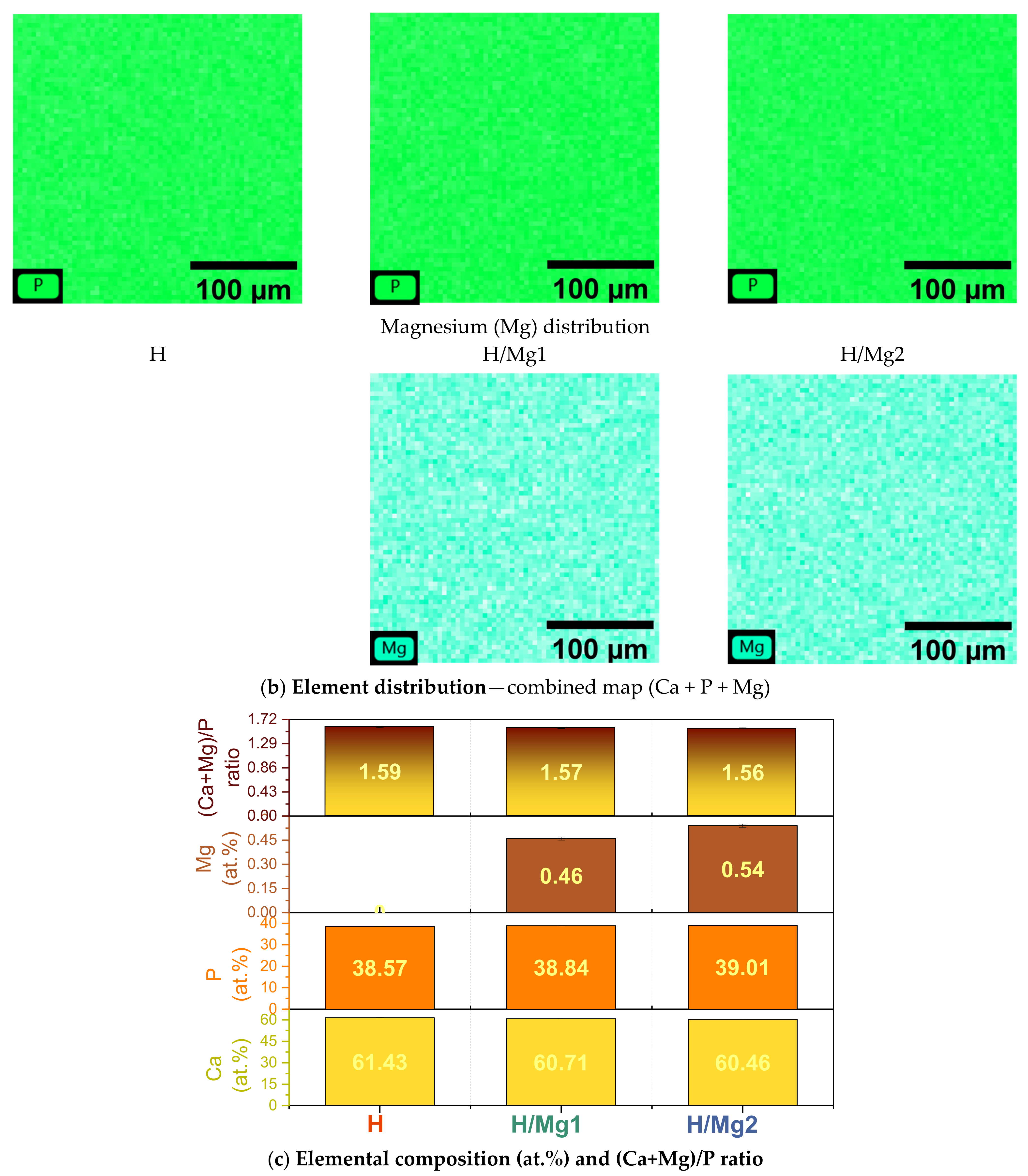
3.2. Elemental Composition

The elemental composition of the coatings was obtained by EDS analysis, and the results are presented in Figure 4 as EDS spectra (a), element distribution on the investigated area (b), and elemental composition and (Ca + Mg)/P ratio (c). The EDS analysis was carried out in triplicate by using different specimens of each material, and the average values along with their standard deviation were taken into consideration.
The EDS spectra achieved on the HAp-based coatings have highlighted the presence of the main elements of the nanostructured surface (Ti and O) and in the coatings (Ca, P, O, and Mg). Moreover, the elemental composition carried out on the annealed nanostructured NT TiO2 surface (results not shown) revealed the presence of fluorine (F) in a concentration of 0.65 wt.% (0.92 at.%). According to the literature, a concentration between 0.5 at% and 3 at.% supports and promotes cell proliferation and differentiation [83]. The residual fluorine found on the NT TiO2 was not further detected in the coatings. Nevertheless, it is worth mentioning that the concentration detected on the nanostructure surfaces is within acceptable limits (0.5–3 at.%).
Since Mg2+ ions are substituting the Ca2+ one in the HAp structure [44], the Mg amount was added to Ca, and the resulting quantity was divided to the P amount.
Thus, the addition of Mg into different concentrations slightly decreased the (Ca + Mg)/P ratio from a value of 1.59, obtained for the H coatings, to a value of 1.57 and 1.56, respectively, for H/Mg1 and H/Mg2 (Figure 4c). Nevertheless, it can be noted that all (Ca + Mg)/P ratios are smaller than that of stoichiometric HAp, indicating the formation of a Ca-deficient apatite.
In terms of element distribution, all the identified elements (Ca, P, and Mg) are uniformly distributed on the investigated areas, with no visible agglomerations, proving a homogeneous coating.

3.3. Phase Composition
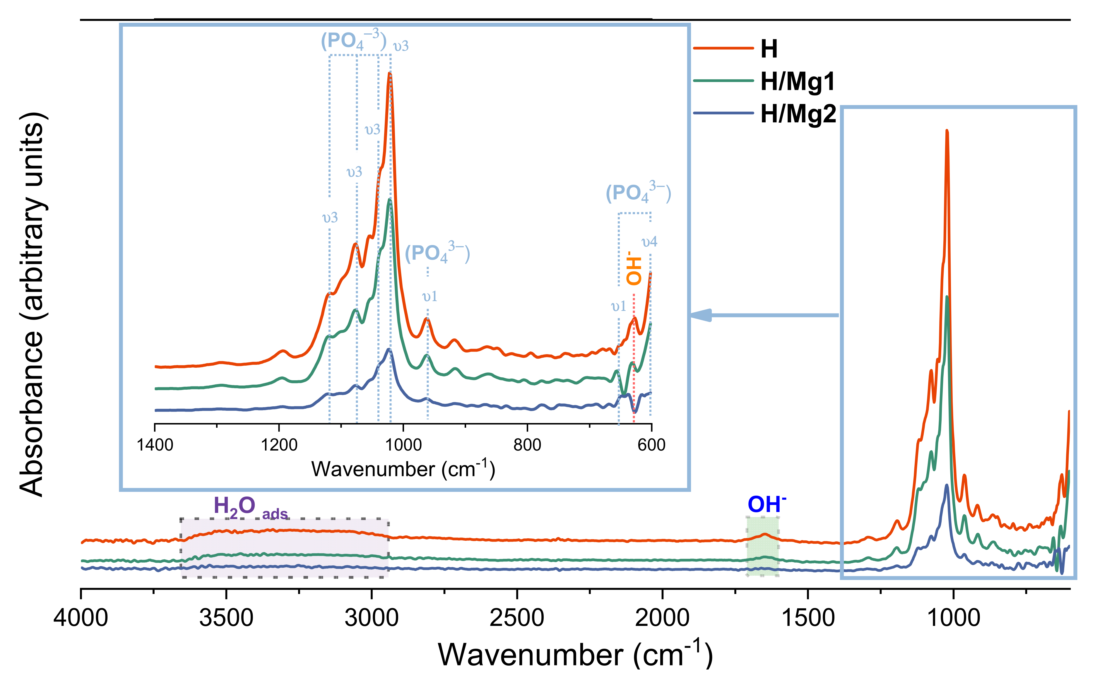
In Figure 5a, the X-ray diffractograms of the undoped and Mg-doped HAp-based coatings are presented along with the computed values of the lattice parameters, the crystallite dimension, and the crystallinity (Figure 5b).
The XRD diffractogram of the HAp-based coatings shows the main diffraction peaks related to the HAp phase according to ICDD card #09-0432. In all the coatings, the presence and the formation of hydroxyapatite as the main phase were found. Two other small diffraction peaks identified at 2θ of approximately 24° and 30°, corresponding to (111) and (120) planes, were attributed to the monetite phase according to ICDD card #1-071-1759, indicating the presence of a secondary phase.
The diffraction peaks corresponding to pure Ti (ICDD card #44-1294) as bulk substrate material and the nanostructured surface made of TiO2 nanotubes with its two phases, namely anatase (ICDD card #21-1272) and rutile (ICDD card #21-1276), were also identified, suggesting that the coatings are to some extent porous due to the presence of the ribbon-like crystals. The diffractogram of the NT surface is available in Ref. [96].
Irrespective of the absence or presence of Mg within the coatings and/or its amount, all diffractograms present the same allure and intensities, with just a few differences, namely the intensity of the peaks associated to the monetite phase which decreased after Mg addition and the peak of HAp identified at ~26°.
The diffraction peak of HAp corresponding to (002) the plane at 2θ of ~26° is the most intense and illustrates that the obtained coatings have a preferential orientation toward the c-axis which is in good agreement with the SEM images.
According to the literature [108,109], the change in crystal growth direction could be due to the incorporation of mineral ions and the increase in their content which may favor the growth of crystals in the (002) direction, which is perpendicular to the substrate surface (along the c-axis). Moreover, if the crystals were all perfectly aligned with the c-axis normal to the substrate, the (002) diffraction peak would be the only one visible. The fact that other HAp peaks are present in diffractograms (Figure 5a) indicates that some crystals are oriented in other directions [110].
Thus, to identify the preferential orientation of the crystallites along a crystal plane (hkl) in the samples, the texture coefficient TC(hkl) of each peak was calculated following the relation [111,112]:
T C h k l = I h k l I 0 , h k l 1 N 0 n I h k l I 0 , h k l
where TC(hkl) is the texture coefficient of plane (hkl), Ihkl is the measured intensity, I0,hkl values are the intensity taken from the ICDD powder diffraction file of hydroxyapatite (ICDD #09-0432), and N is the number of peaks taken into consideration.
Thus, the (002) diffraction peak and three strongest diffraction peaks of HAp ((211), (112) and (213)) were selected; therefore n = 4. A random orientation would lead to a TC(hkl) value of 1, whereas a complete dominance of one peak would return a value of 4, which represents the number of diffraction peaks taken into consideration.
The degree of orientation of each coating was also analyzed from the standard deviation σ of all TC(hkl) values, being calculated based on the following relation [112]:
σ = 1 N T C h k l 1 2
The value of σ is an indicator of the orientation degree, and it can be used to compare different samples. Table 4 presents the values for the texture coefficient and degree of the preferred orientation.
Thus, for a complete randomized sample, the TC(hkl) will be 1, and the preferred orientation σ will be 0, while for a fully aligned material, the TC(hkl) will be 4 and the preferred orientation σ will be 1.73.
By comparing the values of the texture coefficient of all samples, it can be noted that compared to the undoped HAp, the addition and the increment of Mg concentration have led to the increment of the TC(hkl) for the (002) plane and to the decrement of the TC(hkl) for (211), (112) and (213) planes. Thus, the data presented in Table 4 indicate that the preferred orientation is in the (002) direction for all samples, the highest values being obtained for the H/Mg2 samples, followed by the H/Mg1 and H.
Another change that involves the diffraction peak at 26° consists of shifting the HAp main XRD peak toward the right, denoting the inclusion of the doping elements in the HAp structure. This is available when the ionic radius of the doping element is smaller than the one of Ca2+, which is also the case for Mg2+. By increasing the Mg amount, the XRD peaks became more intense, and some of the peak’s position shifted to the right, indicating lattice contraction caused by the Mg substitution. This effect could be explained by the increment of the crystallite size and lattice changes associated with Mg substitution in the HAp structure. As can be observed in Figure 5b(i,ii), compared to H coatings, the addition of Mg led to a slight decrement of the a parameter and an increment of the c parameter along with a higher crystallite dimension.
According to the ISO 13779-3 standard [113], the percentage of crystallinity should be a minimum of 45% for HAp. Thus, it can be observed that the coatings with Mg, codified as H/Mg1 and H/Mg2, are slightly above 45%, reaching values of approx. 47% and 49%, respectively, while the H coating registered a crystallinity of only ~21%. Overall, it was noted that the addition of Mg has a beneficial effect on the crystallinity of HAp-based coatings.
Compared to our previous research in which HAp-doped Mg was electrochemically deposited on a Ti6Al4V alloy [65], in the current research, the electrolyte was slightly adjusted by reducing the concentration of all chemical reagents involved by 5%, coupled with the use of pure Ti with a nanostructured surface as substrate instead of Ti6Al4V, could be the cause that led to higher crystallinity. Thus, it can be assumed that even the slightest change of the electrochemical parameters can affect the characteristics of the deposited materials. Nonetheless, further analyses in this direction are necessary to validate this assumption.

3.4. Chemical Bonds

Figure 6 depicts the FTIR spectra of the undoped and Mg-doped HAp coatings. FTIR spectra are dominated by internal (PO4)3− modes, which typically show a strong molecular character with respect to their vibrational properties [114]. Nonetheless, some variations of the absorption bands, which may indicate the substitutions of Mg ions into the HAp structure, are visible.
The spectrum of HAp shows intense peaks between 1120 and 1020 cm−1, which were assigned to the ν3 asymmetric stretching mode of (PO4)3− [115], while the weak peak at 960 cm−1 can be assigned to the ν1 asymmetric stretching mode of (PO4)3− [65].
Compared with undoped HAp, a decrement in the intensity of the phosphate groups (PO4)3− was associated with the addition and increment of magnesium content. The observed FTIR spectral changes can also be related to the destabilization of the HAp structure by the incorporation of Mg2+ ions.
Nonetheless, the OH stretching vibration identified at 630 cm−1 is unique and indicates that the HAp-based coatings are crystallized [115]. The adsorbed water generates a broad band in the range of 3000–3500 cm−1 [116] and at about 1650 cm−1 [115,117].
Thus, all developed coatings present the peaks assigned to PO43 and OH, which are considered specific to HAp, proving the formation of the HAp phase.

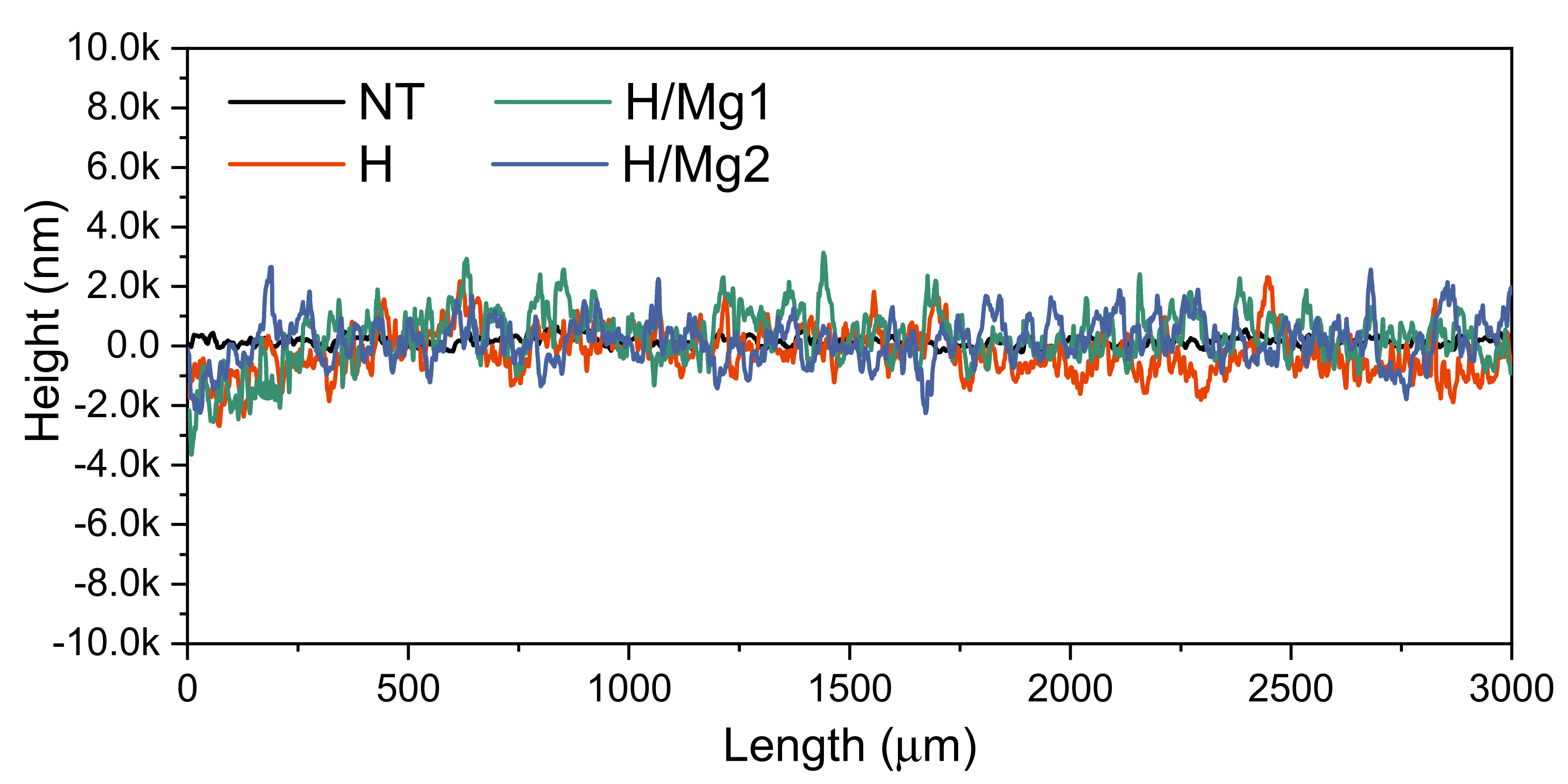
3.5. Surface Roughness
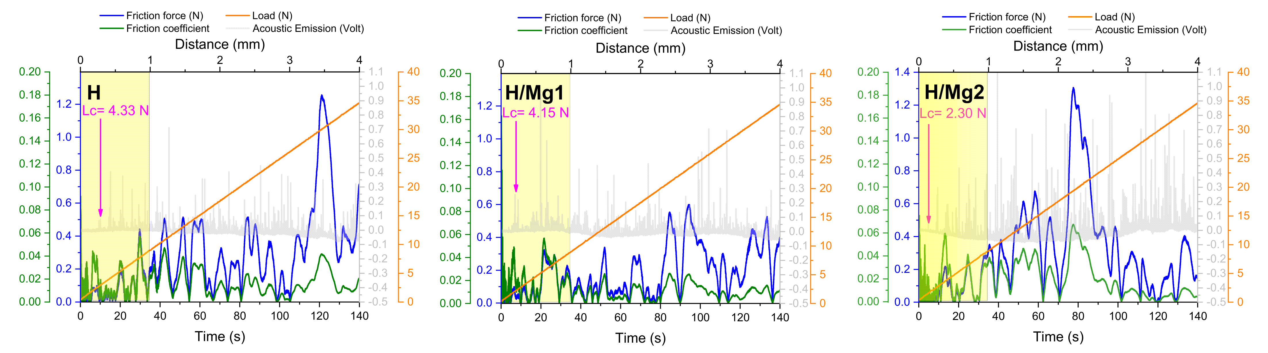
Despite the high number of studies, the results in the literature demonstrate difficulties in deciding the optimum value of surface roughness for better osseointegration and decreased bacterial adhesion [118,119,120].
In Figure 7, specific profiles of the nanostructured surface (NT), uncoated and coated with HAp-based coatings, are presented, while in Table 5 the values of the main roughness parameters along with the coating thicknesses are given.
A comparison of the average roughness (Ra) parameter for all materials indicates that roughness ranged between 490 and 590 nm, with the smallest value being registered for the coatings H/Mg2, showing that high concentrations of Mg (i.e., 1.5 mM Mg in the electrolyte) tend to smoothen the surface. The largest Ra value, of 583 nm was observed for the H/Mg1 coatings, while the H coatings had an Ra of 535 nm. Thus, based on the results of the Ra parameter, it can be noted that the amount of Mg has a direct influence on the coatings roughness.
The coatings thickness was measured, and it was noted that the smallest value of 9.6 μm (±0.9) was obtained for the undoped H followed by H/Mg1 coatings with a thickness of 11.3 μm (±0.6), while the largest one was attributed to the H/Mg2 coating of 13.7 μm (±1.1).
Thus, it can be said that the addition of Mg into HAp increases the deposition rate, leading to higher thicknesses, while in terms of roughness, a higher concentration of Mg leads to a decrement of the average roughness.

3.6. Adhesion

One of the main disadvantages of the HAp-based coatings obtained by electrochemical deposition is their adhesion to the substrate. Thus, coating adhesion to the substrate is very important for the implantable medical devices, and at times, it can be quite challenging to achieve.
The SEM images of lattice pattern indentation after adhesion tests were achieved on areas of 897 × 897 μm2, presented in Figure 8. Furthermore, the SEM images were analyzed by Image J software to quantify the delaminated areas in percentages.
After detaching the scotch tape from the coating surface, a very thin layer of the coating remained on the tape but without affecting the integrity of coatings which is in good agreement with other studies [121].
A comparison of the lattice patterns highlighted that the best adhesion was noted for the H and H/Mg1 coatings, which had very small areas detached (0.31% and 0.44%, respectively), while the H/Mg2 coatings showed the lowest adhesion with large detachments/flakes from their surfaces, which falls into category 3B (5–15% delamination area) with an area of 8% that was detached from the surface. However, it can be observed that on the detached areas of the H/Mg2 sample, a layer of apatite that resembles the bottom layer of HAp remained on the substrate, indicating that the coating was not entirely detached from the surface. Thus, the results of adhesion tests showed that Mg–HAp coatings obtained from an electrolyte that contains at least 1.5 mM Mg can alter the adhesion of the coatings to the substrate.
Complementary to the tape test, we have also performed a micro-scratch test, which showed that the initial penetration of the coating starts within the first mm of the scratch length (Figure 9).
Based on the signal captured by the acoustic emission sensor, the critical load (Lc) was estimated. The highest Lc was registered for the undoped HAp with a value of 4.33 N, closely followed by the H/Mg1 sample with an Lc of 4.15 N, while the smallest value of 2.30 N was obtained for the H/Mg2 sample which is in agreement with the results obtained from the tape test and presented in Figure 8.
Moreover, the elemental composition after the micro-scratch tests was investigated in the center of the scratch and at its end (Figure 10) to identify if some traces of the coatings were still present.
The results presented in Table 6 highlight that the main elements of hydroxyapatite (Ca, P and O) are found in all the investigated areas. However, oxygen is also detected from the titanium dioxide, which occurs instantly on the surface of titanium.
It can also be seen that for all tested HAp coatings, the amount of calcium and phosphorous content decreases at the end compared with the middle of the scratch imprint. The presence of CaP traces within the scratch imprint is beneficial in the case of some damage of the coatings due to friction with hard tissue or careless handling of medical devices.

4. Conclusions

The findings of the present study can be summarized as follows:
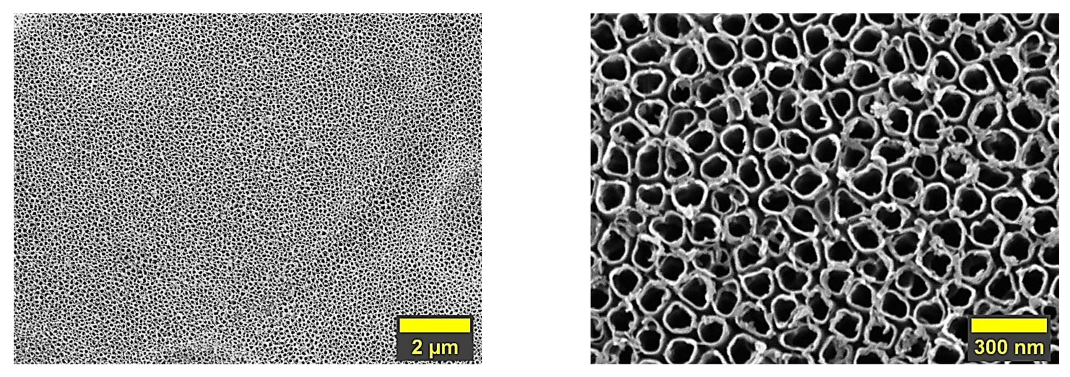
  • Hydroxyapatite-based coatings undoped and doped with Mg in different concentrations were successfully deposited by electrochemical deposition in pulsed galvanostatic mode on a nanostructured surface made of titania nanotubes with a diameter of ~72 nm (±2);
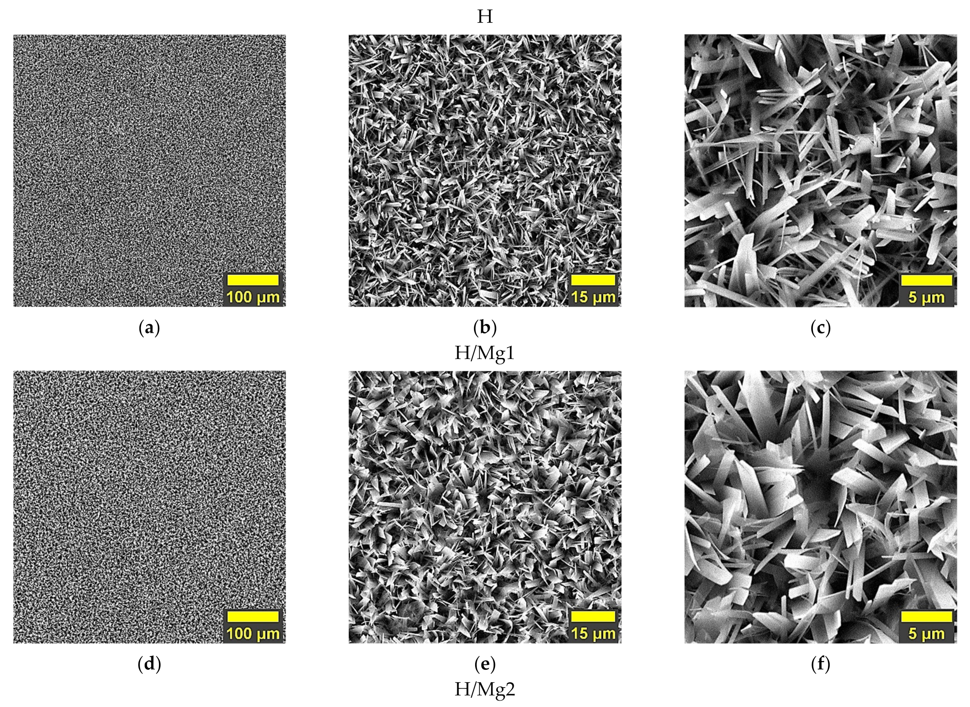
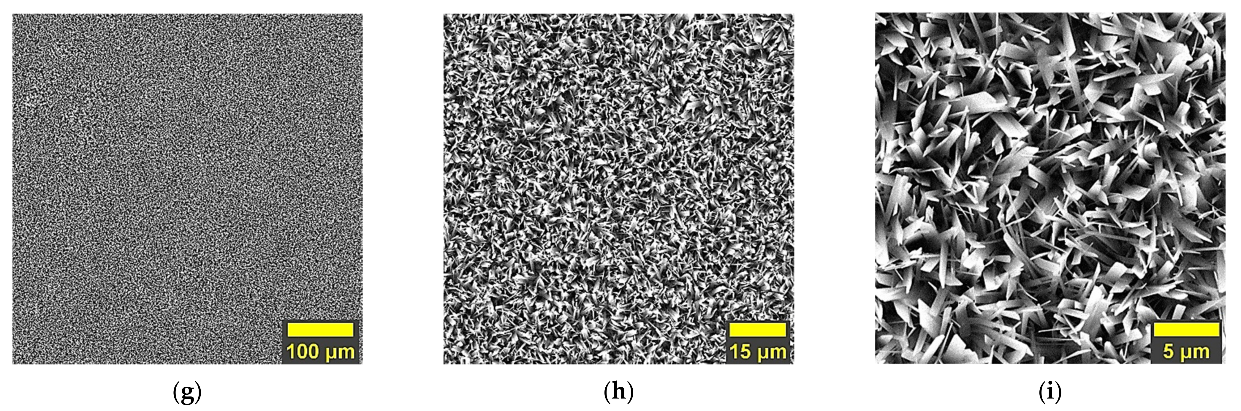
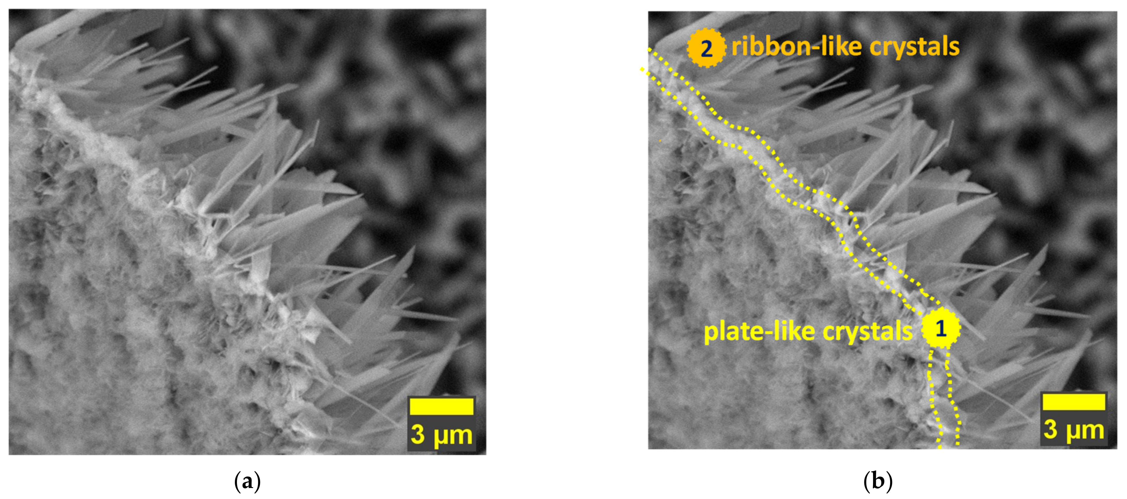
  • In terms of morphology, the present study showed that the undoped HAp and the HAp coatings obtained with a concentration of 1.5 mM of Mg in the electrolyte present similar morphologies, which are made of very thin and narrow ribbon-like crystals, while the morphology of the HAp coatings obtained with a concentration of 1 mM of Mg in the electrolyte, are made of wider and thicker ribbon-like crystals, suggesting that the growth mechanism of the crystals is influenced by the Mg concentration within the electrolyte used to obtain the coatings;
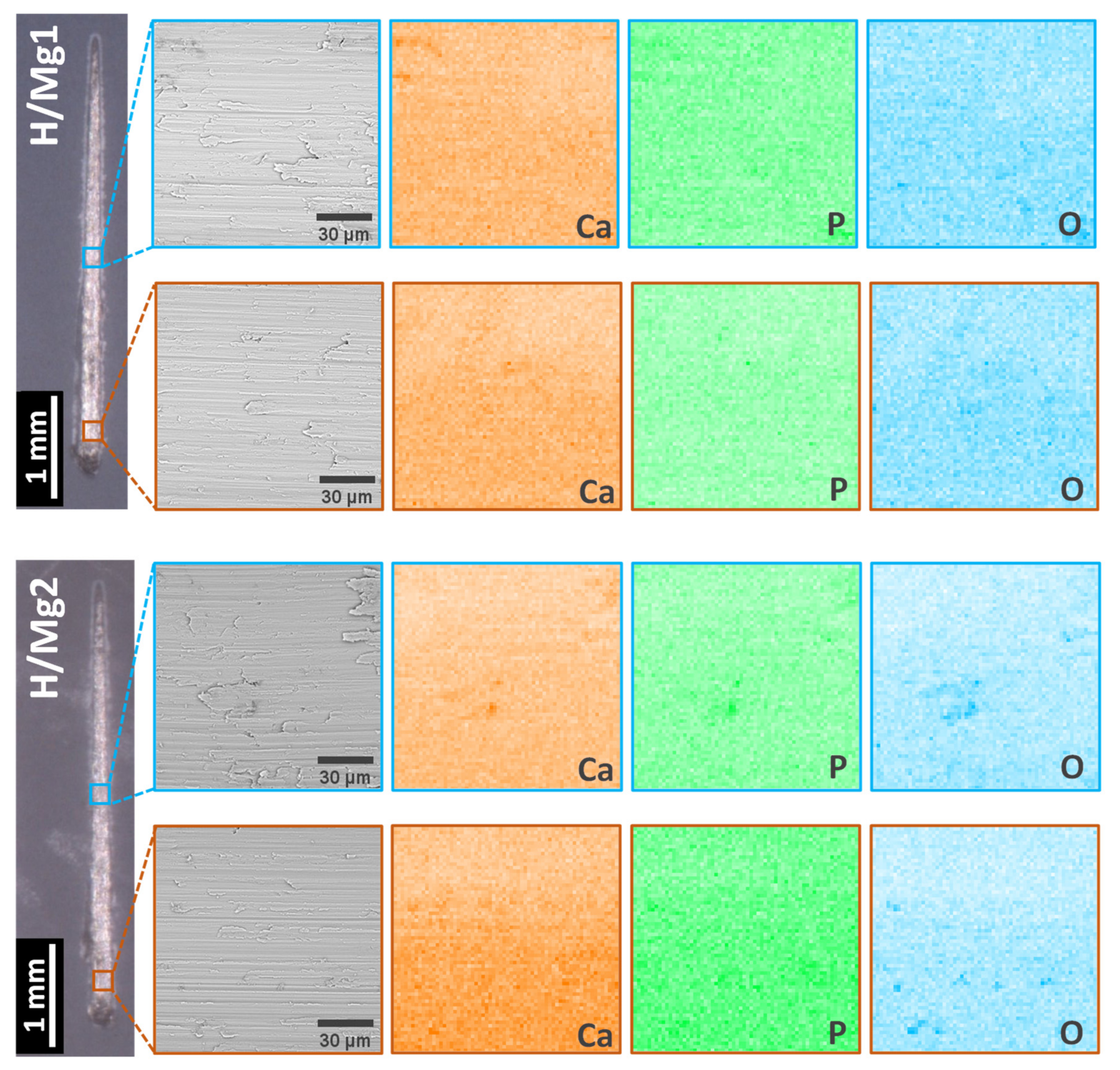
  • EDS analysis revealed that by increasing the Mg concentration in the electrolyte from 1 mM to 1.5 mM, the quantity of Mg identified in the coatings is 0.44 at.% (0.31 wt.%) and 0.54 at.% (0.37 wt.%) which are within the values of Mg found in human hard bone tissue (0.30–0.72 wt.%); also, all elements (Ca, P, and Mg) are uniformly distributed within the coatings, indicating that the pulsed galvanostatic method is a reliable deposition technique;
  • Compared with the undoped HAp, which registered a thickness of 9.6 μm, addition and increment of the Mg concentration in the electrolyte from 1 mM to 1.5 mM, led to thickness of 11.3 and 13.7 μm, indicating a strong association between the Mg amount and the deposition rate. Thus, a concentration of 1.5 mM of Mg in the electrolyte increases the deposition rate, while with a decrement of the Mg concentration, to a value of 1 mM, a less thick coating can be obtained. Based on this, it can be said that the coating thickness can also be controlled through a careful selection of the doping element concentration within the electrolyte. Of course, this should also be associated and correlated with other parameters involved in the electrochemical deposition, such as the deposition technique, pH value, deposition temperature, and so on to design a coating with enhanced and tunable features;
  • The phase composition and chemical bonds analysis confirmed that all coatings consist of the HAp phase and that Mg2+ substituted some of the Ca2+ in the HAp structure. Moreover, the XRD analysis showed that the coatings have a preferred orientation toward the c-axis direction and that the addition of Mg in the proposed concentrations of 1 mM and 1.5 mM in the electrolyte can enhance the crystallinity of the HAp-based coatings;
  • With respect to the Mg concentration in the electrolyte, it was observed that compared to the undoped HAp, the use of 1.5 mM Mg leads to an average roughness of 493 nm, while the use of a concentration of 1 mM Mg leads to an average roughness of 582 nm, suggesting that higher concentrations of Mg tend to smoothen the surface;
  • The “tape test” and the micro-scratch tests highlighted that the adhesion of the HAp-based coatings is in strong correlation with the Mg concentration, indicating that an increment of the Mg can affect the adhesion of the coating even if the coatings are obtained on a nanostructured surface, which is known to enhance the adhesion of the coatings obtained by electrochemical techniques;
  • moreover, the adhesion tests also showed that even if the coatings are being scratched, a small amount of coating will remain on the metallic substrate.
Overall, it can be said that the results of the current investigation indicate that the Mg concentration influences the physico-chemical properties of HAp-based coatings, and a concentration of 1 mM of Mg in the electrolyte used to obtain the coatings can maintain and/or even enhance the properties and behavior of HAp-based coatings. The coatings obtained with the addition of 1 mM of Mg, will be investigated in terms of electrochemical behavior, cell viability and proliferation, biomineralization ability, and degradation to establish if the proposed coatings are suitable for biomedical applications.

Supplementary Materials

The following supporting information can be downloaded at: https://www.mdpi.com/article/10.3390/coatings12081097/s1, Figure S1: Original SEM images found in Figure 1; Figure S2: Original SEM images found in Figure 2; Figure S3: Original SEM images found in Figure 3; Figure S4: Original SEM images found in Figure 8.

Author Contributions

Conceptualization: C.M.C. and D.M.V.; methodology: C.M.C. and D.M.V.; validation: C.M.C.; formal analysis, E.U. and A.S.; investigation: I.C.I., E.U., A.E.K., A.C.P., R.I.Z. and A.V.; writing—original draft preparation, C.M.C. and D.M.V.; writing—review and editing, C.M.C. and A.V.; project administration, D.M.V.; funding acquisition, D.M.V. All authors have read and agreed to the published version of the manuscript.

Funding

This research has been funded by the Romanian Ministry of Education and Research, CNCS-UEFISCDI, projects number PN-III-P1-1.1-TE-2019-1331 (TE 172/2020; 3B-CoatED) and PN-III-P2-2.1-PED-2021-4275 (621PED/2022; BioMimCells), within PNCDI III.

Institutional Review Board Statement

Not applicable.

Informed Consent Statement

Not applicable.

Data Availability Statement

Not applicable.

Acknowledgments

A.V. thanks to Institutional Performance Program—Projects for Financing Excellence 18PFE/30.12.2021.

Conflicts of Interest

The authors declare no conflict of interest.

References

  1. Bannon, B.; Mild, E. Titanium Alloys for Biomaterial Application: An Overview. In Titanium Alloys in Surgical Implants; Luckey, H.A., Kubli, F.J., Eds.; ASTM International: West Conshohocken, PA, USA, 1983; pp. 7–9. [Google Scholar]
  2. Hanawa, T. Biofunctionalization of Titanium for Dental Implant. Jpn. Dent. Sci. Rev. 2010, 46, 93–101. [Google Scholar] [CrossRef] [Green Version]
  3. Navarro, M.; Michiardi, A.; Castaño, O.; Planell, J.A. Biomaterials in Orthopaedics. J. R. Soc. Interface 2008, 5, 1137–1158. [Google Scholar] [CrossRef] [PubMed] [Green Version]
  4. Ferraris, S.; Spriano, S. Antibacterial Titanium Surfaces for Medical Implants. Mater. Sci. Eng. C 2016, 61, 965–978. [Google Scholar] [CrossRef]
  5. Baino, F.; Potestio, I. Orbital Implants: State-of-the-Art Review with Emphasis on Biomaterials and Recent Advances. Mater. Sci. Eng. C 2016, 69, 1410–1428. [Google Scholar] [CrossRef]
  6. Saini, M. Implant Biomaterials: A Comprehensive Review. World J. Clin. Cases 2015, 3, 52–57. [Google Scholar] [CrossRef]
  7. Prasad, K.; Bazaka, O.; Chua, M.; Rochford, M.; Fedrick, L.; Spoor, J.; Symes, R.; Tieppo, M.; Collins, C.; Cao, A.; et al. Metallic Biomaterials: Current Challenges and Opportunities. Materials 2017, 10, 884. [Google Scholar] [CrossRef] [PubMed]
  8. Abdel-Hady Gepreel, M.; Niinomi, M. Biocompatibility of Ti-Alloys for Long-Term Implantation. J. Mech. Behav. Biomed. Mater. 2013, 20, 407–415. [Google Scholar] [CrossRef]
  9. Davis, R.; Singh, A.; Jackson, M.J.; Coelho, R.T.; Prakash, D.; Charalambous, C.P.; Ahmed, W.; da Silva, L.R.R.; Lawrence, A.A. A Comprehensive Review on Metallic Implant Biomaterials and Their Subtractive Manufacturing. Int. J. Adv. Manuf. Technol. 2022, 120, 1473–1530. [Google Scholar] [CrossRef]
  10. Lu, X.; Wu, Z.; Xu, K.; Wang, X.; Wang, S.; Qiu, H.; Li, X.; Chen, J. Multifunctional Coatings of Titanium Implants Toward Promoting Osseointegration and Preventing Infection: Recent Developments. Front. Bioeng. Biotechnol. 2021, 9, 783816. [Google Scholar] [CrossRef]
  11. Zhang, L.-C.; Chen, L.-Y.; Wang, L. Surface Modification of Titanium and Titanium Alloys: Technologies, Developments, and Future Interests. Adv. Eng. Mater. 2020, 22, 1901258. [Google Scholar] [CrossRef]
  12. Madhavi, G.; Kishan, N.; Raghavendra, C.R. A Review on Recent Approaches in the Field of Surface Coating. Mater. Today Proc. 2022, 52, 403–406. [Google Scholar] [CrossRef]
  13. Chen, L.; Song, X.; Xing, F.; Wang, Y.; Wang, Y.; He, Z.; Sun, L. A Review on Antimicrobial Coatings for Biomaterial Implants and Medical Devices. J. Biomed. Nanotechnol. 2020, 16, 789–809. [Google Scholar] [CrossRef] [PubMed]
  14. Civantos, A.; Martínez-Campos, E.; Ramos, V.; Elvira, C.; Gallardo, A.; Abarrategi, A. Titanium Coatings and Surface Modifications: Toward Clinically Useful Bioactive Implants. ACS Biomater. Sci. Eng. 2017, 3, 1245–1261. [Google Scholar] [CrossRef] [PubMed]
  15. Leeuwenhoek, A.V. Microscopical Observations of the Structure of Teeth and Other Bones: Made and Communicated, in a Letter by Mr. Anthony Leeuwenhoeck. Philos. Trans. R. Soc. Lond. 1677, 12, 1002–1003. [Google Scholar] [CrossRef] [Green Version]
  16. Shear, M.J.; Kramer, B. Composition of Bone: I. Analytical Micro Methods. J. Biol. Chem. 1928, 79, 105–120. [Google Scholar] [CrossRef]
  17. Vallet-Regi, M.; González-Calbet, J.M. Calcium Phosphates as Substitution of Bone Tissues. Prog. Solid State Chem. 2004, 32, 1–31. [Google Scholar] [CrossRef]
  18. Narayanan, R.; Seshadri, S.K.; Kwon, T.Y.; Kim, K.H. Calcium Phosphate-Based Coatings on Titanium and Its Alloys. J. Biomed. Mater. Res.—Part B Appl. Biomater. 2008, 85, 279–299. [Google Scholar] [CrossRef] [PubMed]
  19. Koutsopoulos, S. Synthesis and Characterization of Hydroxyapatite Crystals: A Review Study on the Analytical Methods. J. Biomed. Mater. Res. 2002, 62, 600–612. [Google Scholar] [CrossRef] [PubMed]
  20. Chakraborty, R.; Sengupta, S.; Saha, P.; Das, K.; Das, S. Synthesis of Calcium Hydrogen Phosphate and Hydroxyapatite Coating on SS316 Substrate through Pulsed Electrodeposition. Mater. Sci. Eng. C 2016, 69, 875–883. [Google Scholar] [CrossRef]
  21. Mihailescu, N.; Stan, G.E.; Duta, L.; Chifiriuc, M.C.; Bleotu, C.; Sopronyi, M.; Luculescu, C.; Oktar, F.N.; Mihailescu, I.N. Structural, Compositional, Mechanical Characterization and Biological Assessment of Bovine-Derived Hydroxyapatite Coatings Reinforced with MgF 2 or MgO for Implants Functionalization. Mater. Sci. Eng. C 2016, 59, 863–874. [Google Scholar] [CrossRef] [PubMed]
  22. Wang, W.; Yeung, K.W.K. Bone Grafts and Biomaterials Substitutes for Bone Defect Repair: A Review. Bioact. Mater. 2017, 2, 224–247. [Google Scholar] [CrossRef]
  23. Dascălu, C.-A.; Maidaniuc, A.; Pandele, A.M.; Voicu, S.I.; Machedon-Pisu, T.; Stan, G.E.; Cîmpean, A.; Mitran, V.; Antoniac, I.V.; Miculescu, F. Synthesis and Characterization of Biocompatible Polymer-Ceramic Film Structures as Favorable Interface in Guided Bone Regeneration. Appl. Surf. Sci. 2019, 494, 335–352. [Google Scholar] [CrossRef]
  24. Antoniac, I.V.; Antoniac, A.; Vasile, E.; Tecu, C.; Fosca, M.; Yankova, V.G.; Rau, J.V. In Vitro Characterization of Novel Nanostructured Collagen-Hydroxyapatite Composite Scaffolds Doped with Magnesium with Improved Biodegradation Rate for Hard Tissue Regeneration. Bioact. Mater. 2021, 6, 3383–3395. [Google Scholar] [CrossRef] [PubMed]
  25. Yuan, B.; Chen, H.; Zhao, R.; Deng, X.; Chen, G.; Yang, X.; Xiao, Z.; Aurora, A.; Iulia, B.A.; Zhang, K.; et al. Construction of a Magnesium Hydroxide/Graphene Oxide/Hydroxyapatite Composite Coating on Mg-Ca-Zn-Ag Alloy to Inhibit Bacterial Infection and Promote Bone Regeneration. Bioact. Mater. 2022, 18, 354–367. [Google Scholar] [CrossRef]
  26. Antoniac, I.; Miculescu, F.; Cotrut, C.; Ficai, A.; Rau, J.V.; Grosu, E.; Antoniac, A.; Tecu, C.; Cristescu, I. Controlling the Degradation Rate of Biodegradable Mg–Zn-Mn Alloys for Orthopedic Applications by Electrophoretic Deposition of Hydroxyapatite Coating. Materials 2020, 13, 263. [Google Scholar] [CrossRef] [Green Version]
  27. Bryington, M.S.; Hayashi, M.; Kozai, Y.; Vandeweghe, S.; Andersson, M.; Wennerberg, A.; Jimbo, R. The Influence of Nano Hydroxyapatite Coating on Osseointegration after Extended Healing Periods. Dent. Mater. 2013, 29, 514–520. [Google Scholar] [CrossRef]
  28. Gambardella, A.; Bianchi, M.; Kaciulis, S.; Mezzi, A.; Brucale, M.; Cavallini, M.; Herrmannsdoerfer, T.; Chanda, G.; Uhlarz, M.; Cellini, A.; et al. Magnetic Hydroxyapatite Coatings as a New Tool in Medicine: A Scanning Probe Investigation. Mater. Sci. Eng. C 2016, 62, 444–449. [Google Scholar] [CrossRef]
  29. Ressler, A.; Žužić, A.; Ivanišević, I.; Kamboj, N.; Ivanković, H. Ionic Substituted Hydroxyapatite for Bone Regeneration Applications: A Review. Open Ceram. 2021, 6, 100122. [Google Scholar] [CrossRef]
  30. Shepherd, J.H.; Shepherd, D.V.; Best, S.M. Substituted Hydroxyapatites for Bone Repair. J. Mater. Sci. Mater. Med. 2012, 23, 2335–2347. [Google Scholar] [CrossRef] [PubMed]
  31. Qi, G.; Zhang, S.; Khor, K.A.; Lye, S.W.; Zeng, X.; Weng, W.; Liu, C.; Venkatraman, S.S.; Ma, L.L. Osteoblastic Cell Response on Magnesium-Incorporated Apatite Coatings. Appl. Surf. Sci. 2008, 255, 304–307. [Google Scholar] [CrossRef]
  32. Bigi, A.; Foresti, E.; Gregorini, R.; Ripamonti, A.; Roveri, N.; Shah, J.S. The Role of Magnesium on the Structure of Biological Apatites. Calcif. Tissue Int. 1992, 50, 439–444. [Google Scholar] [CrossRef]
  33. Howlett, C.R.; Zreioat, H.; O’Dell, R.; Noorman, J.; Evans, P.; Dalton, B.A.; McFarland, C.; Steele, J.G. The Effect of Magnesium Ion Implantation into Alumina upon the Adhesion of Human Bone Derived Cells. J. Mater. Sci. Mater. Med. 1994, 5, 715–722. [Google Scholar] [CrossRef]
  34. Antoniac, I.; Miculescu, M.; Mănescu (Păltânea), V.; Stere, A.; Quan, P.H.; Păltânea, G.; Robu, A.; Earar, K. Magnesium-Based Alloys Used in Orthopedic Surgery. Materials 2022, 15, 1148. [Google Scholar] [CrossRef] [PubMed]
  35. Peron, M.; Torgersen, J.; Berto, F. Mg and Its Alloys for Biomedical Applications: Exploring Corrosion and Its Interplay with Mechanical Failure. Metals 2017, 7, 252. [Google Scholar] [CrossRef] [Green Version]
  36. Uskoković, V. Ion-Doped Hydroxyapatite: An Impasse or the Road to Follow? Ceram. Int. 2020, 46, 11443–11465. [Google Scholar] [CrossRef]
  37. Ratha, I.; Datta, P.; Balla, V.K.; Nandi, S.K.; Kundu, B. Effect of Doping in Hydroxyapatite as Coating Material on Biomedical Implants by Plasma Spraying Method: A Review. Ceram. Int. 2021, 47, 4426–4445. [Google Scholar] [CrossRef]
  38. Zhao, S.; Jiang, Q.; Peel, S.; Wang, X.; He, F. Effects of Magnesium-Substituted Nanohydroxyapatite Coating on Implant Osseointegration. Clin. Oral Implant. Res. 2013, 24, 34–41. [Google Scholar] [CrossRef]
  39. Tampieri, A.; Celotti, G.C.; Landi, E.; Sandri, M. Magnesium Doped Hydroxyapatite: Synthesis and Characterization. Key Eng. Mater. 2004, 264–268, 2051–2054. [Google Scholar] [CrossRef]
  40. Ren, F.; Leng, Y.; Xin, R.; Ge, X. Synthesis, Characterization and Ab Initio Simulation of Magnesium-Substituted Hydroxyapatite. Acta Biomater. 2010, 6, 2787–2796. [Google Scholar] [CrossRef] [PubMed]
  41. Kannan, S.; Lemos, I.A.F.; Rocha, J.H.G.; Ferreira, J.M.F. Synthesis and Characterization of Magnesium Substituted Biphasic Mixtures of Controlled Hydroxyapatite/β-Tricalcium Phosphate Ratios. J. Solid State Chem. 2005, 178, 3190–3196. [Google Scholar] [CrossRef]
  42. Laurencin, D.; Almora-Barrios, N.; de Leeuw, N.H.; Gervais, C.; Bonhomme, C.; Mauri, F.; Chrzanowski, W.; Knowles, J.C.; Newport, R.J.; Wong, A.; et al. Magnesium Incorporation into Hydroxyapatite. Biomaterials 2011, 32, 1826–1837. [Google Scholar] [CrossRef] [PubMed] [Green Version]
  43. Suchanek, W.L.; Byrappa, K.; Shuk, P.; Riman, R.E.; Janas, V.F.; TenHuisen, K.S. Preparation of Magnesium-Substituted Hydroxyapatite Powders by the Mechanochemical–Hydrothermal Method. Biomaterials 2004, 25, 4647–4657. [Google Scholar] [CrossRef] [PubMed]
  44. Mehrjoo, M.; Javadpour, J.; Shokrgozar, M.A.; Farokhi, M.; Javadian, S.; Bonakdar, S. Effect of Magnesium Substitution on Structural and Biological Properties of Synthetic Hydroxyapatite Powder. Mater. Express 2015, 5, 41–48. [Google Scholar] [CrossRef]
  45. Landi, E.; Logroscino, G.; Proietti, L.; Tampieri, A.; Sandri, M.; Sprio, S. Biomimetic Mg-Substituted Hydroxyapatite: From Synthesis to in Vivo Behaviour. J. Mater. Sci. Mater. Med. 2008, 19, 239–247. [Google Scholar] [CrossRef]
  46. Chirică, I.M.; Enciu, A.-M.; Tite, T.; Dudău, M.; Albulescu, L.; Iconaru, S.L.; Predoi, D.; Pasuk, I.; Enculescu, M.; Radu, C.; et al. The Physico-Chemical Properties and Exploratory Real-Time Cell Analysis of Hydroxyapatite Nanopowders Substituted with Ce, Mg, Sr, and Zn (0.5–5 at.%). Materials 2021, 14, 3808. [Google Scholar] [CrossRef]
  47. Chen, S.; Shi, Y.; Zhang, X.; Ma, J. Biomimetic Synthesis of Mg-substituted Hydroxyapatite Nanocomposites and Three-dimensional Printing of Composite Scaffolds for Bone Regeneration. J. Biomed. Mater. Res. Part A 2019, 107, 2512–2521. [Google Scholar] [CrossRef]
  48. He, L.Y.; Zhang, X.M.; Liu, B.; Tian, Y.; Ma, W.H. Effect of Magnesium Ion on Human Osteoblast Activity. Braz. J. Med. Biol. Res. 2016, 49, 1–6. [Google Scholar] [CrossRef]
  49. Jiao, M.-J.; Wang, X.-X. Electrolytic Deposition of Magnesium-Substituted Hydroxyapatite Crystals on Titanium Substrate. Mater. Lett. 2009, 63, 2286–2289. [Google Scholar] [CrossRef]
  50. Mróz, W.; Bombalska, A.; Burdyńska, S.; Jedyński, M.; Prokopiuk, A.; Budner, B.; Ślósarczyk, A.; Zima, A.; Menaszek, E.; Ścisłowska-Czarnecka, A.; et al. Structural Studies of Magnesium Doped Hydroxyapatite Coatings after Osteoblast Culture. J. Mol. Struct. 2010, 977, 145–152. [Google Scholar] [CrossRef]
  51. Adzila, S.; Kanasan, N.; Hassan, M.F.; Arifin, A.M.T.; Yunos, M.Z.; Rahman, M.N.A.; Haq, R.H.A. Synthesis and Characterization of Magnesium Doped Calcium Phosphatefor Bone Implant Application. ARPN J. Eng. Appl. Sci. 2016, 11, 8694–8697. [Google Scholar]
  52. Leena, M.; Rana, D.; Webster, T.J.; Ramalingam, M. Accelerated Synthesis of Biomimetic Nano Hydroxyapatite Using Simulated Body Fluid. Mater. Chem. Phys. 2016, 180, 166–172. [Google Scholar] [CrossRef]
  53. Ganvir, A.; Nagar, S.; Markocsan, N.; Balani, K. Deposition of Hydroxyapatite Coatings by Axial Plasma Spraying: Influence of Feedstock Characteristics on Coating Microstructure, Phase Content and Mechanical Properties. J. Eur. Ceram. Soc. 2021, 41, 4637–4649. [Google Scholar] [CrossRef]
  54. Bița, A.-I.; Antoniac, I.; Miculescu, M.; Stan, G.E.; Leonat, L.; Antoniac, A.; Constantin, B.; Forna, N. Electrochemical and In Vitro Biological Evaluation of Bio-Active Coatings Deposited by Magnetron Sputtering onto Biocompatible Mg-0.8Ca Alloy. Materials 2022, 15, 3100. [Google Scholar] [CrossRef]
  55. Vranceanu, D.M.; Cotrut, C.M.; Bramowicz, M.; Titorencu, I.; Kulesza, S.; Kiss, A.; Berbecaru, A.; Pruna, V.; Branzei, M.; Vladescu, A. Osseointegration of Sputtered SiC-Added Hydroxyapatite for Orthopaedic Applications. Ceram. Int. 2016, 42, 10085–10093. [Google Scholar] [CrossRef]
  56. Osuchukwu, O.A.; Salihi, A.; Abdullahi, I.; Obada, D.O. Synthesis and Characterization of Sol–Gel Derived Hydroxyapatite from a Novel Mix of Two Natural Biowastes and Their Potentials for Biomedical Applications. Mater. Today Proc. 2022, 62, 4182–4187. [Google Scholar] [CrossRef]
  57. Duta, L.; Oktar, F.N.; Stan, G.E.; Popescu-Pelin, G.; Serban, N.; Luculescu, C.; Mihailescu, I.N. Novel Doped Hydroxyapatite Thin Films Obtained by Pulsed Laser Deposition. Appl. Surf. Sci. 2013, 265, 41–49. [Google Scholar] [CrossRef]
  58. Antoniac, I.V.; Filipescu, M.; Barbaro, K.; Bonciu, A.; Birjega, R.; Cotrut, C.M.; Galvano, E.; Fosca, M.; Fadeeva, I.V.; Vadalà, G.; et al. Iron Ion-Doped Tricalcium Phosphate Coatings Improve the Properties of Biodegradable Magnesium Alloys for Biomedical Implant Application. Adv. Mater. Interfaces 2020, 7, 2000531. [Google Scholar] [CrossRef]
  59. Safavi, M.S.; Walsh, F.C.; Surmeneva, M.A.; Surmenev, R.A.; Khalil-Allafi, J. Electrodeposited Hydroxyapatite-Based Biocoatings: Recent Progress and Future Challenges. Coatings 2021, 11, 110. [Google Scholar] [CrossRef]
  60. Mokabber, T.; Lu, L.Q.; van Rijn, P.; Vakis, A.I.; Pei, Y.T. Crystal Growth Mechanism of Calcium Phosphate Coatings on Titanium by Electrochemical Deposition. Surf. Coat. Technol. 2018, 334, 526–535. [Google Scholar] [CrossRef]
  61. Katić, J.; Metikoš-Huković, M.; Škapin, S.D.; Petravić, M.; Varašanec, M. The Potential-Assisted Deposition as Valuable Tool for Producing Functional Apatite Coatings on Metallic Materials. Electrochim. Acta 2014, 127, 173–179. [Google Scholar] [CrossRef]
  62. Aina, V.; Lusvardi, G.; Annaz, B.; Gibson, I.R.; Imrie, F.E.; Malavasi, G.; Menabue, L.; Cerrato, G.; Martra, G. Magnesium- and Strontium-Co-Substituted Hydroxyapatite: The Effects of Doped-Ions on the Structure and Chemico-Physical Properties. J. Mater. Sci. Mater. Med. 2012, 23, 2867–2879. [Google Scholar] [CrossRef] [PubMed]
  63. Farzadi, A.; Bakhshi, F.; Solati-Hashjin, M.; Asadi-Eydivand, M.; abu Osman, N.A. Magnesium Incorporated Hydroxyapatite: Synthesis and Structural Properties Characterization. Ceram. Int. 2014, 40, 6021–6029. [Google Scholar] [CrossRef] [Green Version]
  64. Bauer, L.; Antunović, M.; Rogina, A.; Ivanković, M.; Ivanković, H. Bone-Mimetic Porous Hydroxyapatite/Whitlockite Scaffolds: Preparation, Characterization and Interactions with Human Mesenchymal Stem Cells. J. Mater. Sci. 2021, 56, 3947–3969. [Google Scholar] [CrossRef]
  65. Vranceanu, D.M.; Ionescu, I.C.; Ungureanu, E.; Cojocaru, M.O.; Vladescu, A.; Cotrut, C.M. Magnesium Doped Hydroxyapatite-Based Coatings Obtained by Pulsed Galvanostatic Electrochemical Deposition with Adjustable Electrochemical Behavior. Coatings 2020, 10, 727. [Google Scholar] [CrossRef]
  66. Yajing, Y.; Qiongqiong, D.; Yong, H.; Han, S.; Pang, X. Magnesium Substituted Hydroxyapatite Coating on Titanium with Nanotublar TiO2 Intermediate Layer via Electrochemical Deposition. Appl. Surf. Sci. 2014, 305, 77–85. [Google Scholar] [CrossRef]
  67. Robertson, S.F.; Bandyopadhyay, A.; Bose, S. Titania Nanotube Interface to Increase Adhesion Strength of Hydroxyapatite Sol-Gel Coatings on Ti-6Al-4V for Orthopedic Applications. Surf. Coat. Technol. 2019, 372, 140–147. [Google Scholar] [CrossRef]
  68. Kar, A.; Raja, K.S.; Misra, M. Electrodeposition of Hydroxyapatite onto Nanotubular TiO2 for Implant Applications. Surf. Coat. Technol. 2006, 201, 3723–3731. [Google Scholar] [CrossRef]
  69. Sul, Y.-T.; Johansson, C.B.; Jeong, Y.; Wennerberg, A.; Albrektsson, T. Resonance Frequency and Removal Torque Analysis of Implants with Turned and Anodized Surface Oxides. Clin. Oral Implant. Res. 2002, 13, 252–259. [Google Scholar] [CrossRef] [PubMed]
  70. Sul, Y. The Significance of the Surface Properties of Oxidized Titanium to the Bone Response: Special Emphasis on Potential Biochemical Bonding of Oxidized Titanium Implant. Biomaterials 2003, 24, 3893–3907. [Google Scholar] [CrossRef]
  71. Yao, C.; Slamovich, E.B.; Webster, T.J. Enhanced Osteoblast Functions on Anodized Titanium with Nanotube-like Structures. J. Biomed. Mater. Res. Part A 2008, 85, 157–166. [Google Scholar] [CrossRef] [PubMed]
  72. Ahn, T.-K.; Lee, D.H.; Kim, T.; Jang, G.; Choi, S.; Oh, J.B.; Ye, G.; Lee, S. Modification of Titanium Implant and Titanium Dioxide for Bone Tissue Engineering. Nov. Biomater. Regen. Med. 2018, 1077, 355–368. [Google Scholar] [CrossRef]
  73. Popat, K.C.; Leoni, L.; Grimes, C.A.; Desai, T.A. Influence of Engineered Titania Nanotubular Surfaces on Bone Cells. Biomaterials 2007, 28, 3188–3197. [Google Scholar] [CrossRef]
  74. Wang, N.; Li, H.; Lü, W.; Li, J.; Wang, J.; Zhang, Z.; Liu, Y. Effects of TiO2 Nanotubes with Different Diameters on Gene Expression and Osseointegration of Implants in Minipigs. Biomaterials 2011, 32, 6900–6911. [Google Scholar] [CrossRef] [PubMed]
  75. Von Wilmowsky, C.; Bauer, S.; Roedl, S.; Neukam, F.W.; Schmuki, P.; Schlegel, K.A. The Diameter of Anodic TiO2 Nanotubes Affects Bone Formation and Correlates with the Bone Morphogenetic Protein-2 Expression in Vivo. Clin. Oral Implant. Res. 2012, 23, 359–366. [Google Scholar] [CrossRef] [PubMed]
  76. Fu, Y.; Mo, A. A Review on the Electrochemically Self-Organized Titania Nanotube Arrays: Synthesis, Modifications, and Biomedical Applications. Nanoscale Res. Lett. 2018, 13, 187. [Google Scholar] [CrossRef] [PubMed]
  77. Macak, J.M.; Hildebrand, H.; Marten-Jahns, U.; Schmuki, P. Mechanistic Aspects and Growth of Large Diameter Self-Organized TiO2 Nanotubes. J. Electroanal. Chem. 2008, 621, 254–266. [Google Scholar] [CrossRef]
  78. Ribeiro, B.; Offoiach, R.; Rossetti, S.; Salatin, E.; Lekka, M.; Fedrizzi, L. On Growth and Morphology of TiO2 Nanotubes on CP-Ti by Anodic Oxidation in Ethylene Glycol Electrolyte: Influence of Electrolyte Aging and Anodization Parameters. Materials 2022, 15, 3338. [Google Scholar] [CrossRef]
  79. Puckett, S.D.; Taylor, E.; Raimondo, T.; Webster, T.J. The Relationship between the Nanostructure of Titanium Surfaces and Bacterial Attachment. Biomaterials 2010, 31, 706–713. [Google Scholar] [CrossRef]
  80. Ercan, B.; Taylor, E.; Alpaslan, E.; Webster, T.J. Diameter of Titanium Nanotubes Influences Anti-Bacterial Efficacy. Nanotechnology 2011, 22, 295102. [Google Scholar] [CrossRef] [PubMed]
  81. Chouhan, S.; Flora, S.J.S. Effects of Fluoride on the Tissue Oxidative Stress and Apoptosis in Rats: Biochemical Assays Supported by IR Spectroscopy Data. Toxicology 2008, 254, 61–67. [Google Scholar] [CrossRef] [PubMed]
  82. Li, Y.; Zou, S.; Wang, D.; Feng, G.; Bao, C.; Hu, J. The Effect of Hydrofluoric Acid Treatment on Titanium Implant Osseointegration in Ovariectomized Rats. Biomaterials 2010, 31, 3266–3273. [Google Scholar] [CrossRef] [PubMed]
  83. Isa, Z.M.; Schneider, G.B.; Zaharias, R.; Seabold, D.; Stanford, C.M. Effects of Fluoride-Modified Titanium Surfaces on Osteoblast Proliferation and Gene Expression. Int. J. Oral Maxillofac. Implant. 2006, 21, 203–211. [Google Scholar]
  84. Cooper, L.; Zhou, Y.; Takebe, J.; Guor, J.; Abron, A.; Holmen, A.; Ellingsen, J. Fluoride Modification Effects on Osteoblast Behavior and Bone Formation at TiO2 Grit-Blasted c.p. Titanium Endosseous Implants. Biomaterials 2006, 27, 926–936. [Google Scholar] [CrossRef]
  85. Ellingsen, J.E.; Johansson, C.B.; Wennerberg, A.; Holmén, A. Improved Retention and Bone-to Lmplant Contact with Fluoride-Modified Titanium Implants. Int. J. Oral Maxillofac. Implant. 2004, 19, 659–666. [Google Scholar]
  86. Abrahamsson, I.; Albouy, J.-P.; Berglundh, T. Healing at Fluoride-Modified Implants Placed in Wide Marginal Defects: An Experimental Study in Dogs. Clin. Oral Implant. Res. 2008, 19, 153–159. [Google Scholar] [CrossRef] [PubMed]
  87. Gazzano, E.; Bergandi, L.; Riganti, C.; Aldieri, E.; Doublier, S.; Costamagna, C.; Bosia, A.; Ghigo, D. Fluoride Effects: The Two Faces of Janus. Curr. Med. Chem. 2010, 17, 2431–2441. [Google Scholar] [CrossRef]
  88. Bačáková, L.; Mareš, V.; Lisá, V.; Švorčıík, V. Molecular Mechanisms of Improved Adhesion and Growth of an Endothelial Cell Line Cultured on Polystyrene Implanted with Fluorine Ions. Biomaterials 2000, 21, 1173–1179. [Google Scholar] [CrossRef]
  89. Li, B.; Logan, B.E. Bacterial Adhesion to Glass and Metal-Oxide Surfaces. Colloids Surf. B Biointerfaces 2004, 36, 81–90. [Google Scholar] [CrossRef]
  90. Katsikogianni, M.; Spiliopoulou, I.; Dowling, D.P.; Missirlis, Y.F. Adhesion of Slime Producing Staphylococcus Epidermidis Strains to PVC and Diamond-like Carbon/Silver/Fluorinated Coatings. J. Mater. Sci. Mater. Med. 2006, 17, 679–689. [Google Scholar] [CrossRef]
  91. Shinonaga, Y.; Arita, K. Antibacterial Effect of Acrylic Dental Devices after Surface Modification by Fluorine and Silver Dual-Ion Implantation. Acta Biomater. 2012, 8, 1388–1393. [Google Scholar] [CrossRef]
  92. ARITA, K.; SHINONAGA, Y.; NISHINO, M. Plasma-Based Fluorine Ion Implantation into Dental Materials for Inhibition of Bacterial Adhesion. Dent. Mater. J. 2006, 25, 684–692. [Google Scholar] [CrossRef] [PubMed] [Green Version]
  93. Zhang, X.; Zhang, D.; Peng, Q.; Lin, J.; Wen, C. Biocompatibility of Nanoscale Hydroxyapatite Coating on TiO2 Nanotubes. Materials 2019, 12, 1979. [Google Scholar] [CrossRef] [PubMed] [Green Version]
  94. Fathyunes, L.; Khalil-Allafi, J.; Sheykholeslami, S.O.R.; Moosavifar, M. Biocompatibility Assessment of Graphene Oxide-Hydroxyapatite Coating Applied on TiO 2 Nanotubes by Ultrasound-Assisted Pulse Electrodeposition. Mater. Sci. Eng. C 2018, 87, 10–21. [Google Scholar] [CrossRef]
  95. Shahmohammadi, P.; Khazaei, B.A. Characterization of Zn/Mg-Enriched Calcium Phosphate Coating Produced by the Two-Step Pulsed Electrodeposition Method on Titanium Substrate. Surf. Interfaces 2021, 22, 100819. [Google Scholar] [CrossRef]
  96. Vranceanu, D.M.; Ungureanu, E.; Ionescu, I.C.; Parau, A.C.; Kiss, A.E.; Vladescu, A.; Cotrut, C.M. Electrochemical Surface Biofunctionalization of Titanium through Growth of TiO2 Nanotubes and Deposition of Zn Doped Hydroxyapatite. Coatings 2022, 12, 69. [Google Scholar] [CrossRef]
  97. Blackwood, D.J.; Seah, K.H.W. Electrochemical Cathodic Deposition of Hydroxyapatite: Improvements in Adhesion and Crystallinity. Mater. Sci. Eng. C 2009, 29, 1233–1238. [Google Scholar] [CrossRef]
  98. Sa, Y.; Guo, Y.; Feng, X.; Wang, M.; Li, P.; Gao, Y.; Yang, X.; Jiang, T. Are Different Crystallinity-Index-Calculating Methods of Hydroxyapatite Efficient and Consistent? New J. Chem. 2017, 41, 5723–5731. [Google Scholar] [CrossRef]
  99. Landi, E.; Tampieri, A.; Celotti, G.; Sprio, S. Densification Behaviour and Mechanisms of Synthetic Hydroxyapatites. J. Eur. Ceram. Soc. 2000, 20, 2377–2387. [Google Scholar] [CrossRef]
  100. Narayanan, R.; Kwon, T.-Y.; Kim, K.-H. Direct Nanocrystalline Hydroxyapatite Formation on Titanium from Ultrasonated Electrochemical Bath at Physiological PH. Mater. Sci. Eng. C 2008, 28, 1265–1270. [Google Scholar] [CrossRef]
  101. ASTM D3359-17; Standard Test Methods for Rating Adhesion by Tape Test. ASTM International: West Conshohocken, PA, USA, 2017; pp. 1–9. [CrossRef]
  102. Mokabber, T.; Zhou, Q.; Vakis, A.I.; van Rijn, P.; Pei, Y.T. Mechanical and Biological Properties of Electrodeposited Calcium Phosphate Coatings. Mater. Sci. Eng. C 2019, 100, 475–484. [Google Scholar] [CrossRef]
  103. Eliaz, N.; Eliyahu, M. Electrochemical Processes of Nucleation and Growth of Hydroxyapatite on Titanium Supported by Real-Time Electrochemical Atomic Force Microscopy. J. Biomed. Mater. Res.—Part A 2007, 80, 621–634. [Google Scholar] [CrossRef] [PubMed]
  104. Dos Santos, E.A.; Moldovan, M.S.; Jacomine, L.; Mateescu, M.; Werckmann, J.; Anselme, K.; Mille, P.; Pelletier, H. Oriented Hydroxyapatite Single Crystals Produced by the Electrodeposition Method. Mater. Sci. Eng. B Solid-State Mater. Adv. Technol. 2010, 169, 138–144. [Google Scholar] [CrossRef]
  105. Saremi, M.; Golshan, B.M. Microstructural Study of Nano Hydroxyapatite Coating Obtained by Pulse Electrodeposition Process on Ti–6Al–4V. Trans. IMF 2007, 85, 99–102. [Google Scholar] [CrossRef]
  106. Hong, Z.; Luan, L.; Paik, S.-B.; Deng, B.; Ellis, D.E.; Ketterson, J.B.; Mello, A.; Eon, J.G.; Terra, J.; Rossi, A. Crystalline Hydroxyapatite Thin Films Produced at Room Temperature—An Opposing Radio Frequency Magnetron Sputtering Approach. Thin Solid Film. 2007, 515, 6773–6780. [Google Scholar] [CrossRef]
  107. López-Tomàs, L.; Claret, J.; Sagués, F. Quasi-Two-Dimensional Electrodeposition under Forced Fluid Flow. Phys. Rev. Lett. 1993, 71, 4373–4376. [Google Scholar] [CrossRef]
  108. Wang, J.; Chao, Y.; Wan, Q.; Zhu, Z.; Yu, H. Fluoridated Hydroxyapatite Coatings on Titanium Obtained by Electrochemical Deposition. Acta Biomater. 2009, 5, 1798–1807. [Google Scholar] [CrossRef]
  109. Huang, Y.; Ding, Q.; Pang, X.; Han, S.; Yan, Y. Corrosion Behavior and Biocompatibility of Strontium and Fluorine Co-Doped Electrodeposited Hydroxyapatite Coatings. Appl. Surf. Sci. 2013, 282, 456–462. [Google Scholar] [CrossRef]
  110. Liu, D.; Savino, K.; Yates, M.Z. Coating of Hydroxyapatite Films on Metal Substrates by Seeded Hydrothermal Deposition. Surf. Coat. Technol. 2011, 205, 3975–3986. [Google Scholar] [CrossRef]
  111. Perry, A.J. The Structure and Colour of Some Nitride Coatings. Thin Solid Film. 1986, 135, 73–85. [Google Scholar] [CrossRef]
  112. Kim, K.H.; Chun, J.S. X-Ray Studies of SnO2 Prepared by Chemical Vapour Deposition. Thin Solid Film. 1986, 141, 287–295. [Google Scholar] [CrossRef]
  113. ISO 13779-3:2018 (E); Implants for Surgery-Hydroxyapatite-Part 3: Chemical Analysis and Characterization of Crystallinity and Phase Purity. International Organization for Standardization: Geneva, Switzerland, 2018.
  114. Jastrzębski, W.; Sitarz, M.; Rokita, M.; Bułat, K. Infrared Spectroscopy of Different Phosphates Structures. Spectrochim. Acta Part A Mol. Biomol. Spectrosc. 2011, 79, 722–727. [Google Scholar] [CrossRef] [PubMed]
  115. Stoch, A.; Jastrzȩbski, W.; Brozek, A.; Trybalska, B.; Cichocińska, M.; Szarawara, E. FTIR Monitoring of the Growth of the Carbonate Containing Apatite Layers from Simulated and Natural Body Fluids. J. Mol. Struct. 1999, 511–512, 287–294. [Google Scholar] [CrossRef]
  116. Ye, H.; Liu, X.Y.; Hong, H. Characterization of Sintered Titanium/Hydroxyapatite Biocomposite Using FTIR Spectroscopy. J. Mater. Sci. Mater. Med. 2009, 20, 843–850. [Google Scholar] [CrossRef] [PubMed] [Green Version]
  117. Hanifi, A.; Fathi, M.H.; Mir Mohammad Sadeghi, H.; Varshosaz, J. Mg2+ Substituted Calcium Phosphate Nano Particles Synthesis for Non Viral Gene Delivery Application. J. Mater. Sci. Mater. Med. 2010, 21, 2393–2401. [Google Scholar] [CrossRef] [PubMed]
  118. Wassmann, T.; Kreis, S.; Behr, M.; Buergers, R. The Influence of Surface Texture and Wettability on Initial Bacterial Adhesion on Titanium and Zirconium Oxide Dental Implants. Int. J. Implant Dent. 2017, 3, 32. [Google Scholar] [CrossRef] [PubMed]
  119. Kligman, S.; Ren, Z.; Chung, C.-H.; Perillo, M.A.; Chang, Y.-C.; Koo, H.; Zheng, Z.; Li, C. The Impact of Dental Implant Surface Modifications on Osseointegration and Biofilm Formation. J. Clin. Med. 2021, 10, 1641. [Google Scholar] [CrossRef] [PubMed]
  120. Jemat, A.; Ghazali, M.J.; Razali, M.; Otsuka, Y. Surface Modifications and Their Effects on Titanium Dental Implants. BioMed Res. Int. 2015, 2015, 791725. [Google Scholar] [CrossRef] [Green Version]
  121. Parcharoen, Y.; Kajitvichyanukul, P.; Sirivisoot, S.; Termsuksawad, P. Hydroxyapatite Electrodeposition on Anodized Titanium Nanotubes for Orthopedic Applications. Appl. Surf. Sci. 2014, 311, 54–61. [Google Scholar] [CrossRef]
Figure 1. SEM images of the nanostructured surface after annealing (see Figure S1 in Supplementary File for the original SEM images).
Coatings 12 01097 g001
Figure 2. Morphology of the H (ac); H/Mg1 (df); and H/Mg2 (gi) coatings deposited on the nanostructured surfaces (see Figure S2 in Supplementary File for the original SEM images).
Coatings 12 01097 g002aCoatings 12 01097 g002b
Figure 3. Representative SEM images of the Mg-doped HAp coatings (H/Mg2) in section without (a) and with (b) annotations (see Figure S3 in supplementary file for the original SEM images).
Coatings 12 01097 g003
Figure 4. EDS spectra (a); element distribution–combined map (Ca+P+Mg) (b); elemental composition (at.%) and (Ca+Mg)/P ratio (c) of the coatings.
Coatings 12 01097 g004aCoatings 12 01097 g004b
Figure 5. XRD diffractograms of the developed coatings (a) and evolution of the computed structural parameters (b): (i–ii)—a and c lattice parameters; (iii) crystallite dimension (L(002)), (iv)—crystallinity (χc).
Coatings 12 01097 g005
Figure 6. FTIR spectra of the HAp-based coatings deposited on the nanostructured surface.
Coatings 12 01097 g006
Figure 7. Representative profile lines of the investigated materials.
Coatings 12 01097 g007
Figure 8. SEM images of the coatings deposited on the nanostructured surface after the adhesion test; the arrows indicate the detached areas (see Figure S4 in supplementary file for the original SEM images).
Coatings 12 01097 g008
Figure 9. Scratch curves for all investigated coatings obtained by micro-scratch tests.
Coatings 12 01097 g009
Figure 10. SEM images and EDS mapping within the scratches of all coatings.
Coatings 12 01097 g010aCoatings 12 01097 g010b
Table 1. Samples codification.
Surface Treatment MaterialSample
Codification
1Anodic oxidationcp-Ti with nanostructured surface consisting in titania nanotubes NT
2Electrochemical deposition in pulsed galvanostatic modeFunctionalization of NT with undoped hydroxyapatite coatings H
3Functionalization of NT with Mg (concentration 1) doped hydroxyapatite coatings H/Mg1
4Functionalization of NT with Mg (concentration 2) doped hydroxyapatite coatingsH/Mg2
Table 2. Main electrochemical parameters involved in the deposition process.
1 cycle ActivationiON = −0.85 mA/cm2, tON = 1 s
RelaxationiOFF = 0.00 mA/cm2, tOFF = 1 s
Cycles number 1200
Temperature (°C)75 °C
Table 3. Chemical composition of the electrolyte.
SamplesChemical Composition (mM)(Ca + Mg)/P Ratio
Ca(NO3)2∙4H2ONH4H2PO4Mg(NO3)2∙6H2O
H10 6 mM0(Ca + Mg)/P = 1.67
H/Mg19 1
H/Mg28.5 1.5
Table 4. Texture coefficients and degree of preferred orientation for the undoped and Mg-doped HAp coatings.
Coating TC(hkl)Degree of
Preferred
Orientation
σ
TC(002)TC(211)TC(112)TC(213)
H2.960.210.350.481.14
H/Mg13.230.150.250.361.29
H/Mg23.480.100.170.261.43
Table 5. Main roughness parameters and coating thickness.
NTHH/Mg1H/Mg2
Ra (nm)132.2 (±9.1)535.6 (±22.8)582.6 (±34.6)493.5 (±61.4)
Rq(nm)154.5 (±12.9)689.8 (±49.3)741.9 (±49.4)644.7 (±59.6)
Coating Thickness (μm)-9.6 (±0.9)11.3 (±0.6)13.7 (±1.1)
Table 6. Chemical composition within the scratches imprint of all investigated coatings.
SampleEDS Mapping on Area inside the Scratch Concentration (at. %)
CaPOTi
HArea 1 *2.432.2341.45Balance
Area 2 **1.911.7839.38Balance
H/Mg1Area 1 *2.232.0240.39Balance
Area 2 **1.911.8438.16Balance
H/Mg2Area 1 *2.011.939.6Balance
Area 2 **1.761.6838.88Balance
* Area 1—in the middle of the scratch blue square in Figure 10; ** Area 2—at the end of the scratch, dark orange square in Figure 10.
Publisher’s Note: MDPI stays neutral with regard to jurisdictional claims in published maps and institutional affiliations.

