A Bioactive Enamel Sealer Can Protect Enamel during Orthodontic Treatment: An In Vitro Study
Abstract
:1. Introduction
2. Materials and Methods
2.1. Synthesis of Bioactive Orthodontic Sealer
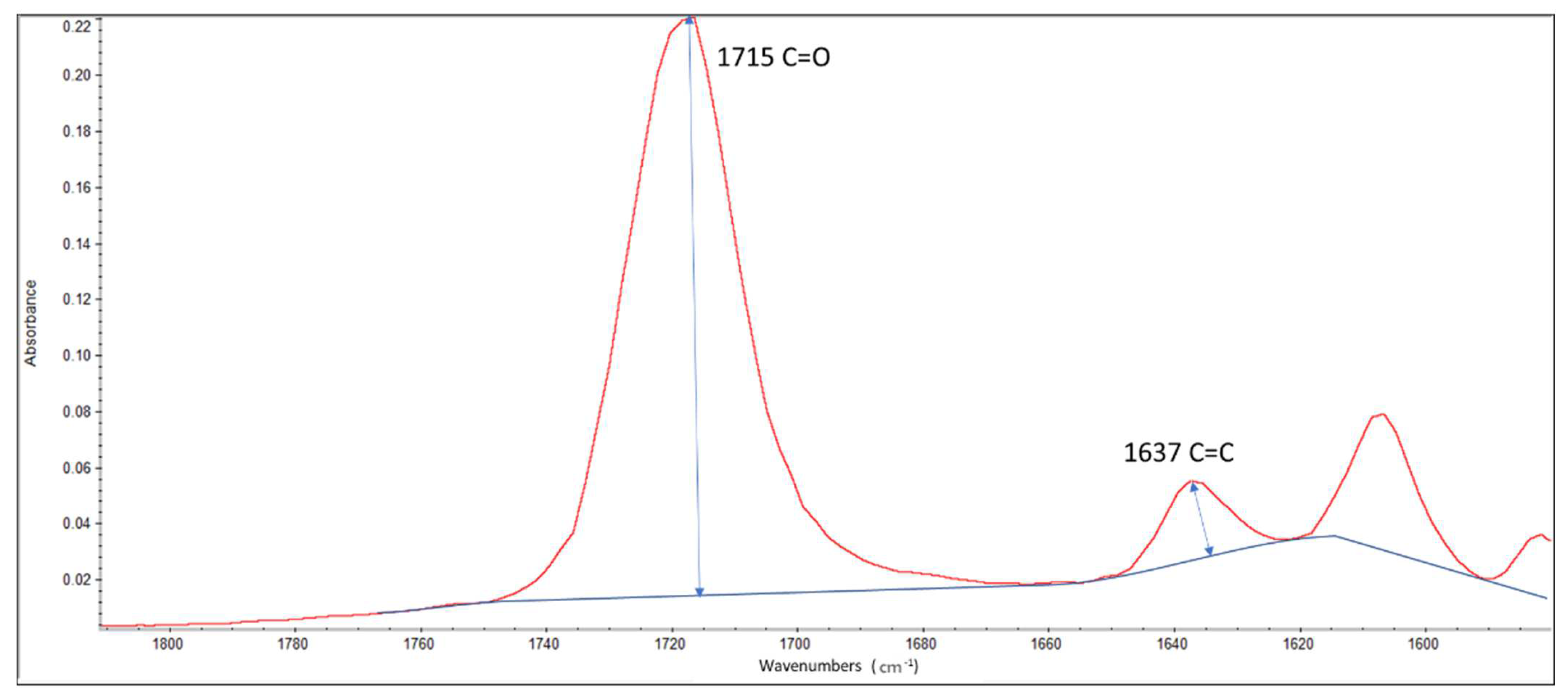
2.2. Attenuated Total Reflectance Fourier Transform Infrared Spectroscopy (FT-IR/ATR) Analysis for the Utilized Resin Systems
2.3. Specimen Preparation
2.4. Bracket Bonding
2.5. Tested Material Application
2.6. Erosion Challenge
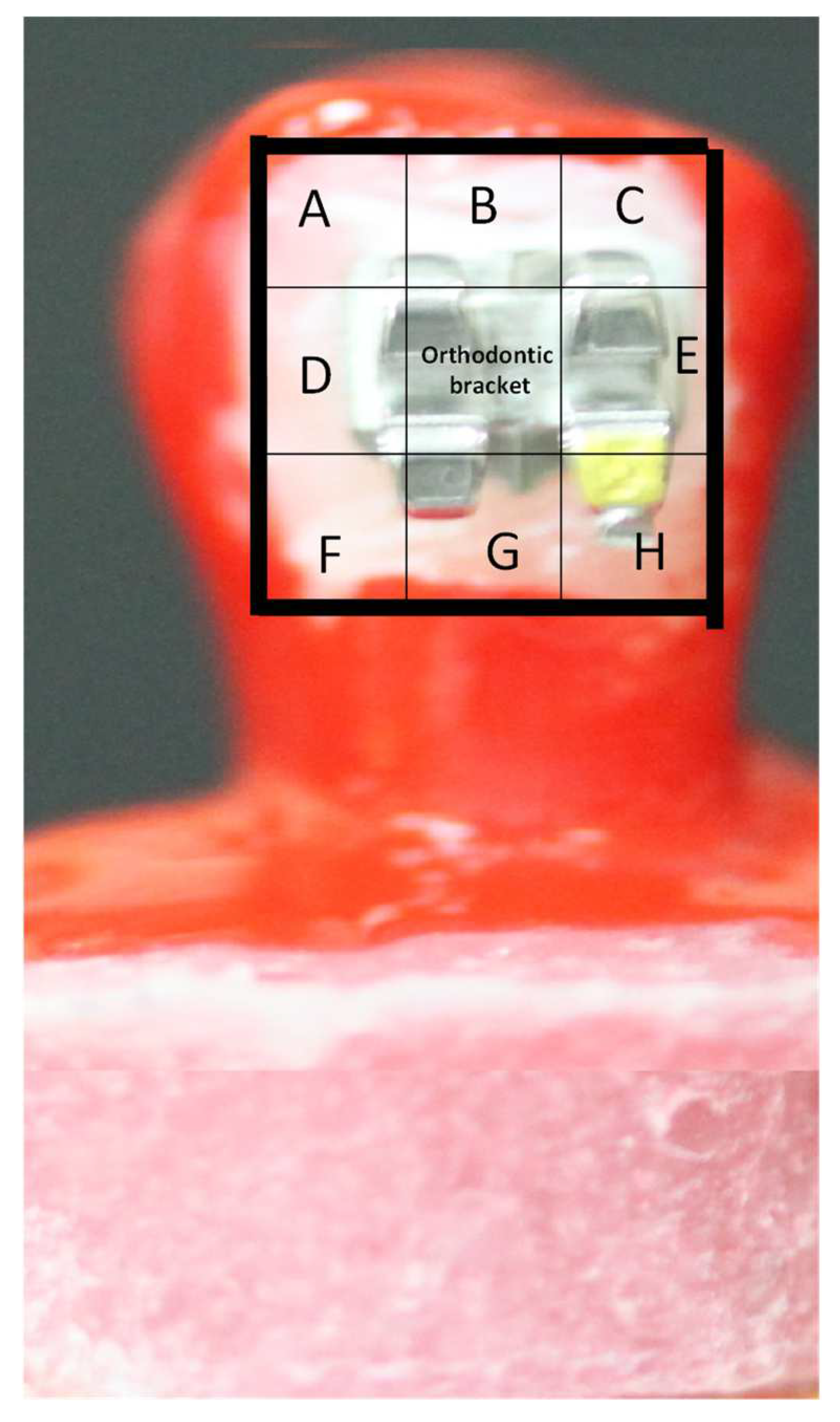
2.7. Scanning Electron Microscope, Equipped with Electron Dispersive Spectroscopy (EDS), Top Surface Examination
2.8. Statistical Analysis
3. Results
3.1. Degree of Conversion
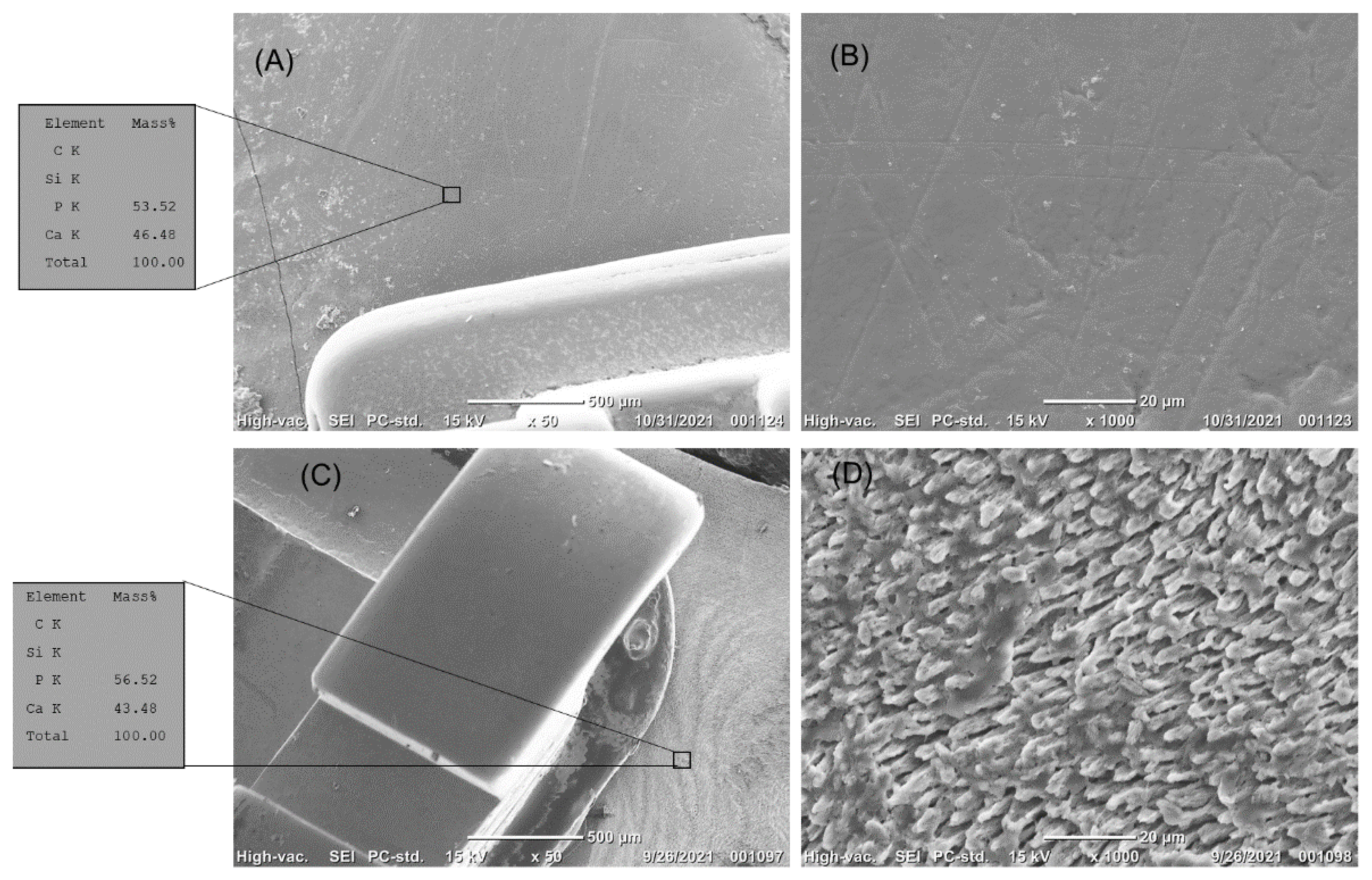
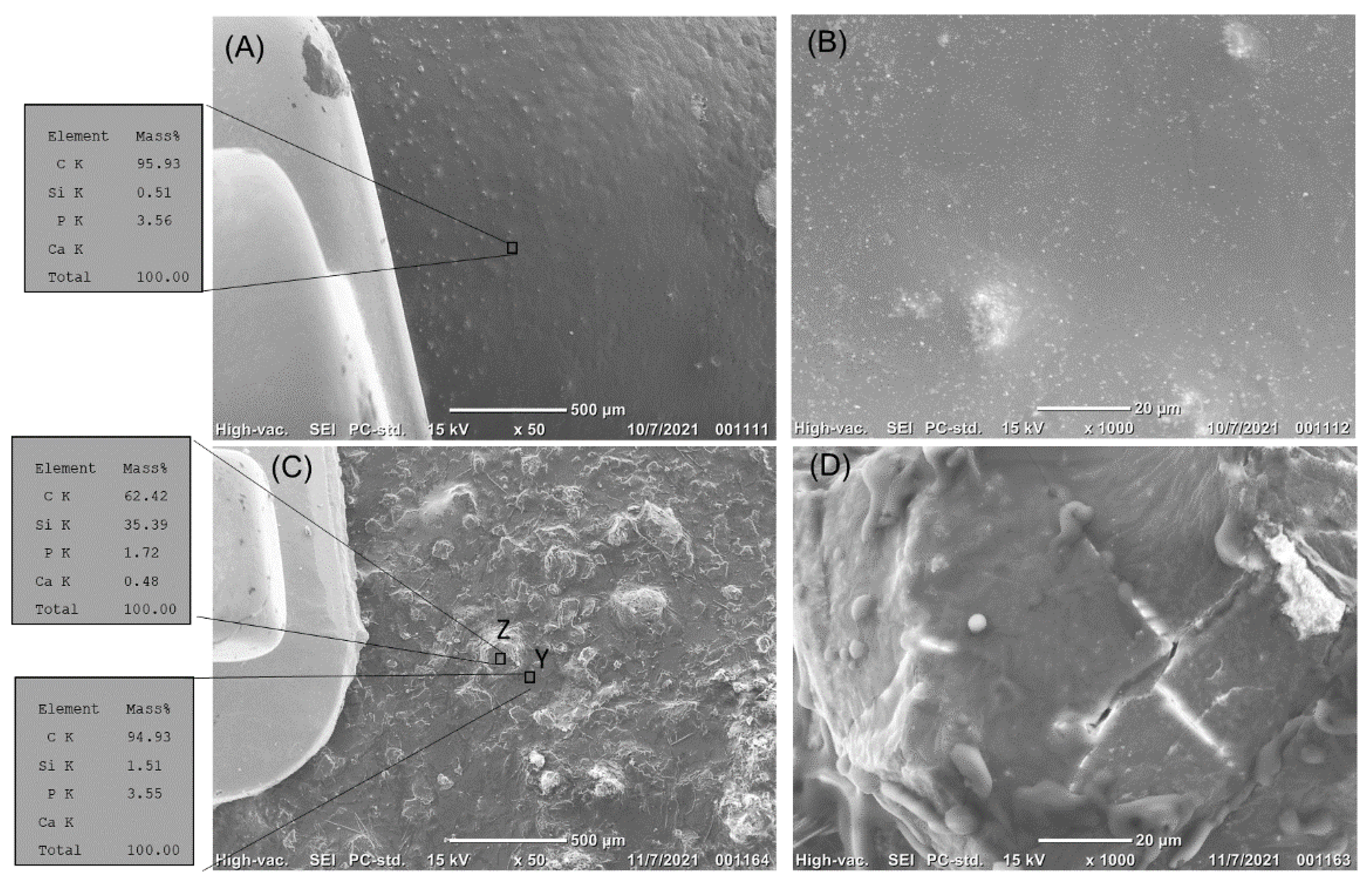
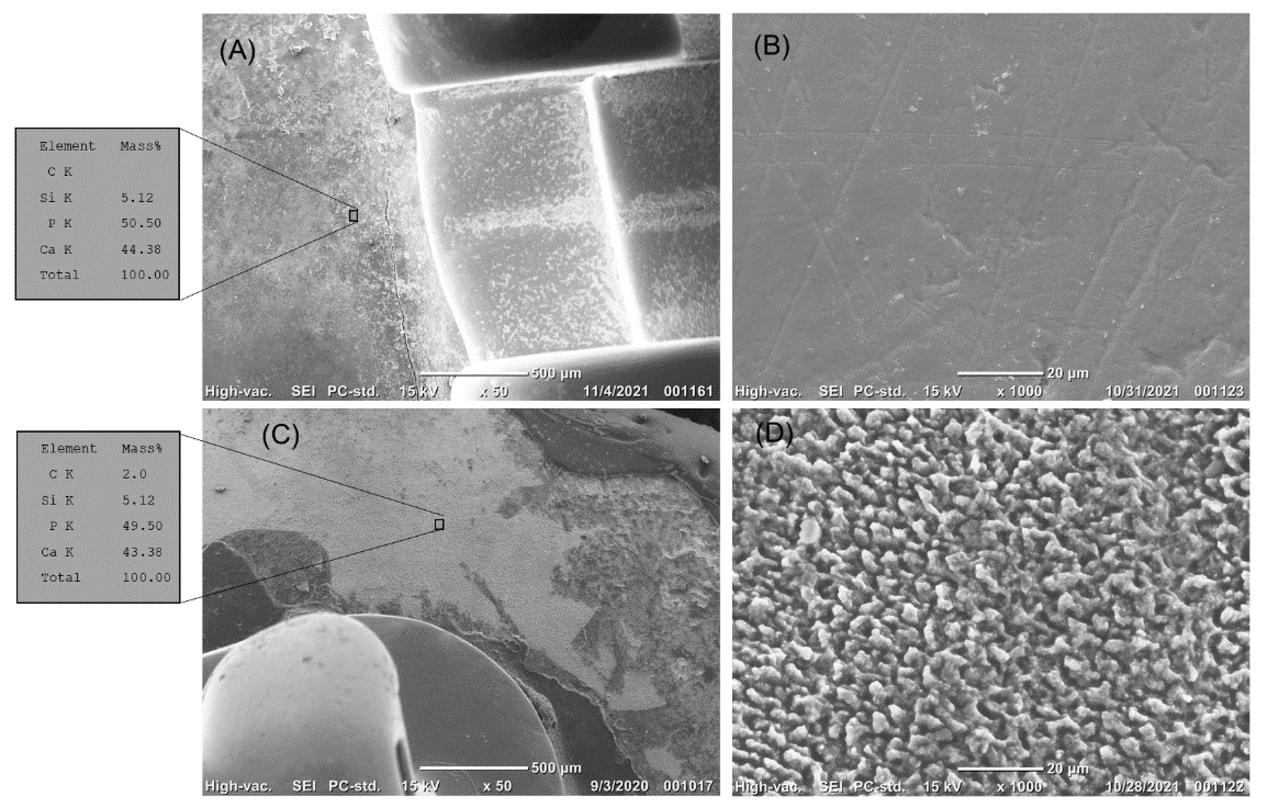
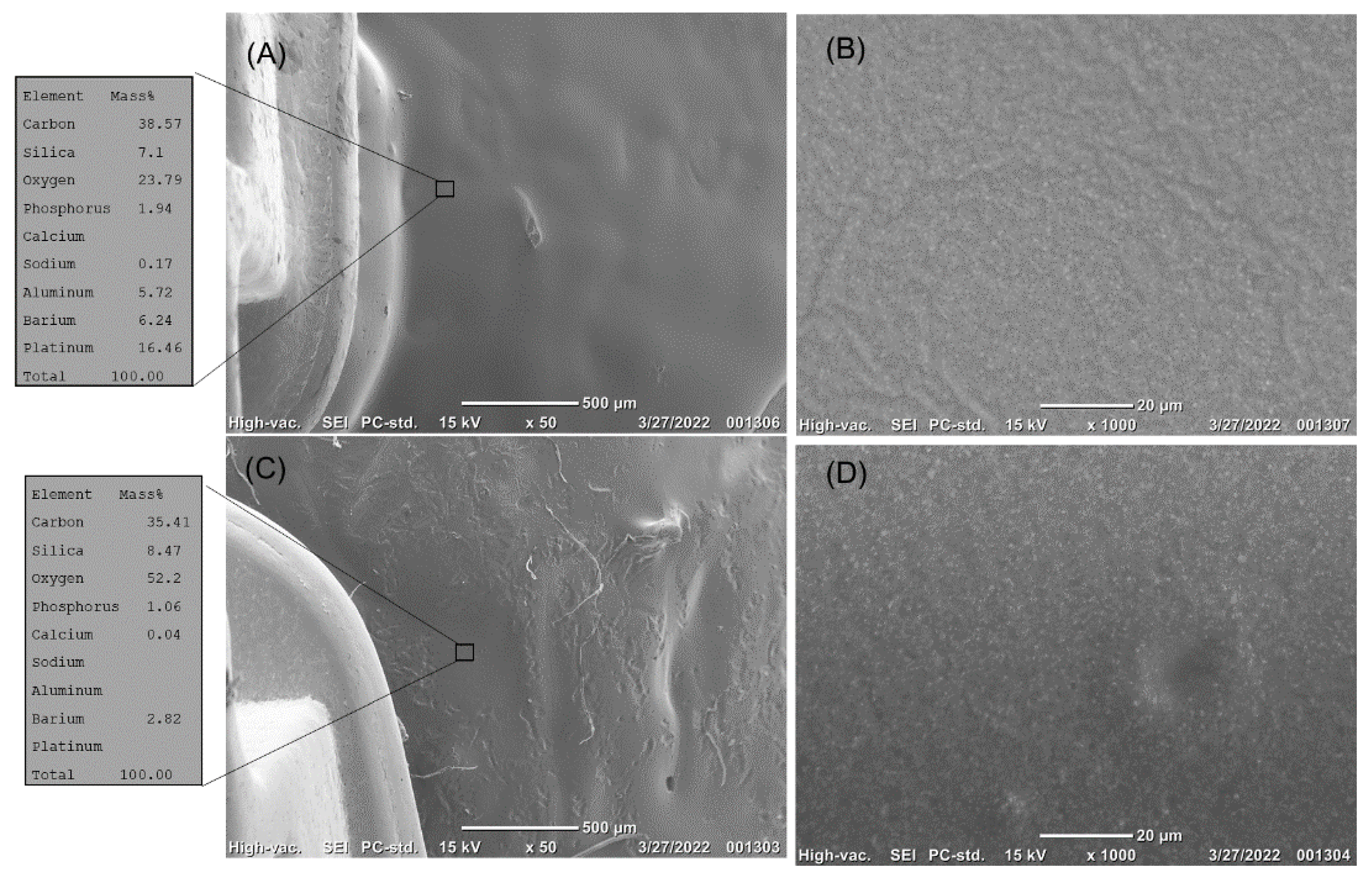
3.2. Scanning Electron Microscope, Equipped with Electron Dispersive Spectroscopy (EDS), Top Surface Examination Results
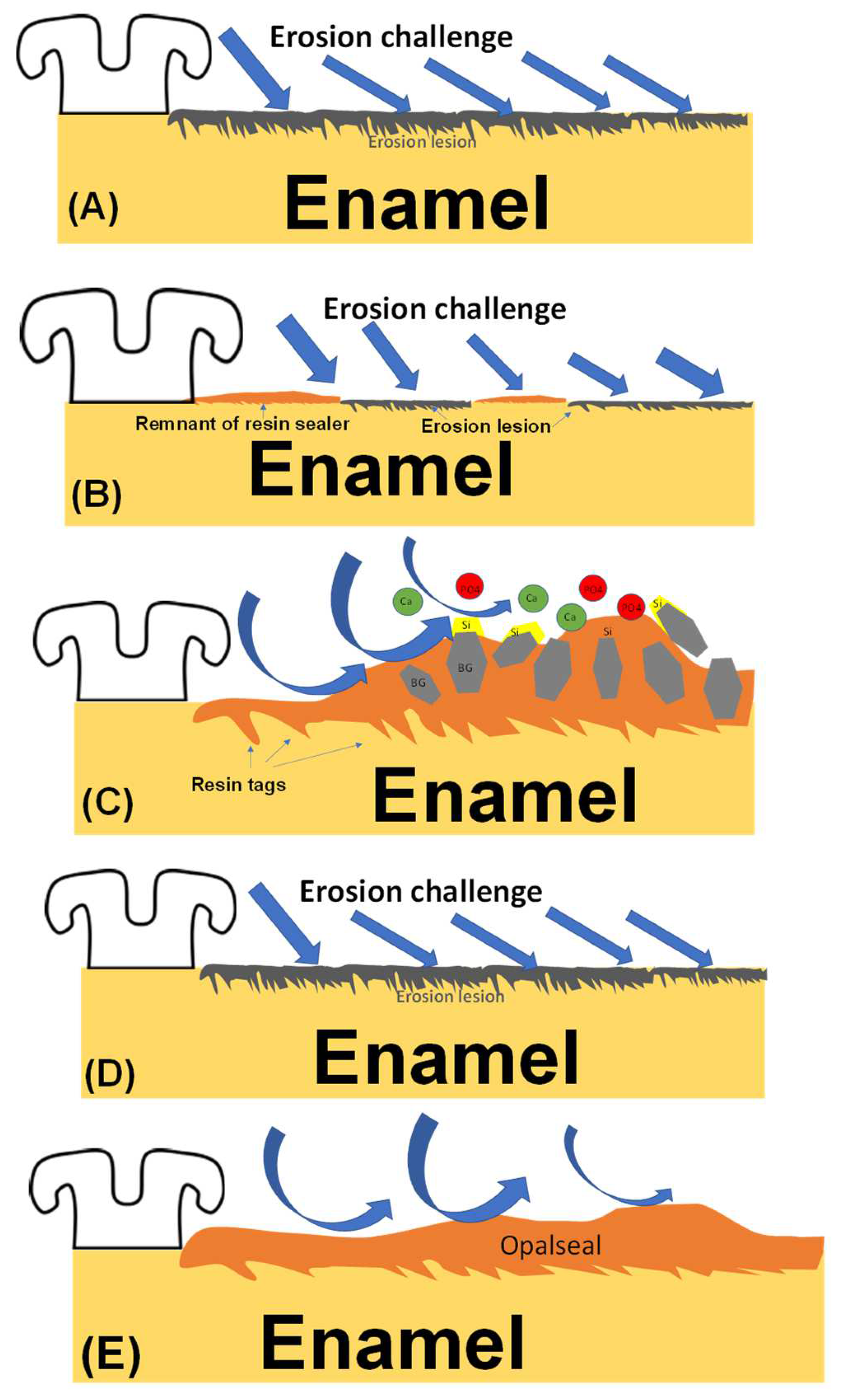
4. Discussion
5. Conclusions
Funding
Institutional Review Board Statement
Informed Consent Statement
Data Availability Statement
Acknowledgments
Conflicts of Interest
References
- Beyer, M.; Reichert, J.; Sigusch, B.W.; Watts, D.C.; Jandt, K.D. Morphology and structure of polymer layers protecting dental enamel against erosion. Dent. Mater. 2012, 28, 1089–1097. [Google Scholar] [CrossRef] [PubMed]
- Al Mulla, A.H.; Kharsa, S.A.; Kjellberg, H.; Birkhed, D. Caries risk profiles in orthodontic patients at follow-up using Cariogram. Angle Orthod. 2009, 79, 323–330. [Google Scholar] [CrossRef] [PubMed] [Green Version]
- Al-Bazi, S.M.; Abbassy, M.A.; Bakry, A.S.; Merdad, L.A.; Hassan, A.H. Effects of chlorhexidine (gel) application on bacterial levels and orthodontic brackets during orthodontic treatment. J. Oral Sci. 2016, 58, 35–42. [Google Scholar] [CrossRef] [Green Version]
- Mizrahi, E. Enamel demineralization following orthodontic treatment. Am. J. Orthod. 1982, 82, 62–67. [Google Scholar] [CrossRef]
- Mitchell, L. Decalcification during orthodontic treatment with fixed appliances—An overview. Br. J. Orthod. 1992, 19, 199–205. [Google Scholar] [CrossRef] [PubMed]
- Rosenbloom, R.G.; Tinanoff, N. Salivary Streptococcus mutans levels in patients before, during, and after orthodontic treatment. Am. J. Orthod. Dentofac. Orthop. 1991, 100, 35–37. [Google Scholar] [CrossRef]
- Abbassy, M.A.; Bakry, A.S.; Hill, R. The efficiency of fluoride bioactive glasses in protecting enamel surrounding orthodontic bracket. Biomed. Res. Int. 2021, 2021, 5544196. [Google Scholar] [CrossRef]
- Scully, M.; Morley, B.; Niven, P.; Crawford, D.; Pratt, I.S.; Wakefield, M. Factors associated with high consumption of soft drinks among Australian secondary-school students. Public Health Nutr. 2017, 20, 2340–2348. [Google Scholar] [CrossRef] [Green Version]
- Abbassy, M.A.; Bakry, A.S.; Alshehri, N.I.; Alghamdi, T.M.; Rafiq, S.A.; Aljeddawi, D.H.; Nujaim, D.S.; Hassan, A.H. 45S5 Bioglass paste is capable of protecting the enamel surrounding orthodontic brackets against erosive challenge. J. Orthod. Sci. 2019, 8, 5. [Google Scholar] [CrossRef]
- Bakhsh, T.A.; Bakry, A.S.; Mandurah, M.M.; Abbassy, M.A. Novel evaluation and treatment techniques for white spot lesions. An In Vitro study. Orthod. Craniofacial Res. 2017, 20, 170–176. [Google Scholar] [CrossRef]
- Bakry, A.S.; Abbassy, M.A. Increasing the efficiency of CPP-ACP to remineralize enamel white spot lesions. J. Dent. 2018, 76, 52–57. [Google Scholar] [CrossRef] [PubMed]
- Bakry, A.S.; Abbassy, M.A.; Alharkan, H.F.; Basuhail, S.; Al-Ghamdi, K.; Hill, R. A novel fluoride containing bioactive glass paste is capable of re-mineralizing early caries lesions. Materials 2018, 11, 1636. [Google Scholar] [CrossRef] [PubMed] [Green Version]
- Cohen, W.J.; Wiltshire, W.A.; Dawes, C.; Lavelle, C.L. Long-term in vitro fluoride release and rerelease from orthodontic bonding materials containing fluoride. Am. J. Orthod. Dentofac. Orthop. 2003, 124, 571–576. [Google Scholar] [CrossRef] [Green Version]
- Benson, P.E.; Alexander-Abt, J.; Cotter, S.; Dyer, F.M.V.; Fenesha, F.; Patel, A.; Campbell, C.; Crowley, N.; Millett, D.T. Resin-modified glass ionomer cement vs composite for orthodontic bonding: A multicenter, single-blind, randomized controlled trial. Am. J. Orthod. Dentofac. Orthop. 2019, 155, 10–18. [Google Scholar] [CrossRef] [PubMed]
- Borzabadi-Farahani, A.; Borzabadi, E.; Lynch, E. Nanoparticles in orthodontics, a review of antimicrobial and anti-caries applications. Acta Odontol. Scand. 2014, 72, 413–417. [Google Scholar] [CrossRef] [PubMed]
- Eslamian, L.; Borzabadi-Farahani, A.; Karimi, S.; Saadat, S.; Badiee, M.R. Evaluation of the shear bond strength and antibacterial activity of orthodontic adhesive containing silver nanoparticle, an in-vitro study. Nanomaterials 2020, 10, 1466. [Google Scholar] [CrossRef]
- Khoroushi, M.; Kachuie, M. Prevention and treatment of white spot lesions in orthodontic patients. Contemp. Clin. Dent. 2017, 8, 11–19. [Google Scholar] [CrossRef]
- Hench, L.L. Special report: The interfacial behavior of biomaterials, 1979. J. Biomed. Mater. Res. 1980, 14, 803–811. [Google Scholar] [CrossRef]
- Al-Harbi, N.; Mohammed, H.; Al-Hadeethi, Y.; Bakry, A.S.; Umar, A.; Hussein, M.A.; Abbassy, M.A.; Vaidya, K.G.; Berakdar, G.A.; Mkawi, E.M.; et al. Silica-based bioactive glasses and their applications in hard tissue regeneration: A review. Pharmaceuticals 2021, 14, 75. [Google Scholar] [CrossRef]
- Abbassy, M.A.; Bakry, A.S.; Hill, R.; Habib Hassan, A. Fluoride bioactive glass paste improves bond durability and remineralizes tooth structure prior to adhesive restoration. Dent. Mater. 2021, 37, 71–80. [Google Scholar] [CrossRef]
- Abbassy, M.A.; Bakry, A.S.; Almoabady, E.H.; Almusally, S.M.; Hassan, A.H. Characterization of a novel enamel sealer for bioactive remineralization of white spot lesions. J. Dent. 2021, 109, 103663. [Google Scholar] [CrossRef] [PubMed]
- Bakry, A.S.; Tamura, Y.; Otsuki, M.; Kasugai, S.; Ohya, K.; Tagami, J. Cytotoxicity of 45S5 bioglass paste used for dentine hypersensitivity treatment. J. Dent. 2011, 39, 599–603. [Google Scholar] [CrossRef] [PubMed]
- Bakry, A.S.; Takahashi, H.; Otsuki, M.; Tagami, J. The durability of phosphoric acid promoted bioglass-dentin interaction layer. Dent. Mater. 2013, 29, 357–364. [Google Scholar] [CrossRef] [PubMed]
- Par, M.; Spanovic, N.; Taubock, T.T.; Attin, T.; Tarle, Z. Degree of conversion of experimental resin composites containing bioactive glass 45S5: The effect of post-cure heating. Sci. Rep. 2019, 9, 17245. [Google Scholar] [CrossRef] [PubMed]
- Lee, S.M.; Yoo, K.H.; Yoon, S.Y.; Kim, I.R.; Park, B.S.; Son, W.S.; Ko, C.C.; Son, S.A.; Kim, Y.I. Enamel Anti-demineralization effect of orthodontic adhesive containing bioactive glass and graphene oxide: An in-vitro study. Materials 2018, 11, 1728. [Google Scholar] [CrossRef] [PubMed] [Green Version]
- Lee, S.M.; Kim, I.R.; Park, B.S.; Lee, D.J.; Ko, C.C.; Son, W.S.; Kim, Y.I. Remineralization property of an orthodontic primer containing a bioactive glass with silver and zinc. Materials 2017, 10, 1253. [Google Scholar] [CrossRef] [Green Version]
- Sales-Peres, S.H.; Brianezzi, L.F.; Marsicano, J.A.; Forim, M.R.; da Silva, M.F.; Sales-Peres, A. Evaluation of an experimental gel containing euclea natalensis: An in vitro study. Evid. Based Complement. Altern. Med. 2012, 2012, 184346. [Google Scholar] [CrossRef] [Green Version]
- Bakry, A.S.; Takahashi, H.; Otsuki, M.; Tagami, J. Evaluation of new treatment for incipient enamel demineralization using 45S5 bioglass. Dent. Mater. 2014, 30, 314–320. [Google Scholar] [CrossRef]
- Magalhaes, A.C.; Moraes, S.M.; Rios, D.; Wiegand, A.; Buzalaf, M.A. The erosive potential of 1% citric acid supplemented by different minerals: An in vitro study. Oral Health Prev. Dent. 2010, 8, 41–45. [Google Scholar]
- Yu, H.; Wegehaupt, F.J.; Wiegand, A.; Roos, M.; Attin, T.; Buchalla, W. Erosion and abrasion of tooth-colored restorative materials and human enamel. J. Dent. 2009, 37, 913–922. [Google Scholar] [CrossRef] [Green Version]
- Bakry, A.S.; Marghalani, H.Y.; Amin, O.A.; Tagami, J. The effect of a bioglass paste on enamel exposed to erosive challenge. J. Dent. 2014, 42, 1458–1463. [Google Scholar] [CrossRef] [PubMed]
- Bakry, A.S.; Abbassy, M.A. The efficacy of a bioglass (45S5) paste temporary filling used to remineralize enamel surfaces prior to bonding procedures. J. Dent. 2019, 85, 33–38. [Google Scholar] [CrossRef] [PubMed]
- Shinya, M.; Shinya, A.; Lassila, L.V.; Varrela, J.; Vallittu, P.K. Enhanced degree of monomer conversion of orthodontic adhesives using a glass-fiber layer under the bracket. Angle Orthod. 2009, 79, 546–550. [Google Scholar] [CrossRef] [PubMed] [Green Version]
- Barbour, M.E.; Parker, D.M.; Allen, G.C.; Jandt, K.D. Enamel dissolution in citric acid as a function of calcium and phosphate concentrations and degree of saturation with respect to hydroxyapatite. Eur. J. Oral Sci. 2003, 111, 428–433. [Google Scholar] [CrossRef]


| Materials | Composition |
|---|---|
| 45S5 Bioglass | 45% SiO2, 24.5% Na2O, 24.4% CaO, 6% P2O5 |
| Bioactive glass sealer | Bis-GMA (44% wht), TEGDMA (20% wht), 45S5 Bioglass powder (35% wht), light cure intiator system (1% wht) Camphor quinone, (DMAB) |
| Resin sealer | Bis-GMA (69% wht), TEGDMA (30% wht), light cure intiator system (1% wht) Camphor quinone, (DMAB) |
| Transbond XT Adhesive Primer | TEGDMA, Bis-GMA Triphenylantimony, 4-(Dimethylamino)-Benzeneethanol, Dl-Camphorquinone, Hydroquinone |
| Transbond PLUS adhesive | Silane-treated quartz, Bis-GMA, dichlorodi-methylsilane reaction product with silica; 77% quartz (silica) filler. |
| Nupro® Acidulated Phosphate Fluoride | 2.59% sodium fluoride (1.23% fluoride ion) |
| Opal Seal | HPMA, Bis-GMA, fluoride; glass ceramic and nanofillers (38% wht). |
| Condition of Dentin | Control | Bioactive Resin Sealer | Resin Sealer | Opalseal | Fluoride |
|---|---|---|---|---|---|
| Before acid challenge (% of Areas showing intact enamel) | 100 * | 100 | 100 * | 100 | 100 * |
| After acid challenge (% of Areas showing intact enamel) | 5 ± 2 *,a | 97 ± 3 a | 50 ± 4 *,a | 100 | 10 ± 2 *,a |
Publisher’s Note: MDPI stays neutral with regard to jurisdictional claims in published maps and institutional affiliations. |
© 2022 by the author. Licensee MDPI, Basel, Switzerland. This article is an open access article distributed under the terms and conditions of the Creative Commons Attribution (CC BY) license (https://creativecommons.org/licenses/by/4.0/).
Share and Cite
Abbassy, M.A. A Bioactive Enamel Sealer Can Protect Enamel during Orthodontic Treatment: An In Vitro Study. Coatings 2022, 12, 550. https://doi.org/10.3390/coatings12050550
Abbassy MA. A Bioactive Enamel Sealer Can Protect Enamel during Orthodontic Treatment: An In Vitro Study. Coatings. 2022; 12(5):550. https://doi.org/10.3390/coatings12050550
Chicago/Turabian StyleAbbassy, Mona Aly. 2022. "A Bioactive Enamel Sealer Can Protect Enamel during Orthodontic Treatment: An In Vitro Study" Coatings 12, no. 5: 550. https://doi.org/10.3390/coatings12050550
APA StyleAbbassy, M. A. (2022). A Bioactive Enamel Sealer Can Protect Enamel during Orthodontic Treatment: An In Vitro Study. Coatings, 12(5), 550. https://doi.org/10.3390/coatings12050550


