
Corrosion Behavior of NiTi Alloys Fabricate by Selective Laser Melting Subjected to Femtosecond Laser Shock Peening

Abstract
:1. Introduction
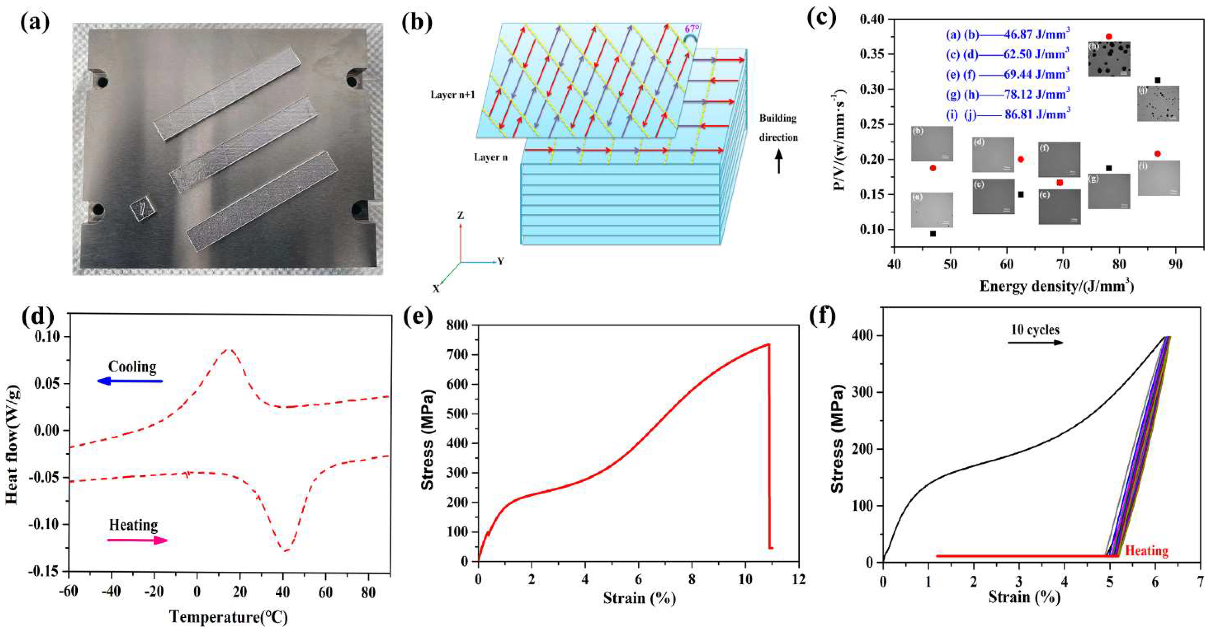
2. Fabrication and Experimental Methods
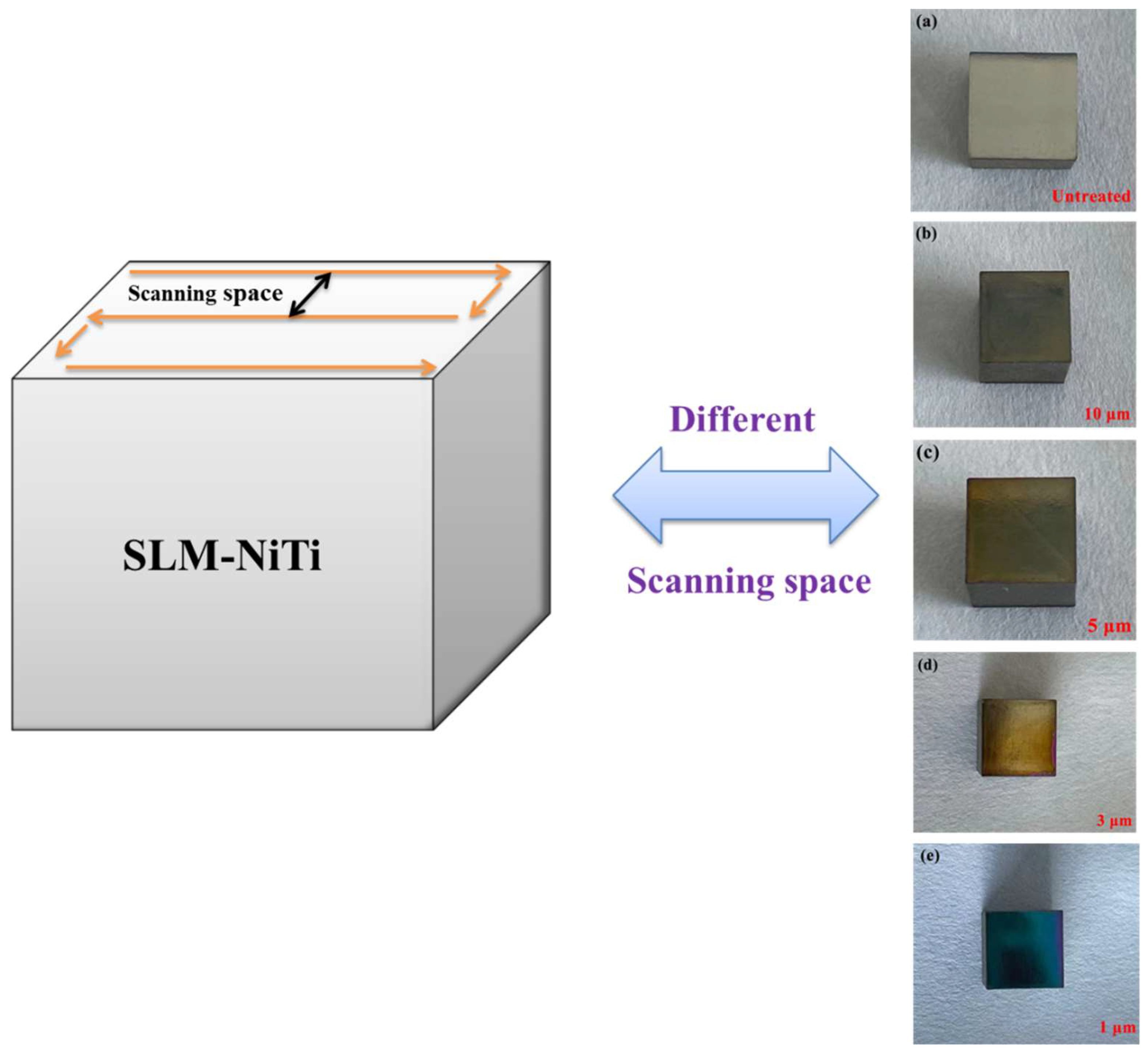
2.1. Femtosecond Laser Shock Peening Experiments
2.2. Test Methods
3. Results
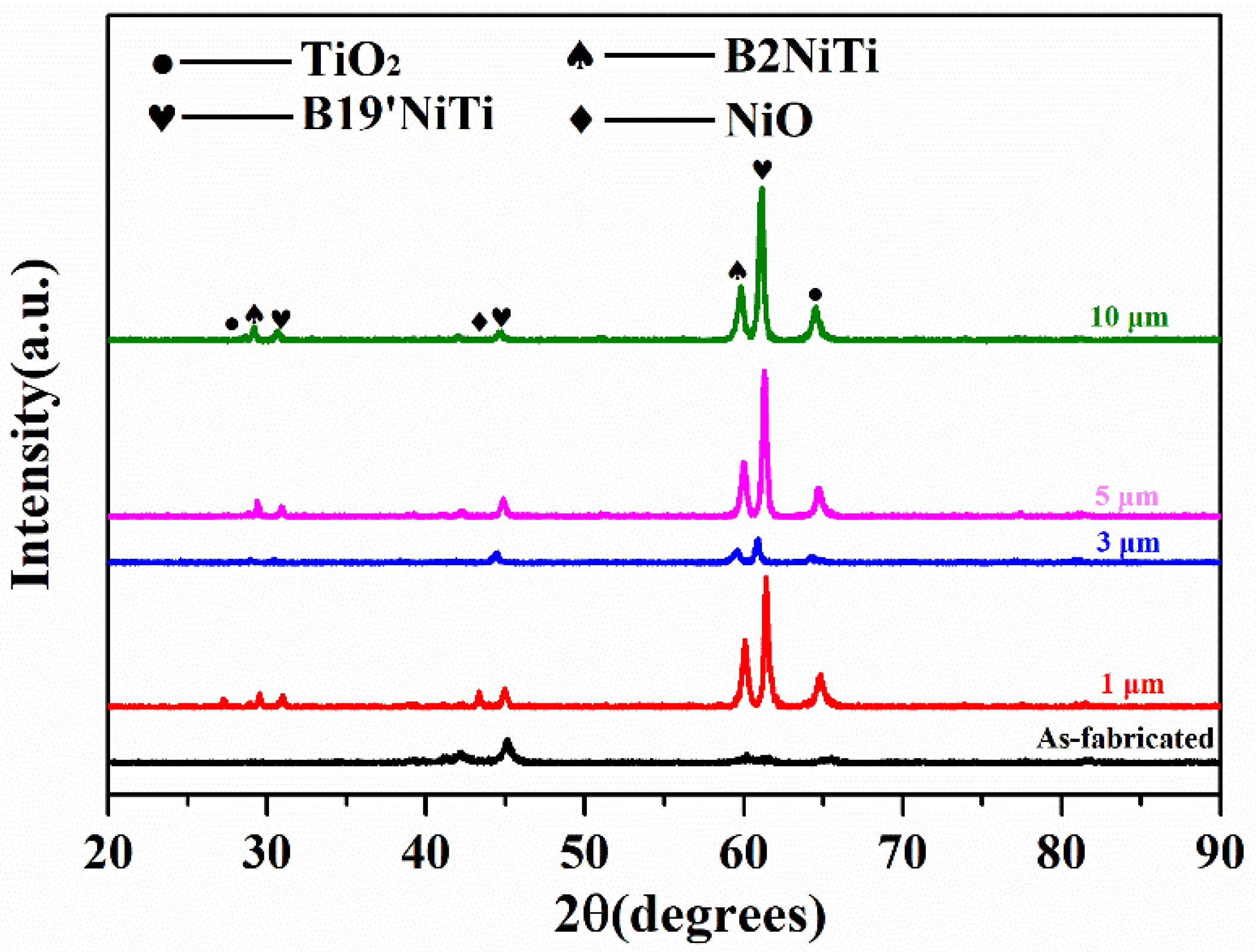
3.1. XRD Analysis
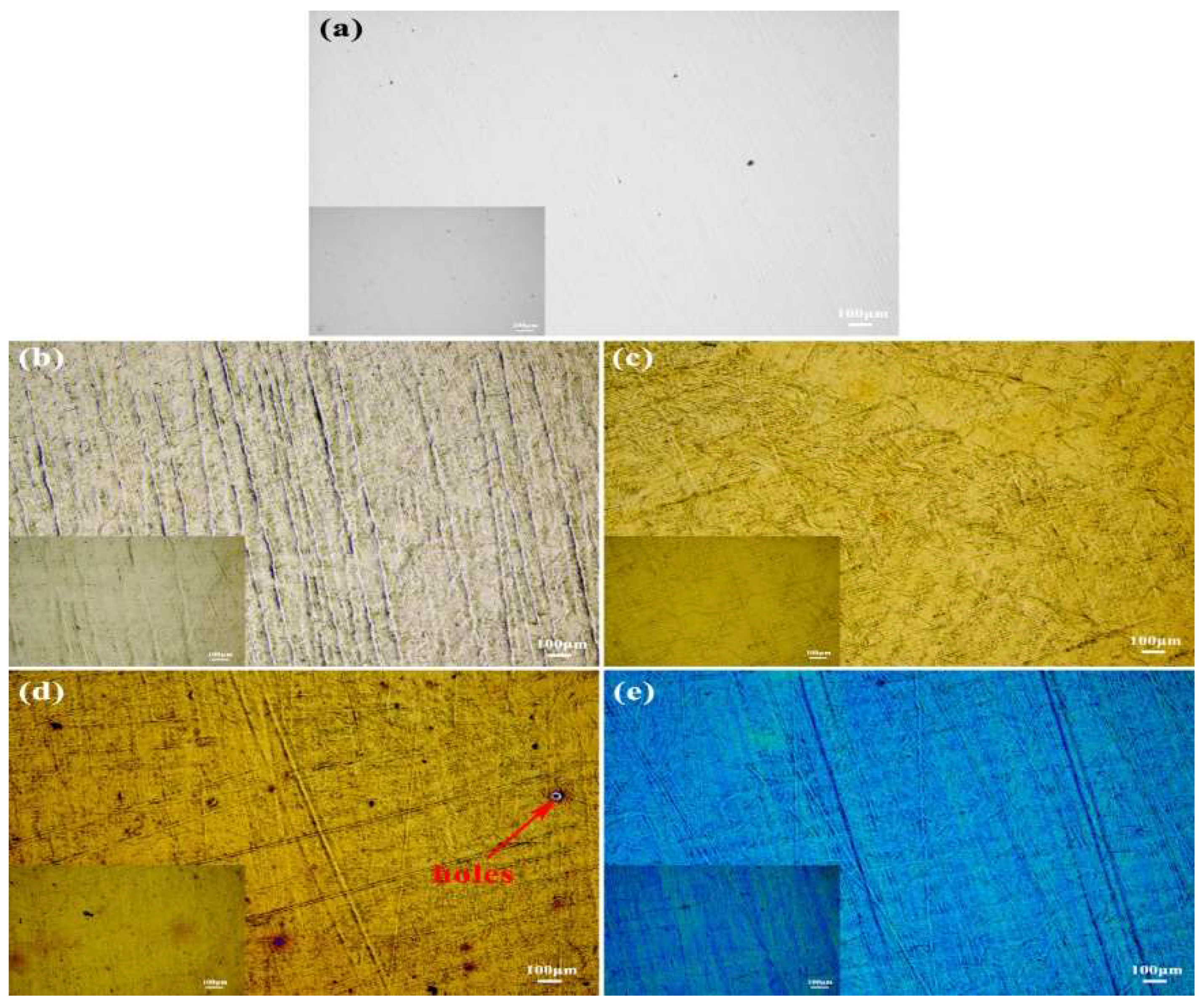
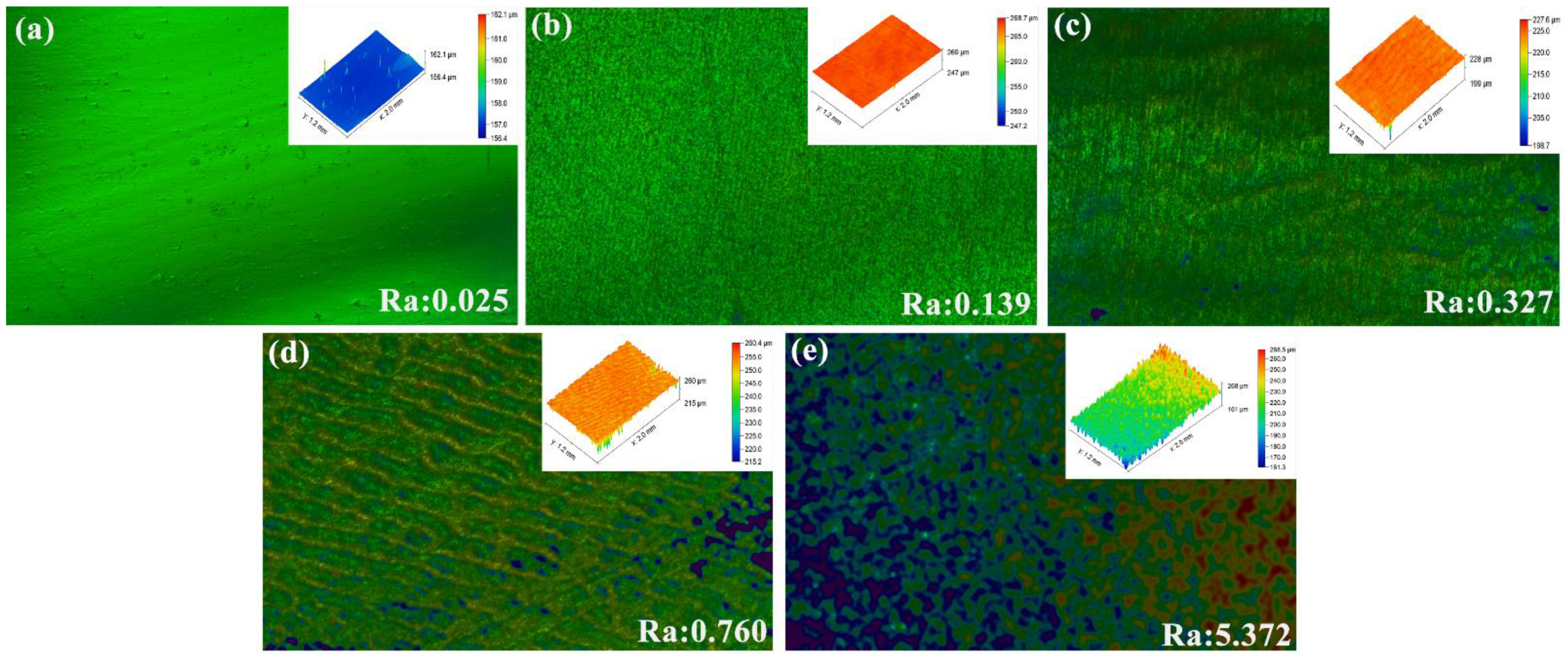
3.2. White Light Interference and Microstructure
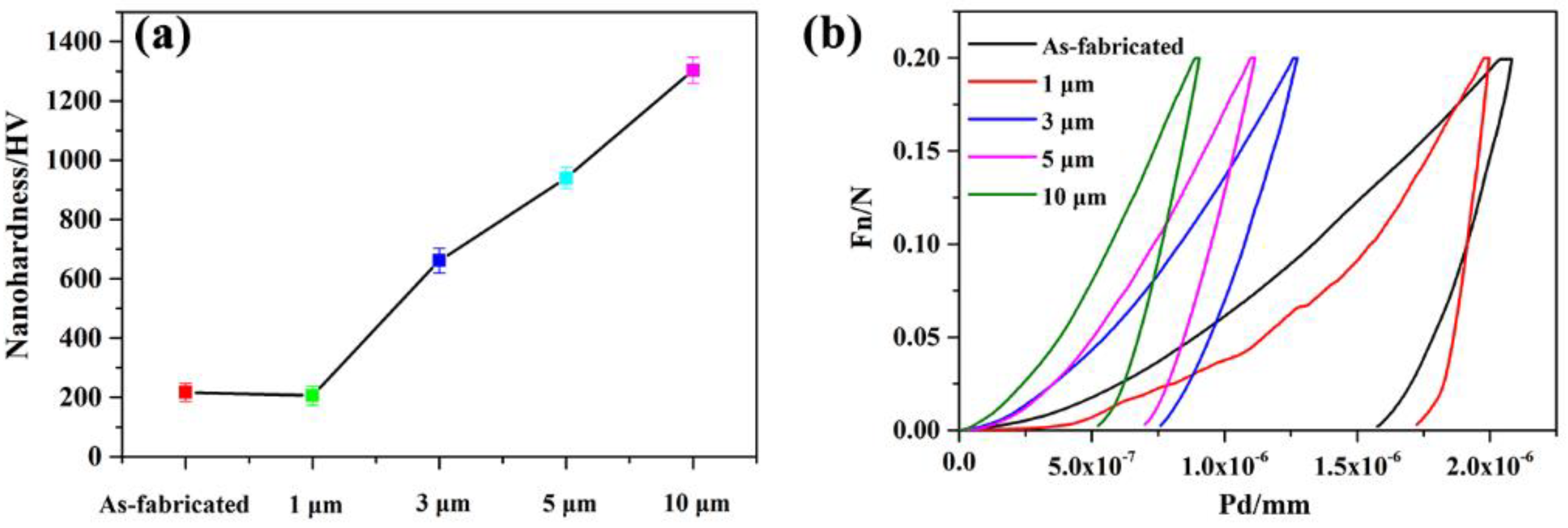
3.3. Nanoindentation Hardness Analysis
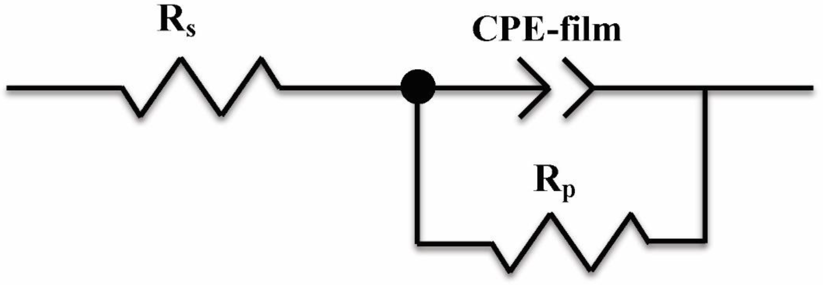
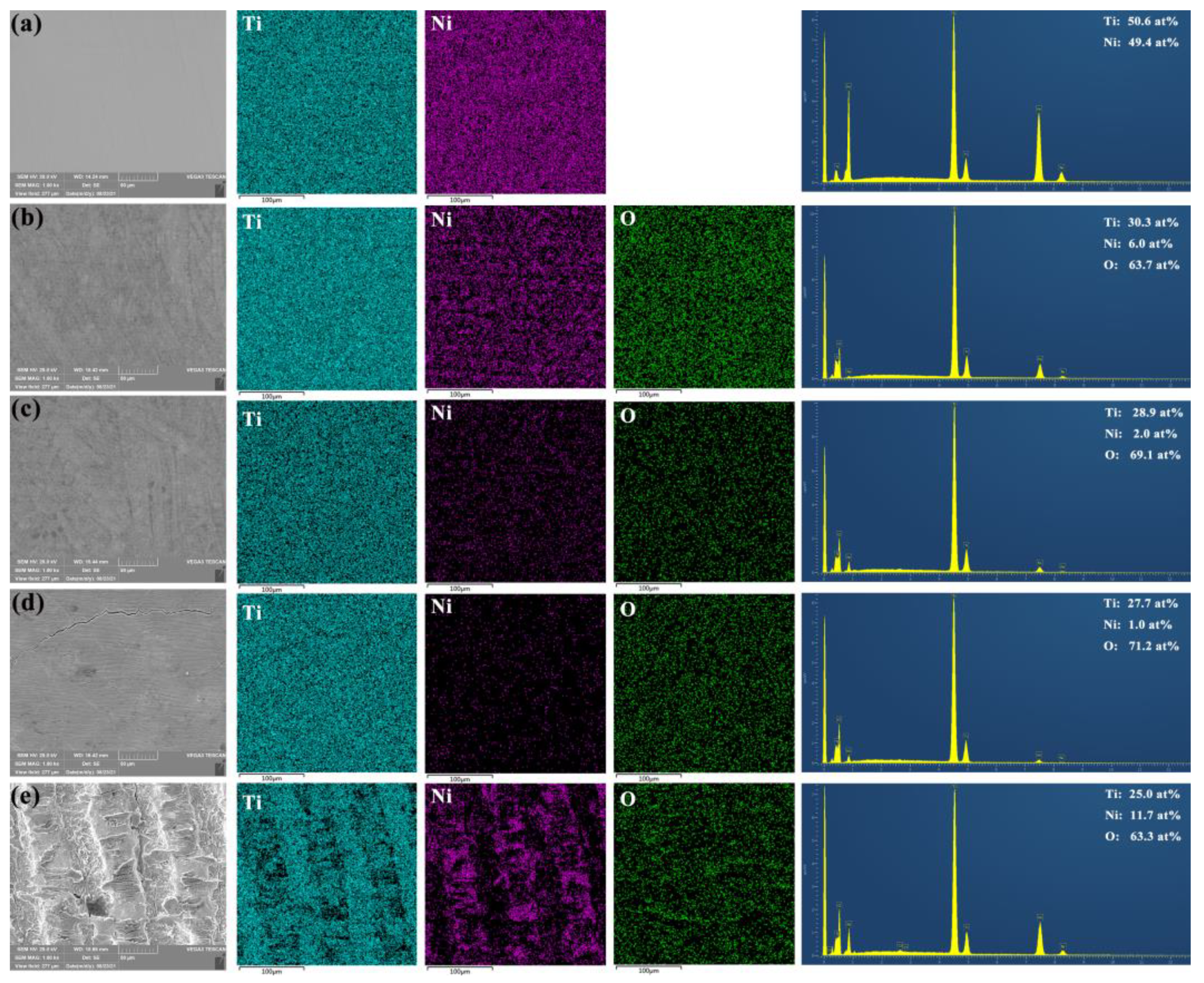
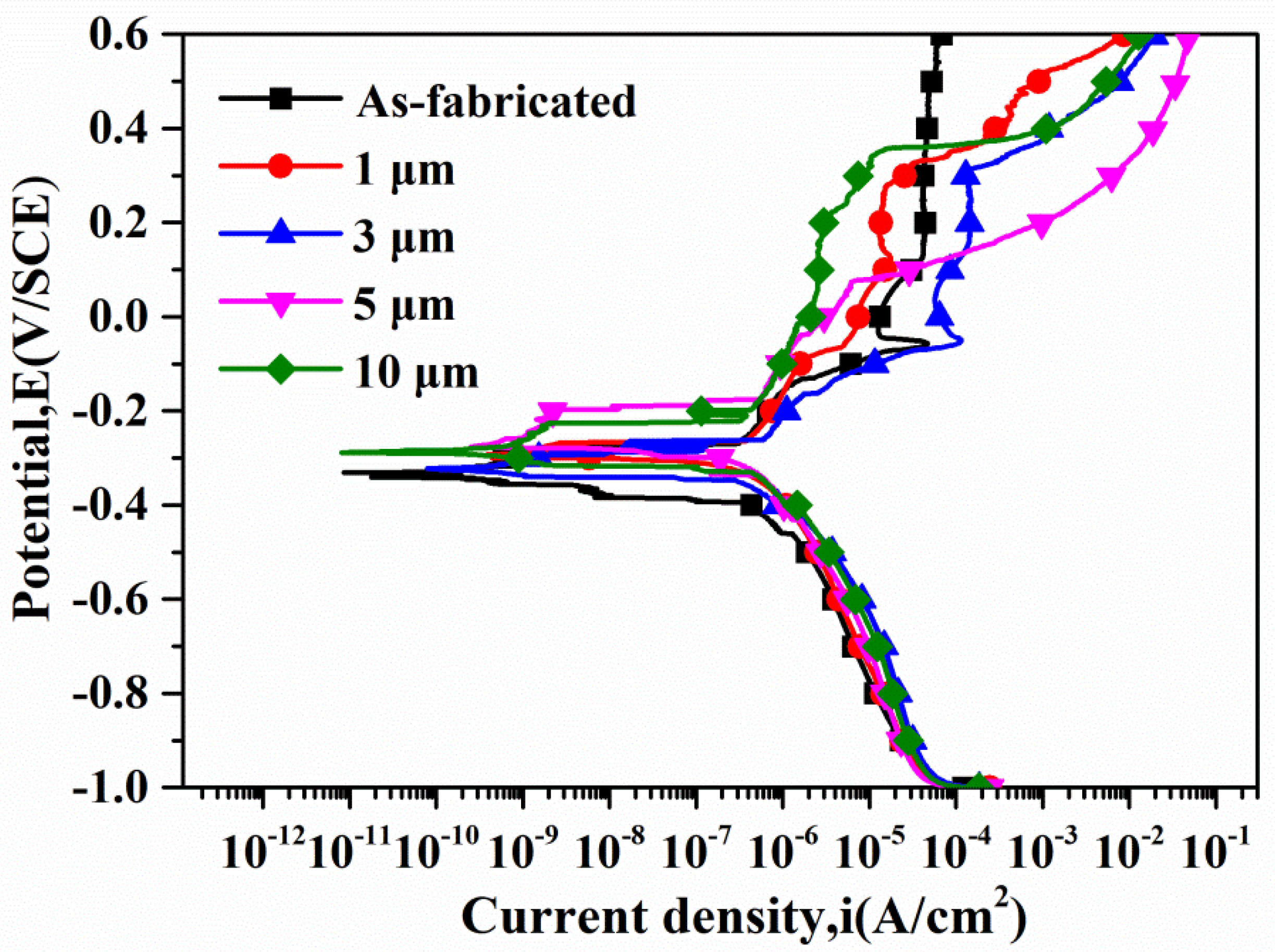
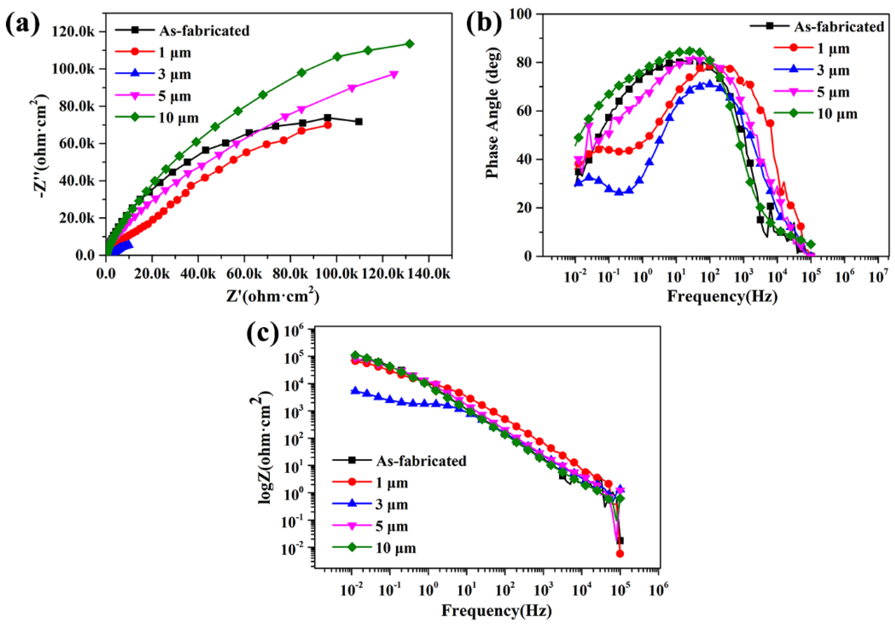
3.4. Electrochemical Corrosion Behavior
4. Conclusions
Author Contributions
Funding
Institutional Review Board Statement
Informed Consent Statement
Data Availability Statement
Conflicts of Interest
References
- Zhao, M.; Shao, Y.; Zheng, W.; Luo, Y.; Qiao, J.; Wu, S.; Yan, Y.; Guo, W. Tailoring the damping and mechanical properties of porous NiTi by a phase leaching process. J. Alloys Compd. 2021, 855, 157471. [Google Scholar] [CrossRef]
- Yu, H.; Qiu, Y.; Young, M.L. Influence of Ni4Ti3 precipitate on pseudoelasticity of austenitic NiTi shape memory alloys deformed at high strain rate. Mater. Sci. Eng. A 2021, 804, 140753. [Google Scholar] [CrossRef]
- Zhang, Q.; Hao, S.; Liu, Y.; Xiong, Z.; Guo, W.; Yang, Y.; Ren, Y.; Cui, L.; Ren, L.; Zhang, Z. The microstructure of a selective laser melting (SLM)-fabricated NiTi shape memory alloy with superior tensile property and shape memory recoverability. Appl. Mater. Today 2020, 19, 100547. [Google Scholar] [CrossRef]
- Elahinia, M.; Moghaddam, N.S.; Andani, M.T.; Amerinatanzi, A.; Bimber, B.A.; Hamilton, R.F. Fabrication of NiTi through additive manufacturing: A review. Prog. Mater. Sci. 2016, 83, 630–663. [Google Scholar] [CrossRef] [Green Version]
- Saedi, S.; Turabi, A.S.; Andani, M.T.; Haberland, C.; Elahinia, M.; Karaca, H. Thermomechanical characterization of Ni-rich NiTi fabricated by selective laser melting. Smart Mater. Struct. 2016, 25, 035005. [Google Scholar] [CrossRef]
- Wang, C.; Tan, X.; Du, Z.; Chandra, S.; Sun, Z.; Lim, C.; Tor, S.B.; Wong, C. Additive manufacturing of NiTi shape memory alloys using pre-mixed powders. J. Mater. Process. Technol. 2019, 271, 152–161. [Google Scholar] [CrossRef]
- Yan, Y.X.; Ahmad, T.; Zhang, X.; Liang, T.; Rehman, S.U.; Manzoor, M.U.; Liu, W.; Basit, M.A.; Saim, A.B. Microstructure, hardness and corrosion behavior of Ni-Ti alloy with the addition of rare earth metal oxide (Gd2O3). Mater. Res. Express 2019, 6, 076513. [Google Scholar] [CrossRef]
- Khoo, Z.X.; Liu, Y.; An, J.; Chua, C.K.; Shen, Y.F.; Kuo, C.N. A review of selective laser melted NiTi shape memory alloy. Materials 2018, 11, 519. [Google Scholar] [CrossRef] [Green Version]
- Chen, X.; Liu, K.; Guo, W.; Gangil, N.; Siddiquee, A.N.; Konovalov, S. The fabrication of NiTi shape memory alloy by selective laser melting: A review. Rapid Prototyp. J. 2019, 25, 1421–1432. [Google Scholar] [CrossRef]
- Yu, Z.; Xu, Z.; Guo, Y.; Xin, R.; Liu, R.; Jiang, C.; Li, L.; Zhang, Z.; Ren, L. Study on properties of SLM-NiTi shape memory alloy under the same energy density. J. Mater. Res. Technol. 2021, 13, 241–250. [Google Scholar] [CrossRef]
- Saugo, M.; Flamini, D.O.; Saidman, S.B. Low-voltage polarization in AOT Solution to enhance the corrosion resistance of nitinol. J. Mater. Eng. Perform. 2021, 30, 1816–1824. [Google Scholar] [CrossRef]
- Veverkova, J.; Bartkova, D.; Weiser, A.; Dlouhy, A.; Babula, P.; Štěpka, P.; Goldbergova, M.P. Effect of Ni ion release on the cells in contact with NiTi alloys. Environ. Sci. Pollut. Res. 2020, 27, 7934–7942. [Google Scholar] [CrossRef]
- Velmurugan, C.; Senthilkumar, V.; Kamala, P.S. Microstructure and corrosion behavior of NiTi shape memory alloys sintered in the SPS process. Int. J. Miner. Met. Mater. 2019, 26, 1311–1321. [Google Scholar] [CrossRef]
- Qiu, P.; Gao, P.; Wang, S.; Li, Z.; Yang, Y.; Zhang, Q.; Xiong, Z.; Hao, S. Study on corrosion behavior of the selective laser melted NiTi alloy with superior tensile property and shape memory effect. Corros. Sci. 2020, 175, 108891. [Google Scholar] [CrossRef]
- Yuan, W.; Li, R.; Chen, Z.; Gu, J.; Tian, Y. A comparative study on microstructure and properties of traditional laser cladding and high-speed laser cladding of Ni45 alloy coatings. Surf. Coat. Technol. 2021, 405, 126582. [Google Scholar] [CrossRef]
- Wang, L.; Chen, M.; Liu, H.; Jiang, C.; Ji, V.; Moreira, F. Optimisation of microstructure and corrosion resistance of Ni-Ti composite coatings by the addition of CeO2 nanoparticles. Surf. Coat. Technol. 2017, 331, 196–205. [Google Scholar] [CrossRef]
- Nishimura, A.; Minehara, E.; Tsukada, T.; Kikuchi, M.; Nakano, J. Ablation of work hardening layers against stress corrosion cracking of stainless steel by repetitive femtosecond laser pulses. In Fifth International Symposium on Laser Precision Microfabrication; Miyamoto, I., Helvajian, H., Itoh, K., Kobayashi, K.F., Ostendorf, A., Sugioka, K., Eds.; International Society for Optics and Photonics: Bellingham, WA, USA, 2004; pp. 673–677. [Google Scholar]
- Jeong, Y.-H.; Kim, W.-G.; Choe, H.-C. Electrochemical behavior of nano and femtosecond laser textured titanium alloy for implant surface modification. J. Nanosci. Nanotechnol. 2011, 11, 1581–1584. [Google Scholar] [CrossRef]
- Park, J.; Han, H.-S.; Park, J.; Seo, H.; Edwards, J.; Kim, Y.-C.; Ok, M.-R.; Seok, H.-K.; Jeon, H. Corrosion behavior of biodegradable Mg-based alloys via femtosecond laser surface melting. Appl. Surf. Sci. 2018, 448, 424–434. [Google Scholar] [CrossRef]
- Kawashima, T.; Sano, T.; Hirose, A.; Tsutsumi, S.; Masaki, K.; Arakawa, K.; Hori, H. femtosecond laser peening of friction stir welded 7075-T73 aluminum alloys. J. Mater. Process. Technol. 2018, 262, 111–122. [Google Scholar] [CrossRef]
- Trdan, U.; Sano, T.; Klobčar, D.; Sano, Y.; Grum, J.; Šturm, R. Improvement of corrosion resistance of AA2024-T3 using femtosecond laser peening without protective and confining medium. Corros. Sci. 2018, 143, 46–55. [Google Scholar] [CrossRef]
- Kolobov, Y.R.; Zhidkov, M.V.; Golosov, E.V.; Vershinina, T.; Kudryashov, S.I.; Makarov, S.V.; Ionin, A.A.; Ligachev, A.E. Phase composition and structure of femtosecond laser-produced oxide layer on VT6 alloy surface. Laser Phys. Lett. 2016, 13, 76103. [Google Scholar] [CrossRef]
- Boinovich, L.B.; Emelyanenko, A.M.; Modestov, A.; Domantovsky, A.G.; Emelyanenko, K.A. Synergistic effect of superhydrophobicity and oxidized layers on corrosion resistance of aluminum alloy surface textured by nanosecond laser treatment. ACS Appl. Mater. Interfaces 2015, 7, 19500–19508. [Google Scholar] [CrossRef] [PubMed] [Green Version]
- Wang, H.; Gurevich, E.L.; Ostendorf, A. Microhardness and microabrasion behaviour of NiTi shape memory alloy after femtosecond laser shock peening without coating in air. In High-Power Laser Materials Processing: Applications, Diagnostics, and Systems IX; Kaierle, S., Heinemann, S.W., Eds.; International Society for Optics and Photonics: Bellingham, WA, USA, 2020; Volume 11273, p. 1127301. [Google Scholar] [CrossRef]
- Wang, H.; Jürgensen, J.; Decker, P.; Hu, Z.; Yan, K.; Gurevich, E.; Ostendorf, A. Corrosion behavior of NiTi alloy subjected to femtosecond laser shock peening without protective coating in air environment. Appl. Surf. Sci. 2020, 501, 144338. [Google Scholar] [CrossRef]
- Zhang, C.; Zhu, J.; Zheng, H.; Li, H.; Liu, S.; Cheng, G. A review on microstructures and properties of high entropy alloys manufactured by selective laser melting. Int. J. Extrem. Manuf. 2020, 2, 032003. [Google Scholar] [CrossRef]
- Yang, Y.; Zhan, J.; Sui, J.; Li, C.; Yang, K.; Castany, P.; Gloriant, T. Functionally graded NiTi alloy with exceptional strain-hardening effect fabricated by SLM method. Scr. Mater. 2020, 188, 130–134. [Google Scholar] [CrossRef]
- Biffi, C.A.; Fiocchi, J.; Valenza, F.; Bassani, P.; Tuissi, A. selective laser melting of NiTi shape memory alloy: Processability, microstructure, and superelasticity. Shape Mem. Superelasticity 2020, 6, 342–353. [Google Scholar] [CrossRef]
- Ibrahim, H.; Jahadakbar, A.; Dehghan, A.; Moghaddam, N.S.; Amerinatanzi, A.; Elahinia, M. In vitro corrosion assessment of additively manufactured porous NiTi structures for bone fixation applications. Metals 2018, 8, 164. [Google Scholar] [CrossRef] [Green Version]
- Norouzi, N.; Nouri, Z. The effect of two-stage acid treatment on surface behavior and improvement of bioactivity of nitinol alloy. Biointerface Res. Appl. Chem. 2021, 11, 10690–10702. [Google Scholar]
- Zhao, T.; Cai, X.; Wang, S.X.; Zheng, S. Effect of CeO2 on microstructure and corrosive wear behavior of laser-cladded Ni/WC coating. Thin Solid Films 2000, 379, 128–132. [Google Scholar]
- Guo, Y.; Jia, S.; Qiao, L.; Su, Y.; Gu, R.; Li, G.; Lian, J. Enhanced corrosion resistance and biocompatibility of polydopamine/dicalcium phosphate dihydrate/collagen composite coating on magnesium alloy for orthopedic applications. J. Alloys Compd. 2019, 817, 152782. [Google Scholar] [CrossRef]
- Su, Y.; Lu, C.; Hu, X.; Guo, Y.; Xun, X.; Zhang, Z.; Li, G.; Lian, J.; Ren, L. Improving the degradation resistance and surface biomineralization ability of calcium phosphate coatings on a biodegradable magnesium alloy via a sol-gel spin coating method. J. Electrochem. Soc. 2018, 165, C155–C161. [Google Scholar] [CrossRef]
- Guo, Y.; Su, Y.; Jia, S.; Sun, G.; Gu, R.; Zhu, D.; Li, G.; Lian, J. Hydroxyapatite/titania composite coatings on biodegradable magnesium alloy for enhanced corrosion resistance, cytocompatibility and antibacterial properties. J. Electrochem. Soc. 2018, 165, C962–C972. [Google Scholar] [CrossRef]
- Huang, Y.S.; Zeng, X.T.; Hu, X.F.; Liu, F.M. Heat treatment effects on EN-PTFE-SiC composite coatings. Surf. Coat. Technol. 2005, 198, 173–177. [Google Scholar] [CrossRef]


| Sample | Powder (w) | Scanning Speed (mm/s) | Frequency (kHz) | Scanning Space (μm) |
|---|---|---|---|---|
| 1 μm | 8 | 60 | 20 | 1 |
| 3 μm | 8 | 60 | 20 | 3 |
| 5 μm | 8 | 60 | 20 | 5 |
| 10 μm | 8 | 60 | 20 | 10 |
| Samples | Ba/mV | Bc/mV | Ecorr/V | Icorr/A·cm−2 |
|---|---|---|---|---|
| As-fabricated | 279.46 ± 2.5 | 169.68 ± 4.1 | −0.32 ± 0.02 | 1.00 ± 0.2 × 10⁻⁸ |
| 1 μm | 56.86 ± 2.1 | 78.77 ± 2.5 | −0.29 ± 0.04 | 4.95 ± 0.3 × 10⁻⁸ |
| 3 μm | 61.43 ± 1.3 | 74.15 ± 3.3 | −0.31 ± 0.08 | 4.08 ± 1.1 × 10⁻⁷ |
| 5 μm | 66.19 ± 2.2 | 81.98 ± 2.5 | −0.24 ± 0.03 | 1.97 ± 0.3 × 10⁻⁹ |
| 10 μm | 74.71 ± 2.3 | 71.53 ± 3.4 | −0.27 ± 0.05 | 1.67 ± 0.1 × 10⁻⁹ |
| Samples | Rs (Ω·cm2) | CPE-film | Rp (Ω·cm2) | n |
|---|---|---|---|---|
| As-fabricated | 57.11 ± 2.5 | (2.168 ± 0.3) × 10−5 | (1.873 ± 0.4) × 105 | 0.8412 ± 0.04 |
| 1 μm | 56.23 ± 3.5 | (2.302 ± 0.2) × 10−5 | (1.285 ± 0.3) × 105 | 0.8008 ± 0.03 |
| 3 μm | 51.17 ± 4.8 | (2.779 ± 0.5) × 10−5 | (1.685 ± 0.2) × 104 | 0.7187 ± 0.01 |
| 5 μm | 48.24 ± 2.7 | (1.681 ± 0.4) × 10−5 | (2.439 ± 0.5) × 105 | 0.8596 ± 0.03 |
| 10 μm | 53.18 ± 2.3 | (1.365 ± 0.3) × 10−5 | (3.439 ± 0.4) × 105 | 0.8865 ± 0.05 |
Publisher’s Note: MDPI stays neutral with regard to jurisdictional claims in published maps and institutional affiliations. |
© 2021 by the authors. Licensee MDPI, Basel, Switzerland. This article is an open access article distributed under the terms and conditions of the Creative Commons Attribution (CC BY) license (https://creativecommons.org/licenses/by/4.0/).
Share and Cite
Ma, L.; Li, W.; Yang, Y.; Ma, Y.; Luo, K.; Jia, B.; Xu, Z.; Yu, Z. Corrosion Behavior of NiTi Alloys Fabricate by Selective Laser Melting Subjected to Femtosecond Laser Shock Peening. Coatings 2021, 11, 1078. https://doi.org/10.3390/coatings11091078
Ma L, Li W, Yang Y, Ma Y, Luo K, Jia B, Xu Z, Yu Z. Corrosion Behavior of NiTi Alloys Fabricate by Selective Laser Melting Subjected to Femtosecond Laser Shock Peening. Coatings. 2021; 11(9):1078. https://doi.org/10.3390/coatings11091078
Chicago/Turabian StyleMa, Long, Wanqing Li, Yongzhi Yang, Yuanxue Ma, Kai Luo, Bochao Jia, Zezhou Xu, and Zhenglei Yu. 2021. "Corrosion Behavior of NiTi Alloys Fabricate by Selective Laser Melting Subjected to Femtosecond Laser Shock Peening" Coatings 11, no. 9: 1078. https://doi.org/10.3390/coatings11091078
APA StyleMa, L., Li, W., Yang, Y., Ma, Y., Luo, K., Jia, B., Xu, Z., & Yu, Z. (2021). Corrosion Behavior of NiTi Alloys Fabricate by Selective Laser Melting Subjected to Femtosecond Laser Shock Peening. Coatings, 11(9), 1078. https://doi.org/10.3390/coatings11091078