Article

Shear Bond Strength of Ceramic Veneers to Zirconia–Calcium Silicate Cores

1
Department of Dental Technology and Materials Science, Central Taiwan University of Science and Technology, Taichung 406, Taiwan
2
Department of Dental Technology, Shu Zen Junior College of Medicine and Management, Kaohsiung 821, Taiwan
3
Institute of Oral Sciences, Chung Shan Medical University, Taichung 402, Taiwan
4
Department of Dentistry, Chung Shan Medical University Hospital, Taichung 402, Taiwan
*
Authors to whom correspondence should be addressed.
Coatings 2021, 11(11), 1326; https://doi.org/10.3390/coatings11111326
Submission received: 29 September 2021 / Revised: 27 October 2021 / Accepted: 28 October 2021 / Published: 29 October 2021
(This article belongs to the Special Issue Surface Functionalization of Implant Materials)

Abstract

:
Improving the bond strength of veneering ceramics to ZrO2-based cores remains a challenge. The purpose of this study was to evaluate the shear bond strength of different ZrO2 cores containing calcium silicate (CaSi) to veneering ceramics. Five types of ZrO2-based cores (n = 230) were divided into two groups: with or without oxygen plasma treatment. These were bound to two veneering ceramics (IPS e.max Ceram or VITA VM9). Shear bond strength of veneering ceramics to various cores was measured (n = 10), in addition to phase composition, surface morphology and contact angle of the cores. The results indicated that the plasma treatment had a significant effect on the water contact angle of the ZrO2-based cores, but had little effect on the bond strength. Regardless of plasma treatment, the highest strength value was recorded in the ZrO2 core specimen containing 20 wt % CaSi, when all cores were adhered to VITA VM 9 veneer. When using IPS e.max Ceram veneer, the shear bond strength of the plasma-treated 20 wt % CaSi-containing ZrO2 core was 16.6 ± 0.9 MPa higher than that of VITA In-Ceram YZ core control (13.4 ± 1.0 MPa) (p < 0.05). We conclude that the presence of 20 wt % CaSi in ZrO2 can improve the shear bond strength of zirconia-based cores to veneering ceramic.

1. Introduction

Zirconia (ZrO2)-based ceramics have been widely used in implant systems and dental restorations because of their excellent mechanical properties, biocompatibility and chemical stability [1,2,3,4,5]. The increasing demand of patients for esthetic and metal-free restorations has driven the development of all-ceramic restorations consisting of zirconia core and ceramic veneer in clinical fixed prosthetics [4,6]. However, the failure rate of the veneer-core dental restorations is very high [2]. Previous studies have reported that 15–25% of failure cases in all-ceramic restorations were due to chipping and cracking of the veneer layer [2]. This is because of the poor bond strength between the ZrO2-based core material and the veneering ceramic, which obviously affects the survival rate of the all-ceramic restoration [7]. The bonding efficacy between the ZrO2-based core and the ceramic veneer is related to several factors, such as chemical bonding, mechanical interlocking and wettability [2,7]. For example, since the ZrO2 matrix does not have inherent glass components or nonpolar covalent bonds [8], it is difficult to chemically bond to veneering ceramic [8,9,10]. For this reason, new ways should be developed to improve the bond strength between zirconia cores and ceramic veneers.
Currently, a wealth of surface treatment technologies such as etching/sandblasting [11], plasma cleaning [12] and coating deposition [6,13,14,15] have been used to increase mechanical interlocking area, improve hydrophilicity or enhance the chemical reactivity of the ZrO2-based core substrates. In the case of commonly used sandblasting, roughening the ZrO2 surface could increase the mechanical interlocking between ZrO2 and ceramics, thereby improving the bond strength [16,17]. However, the residual stress caused by sandblasting process results in a tetragonal-to-monolithic (t→m) phase transformation [11,17,18] and introduces micro-cracks on the zirconia surface [19], which in turn may weaken the mechanical stability between the zirconia core and the ceramic veneer [2,11,18].
When two different ceramic materials are combined, interface bonding becomes the main factor that can lead to failure [1,20]. Chemical bonding is essential to improve the bonding between two materials [20]. In fact, it has been proved that surface conditioning of zirconia with optimal silica or aluminosilicate can effectively improve interfacial bond strength [15,21]. The presence of Si on the zirconia surface plays an important role in promoting the formation of covalent bonds between the zirconia core and the ceramic veneer layer [8,21]. Kim et al. pointed out that the bond strength between zirconia and veneering lithium disilicate was significantly increased by applying a silica-containing glass–ceramic liner, which contradicted findings that use of airborne–particle abrasion decreased bond strength [21]. Farhan et al. coated the yttria-stabilized zirconia (YSZ) core with a mixture of glazed ceramic and YSZ powder to improve the interfacial strength of the core–veneer restoration [8], because Si is the main component of the veneer ceramic material [16].
In addition to applying a Si-containing coating on the surface of the core, the use of a Si-containing core substructure seems to be a promising alternative to improve interfacial bonding. Recently, we have developed a new type of zirconia containing calcium silicate (CaSi), which not only has antibacterial and osteogenic activity, but also possesses mechanical compatibility with cortical bone [22]. However, limited information was available on the bonding quality of the CaSi-ZrO2-based core to the ceramic veneer. This study aimed to explore the shear bond strength between the core and the ceramic veneer, to verify the hypothesis that the introduction of CaSi into ZrO2 improved the binding efficacy to the veneering ceramic.

2. Materials and Methods

2.1. Preparation of ZrO2-Based Cores

The sol–gel method was used to prepare calcium silicate powders with a Ca/Si molar ratio of 1, which has been described in a previous study [22]. In this study, tetraethyl orthosilicate (Sigma-Aldrich, St. Louis, MO, USA) and calcium nitrate (Showa, Tokyo, Japan) were used as precursors. After the sol–gel route of mixing, aging and drying, the powder was sintered in air at 800 °C and then ball-milled in ethanol for 24 h by using a Retsch S 100 centrifugal ball mill (Hann, Germany). CaSi powder was mixed with 3 mol % Y2O3-stabilized ZrO2 powder (Tosoh, Tokyo, Japan) at 0 (ZCS0), 10 wt % (ZCS10), 20 wt % (ZCS20) and 30 wt % (ZCS30) by using a Thinky ARE-250 mixer (Tokyo, Japan) for 5 min. After the mixed powder was uniaxially pressed at 100 MPa for 2 min, the green body was sintered at 1350 °C at a heating rate of 10 °C/min for 3 h, and then cooled to room temperature. In addition, a commercially available In-Ceram YZ zirconia ceramic for CAD/CAM system (VITA Zahnfabrik, Bad Säckingen, Germany) (Table 1) was used as control and was sintered according to the instructions of the manufacturers. The final dimensions of all specimens were about 10 mm in length, 5 mm in width and 3 mm in thickness measured by a digital caliper.

2.2. Plasma Treatment

The surface of the sintered specimen was polished with grit 800 SiC sandpaper, cleaned ultrasonically in deionized water for 10 min and then dried at 60 °C for 30 min for subsequent oxygen plasma treatment. A Femto plasmochemical generator (Diener Electronic, Ebhausen, Germany) was used at the working pressure of 0.6 mbar and a source power of 400 W for 1 min of treatment.

2.3. Phase and Morphology

The phase composition of the plasma-treated ZrO2-based sample was analyzed by using an X-ray diffractometry instrument (XRD; Bruker D8 SSS, Karlsruhe, Germany) with Ni-filtered CuKα radiation operating at 40 kV, 100 mA and a scanning speed of 1°/min. After gold coating using a JFC-1600 coater (JEOL, Tokyo, Japan), the surface morphology was observed under a field-emission scanning electron microscope (FESEM; JEOL JSM-7800F). Energy dispersive X-ray spectroscopy (EDS) was used to detect the element mapping of Zr, Ca and Si in specimens containing CaSi.

2.4. Contact Angle

To understand effect of oxygen plasma treatment on surface hydrophilicity, the static water contact angle of all specimens was determined by the sessile drop method at room temperature (25 °C, 65% relative humidity). A droplet (5 μL) of water was placed on the specimen surface by using a micropipette. The water contact angle was calculated from ten specimens.

2.5. Veneering Ceramic Binding to Zirconia

Ten core specimens were used in each group. IPS e.max Ceram (Ivoclar Vivadent AG, Schaan, Liechtenstein) and VITA VM9 (Zahnfabrik, Bad Säckingen, Germany) (Table 1) systems were used as veneering ceramics. The former contains Dentin A2/T1 powder with the corresponding liquid (Build-Up liquid allround), and the latter contains powdered Transpa Dentine 5M1 and liquid water. In order to obtain a constant specimen size, a square steel mold with a hole size of 4 × 4 × 2 mm3 was placed on the upper surface of the zirconia core. The veneering ceramic powder was mixed with the liquid, and then the mixture slurry was condensed into the mold. The top surface was flattened by cement glass. According to the manufacturer’s instructions, the prepared discs were fired in a WHIP MIX FireLite porcelain furnace (Louisville, KY, USA). In the VM9 group, the disk was processed at a starting temperature of 450 °C and a final temperature of 950 °C for 10 min of hold time, while IPS e.max Ceram was at 403 °C and 750 °C. The cross-sectional morphology was observed by using FESEM.

2.6. Shear Bond Strength

An AGS-10KNX universal testing machine (Shimadzu, Kyoto, Japan) was used for shear bond strength test. A 1 mm-thick blade was employed to apply a vertical loading force at a crosshead rate of 1.0 mm/min until the failure of the bonding between zirconia and veneering ceramic. The shear bond strength (SBS; MPa) was calculated by the formula of SBS = F/A, where F (N) is the breaking load and A (mm2) is the bonded area.

2.7. Fractured Surface Analysis

After the shear bond strength test, an iPhone 11 pro was used to examine the optical image of the veneering ceramic that remained on the surface of the zirconia core to evaluate the fracture modes (adhesive, mixed, and cohesive).

2.8. Statistical Analysis

A one-way analysis of variance (ANOVA) statistical analysis and post-hoc Scheffé’s multiple comparison test were used to evaluate significant differences in contact angle and shear bond strength. Statistical analysis was performed by using IBM SPSS 14.0 software for Windows (Armonk, New York, NY, USA). The significance (p value) was set as 0.05.

3. Results

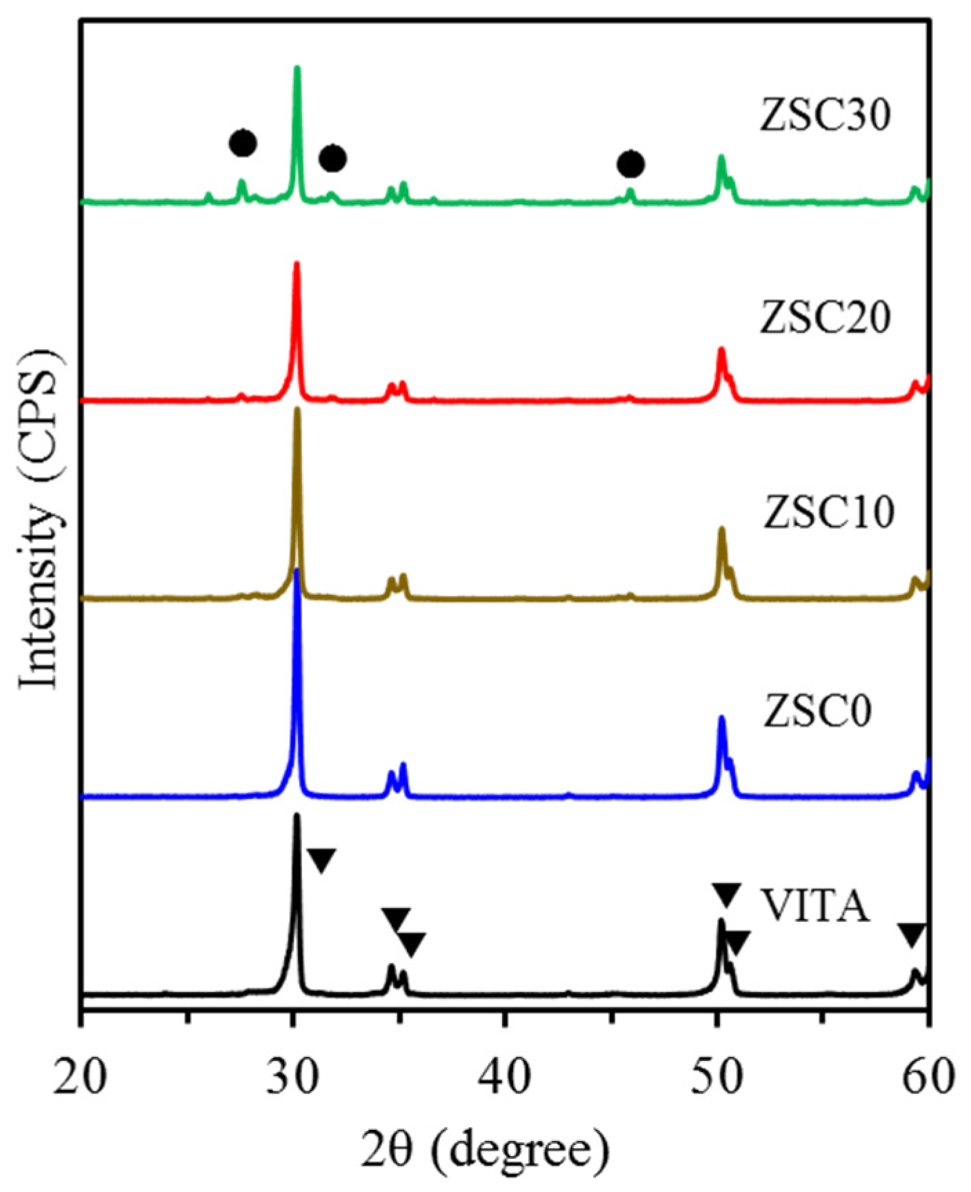
3.1. Phase Composition

The XRD patterns of all core materials after plasma treatment are shown in Figure 1. VITA zirconia revealed a highly crystalline tetragonal phase (t-ZrO2) at 2θ = 30.18°, 34.64°, 35.18°, 50.18°, 50.60° and 59.34°, which were assigned to (101), (002), (110), (112), (200) and (103) crystal planes (JCPDS card no. 48-0224), respectively. ZCS0 without CaSi additive was also attributed to t-ZrO2. On the contrary, the higher CaSi content in ZrO2 resulted in lower peak intensity of all t-ZrO2 phases. In the case of ZCS30, the peaks at 2θ = 27.56°, 31.76° and 45.88° may be attributed to CaZrO3 and Ca3ZrSi2O9 phases [22], in addition to the low peak intensity of t-ZrO2 phases.

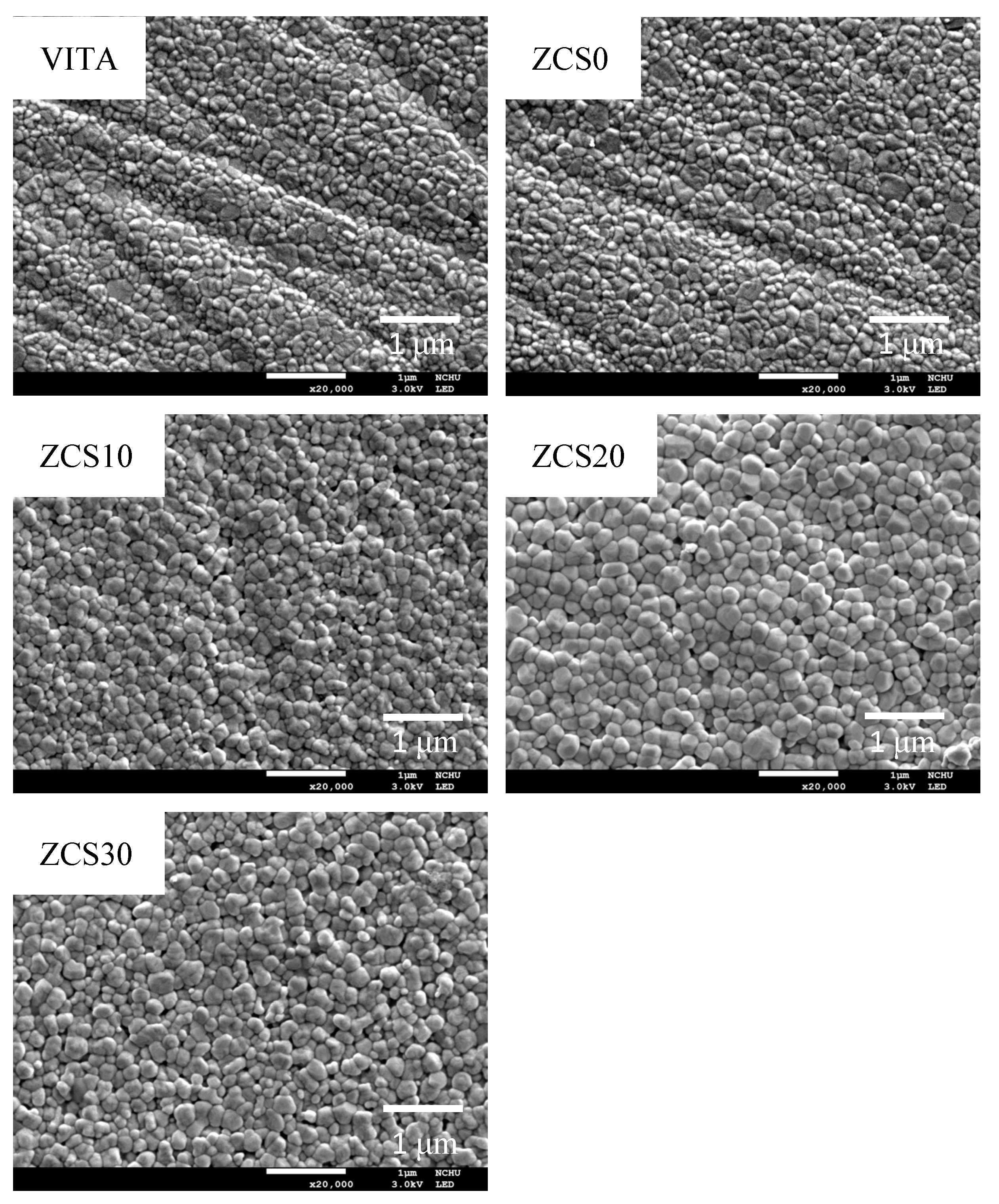
3.2. Surface Morphology

Figure 2 shows the surface morphology at high magnification to show the effect of the CaSi dopant on the grain size of ZrO2. Both the VITA control group and the ZCS0 group showed highly dense bodies with a uniform grain size of approximately 100 nm. With the increase in CaSi content, the grain size of the sintered body increased to about 200 nm.

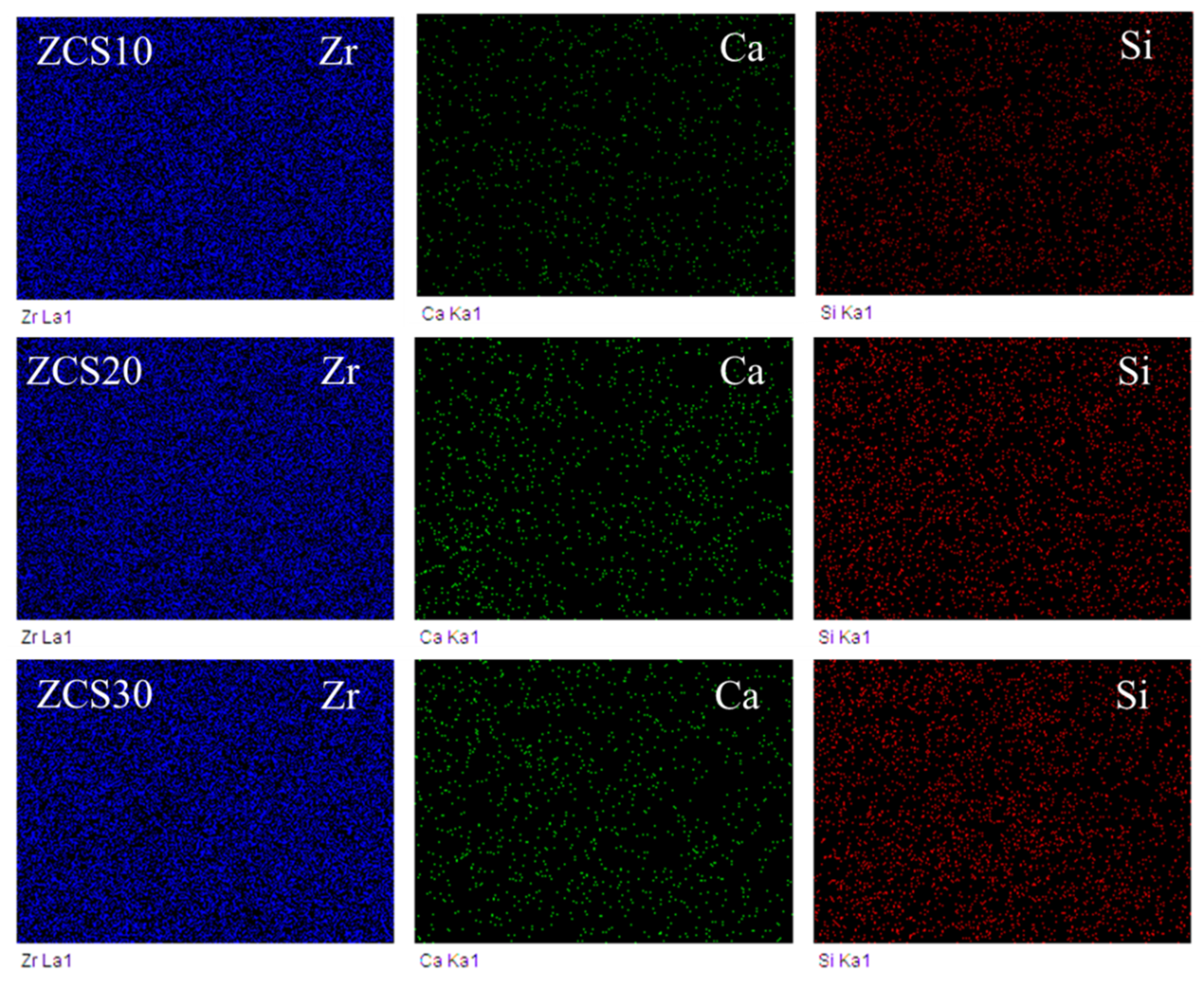
3.3. Elemental Mapping
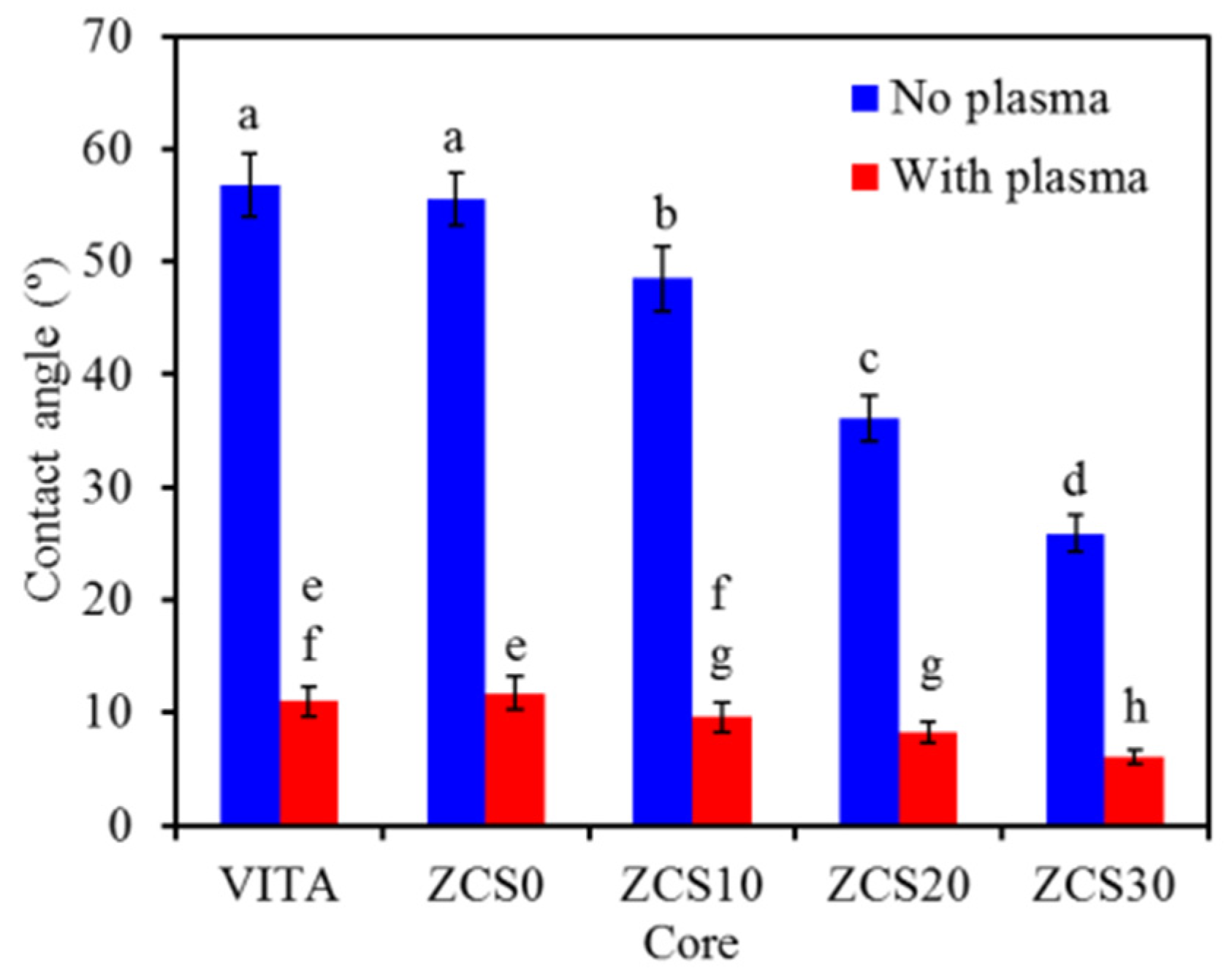
The EDS/element mapping shows the uniform distribution of the Ca and Si elements in the CaSi-containing specimens, in addition to the main component of the Zr element, as shown in Figure 3. Compared with ZCS10, higher Ca and Si elements on the surface can be found in ZCS30.

3.4. Contact Angle

Before plasma treatment, the water contact angle of ZCS0 (56° ± 2°) was not significantly different (p = 0.990) from that of VITA (57° ± 3°) (Figure 4). However, as the CaSi content in the ZrO2-based core substructures increased, their contact angle values decreased significantly (p < 0.05), from 49° ± 3° (ZSC10) to 26° ± 2° (ZCS30). As expected, after the oxygen plasma treatment, all core substructures indicated significantly lower (p < 0.05) contact angles (6–12°) compared to the corresponding substructure without plasma treatment.

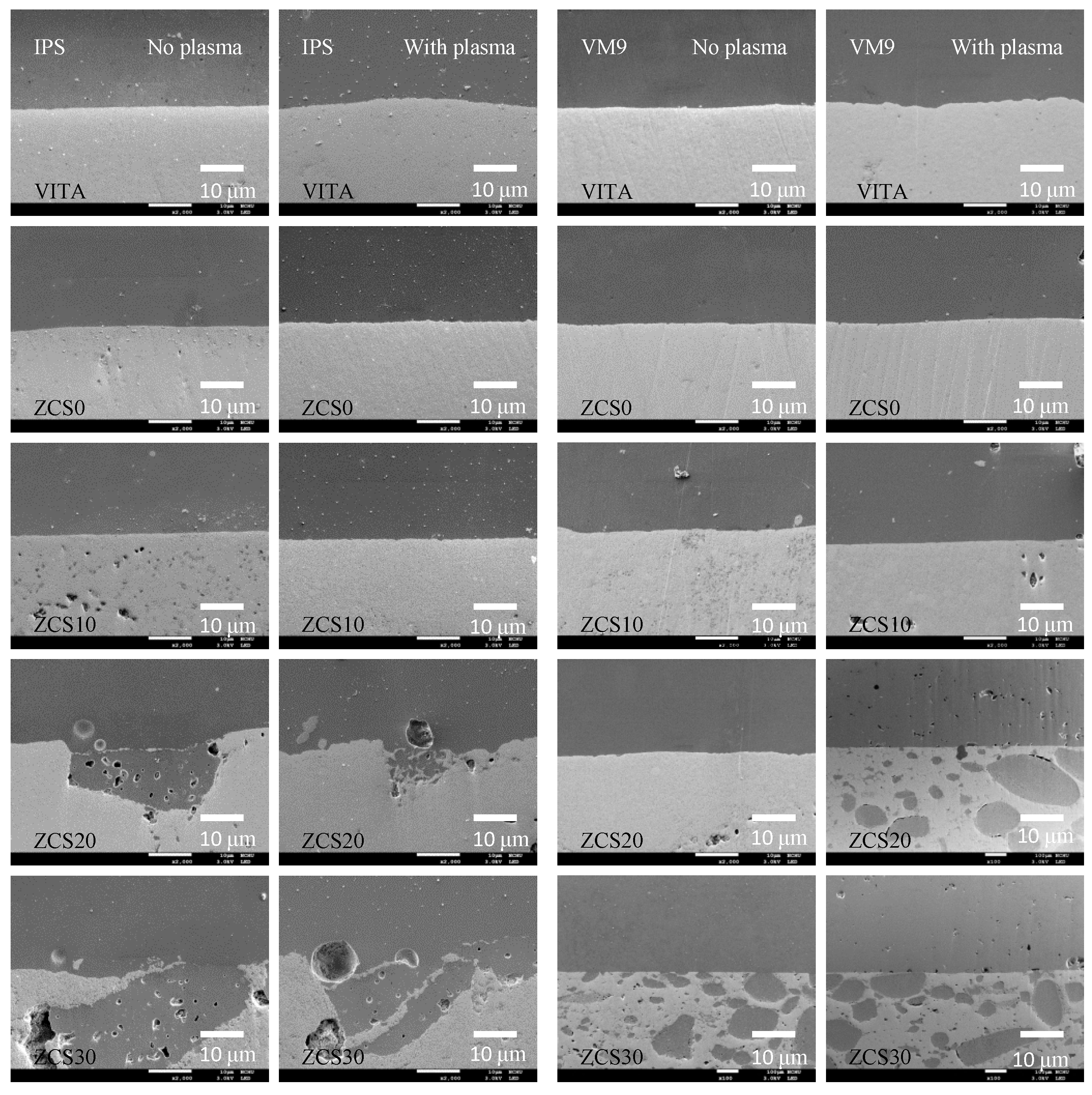
3.5. Interfacial Morphology

Even without oxygen plasma treatment, SEM images indicated that all the ZrO2-based substructures were in good contact with the two veneer ceramics (VM9 and IPS e. max Ceram) (Figure 5). In the CaSi-ZrO2 core, a dual microstructure consisting of a very fine ZrO2 matrix (bright area) and particle-shape CaSi-rich additive (dark area) can be found. The CaSi-rich area displayed certain polishing-induced pores during the preparation process, which may have been caused by irregularly shaped brittle CaSi microparticles and bubbles. However, it is worth noting that there was also a binding between the CaSi-rich area in the core and the veneering ceramic, which was the purpose of this study. Similarly, regardless of using VM9 or IPS e. max Ceram, there was no obvious structural interruption in the veneer–ceramic interface of all plasma treatment groups.

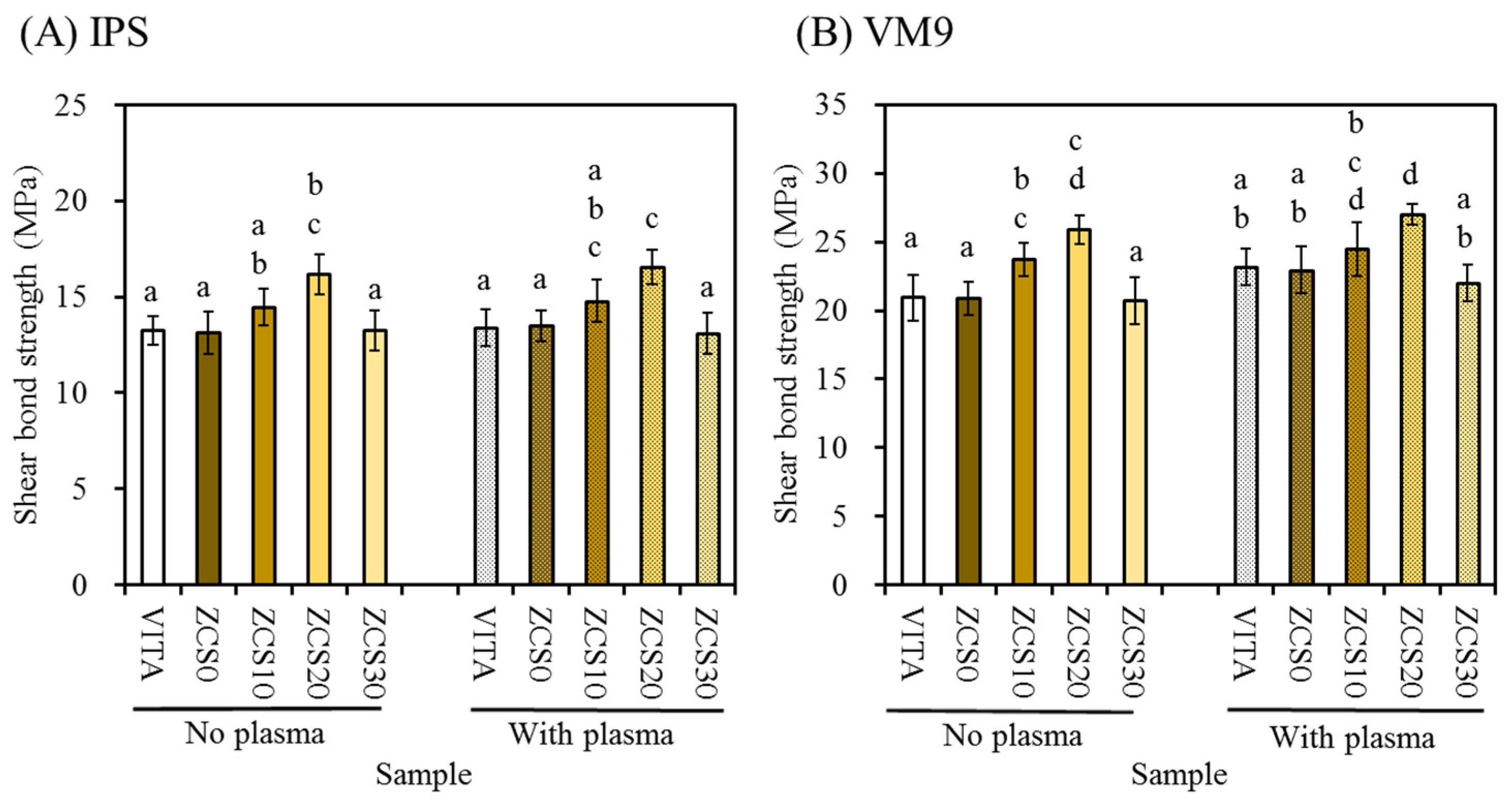
3.6. Shear Bond Strength

The shear bond strength of the veneer–core specimens is shown in Figure 6. When IPS e.max Ceram was used as a ceramic veneer to bond to the non-plasma-treated cores (Figure 6A), the ZCS20 group had a significantly higher (p < 0.05) shear bond strength than the VITA, ZCS0 and ZCS30 groups. In the case of VM9 veneering ceramic (Figure 6B), the bond strength to the core was significantly (p < 0.05) greater than the core with IPS e.max Ceram. For example, the bond strength of VITA-VM9 was 20.9 ± 1.7 MPa, while the bond strength of VITA-IPS e.max Ceram was 13.3 ± 0.7 MPa. Regarding the plasma treatment effect, the bond strength of core-IPS e.max Ceram was in the range of 13.1–16.6 MPa, while the core-VM9 restorations ranged from 22.0 MPa to 27.0 MPa. The plasma treatment of the ZrO2-based core did not affect the bond strength. As an example, the bond strength of the ZSC20 core to VM9 was 25.9 ± 1.0 and 27.0 ± 0.7 MPa before and after plasma treatment, respectively, showing no significant difference (p = 0.963). Similarly, when bonded to IPS e.max Ceram, the values were 16.2 ± 1.0 and 16.6 ± 0.9 MPa, respectively. More importantly, the ZSC20 had the highest shear bond strength among the all-core substructures.

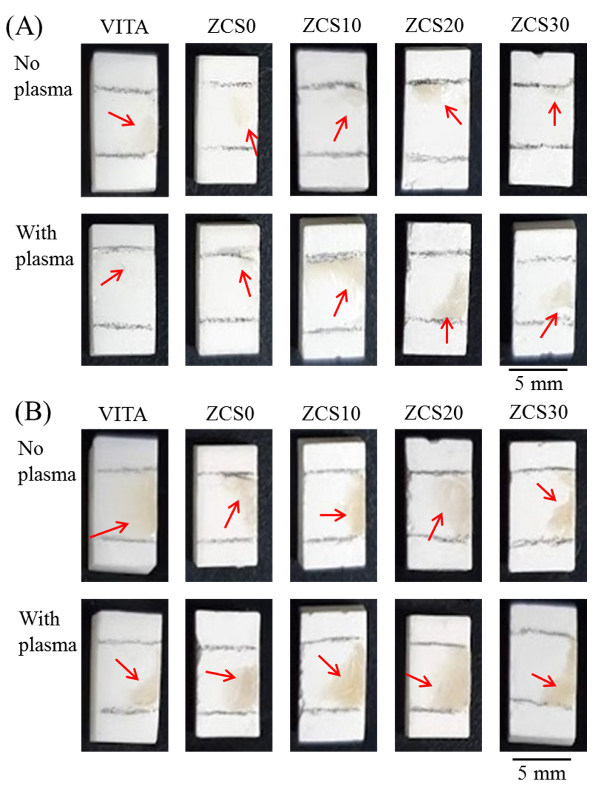
3.7. Fracture Analysis

Generally speaking, the classification of fractures can be divided into three modes: adhesive mode, mixed mode and cohesive mode. Adhesive failure occurs on the surface of the core material, cohesive failure occurs within the veneer and the mixed mode is considered to be a combination of adhesive and cohesive failure. Figure 7 shows the optical fracture images of two veneering ceramics adhered to the surfaces of various core specimens after the shear bond test, indicating no sign of cohesive failure. It can be also seen in color differences: the residual off-white ceramic veneer was different to the white zirconia-based core. When IPS e.max Ceram was adhered to various ZrO2-based cores, the plasma treatment did not result in obvious differences (Figure 7A). An interesting observation was that the residual VM9 adhered to the core surface (Figure 7B) with or without plasma treatment, as shown by the arrow, indicating a mixed mode of cohesive and interfacial fracture. In addition, the residual amount was higher than the corresponding IPS e.max Ceramic veneer.

4. Discussion

Improving the bond strength between the zirconia core and the ceramic veneer is still a major challenge for all-ceramic restoration. This study aimed to evaluate the influence of the CaSi dopant in the ZrO2 matrix on its shear bond strength to veneering ceramics. The veneering ceramics used in this study consisted of SiO2-based feldspathic porcelains such as IPS e.max Ceram and VITA VM9. The former is an ultralow-fusing nano-fluorapatite glass–ceramic that is designed for the veneering of lithium disilicate glass–ceramic and zirconium oxide, and the latter is a low-fusing dental ceramic.
XRD results revealed that the home-made ZrO2 core (ZCS0) displayed a highly crystalline t-ZrO2 phase without t→m phase transformation, which was the same with VITA In-Ceram YZ. On the other hand, when the CaSi component was added to the ZrO2, the highly crystalline zirconia phase was reduced, accompanied by minor secondary phases such as CaZrO3 or Ca3ZrSi2O9 due to the diffusion reaction [22]. Si and Ca elements were also detected by means of EDS mapping. The microscale CaSi dopant used was composed of β-Ca2SiO4 (β-dicalcium silicate), CaO and amorphous SiO2 phases [23], which may be embedded in the ZrO2 matrix after high-temperature sintering. However, the incorporation of CaSi did not result in the t→m transformation of ZrO2, although it did cause an increase in grain size. It is well known that oxides such as CaO, MgO or Y2O3 can stabilize the t-ZrO2 phase at room temperature by controlling the stress-induced t→m transformation [22]. Shuai et al. pointed out that the introduction of CaSiO3 into nano-ZrO2 was beneficial to the m→t transformation [24]. Regarding the grain size, Lung et al. found that Si caused the grain growth of ZrO2 [13]. In addition, SiO2 can be used as a sintering aid to induce liquid phase sintering [25]. Obviously, the presence of Si on the surface of zirconia may have an important role in promoting the formation of covalent bonds between the zirconia-based core and the ceramic veneer, as discussed below.
Plasma treatment on hard materials such as metals or ceramics can be regarded as effective drying and cleaning methods that can remove contaminants such as carbon adsorption on surfaces [26], thereby improving the hydrophilicity and possible bonding efficiency between heterostructures [12,27]. However, air or oxygen plasma treatment has been proven to not cause surface defects and phase changes [17,28]. In this study, the water contact angle of commercial VITA or home-made ZrO2 control (ZCS0) was about 57°, which was similar to the data published in other studies [29]. In contrast, the presence of CaSi effectively improved the hydrophilicity of ZrO2 materials because CaSi is a hydraulic cement material component [30]. After plasma treatment, regardless of the core substructures, the contact angle was less than 12°, displaying good hydrophilicity. The current results of the plasma-induced contact angle reduction were consistent with previous studies [27]. When the surface of the material had more ionizable groups, hydrogen bonding with water became easier, resulting in a decrease in contact angle [27].
The bonding between veneering ceramics and core substructures plays an important role in long-term clinical use. This study aimed to verify the hypothesis that optimal CaSi content in ZrO2-based substructures can enhance the interfacial bond strength between a zirconia core and veneering ceramic. Before exploring the CaSi composition effect, it is clear that there was no significant difference in the bond strength between VITA and ZCS0, regardless of binding to IPS e.max Ceram and VM9 veneering ceramics. However, it was found that the type of veneering ceramics caused significant differences in bond strength. Compared with the IPS e.max Ceram–core assemble, the higher bond strength of the VM9–core assemble may have been due to thermal diffusion at its higher firing temperature, which reinforced the chemical bonding at the interface. Fracture analysis supported insights that revealed the interplays between core–veneer bond strength and surface features of the ZrO2-based substructure. Adhesive failure almost occurred at the core/IPS e.max Ceram interface, due to a weak bonding ability between the veneer and the core, no matter whether the plasma treatment was applied or not. Oppositely, the incidence of mixed failure in VM9 veneer–core groups increased. Mixed failure, consisting of adhesive and cohesive failure, can indicate good bonding quality due to chemical bonds.
Studies have pointed out that plasma treatment can effectively improve the bond strength of ZrO2 and resin [17,20]. However, within the limits of this in vitro study, plasma treatment did not cause any obvious sign of improvement in the shear bond strength between the ZrO2-based core and the ceramic veneer. Chuang’s group [12] also reported that there was no statistical difference in the shear bond strength between the as-polished group and oxygen plasma-treated group. This is possibly due to heat treatment during the preparation of the ZrO2-based core and veneering ceramic. The chemical bonding efficacy induced by thermal diffusion was more effective than the physical cleaning efficacy of plasma treatment.
It is speculated that the content and particle size distribution of the CaSi component as an interplay factor can tailor the shear bond strength of the core–veneer restoration [6]. When the content of CaSi in the ZrO2 matrix was 20 wt %, the chemical bond originating from ion diffusion after firing may have been the reason for the high interfacial strength [31]. Due to the formation of –Si–O–Si– bonds [32], silica has been proven to be a very important factor in enhancing bond strength [8,21]. Liu et al. infiltrated glass into ZrO2 and improved the bond strength of the ceramic veneer–core by ion diffusion-induced chemical bonding [33]. However, excessive CaSi content (for example, 30 wt %) may cause the more failures at the interface between the core and the veneer. The CaSi-containing ZrO2 ceramics were heterogeneous because they were comprised of a continuous high-crystalline t-ZrO2 phase and a low-crystalline CaSi-containing phase. Compared with tough ZrO2, the CaSi-induced phase may have the usual properties of glass, such as brittleness [34]. During high temperature treatment, the interaction of microscale CaSi dopant and ZrO2 led to either the grain growth of zirconia crystals or the aggregation of some CaSi particles. The former could be confirmed by surface morphology, while the latter could be verified by the cross-sectional images. Therefore, the presence of excessive CaSi aggregates at the interface adversely affects the chemical bonding of the ZrO2-based core to veneering ceramic.
We have clarified that in plasma treatment and composition effects, bond affinity was impacted more by CaSi dopant than by plasma treatment. Although the 20 wt % CaSi microparticles in the ZrO2 matrix can enhance the bond strength of a ZrO2-based core to veneering ceramics, the use of nanoscale CaSi instead of microscale CaSi may allow the production of reliable and uniform CaSi-ZrO2-based substructures, which may greatly improve interfacial bond strength. Therefore, further research on the introduction of CaSi nanoparticles to ZrO2 will be required to explore the strength enhancement efficacy of all-ceramic restorations.

5. Conclusions

The present results support the research hypothesis that, at the optimal CaSi content, the shear bond strength of CaSi-ZrO2 to veneering ceramic can be improved. However, oxygen plasma treatment of the core material had no significant impact on the shear bond strength. Within the limits of this study, the ZrO2 core containing 20 wt % CaSi had appropriate structural and chemical properties, which were conducive to interfacial bonding.

Author Contributions

Conceptualization, T.-Y.C. and S.-J.D.; methodology, T.-Y.C., C.-C.Y. and Y.-H.C.; validation, T.-Y.C. and Y.-H.C.; formal analysis, T.-Y.C. and C.-C.Y.; data curation, C.-C.Y.; writing—original draft preparation, T.-Y.C.; writing—review and editing, M.Y. and S.-J.D.; visualization, T.-Y.C., C.-C.Y. and M.Y.; project administration, S.-J.D.; funding acquisition, S.-J.D. All authors have read and agreed to the published version of the manuscript.

Funding

This research was funded by Ministry of Science and Technology (Taiwan), grant number MOST 108-2314-B-040-006-MY3.

Institutional Review Board Statement

Not applicable.

Informed Consent Statement

Not applicable.

Data Availability Statement

Not applicable.

Conflicts of Interest

The authors declare no conflict of interest.

References

  1. Miyazaki, T.; Nakamura, T.; Matsumura, H.; Ban, S.; Kobayashi, T. Current status of zirconia restoration. J. Prosthodont. Res. 2013, 57, 236–261. [Google Scholar] [CrossRef] [PubMed] [Green Version]
  2. El-Ghany, G.O.S.A.; Sherief, A.H. Zirconia based ceramics, some clinical and biological aspects: Review. Future Dent. J. 2016, 2, 55–64. [Google Scholar] [CrossRef]
  3. Yan, M.; Wei, C.K.; Lin, Y.Y.; Hu, S.W.; Ding, S.J. Impact behavior of three notched all-ceramic restorations after soaking in artificial saliva. Materials 2015, 8, 4479–4490. [Google Scholar] [CrossRef] [PubMed] [Green Version]
  4. Soon, G.; Pingguan-Murphy, B.; Lai, K.W.; Akbar, S.A. Review of zirconia-based bioceramic: Surface modification and cellular response. Ceram. Int. 2016, 42, 12543–12555. [Google Scholar] [CrossRef]
  5. Ding, S.J.; Chu, Y.H.; Wang, D.Y. Enhanced properties of novel zirconia-based osteo-implant systems. Appl. Mater. Today 2017, 9, 622–632. [Google Scholar] [CrossRef]
  6. Yan, M.; Csík, A.; Yang, C.C.; Luo, Y.; Fodor, T.; Ding, S.J. Synergistic reinforcement of surface modification on improving the bonding of veneering ceramics to zirconia. Ceram. Int. 2018, 44, 19665–19671. [Google Scholar] [CrossRef] [Green Version]
  7. Cheng, C.W.; Yang, C.C.; Yan, M. Bond strength of heat-pressed veneer ceramics to zirconia with various blasting conditions. J. Dent. Sci. 2018, 13, 301–310. [Google Scholar] [CrossRef]
  8. Farhan, F.A.; Sulaiman, E.; Kutty, M.G. Effect of new zirconia surface coatings on the surface properties and bonding strength of veneering zirconia substrate. Surf. Coat. Technol. 2018, 333, 247–258. [Google Scholar] [CrossRef]
  9. Zaher, A.M.; Hochstedler, J.L.; Rueggeberg, F.A.; Kee, E.L. Shear bond strength of zirconia-based ceramics veneered with 2 different techniques. J. Prosthet. Dent. 2017, 118, 221–227. [Google Scholar] [CrossRef]
  10. Song, K.H.; Im, Y.W.; Lee, J.H.; Lee, J.; Lee, H.H. Evaluation of mold-enclosed shear bond strength between zirconia core and porcelain veneer. Dent. Mater. J. 2018, 37, 783–788. [Google Scholar] [CrossRef] [Green Version]
  11. Elsaka, S.E. Influence of surface treatments on the surface properties of different zirconia cores and adhesion of zirconia-veneering ceramic systems. Dent. Mater. 2013, 29, e239–e251. [Google Scholar] [CrossRef]
  12. Liu, Y.C.; Hsieh, J.P.; Chen, Y.C.; Kang, L.L.; Hwang, C.S.; Chuang, S.F. Promoting porcelain–zirconia bonding using different atmospheric pressure gas plasmas. Dent. Mater. 2018, 34, 1188–1198. [Google Scholar] [CrossRef]
  13. Lung, C.Y.K.; Liu, D.; Matinlinna, J.P. Silica coating of zirconia by silicon nitride hydrolysis on adhesion promotion of resin to zirconia. Mater. Sci. Eng. C 2015, 46, 103–110. [Google Scholar] [CrossRef]
  14. Campos, T.M.B.; Ramos, N.C.; Machado, J.P.B.; Bottino, M.A.; Souza, R.O.A.; Melo, R.M. A new silica-infiltrated Y-TZP obtained by the sol-gel method. J. Dent. 2016, 48, 55–61. [Google Scholar] [CrossRef] [Green Version]
  15. Madani, A.; Nakhaei, M.; Karami, P.; Rajabzadeh, G.; Salehi, S.; Bagheri, H. Sol-gel dip coating of yttria-stabilized tetragonal zirconia dental ceramic by aluminosilicate nanocomposite as a novel technique to improve the bonding of veneering porcelain. Int J. Nanomed. 2016, 11, 3215–3223. [Google Scholar]
  16. Abdullah, A.O.; Hui, Y.; Pollington, S.; Muhammed, F.K.; Sun, X.; Liu, Y. Comparative effectiveness of multiple laser scanning and conventional techniques on zirconia shear bond strength. Coatings 2019, 9, 422. [Google Scholar] [CrossRef] [Green Version]
  17. Yan, M.; Yang, C.C.; Chen, Y.H.; Ding, S.J. Oxygen plasma improved shear bond strength between zirconia and composite resin. Coatings 2020, 10, 635. [Google Scholar] [CrossRef]
  18. Juy, A.; Anglada, M. Surface phase transformation during grinding of Y-TZP. J. Am. Ceram. Soc. 2007, 90, 2618–2621. [Google Scholar] [CrossRef]
  19. Chintapalli, R.K.; Marro, F.G.; Jimenez-Pique, E.; Anglada, M. Phase transformation and subsurface damage in 3Y-TZP after sandblasting. Dent. Mater. 2013, 29, 566–572. [Google Scholar] [CrossRef] [PubMed]
  20. Elias, A.B.; Simao, R.A.; Prado, M.; Cesar, P.F.; Dos Santos, G.B.; da Silva, E.M. Effect of different times of nonthermal argon plasma treatment on the microtensile bond strength of self-adhesive resin cement to yttria-stabilized tetragonal zirconia polycrystal ceramic. J. Prosthet. Dent. 2019, 121, 485–491. [Google Scholar] [CrossRef]
  21. Kim, S.H.; Park, C.J.; Cho, L.R.; Huh, Y.H. Evaluation of the ceramic liner bonding effect between zirconia and lithium disilicate. J. Prosthet. Dent. 2018, 120, 282–289. [Google Scholar] [CrossRef] [PubMed]
  22. Ding, S.J.; Chu, Y.H.; Chen, P.T. Mechanical biocompatibility, osteogenic activity and antibacterial efficacy of calcium silicate-zirconia biocomposites. ACS Omega 2021, 6, 7106–7118. [Google Scholar] [CrossRef]
  23. Huang, Y.R.; Wu, I.T.; Chen, C.C.; Ding, S.J. In vitro comparisons of microscale and nanoscale calcium silicate particles. J. Mater. Chem. B 2020, 8, 6034–6047. [Google Scholar] [CrossRef]
  24. Shuai, C.; Feng, P.; Yang, B.; Cao, Y.; Min, A.; Peng, S. Effect of nano-zirconia on the mechanical and biological properties of calcium silicate scaffolds. Int. J. Appl. Ceram. Technol. 2015, 12, 1148–1156. [Google Scholar] [CrossRef]
  25. Chen, C.C.; Lai, M.H.; Wang, W.C.; Ding, S.J. Properties of anti-washout-type calcium silicate bone cements containing gelatin. J. Mater. Sci. Mater. Med. 2010, 21, 1057–1068. [Google Scholar] [CrossRef]
  26. Weltmann, K.D.; Kolb, J.F.; Holub, M.; Uhrlandt, D.; Šimek, M.; Ostrikov, K.; Hamaguchi, S.; Cvelbar, U.; Černák, M.; Locke, B.; et al. The future for plasma science and technology. Plasma. Process. Polym. 2019, 16, e1800118. [Google Scholar] [CrossRef] [Green Version]
  27. Wu, C.C.; Wei, C.K.; Ho, C.C.; Ding, S.J. Enhanced hydrophilicity and biocompatibility of dental zirconia ceramics by oxygen plasma treatment. Materials 2015, 8, 684–699. [Google Scholar] [CrossRef] [Green Version]
  28. Park, C.; Park, S.W.; Yun, K.D.; Ji, M.K.; Kim, S.; Yang, Y.P.; Lim, H.P. Effect of plasma treatment and its post process duration on shear bonding strength and antibacterial effect of dental zirconia. Materials 2018, 11, 2233. [Google Scholar] [CrossRef] [Green Version]
  29. Yang, C.C.; Ding, S.J.; Lin, T.H.; Yan, M. Mechanical and optical properties evaluation of rapid sintered dental zirconia. Ceram. Int. 2020, 46, 26668–26674. [Google Scholar] [CrossRef]
  30. Wu, I.T.; Kao, P.F.; Huang, Y.R.; Ding, S.J. In vitro and in vivo osteogenesis of gelatin-modified calcium silicate cement with washout resistance. Mater. Sci. Eng. C 2020, 117, 111297. [Google Scholar] [CrossRef] [PubMed]
  31. Ferraris, M.; Verné, E.; Appendino, P.; Moisescu, C.; Krajewski, A.; Ravaglioli, A.; Piancastelli, A. Coatings on zirconia for medical applications. Biomaterials 2000, 21, 765–773. [Google Scholar] [CrossRef]
  32. Abdalla, M.M.; Lung, C.Y.K.; Tsoi, J.K.H.; Matinlinna, J.P. Dental resin-zirconia bonding promotion using high-silica PVD coating with high ionization sputtering processing. Coatings 2019, 9, 182. [Google Scholar] [CrossRef] [Green Version]
  33. Liu, R.; Sun, T.; Zhang, Y.; Zhang, Y.; Jiang, D.; Shao, L. The effect of graded glass–zirconia structure on the bond between core and veneer in layered zirconia restorations. J. Mech. Behav. Biomed. Mater. 2015, 46, 197–204. [Google Scholar] [CrossRef] [PubMed]
  34. Xiang, Z.X.; Chen, X.P.; Song, X.F.; Yin, L. Responses of pre-crystallized and crystallized zirconia-containing lithium silicate glass ceramics to diamond machining. Ceram. Int. 2020, 46, 1924–1933. [Google Scholar] [CrossRef]
Figure 1. XRD patterns of all core materials after oxygen plasma treatment: ▼: ZrO2; ●: CaZrO3 or Ca3ZrSi2O9.
Coatings 11 01326 g001
Figure 2. Surface images of all core materials after oxygen plasma treatment at high magnification (20 k×).
Coatings 11 01326 g002
Figure 3. Zr, Ca and Si element mappings of CaSi-containing core specimen surfaces.
Coatings 11 01326 g003
Figure 4. Water contact angle values of different core substructures before and after plasma treatment. Different lowercase letters show statistically significant differences at p < 0.05 (n = 10).
Coatings 11 01326 g004
Figure 5. Cross-sectional SEM images of various core substructures bonded to VM9 or IPS e.max Ceram veneer ceramics before and after plasma treatment.
Coatings 11 01326 g005
Figure 6. Shear bond strength of (A) IPS e.max Ceram and (B) VM9 veneering ceramics to various core specimens. Different lowercase letters showed statistically significant differences (p < 0.05) (n = 10).
Coatings 11 01326 g006
Figure 7. Fracture images of (A) IPS e.max Ceram and (B) VITA VM9 veneering ceramics bonded to the surface of various core specimens after shear bond testing. The two lines represent the contact zone of the core-veneer bonding before failure. The arrow indicates the remaining veneering ceramics.
Coatings 11 01326 g007
Table 1. Core and veneering ceramics used in study.
Core or VeneerCompositionManufacturerLot No.
Core
In-Ceram YZZrO2-Y2O3-HfO2VITA Zahnfabrik30030
ZSC0ZrO2 powderTosohZ305653B
ZCS10 10 wt % CaSi-90 wt % ZrO2 powder
ZCS2020 wt % CaSi-80 wt % ZrO2 powder
ZCS3030 wt % CaSi-70 wt % ZrO2 powder
Veneer
IPS e.max CeramSiO2-LiO2-Na2O-K2O-Al2O3-CaO-P2O5-FIvoclar Vivadent AGV45249
VM9SiO2-Al2O3-K2O-Na2O-B2OVITA Zahnfabrik11040
Publisher’s Note: MDPI stays neutral with regard to jurisdictional claims in published maps and institutional affiliations.

