Article

Biomechanical Interfaces of Corticotomies on Periodontal Tissue Remodeling during Orthodontic Tooth Movement

1
State Key Laboratory of Oral Diseases & National Clinical Research Center for Oral Diseases, West China Hospital of Stomatology, Sichuan University, Chengdu 610041, Sichuan, China
2
Center of Regenerative Medicine, Renmin Hospital of Wuhan University, Wuhan 430060, Hubei, China
*
Authors to whom correspondence should be addressed.
Coatings 2021, 11(1), 1; https://doi.org/10.3390/coatings11010001
Submission received: 21 November 2020 / Revised: 16 December 2020 / Accepted: 17 December 2020 / Published: 22 December 2020

Abstract

:
Corticotomy is an effective approach in accelerating orthodontic tooth movement (OTM) in clinical treatment. Corticotomy causes regional acceleratory phenomenon (RAP) in the alveolar bone of surgical sites. However, the molecular mechanism of RAP after corticotomy remains unclear. Herein, we established a mouse model to study the biomechanical interfaces of corticotomy-assisted OTM and to investigate the histological responses and underlying cellular mechanism. A total of 144 male C57BL/6 mice were randomly assigned into four groups: corticotomy alone (Corti), sham operation (Sham), corticotomy with tooth movement (Corti + TM), and sham operation with tooth movement (Sham + TM). Nickel–titanium orthodontic springs were applied to trigger tooth movement. Mice were sacrificed on Post-Surgery Day (PSD) 3, 7, 14, 21, and 28 for radiographic, histological, immunohistochemical, and molecular biological analyses. The results reveal that corticotomy significantly promoted alveolar bone turnover and periodontal tissue remodeling. During orthodontic tooth movement, corticotomy significantly promoted osteogenic and proliferative activity, accelerated tooth movement, and eliminated root resorption by upregulating Wnt signal pathway.

1. Introduction

Completion of a comprehensive orthodontic treatment for moderate to severe malocclusions usually requires 20–36 months [1]. Orthodontic appliances cause inconveniences in patients’ daily life, especially in oral hygiene. Prolonged treatment also increases the risk of dental caries and root resorption. Over the last 20 years, the number of adults who seek orthodontic treatment has been growing. Yet treatment duration and risks increase as an individual age due to the declining metabolic activity in adults [2]. Furthermore, orthodontists are usually confronted with complicated situations resulting in ineffective tooth movement, including poor patient compliance and bracket fall-off. Therefore, acceleration of orthodontic tooth movement (OTM) would be desirable for both patients and orthodontists.
Plenty of research has been carried out over the years, and several modalities have been suggested to accelerate OTM, e.g., low-intensity laser treatment, photobiomodulation, pulsed electromagnetic fields, corticotomies, and pharmacological approaches [3]. However, recent studies suggest that surgical adjunctive procedures have the best potential in reducing treatment time [4], and surgical corticotomy has been confirmed by high-quality randomized clinical to be the only effective approach in accelerating OTM [5,6,7].
Corticotomy causes a regional acceleratory phenomenon (RAP), first introduced by Frost [8]. RAP involves a localized demineralization-remineralization period in the alveolar bone that intensifies bone remodeling and eliminates the resistance of dense cortical bone. It allows rapid movement in the demineralized alveolar bone [9]. Yet, the molecular mechanism of RAP after corticotomy remains unclear. The significance of this research was to deepen the current understanding of the biomechanical interfaces of corticotomy-assisted OTM and to discover the histological responses and underlying cellular mechanism of OTM with corticotomy, thus providing a solid molecular basis for the clinical applications of corticotomy in OTM.
Corticotomy was first applied to orthodontics by Köle et al. in 1959 [10]. Düker established an experimental animal model to study the effects of corticotomy on tooth movement (TM) rate and periodontal tissue [11]. Düker used Köle’s method, including the full flap and vertical incision of cortical bone, which was similar to osteotomy. Recently, Wilcko [12] improved this surgery method by making continuous holes in the cortical bone. Since this method reduced the trauma of surgery, in this study, we performed the surgery with three continuous cortical miniholes, as reported by Wilcko et al. Wang et al. built a rat corticotomy model with tooth movement to examine underlying cellular responses to corticotomy-assisted tooth movements [13]. However, previous research found that mouse and human genomes are genetically similar—about 90% of human genes have direct orthologues with mice [14]. Hence, we aimed to establish a mouse model with corticotomy-assisted tooth movement.

2. Materials and Methods

2.1. Animals

All experimental procedures were approved by the animal experiment ethics committee of the State Key Laboratory of Oral Diseases of Sichuan University in China. A total of 144 8-week-old male C57BL/6 mice weighing 22–25 g was obtained from the university’s experimental animal center. They were housed in the environment with a standard 12-h light-and-dark cycle and fed a soft diet with water ad libitum. The animals were acclimatized for at least 1 week before the start of the experiment.
All the mice were randomly assigned to 4 groups: corticotomy alone (Corti), sham operation (Sham), corticotomy with tooth movement (Corti + TM), and sham operation with tooth movement (Sham + TM).

2.2. Corticotomy and Tooth Movement

Mice were anaesthetized via intraperitoneal injection of Ketamine (80 mg/kg) and Xylazine (16 mg/kg). After that, their mouths were disinfected by a povidoneiodine solution. Full-thickness maxillary buccal and palatal flaps adjacent to the upper left first molar were elevated. The Corti group and the Corti + TM group were then subjected to corticotomy with cortical miniholes created by a round bur (Ø = 0.5 mm; depth = 0.5 mm), three holes per buccal and palatal sides. In sham-operated groups, only flap surgeries were carried out. Flaps were repositioned and sutured after the surgery. Only one side of the maxilla was operated on, and the opposite side was allowed to chew normally.
Immediately after surgery, orthodontic appliances were applied to the TM groups. Nickel–titanium orthodontic springs were fixed between the upper left incisor and the operated molar with ultraviolet curable resin (MB 4403; 3M Unitek, Monrovia, California) generating 4 g of force to move the upper first molar mesially. No re-activation of spring was performed. Following post-anesthesia recovery, animals were kept in a heat-controlled environment. All mice were fed a soft food diet and received daily subcutaneous injections of buprenorphine (0.05–0.1 mg/kg) for analgesia for 3 days after surgery and tooth movement initiation. No evidence of infection or prolonged inflammation at surgical sites was detected.

2.3. Sample Preparation, Micro-CT Test and Tissue Processing

For Corti and Sham groups, mice were sacrificed on Post-Surgery Day 7 (PSD 7). For Corti+TM and Sham+TM groups, mice were sacrificed at multiple time points (PSD 3, 7, 14, 21, 28; n = 12 for each time points). Each molar-bearing segment of the left maxilla was harvested and fixed in 4% paraformaldehyde overnight at 4 °C.
The micro-computed tomographic (micro-CT) scanning and analyses followed published guidelines [15]. Samples of maxilla were scanned using a high-resolution micro-CT 50 system (Scanco Medical, Brüttisellen, Switzerland) at a 10 μm voxel size (70 kV, 115 μA, and 300 ms integration time). Three-dimensional reconstruction and analyses were carried out using VG Studio Max software (version 2.2; Volume Graphics, Heidelberg, Germany). The gap between the upper first and second molar was measured to represent the tooth movement distance of the first molar. For bone morphometry studies, the volumes of interest were the inter-radicular alveolar bone of the upper first molar. Hereafter, we calculated and compared the trabecular bone mineral density (BMD), bone volume to total volume (BV/TV) ratio, trabecular thickness (Tb.Th), trabecular number (Tb.N), trabecular spacing (Tb.Sp), and cortex thickness.
After micro-CT scans, specimens were decalcified in 19% EDTA for 3 weeks followed by dehydration through an ascending ethanol series, and they were then cleared in xylene and embedded in paraffin. Serial 4 μm longitudinal sections were cut and mounted on microscopic glass slides. Tissue collection and sectioning prepared for histology, immunohistochemistry, and immunofluorescence were prepared by one individual and then assessed by another, blinded individual.

2.4. Histology and Cellular Analyses

Movat’s pentachrome staining was performed to observe vascular structures including nuclei, muscle, collagen, and fibrin. Aniline blue staining was used to detect the osteoid matrix. Picrosirius red staining was carried out to identify tightly packed and aligned collagen fibers.
Alkaline phosphatase (ALP) activity and tartrate-resistant acid phosphatase (TRAP) were examined on the molar-bearing tissue sections. For ALP, sections were treated with NBT/BCIP substrate (#34043, Thermo Fisher Scientific, Waltham, MA, USA). TRAP activity was detected by a TRAP staining kit (386A, Sigma, Ronkonkoma, NY, USA) and kept in a chamber with moist air at 37 °C. The number of TRAP positive cells at the apical part of the distal buccal root of the maxillary first molar were quantified and subjected to statistical analysis.

2.5. Immunohistochemistry and Immunofluorescence

Tissue sections were deparaffinized and hydrated following standard procedures. Endogenous peroxidase activity was quenched by 3% hydrogen peroxide for 30 min in darkness and then washed with PBS. Subsequently, sections were blocked with 5% goat serum (Vector S-1000) for 30 min at 37 °C to prevent unspecific background staining.
For immunohistochemistry staining of Osterix and PCNA, appropriate polyclonal primary antibodies were added and incubated overnight at 4 °C in a moist chamber. The dilution rate for PCNA was 1:10,000 and 1:500 for Osterix. Sections were incubated with a secondary antibody (Vector BA-x) for 30 min at room temperature and rinsed with PBS. An advidin/biotinylated enzyme complex (Kit ABC Peroxidase Standard Vectastain PK-4000) was then added and incubated for 30 min followed by a DAB substrate kit (Vector Peroxidase substrate DAB SK-4100) for coloration.
For immunofluorescence staining of Runx2, after the polyclonal primary antibody (Runx2 1:200 diluted) was added and incubated overnight at 4 °C in a moist chamber, the samples were incubated with a fluorochrome-conjugated secondary antibody (1:500) for 30 min at room temperature in the dark. The slides were finally mounted with a DAPI mounting medium (Vector Laboratories).
To prove the specificity, a negative control slide was set for each immunostaining reaction where primary antibodies were omitted. The results were determined by measuring the number of immune-positive pixels of periodontal membrane and alveolar bone on both tensile and compressive sides.

2.6. Quantitative Real-Time Polymerase Chain Reaction(qRT-PCR)

For qRT-PCR, osseous tissues surrounding the roots of the upper first molar were collected and snap-frozen in liquid nitrogen. The total RNA was extracted and quantified by spectrometry between 260 and 280 nm. Subsequently, the reverse transcription was carried out using the SYBR PrimeScript RT-PCR Kit (TaKaRa, Dalian, Japan), and cDNA was the template for further PCR reaction. Each reaction was conducted in triplicate. The ΔΔCt method was used to determine the gene expression, and results were normalized to GAPDH. The Primer sequences are given in the Table 1.

2.7. Statistical Analyses

Results are presented as the mean ± standard error values of independent replicates. A Student’s t test was used to analyze and quantify the differences between groups. All statistical analyses were performed with SPSS software (IBM, Armonk, New York, NY, USA). p-value < 0.05 was considered significant.

3. Results

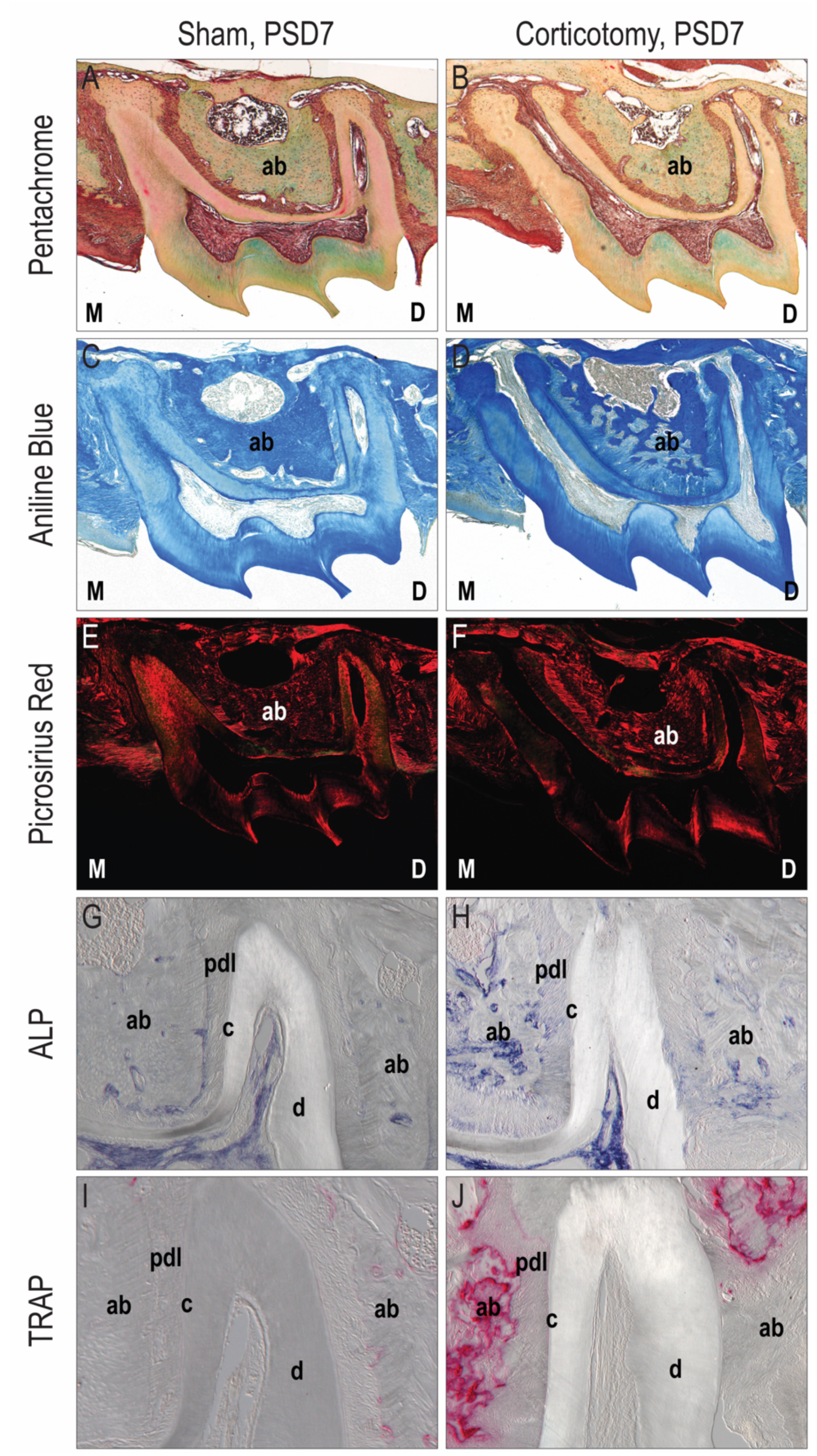
3.1. Corticotomy Promoted Local Alveolar Bone Remodeling

Histological staining was performed to observe mainly the collagen and fiber in the non-tooth-movement group. For example, compared with Sham group (Figure 1A), the Corti group (Figure 1B) presented more inflammatory infiltration and irregular marrow cavity on PSD 7. As shown by Aniline blue staining, corticotomy caused demineralization of the local alveolar bone (Figure 1C,D). Picrosirius red staining did not indicate any differences (Figure 1E,F). Abundant ALP/TRAP activity was observed in the corticotomy area (Figure 1H,J). In contrast, the Sham group showed minimal ALP/TRAP activity (Figure 1G,I), which indicated the stimulation of bone turnover with corticotomy surgery.

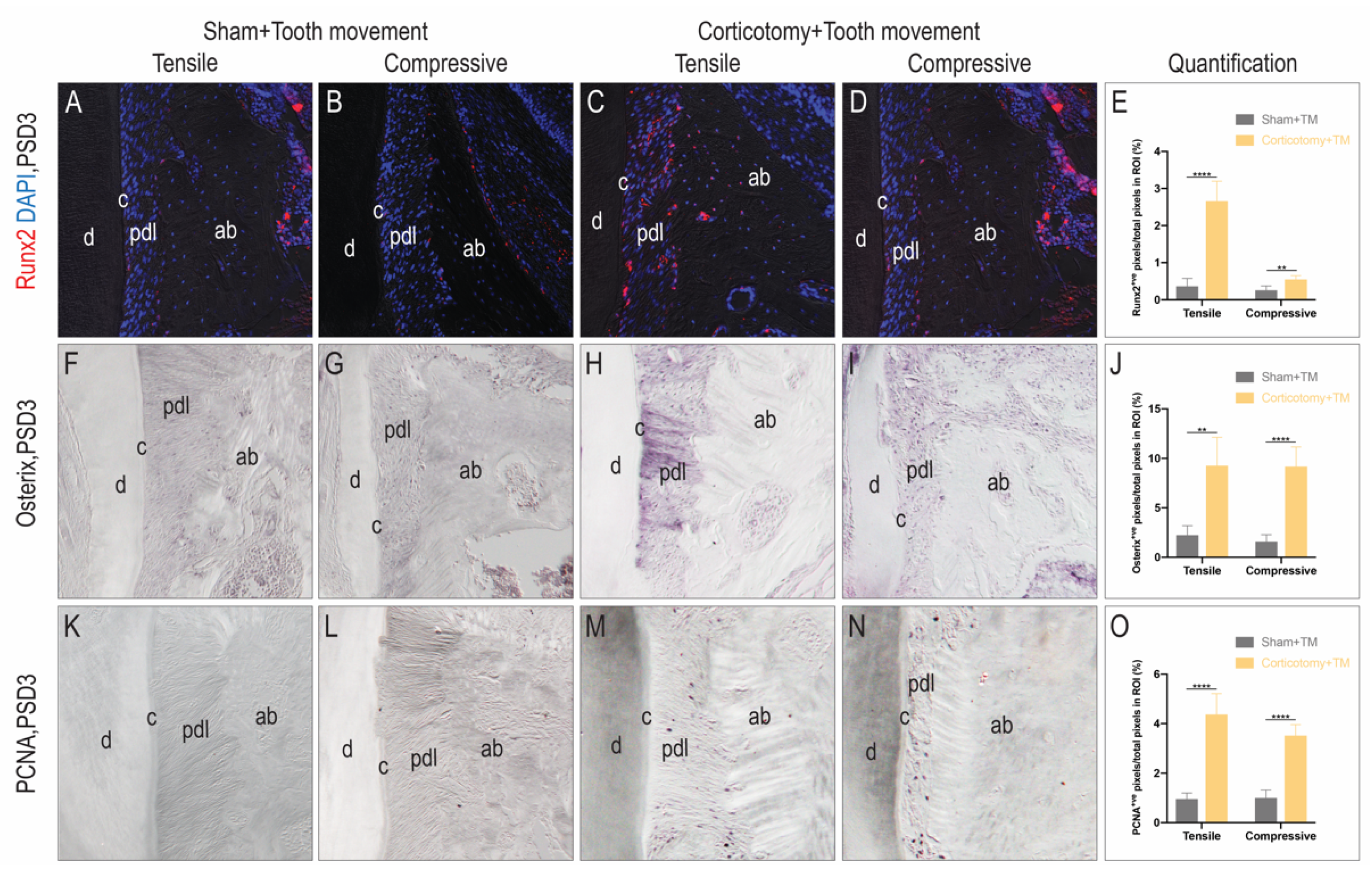
3.2. Corticotomy Promoted Cell Proliferation and Osteogenic Mineralization during Tooth Movement in the Alveolar Bone

Immunofluorescence and immunohistochemistry staining were carried out to determine the osteogenic mineralization in the tooth-movement period. On PSD 3, the pre-osteoblast maker Runx2 was expressed at much higher levels in the periodontal ligament on the tensile side of the Corti group (Figure 2A), compared to the Sham group (Figure 2C). Nevertheless, there was minimal Runx2 expression on the compressive side of both groups (Figure 2B,D). Simultaneously, the positive cells of the transcription factor Osterix and the cell proliferating maker PCNA were more abundant on both sides of the Corti group than the Sham group. We calculated the antigen-positive pixels in periodontal ligament, and the difference between the two groups was significant. These results demonstrate that corticotomy can promote cell proliferation and osteogenic mineralization during tooth movement.

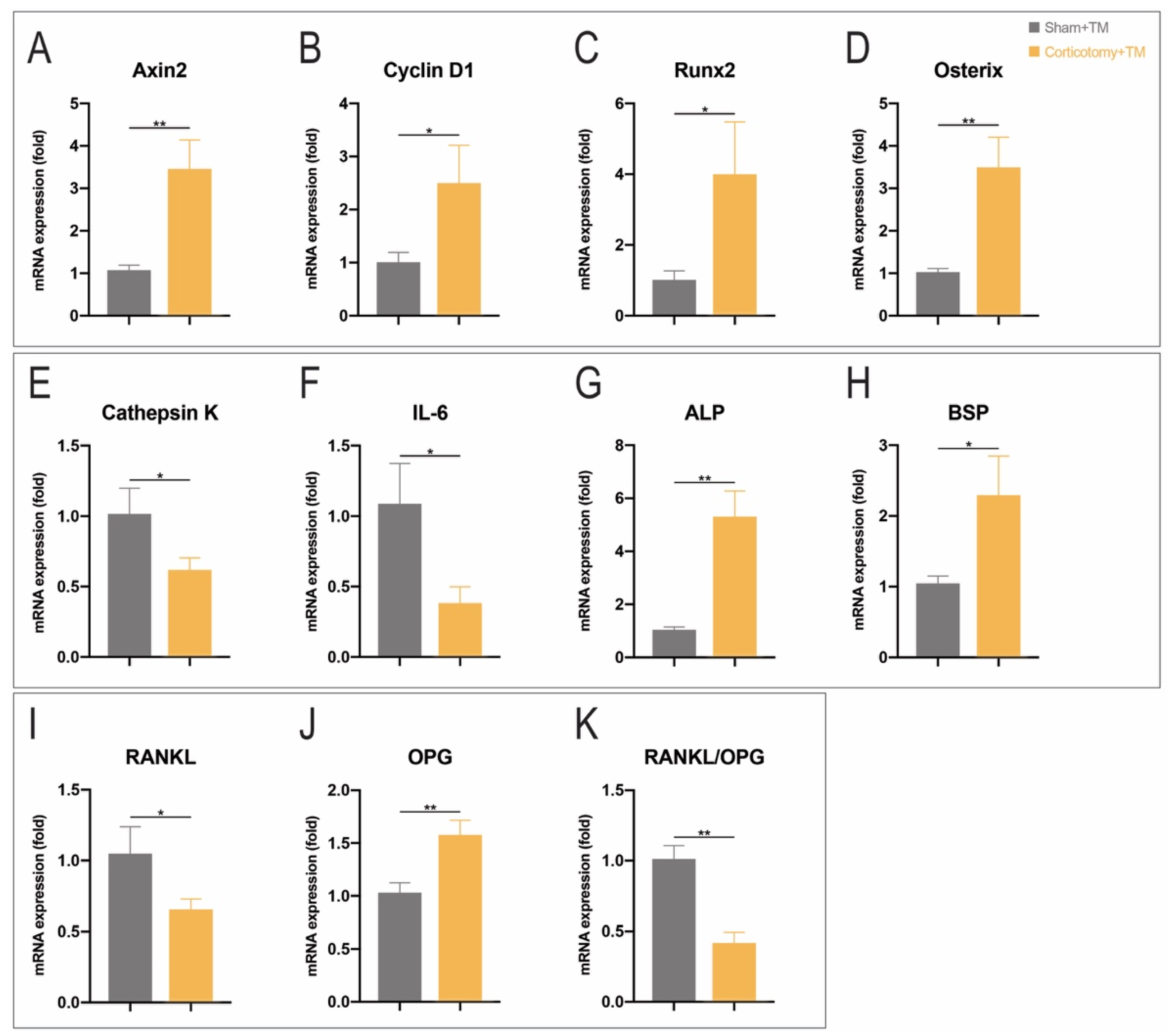
3.3. Corticotomy Promoted Osteogenesis and Inhibits Inflammation by Upregulating Wnt Signaling Pathway in Periodontal Tissue

PCR results are shown in Figure 3. The expression levels of Runx2 mRNA, Osterix mRNA, and ALP mRNA in the Corti + TM group were remarkably higher than in the Sham + TM group (Figure 3C,D,G), which is in accordance with the immunohistochemistry staining results. Cell cycle-related gene (Cyclin D1) and Bone sialoprotein (BSP) showed higher expressions after corticotomy as well (Figure 3B,H).
Axin2 served as a member of a negative feedback regulator in Wnt signaling pathway and was expressed more in the Corti + TM group (Figure 3A). Cathepsin K, a protease of the cysteine family, plays an important role in cleaving bone proteins. IL-6 is a primary cytokine in the inflammatory process. IL-6 and Cathepsin K were expressed more in the Sham + TM group (Figure 3E,F), which indicated the inflammation level and osteoclast number. The RANKL/OPG ratio was calculated in order to understand the osteoclast activity. The ratio was decreased in the Corti + TM group after tooth movement, showing that the osteoclast activity was inhibited (Figure 3I–K).

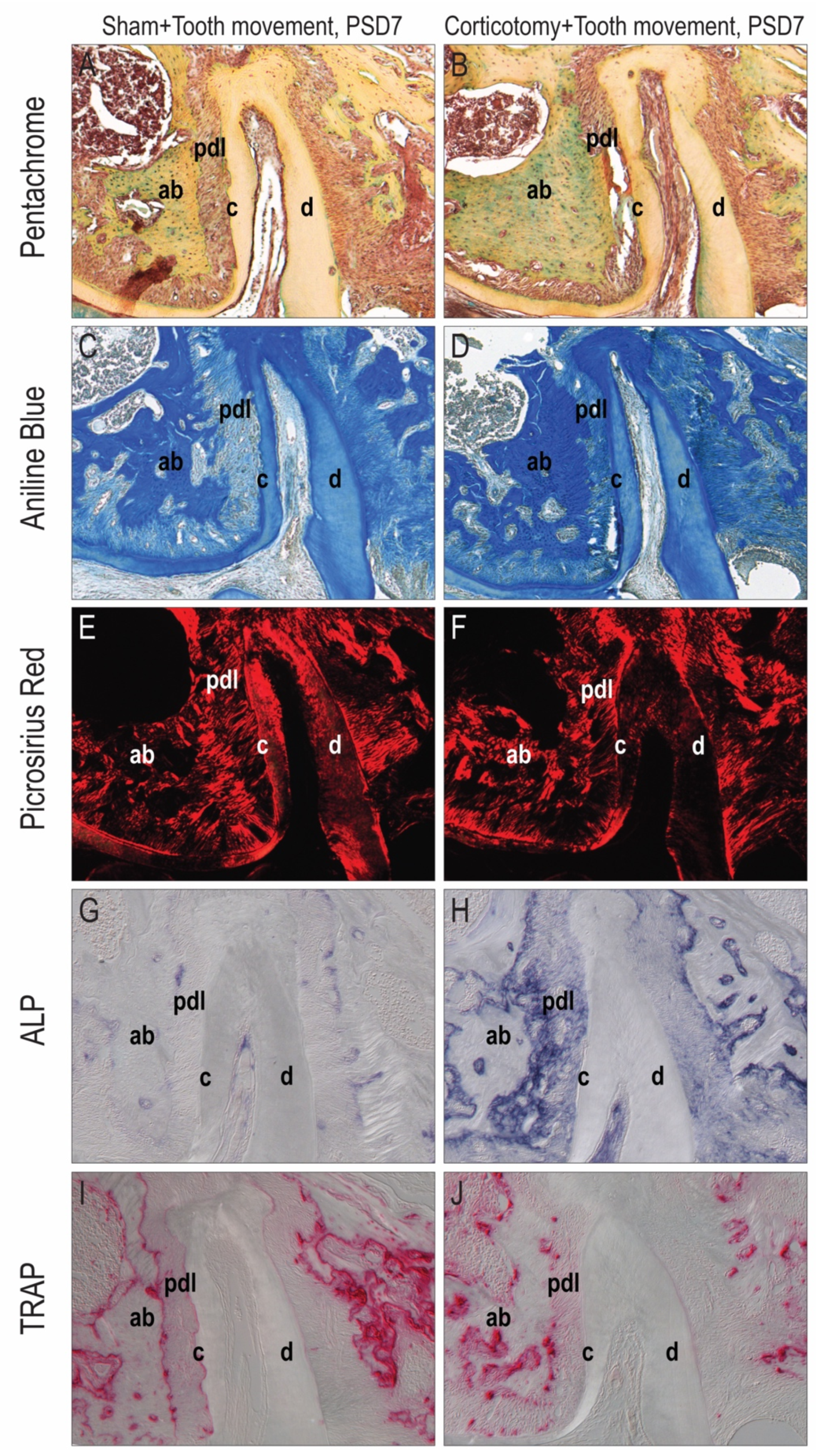
3.4. Corticotomy Eliminated Root Resorption during Tooth Movement

Significant differences were observed between the Corti + TM and Sham + TM groups (Figure 4A,B). The periodontal ligament presented a disorderly arrangement and was compressed on the compressive side but stretched on the tensile side (Figure 4E,F). In the Sham+TM group, discontinuous cementum implied the resorption of tooth root (Figure 4A,C). On the contrary, the cementum was much more continuous, and there was no significant root-resorption in the Corti + TM group (Figure 4B,D). ALP activity was hardly found in the Sham + TM group, but TRAP activity was abundant on both compressed and tensile sides (Figure 4G,I). Nevertheless, in the Corti + TM group, ALP activity was significantly increased, but TRAP activity was remarkably decreased (Figure 4H,J).

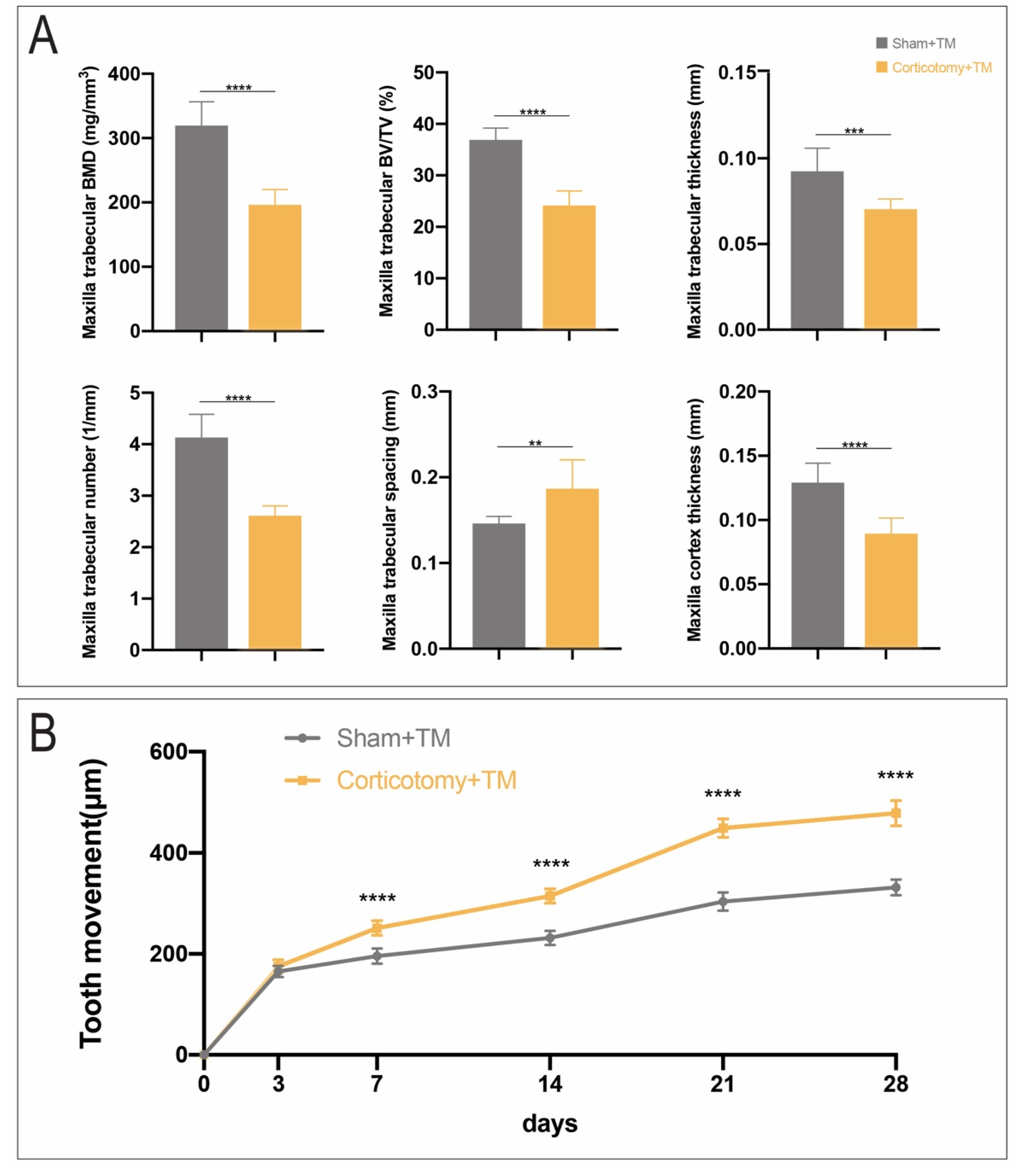
3.5. Corticotomy Led to Transient Osteoporosis in Alveolar Bone and Accelerated Orthodontic Tooth Movement

As micro CT results showed, trabecular structural parameters such as BMD, BV/TV, Tb.Th, Tb.N, Tb.Sp, and cortex thickness presented significant differences between the two groups (Figure 5A), which implied a transient phenomenon of osteoporosis in response to corticotomy. The tooth movement ratio also demonstrated that there was an accelerating effect in the Corti + TM group because of the reduction in resistance in the local alveolar bone (Figure 5B).

4. Discussion

Orthodontic tooth movement is a result of external mechanical stimulation. The rate of bone remodeling is the key factor for the speed of tooth movement [16]. Based on previous study reports, in selective corticotomy, alveolar bone remodeling and bone resorption are coupled [17].
ALP is a systemic maker for new bone formation, which is associated with the calcification process. An elevated ALP level is commensurate with active bone remodeling. TRAP is a specific biochemical marker of osteoclast differentiation and function [18]. Demineralization of local alveolar bone was concentrated around the root on PSD 7, as we observed, and abundant ALP/TRAP positive cells were expressed in the Corti group, which indicated the promotion of bone remodeling as a result of corticotomy. After 4 g of orthodontic force was applied, we focused on the distal root of upper first molar because it is perpendicular to the direction of TM, which simulates the parallel movement of tooth in an orthodontic clinic. Evident resorption lacuna was found on the root surface of the compressed side in the Sham group. By contrast, the cementum was continuous and smooth with corticotomy, and resorption was hardly found. The difference in the ALP/TRAP ratio also accounts for the role of corticotomy and thus the inhibition of root resorption during TM.
We performed qRT-PCR to detect the molecular expression level of the alveolar bone around the root. The results showed osteoblast-related factors; e.g., Axin2 ALP, Osterix, Runx2, and BSP were significantly more expressed in the Corti group. OPG is a soluble inhibitor of the receptor antagonism against RANK, which can bind to RANKL and inhibit osteoclast differentiation [19,20]. RANKL/OPG is an essential molecule that affects the metabolism of the alveolar bone and mediates osteoclast formation. We also investigated their expression to reflect the effect of corticotomy on osteoclasts. The results showed that the expression level of OPG increased in the Corti group, and RANKL expression decreased. Therefore, the resultant RANKL/OPG ratio was remarkably decreased, which reflected the actual functional RANKL molecules and the activity of osteoclastogenesis. These results are consistent with a previous study using rat models [21].
Wnt signaling pathway is involved in regulating the proliferation and differentiation of osteoblasts and other functional activities [22]. The importance of Wnt signaling during bone formation has been well verified [23]. Axin2, a direct downstream gene, is a negative feedback regulator of the classic Wnt signaling pathway, and its expression can reflect the activation level of Wnt signaling pathway [24]. In our study, the Axin2 gene expressed at a remarkable higher level in the Corti group. Thus, we confirmed that corticotomy stimulates osteogenesis and inhibits root resorption by upregulating Wnt signaling pathway in periodontal tissue.
Runx2 is the master transcription factor essential for osteogenic differentiation and bone formation [25]. It plays an important role in all stages of bone formation, mainly in the early stage of osteoblast differentiation [26]. Osterix is a specific osteoblast transcription factor that was first discovered by Nakashima et al. [27] in 2002. It is the downstream of Runx2 in osteoblast differentiation [28]. Studies have found that Runx2 promotes the early immature differentiation of osteoblasts [29], while Osterix dominates the final mature differentiation of osteoblasts [30]. In our study, the immunostaining of Runx2 and Osterix were stronger in the Corti group, and the results were consistent with the PCR results. We also applied immunohistochemical staining to test PCNA, which is a factor indicating the cell proliferation rate. Both PCNA immunostaining results and the expression level of Cyclin D1 in the PCR test confirmed that corticotomy can promote cell proliferation in the alveolar bone.
Our micro-CT study suggested that, after osteocortical incision, the maxillary alveolar bone experienced a process of osteoporosis. However, demineralization was just a transient period. A previous study showed that bone loss was more severe in corticotomy-alone group than in Corti + TM groups [31]. Other researchers found that demineralization occurred before PSD 14, and re-mineralization then began [32]. The difference in TM ratio in our study confirmed that osteoporosis leads to an acceleration of tooth movement at an early stage, which is a result of a decrease in resistance in the local alveolar bone.
Taken together, the results demonstrate that corticotomy caused demineralization and remineralization in the local alveolar bone to stimulate bone remodeling. During orthodontic tooth movement, cell proliferation and osteogenic mineralization were promoted, and root resorption was inhibited through upregulation of Wnt signaling pathway. As a result, corticotomy had an acceleration effect on the tooth movement rate and reduced side effects in orthodontics, e.g., root resorption.

5. Conclusions

This study demonstrated that corticotomy promoted periodontal tissue remodeling and accelerated tooth movement by upregulating Wnt signal pathway. Plenty of clinical evidence has suggested that corticotomy of the alveolar bone can accelerate local orthodontic tooth movement. We detected the underlying cell and molecular mechanisms and revealed that these regulations may be via Wnt signaling pathway.

Author Contributions

Conceptualization: R.L., X.Y. and S.Z.; methodology: R.L. and X.Y.; validation: R.L., L.H. and X.Y.; data curation: R.L., L.H. and X.Y.; formal analysis: R.L., X.X., Y.G. and Y.J.; investigation: R.L. and L.H.; writing—original draft preparation: R.L. and X.Y.; writing—review and editing: R.L., X.Y., Q.Y. and S.Z.; funding acquisition: X.Y. and S.Z. All authors gave final approval and agree to be accountable for all aspects of the work. All authors have read and agreed to the published version of the manuscript.

Funding

This research project was supported by grants from the National Natural Science Foundation of China (81801019 and 82071150).

Institutional Review Board Statement

The study was conducted according to the guidelines of the Declaration of Helsinki, and approved by the Ethics Committee of the State Key Laboratory of Oral Diseases of Sichuan University in China. (Protocol code WCHSIRB-D-2018-059 and date of approval 01 March 2018).

Informed Consent Statement

“Not applicable” for studies not involving humans.

Data Availability Statement

The data that support the findings of this study are available from the corresponding author upon reasonable request.

Conflicts of Interest

The authors declare no conflict of interest.

References

  1. Gil, A.P.S.; Haas, O.L., Jr.; Méndez-Manjón, I.; Masiá-Gridilla, J.; Valls-Ontañón, A.; Hernández-Alfaro, F.; Guijarro-Martínez, R. Alveolar corticotomies for accelerated orthodontics: A systematic review. J. Craniomaxillofac. Surg. 2018, 46, 438–445. [Google Scholar] [PubMed]
  2. Jawad, Z.; Bates, C.; Hodge, T. Who needs orthodontic treatment? Who gets it? And who wants it? Br. Dent. J. 2015, 218, 99. [Google Scholar] [PubMed]
  3. Gkantidis, N.; Mistakidis, I.; Kouskoura, T.; Pandis, N. Effectiveness of non-conventional methods for accelerated orthodontic tooth movement: A systematic review and meta-analysis. J. Dent. 2014, 42, 1300–1319. [Google Scholar] [PubMed]
  4. Nimeri, G.; Kau, C.H.; Abou-Kheir, N.S.; Corona, R. Acceleration of tooth movement during orthodontic treatment—A frontier in Orthodontics. Prog. Orthod. 2013, 14, 42. [Google Scholar]
  5. Al-Naoum, F.; Hajeer, M.Y.; Al-Jundi, A. Does Alveolar Corticotomy Accelerate Orthodontic Tooth Movement When Retracting Upper Canines? A Split-Mouth Design Randomized Controlled Trial. J. Oral Maxillofac. Surg. 2014, 72, 1880–1889. [Google Scholar]
  6. Bhattacharya, P.; Bhattacharya, H.; Anjum, A.; Bhandari, R.; Agarwal, D.K.; Gupta, A.; Ansar, J. Assessment of Corticotomy Facilitated Tooth Movement and Changes in Alveolar Bone Thickness—A CT Scan Study. J. Clin. Diagn. Res. 2014, 8, 26–30. [Google Scholar]
  7. Wilcko, M.T.; Ferguson, D.J.; Makki, L.; Wilcko, W.M. Keratinized Gingiva Height Increases After Alveolar Corticotomy and Augmentation Bone Grafting. J. Periodontol. 2015, 86, 1107–1115. [Google Scholar]
  8. Frost, H.M. The regional acceleratory phenomenon: A review. Henry Ford Hosp. Med. J. 1983, 31, 3–9. [Google Scholar]
  9. Umar, R.; Queiroz, C.R.; Prasad, G.M. Surgically accelerated orthodontic techniques and periodontal response: A systematic review. Eur. J. Orthod. 2020, 42, 635–642. [Google Scholar]
  10. KoLe, H. Surgical operations on the alveolar ridge to correct occlusal abnormalities. Oral Surg. Oral Med. Oral Pathol. 1959, 12, 515–529. [Google Scholar]
  11. Serge, K.; Duke, R. Mouth: Experimental animal research into segmental alveolar movement after corticotomy. Afr. Zool. 1976, 40, 127–142. [Google Scholar]
  12. Wilcko, W.M.; Wilcko, T.; Bouquot, J.E.; Ferguson, D.J. Rapid orthodontics with alveolar reshaping: Two case reports of decrowding. Int. J. Periodontics Restor. Dent. 2001, 21, 9–19. [Google Scholar]
  13. Wang, L.; Lee, W.; Lei, D.L.; Liu, Y.P.; Yamashita, D.D.; Yen, S.L.K. Tissue responses in corticotomy- and osteotomy-assisted tooth movements in rats: Histology and immunostaining. Am. J. Orthod. Dentofac. Orthop. 2009, 136, 1–11. [Google Scholar]
  14. Yue, F.; Cheng, Y.; Breschi, A.; Vierstra, J.; Wu, W.; Ryba, T.; Sandstrom, R.; Ma, Z.; Davis, C.; Pope, B.D.; et al. A comparative encyclopedia of DNA elements in the mouse genome. Nature 2014, 515, 355–364. [Google Scholar]
  15. Bouxsein, M.L.; Boyd, S.K.; Christiansen, B.A.; Guldberg, R.E.; Jepsen, K.J.; Müller, R. Guidelines for assessment of bone microstructure in rodents using micro-computed tomography. J. Bone Miner. Res. 2010, 25, 1468–1486. [Google Scholar]
  16. Baloul, S.S.; Gerstenfeld, L.C.; Morgan, E.F.; Carvalho, R.S.; Van Dyke, T.E.; Kantarci, A. Mechanism of action and morphologic changes in the alveolar bone in response to selective alveolar decortication-facilitated tooth movement. Am. J. Orthod. Dentofac. Orthop. 2011, 139, S83–S101. [Google Scholar]
  17. Sebaoun, J.D.; Kantarci, A.; Turner, J.W.; Carvalho, R.S.; Van Dyke, T.E.; Ferguson, D.J. Modeling of trabecular bone and lamina dura following selective alveolar decortication in rats. J. Periodontol. 2008, 79, 1679–1688. [Google Scholar]
  18. Minkin, C. Bone acid phosphatase: Tartrate-resistant acid phosphatase as a marker of osteoclast function. Calcif. Tissue Int. 1982, 34, 285–290. [Google Scholar]
  19. Aubin, J.E.; Bonnelye, E. Osteoprotegerin and its Ligand: A New Paradigm for Regulation of Osteoclastogenesis and Bone Resorption. Osteoporos. Int. 2000, 11, 905–913. [Google Scholar]
  20. Kostenuik, P.; Shalhoub, V. Osteoprotegerin: A physiological and pharmacological inhibitor of bone resorption. Curr. Pharm. Des. 2001, 7, 613–635. [Google Scholar]
  21. Zhou, Y.; He, X.; Zhang, D. Study of bone remodeling in corticotomy-assisted orthodontic tooth movement in rats. J. Cell. Biochem. 2019, 120, 15952–15962. [Google Scholar] [CrossRef] [PubMed]
  22. Karner, C.M.; Long, F. Wnt signaling and cellular metabolism in osteoblasts. Cell. Mol. Life Sci. 2017, 74, 1649–1657. [Google Scholar] [CrossRef] [PubMed]
  23. Maupin, K.A.; Droscha, C.J.; Williams, B.O. A comprehensive overview of skeletal phenotypes associated with alterations in Wnt/β-catenin signaling in humans and mice. Bone Res. 2013, 1, 27–71. [Google Scholar] [CrossRef] [PubMed] [Green Version]
  24. Zhao, C.; Li, Y.; Wang, X.; Zou, S.; Hu, J.; Luo, E. The Effect of Uniaxial Mechanical Stretch on Wnt/β-Catenin Pathway in Bone Mesenchymal Stem Cells. J. Craniofac. Surg. 2016, 28, 113. [Google Scholar] [CrossRef]
  25. Han, Y.; Bai, T.; Liu, W. Controlled Heterogeneous Stem Cell Differentiation on a Shape Memory Hydrogel Surface. Sci. Rep. 2014, 4, 5815. [Google Scholar] [CrossRef]
  26. Lee, M.H.; Javed, A.; Kim, H.J.; Shin, H.I.; Gutierrez, S.; Choi, J.Y.; Rosen, V.; Stein, J.L.; van Wijnen, A.J.; Stein, G.S.; et al. Transient upregulation of CBFA1 in response to bone morphogenetic protein-2 and transforming growth factor beta1 in C2C12 myogenic cells coincides with suppression of the myogenic phenotype but is not sufficient for osteoblast differentiation. J. Cell. Biochem. 1999, 73, 114–125. [Google Scholar] [CrossRef]
  27. Nakashima, K.; Zhou, X.; Kunkel, G.; Zhang, Z.; Deng, J.M.; Behringer, R.R.; de Crombrugghe, B. The novel zinc finger-containing transcription factor osterix is required for osteoblast differentiation and bone formation. Cell 2002, 108, 17–29. [Google Scholar] [CrossRef] [Green Version]
  28. Wong, S.K.; Mohamad, N.; Ibrahim, N.; Chin, K.; Shuid, A.N.; Ima-Nirwana, S. The Molecular Mechanism of Vitamin E as a Bone-Protecting Agent: A Review on Current Evidence. Int. J. Mol. Sci. 2019, 20, 1453. [Google Scholar] [CrossRef] [Green Version]
  29. Komori, T. Regulation of osteoblast differentiation by Runx2. Adv. Exp. Med. Biol. 2010, 658, 43–49. [Google Scholar]
  30. Zhang, C. Molecular mechanisms of osteoblast-specific transcription factor Osterix effect on bone formation. J. Peking Univ. 2012, 44, 659–665. [Google Scholar]
  31. Lee, W.; Karapetyan, G.; Moats, R.; Yamashita, D.D.; Moon, H.B.; Ferguson, D.J.; Yen, S. Corticotomy-/osteotomy-assisted tooth movement microCTs differ. J. Dent. Res. 2008, 87, 861–867. [Google Scholar] [CrossRef] [PubMed]
  32. Zou, M.; Li, C.; Zheng, Z. Remote Corticotomy Accelerates Orthodontic Tooth Movement in a Rat Model. BioMed Res. Int. 2019, 2019, 4934128. [Google Scholar] [CrossRef] [PubMed]
Figure 1. Histological sections from the maxillary first molar on PSD 7 (n = 12). (A) Tissue sections stained with Movat’s pentachrome after sham operation and (B) corticotomy. (Elastic Fibers: Black to Blue/Black; Nuclei: Blue/Black; Collagen: Yellow; Reticular Fibers: Yellow; Mucin: Bright Blue; Fibrin: Bright Red; Muscle: Red.) (C) Aniline blue staining to detect a new osteoid matrix after sham operation and (D) corticotomy. (E) Tissues stained with picrosirius red after sham operation and (F) corticotomy. Representative sagittal sections captured from the distal root of M1. (G) Tissue sections stained with ALP to detect mineralized activity after sham operation and (H) corticotomy. (I) Tissue sections stained with TRAP to detect osteoclast activity after sham operation and (J) corticotomy. Abbreviations: PSD: post-surgery day; M: mesial; D: distal; pdl: periodontal ligament; ab: alveolar bone; d: dentin; c: cementum. Scale bar = 100 μm (AF). Scale bar = 50 μm (GJ).
Coatings 11 00001 g001
Figure 2. Representative sagittal tissue sections immunostained to detect pre-osteoblasts and cell proliferation on PSD 3. (A) Immunostaining with Runx2 on the tensile side of the Sham+TM group (n = 12), (B) on the compressive side of the Sham + TM group (n = 12), (C) on the tensile side of the Corti+TM group (n = 12), and (D) on the compressive side of the Corti + TM group (n = 12) (DAPI, blue; Runx2, red), quantified in (E). (F) Immunostaining with Osterix on the tensile side of the Sham+TM group (n = 12), (G) on the compressive side of the Sham + TM group (n = 12), (H) on the tensile side of the Corti + TM group (n = 12), and (I) on the compressive side of the Corti + TM group (n = 12), quantified in (J). (K) Immunostaining with PCNA on the tensile side of the Sham+TM group (n = 12), (L) on the compressive side of the Sham + TM group (n = 12), (M) on the tensile side of the Corti + TM group (n = 12), and (N) on the compressive side of the Corti+TM group (n = 12), quantified in (O). Abbreviations: ab: alveolar bone; pdl: periodontal ligament; d: dentin; c: cementum; PSD: post-surgery day; ROI: region of interest. *: p < 0.05; **: p < 0.01; ***: p < 0.001; ****: p < 0.0001. Scale bar = 50 μm for all panels.
Coatings 11 00001 g002
Figure 3. Quantitative real-time polymerase chain reaction of gene expression surrounding the roots of the upper first molar in the Sham + TM and Corti + TM groups (n = 12). (A) Expression levels of Axin2, (B) Cyclin D1, (C) Runx2, (D) Osterix, (E) Cathepsin K, (F) IL-6, (G) ALP, (H) BSP, (I) RANKL, and (J) OPG. (K) Expression level ratio of RANKL/OPG. Abbreviations: *: p < 0.05; **: p < 0.01; ***: p < 0.001; ****: p < 0.0001.
Coatings 11 00001 g003
Figure 4. Histological sections from the distal roots of the maxillary first molar on PSD 7 (n = 12). (A) Tissue sections stained with Movat’s pentachrome after Sham + TM and (B) Corti+TM. (C) Aniline blue staining after Sham + TM and (D) Corti + TM. (E) Tissues stained with picrosirius red after Sham + TM and (F) Corti + TM. (G) Tissue sections stained with ALP to detect mineralized activity after Sham + TM and (H) Corti + TM. (I) Tissue sections stained with TRAP to detect osteoclast activity after Sham + TM and (J) Corti + TM. Abbreviations: PSD: post-surgery day. M: mesial. D: distal. pdl: periodontal ligament. ab: alveolar bone. d: dentin. c: cementum. Scale bar = 50 um for all panels.
Coatings 11 00001 g004
Figure 5. Comparative analyses of the micro-CT measurements. (A) Histomorphometric parameters of inter-radicular alveolar bone of the maxillary first molar (n = 12). The Sham + TM group vs. the Corti + TM group. (B) Distance of orthodontic tooth movement of the maxillary first molar (n = 12). The Sham+TM group vs. the Corti + TM group. Abbreviations: BMD: Bone Mineral Density; BV/TV: Bone Volumn/Tissue Volumn; *: p < 0.05; **: p < 0.01; ***: p < 0.001; ****: p < 0.0001.
Coatings 11 00001 g005
Table 1. Gene nomenclature and primer sequences.
GeneForward Primer (5′→3′)Reverse Primer (3′→5′)
Axin2CCTGGCTCCAGAAGATCACTAGGTGACAACCAGCTCAC
Cyclin D1GCGTACCCTGACACCAATCTCACTTGAAGTAAGATACGGAGGGC
Runx2GCCGGGAATGATGAGAACTGGGGAGGATTTGTGAAGA
OsterixGCTGGAGAGGGAAAGGGGCCGAACAACCCAAAACT
Cathepsin KACTTGGGAGACATGACCAGCATTACTGTAGGATCGAGAGGG
IL-6TACCACTTCACAAGTCGGAAATTGCCATTGCACAACTC
ALPTGGGGAGGATTTGTGAAGATGGAGACATTTTCCCGTTC
BSPGGTGAAAGTGACTGATTCTGGGAGGACACAGCATTCTGTG
RANKLACTCTGGAGAGTGAAGACACGTTGCAGTTCCTTCTGCAC
OPGGAAAGGAAATGCAACACATGACCACAGGGTGACATCTATTCCA
GAPDHAGGTCGGTGTGAACGGATTTGTGTAGACCATGTAGTTGAGGTCA
Publisher’s Note: MDPI stays neutral with regard to jurisdictional claims in published maps and institutional affiliations.

