Article

Leaf Extract of Dillenia indica as a Source of Selenium Nanoparticles with Larvicidal and Antimicrobial Potential toward Vector Mosquitoes and Pathogenic Microbes

by
Meenambigai Krishnan
1,
Kokila Ranganathan
1,
Premkumar Maadhu
1,
Pazhanivel Thangavelu
2,
Sivashanmugan Kundan
3,* and
Nareshkumar Arjunan
1,*
1
Department of Zoology, School of Life Sciences, Periyar University, Salem 636011, India
2
Department of Physics, School of Physical Sciences, Periyar University, Salem 636011, India
3
School of Electrical Engineering and Computer Science, Oregon State University, Corvallis, OR 97331, USA
*
Authors to whom correspondence should be addressed.
Coatings 2020, 10(7), 626; https://doi.org/10.3390/coatings10070626
Submission received: 14 April 2020 / Revised: 13 June 2020 / Accepted: 19 June 2020 / Published: 30 June 2020
(This article belongs to the Special Issue Nanofabricated Surfaces for Biomedical Applications)

Abstract

:
Chikungunya, dengue, Zika, malaria, Japanese encephalitis, filariasis, West Nile, etc. are mosquito transmitted diseases that have killed millions of people worldwide, and millions of people are at risk of these diseases. Control of the mosquitoes, such as Aedes aegypti and Culex quinquefasciatus, is challenging due to their development of resistance to synthetic insecticides. The habitats of the young mosquitoes are also the habitats for foodborne pathogens like Staphylococcus aureus (MTCC96) and Serratia marcescens (MTCC4822). The present study was aimed at synthesizing eco-friendly green nanoparticles using Dillenia indica leaf broth and analyzing its efficacy in controlling the vector mosquitoes A. aegypti and C. quinquefasciatus, as well as the microbial pathogens St. aureus and Se. marcescens. The formation of selenium nanoparticles (SeNps) was confirmed using UV-Vis spectroscopy (absorption peak at 383.00 nm), Fourier transform infrared radiation (FTIR spectrum peaks at 3177, 2114, 1614, 1502, 1340, 1097, 901, 705, and 508 cm−1), X-ray diffraction (diffraction peaks at 23.3 (100), 29.6 (101), 43.5 (012), and 50.05 (201)), and scanning electron microscopy (oval shaped). The size of the nanoparticles and their stability were analyzed using dynamic light scattering (Z-Average value of 248.0 nm) and zeta potential (−13.2 mV). The SeNps disorganized the epithelial layers and have broken the peritrophic membrane. Histopathological changes were also observed in the midgut and caeca regions of the SeNPs treated A. aegypti and C. quinquefasciatus larvae. The SeNps were also active on both the bacterial species showing strong inhibitory zones. The present results will explain the ability of SeNps in controlling the mosquitoes as well as the bacteria and will contribute to the development of multi potent eco-friendly compounds.

1. Introduction

The genus Dillenia belongs to the family Dilleniaceae which has 60 species, of which Dillenia indica Linnaeus is one of the common edible species. D. indica, also known as elephant apple, is an evergreen tree. They grow up to 15 m tall, with spreading branches and leaves that are 20–30 cm long, serrate, oblong-lanceolate, acuminate and clustered towards the branch endings [1]. Though this evergreen tropical tree is native to Indonesia, it is distributed throughout India, China and the neighboring countries [2]. In India it is distributed throughout from the North to South [3]. The leaves and fruits of D. indica were traditionally used to cure fever, constipation, diarrhea, stomach pain, etc. [4]. The decoction of this plant is used as an universal antidote ([3]). In addition to the fruits that are rich in fiber, the leaves also possess good pharmacological activities. This plant contains ascorbic acid, tocopherol, carotene, [5] and is also rich in alkaloids, glycosides, steroids, flavonoids, tannins, saponins and phenolic compounds [6]. Mosquitoes (Diptera: Culicidae) are a huge threat to humans, affecting millions of people throughout the world; thousands of people have been killed by this insect from the day it emerged as a vector for devastating parasites and pathogens [7]. Mosquitoes transmit malaria, filariasis, West Nile, Zika virus, dengue, Japanese encephalitis, chikungunya and many others human and animal diseases [8,9,10,11] In India, there are 1.4 million suspected cases and 11,985 confirmed cases of dengue and chikungunya and Japanese encephalitis, another vector borne disease, affected more than 5000 people and killed approximately 1000 during 2006 and 2007 [12,13].
Aedes aegypti (Diptera: Culicidae) is the primary vector of dengue and chikungunya, the deadly viral disease infecting more than 50 million people every year [14]. This mosquito species is widely distributed in the tropical and subtropical zones. Culex quinquefasciatus acts as an important vector for some parasitic and arboviral diseases [15]. Microfilariaea, a disease causing worm, is transmitted to humans by the Culex mosquito—especially by the C. quinquefasciatus. Lymphatic filariasis (elephantiasis), a C. quinquefasciatus transmitted disease, infected around 120 million people in tropical areas of India, Africa and South-East Asia, and around 72 countries worldwide are threatened by this disease [16]. The bites of this particular species are more painful and constant [17]. This species can also carry pathogens and parasitic worms to birds, domestic animals, livestock and wild animals, causing avian malaria and zoonotic dirofilariasis that leads to decrease in productivity and death [18]. This species breeds in polluted gutters, blocked drains, and other water retention habitats containing organic matter [19].
Infectious bacterial disease has been given more attention during the past few decades due to the reports that it affects millions of people worldwide each year [20,21]. Staphylococcus aureus is a Gram-positive pathogenic bacterium which often causes the nosocomial infection [22]. This bacterial species is also responsible for illnesses through suppurative or nonsuppurative (toxin-mediated) means [23]. Serratia marcescens is a Gram-negative human pathogenic bacterium responsible for frequent hospital acquired infections including urinary tract infections [24]. In addition to the urinary tract infections, this species is also a main cause for wound infections, respiratory infections, eye infections, meningitis, osteomyelitis, endocarditis and septicemia [25].
Control of any vectors or the bacterial pathogens solely depend on chemical and synthetic agents that cause severe side effects and pollute the environment. Moreover, the insects and bacterial pathogens develop resistance to the synthetic agents in nearly five to six repetitive applications [26]. These chemicals also lead to the bio-magnification of toxic substances through the food chain, also affecting non-target organisms including human beings [27]. Hence, effective eco-friendly control agents against mosquito vectors and microbial pathogens are given more priority [7].
Eighty percent of the population worldwide depend mostly on plants for their various needs, due to plants’ multipotent activities [28], that interest eco-friendly researchers and even the nanotechnology field. Nanotechnology has transformed the medical field and many other industries via innovative new techniques that have led to disease detection, disease control, targeted treatments, etc. [29]. Metals at the nano scale have various important properties that are absent in the large scale [30,31]. In the present decade, scientists are attracted toward research on nanoparticles because of their exclusive catalytic, optical, electronic, magnetic, antimicrobial, wound healing, anti-inflammatory and pesticidal properties [32,33,34]
The nanoparticles, especially the plant mediated nanoparticles, are promising toxic agents against pre-adult mosquito developmental stages. The silver [35], silica [36] Au [37] iron and iron oxide [38] nanoparticles were reported as toxic agents against different vector mosquito species. The toxicity of these nanoparticles is possibly due to their ability to penetrate the insect exoskeleton reaching the cells, where they interact with the proteins, affecting their functions [39].
Selenium’s properties at the nanoscale level have attracted the attention of the whole scientific community due to having unique bioactivities [40]. Selenium nanoparticles have gained an important role in the field of medicine, due to novel properties that are not available in other selenium compounds. Selenium is an essential trace element in human and animal bodies, which plays vital role in metabolism and protects from oxidative stress, aging, tumor, and immune regulatory functions [41,42].
The synthesis of selenium nanoparticles can be done by chemical reduction or oxidation techniques, using a suitable precursor [43], but a reliable and environmentally friendly technique to synthesize selenium nanoparticles is an important application [44,45]. The plant material reduces the selenite to elemental red selenium and when developed into a nanometer range, it becomes insoluble and nontoxic [46]. The preparation of selenium nanoparticles using plants is very successful due to the presence of active phytoconsitutents that act as reducing and capping agents. Few plants viz., Withania somnifera [47], Emblica officinalis [48], fenugreek seed extract [49], hawthorn fruit extract [50], Aloe vera leaf extract [51], Vitis vinifera [52] etc., were successful in preparation of stable selenium nanoparticles.
Based on the above mentioned facts, the present study is aimed to biologically synthesize selenium nanoparticles using the leaves extract of D. indica, which possibly could be the result of a reduction process carried out by any one or many of the compounds such as flavonoids and terpenoids present in the plant extract. Further, the synthesized nanoparticles were subjected to various characterization techniques such as UV-Vis spectroscopy, Fourier transform infrared radiation, X-ray diffraction, scanning electron microscopy, dynamic light scattering and zeta potential to confirm the formation of nanoparticles. The biologically synthesized nanoparticles are formulated at different concentrations to test their ability in killing pre-adult developmental stages of A. aegypti and C. quinquefasciatus larvae, and also to test its inhibitory effect on the growth and development of microbial pathogens, St. aureus (MTCC96) and Se. marcescens (MTCC4822). Histopathological analysis was performed to visualize the effect of the nanoparticles on mosquitoes.

2. Materials and Methods

2.1. Collection and Preparation of Leaves Broth

Dillenia indica leaves were collected from the ABS Garden, Karriyapatty, Salem, Tamil Nadu, India. The D. indica leaves were washed with water and dried in shade at room temperature. The dried leaves were powdered using an electric blender. Then 5 g of the powder was mixed with 100 mL of sterile distilled water in a 300 mL Erlenmeyer flask and boiled for 5 min for final extraction of the broth solution.

2.2. Green Synthesis of Selenium Nanoparticles

SeNPs were synthesized by taking 10 mL of the broth mentioned above mixed with 100 mL of 10 mM selenious acid and 40 mL of 40 mM ascorbic acid, which was used as an initiator of the reduction reaction. The mixture was incubated at room temperature, and after 24 h of incubation the mixture was centrifuged at 5000 rpm for 15 min. The pellet was repeatedly washed thrice with double-distilled water and dried overnight. The powder form of the selenium nanoparticles was used for further analysis [53].

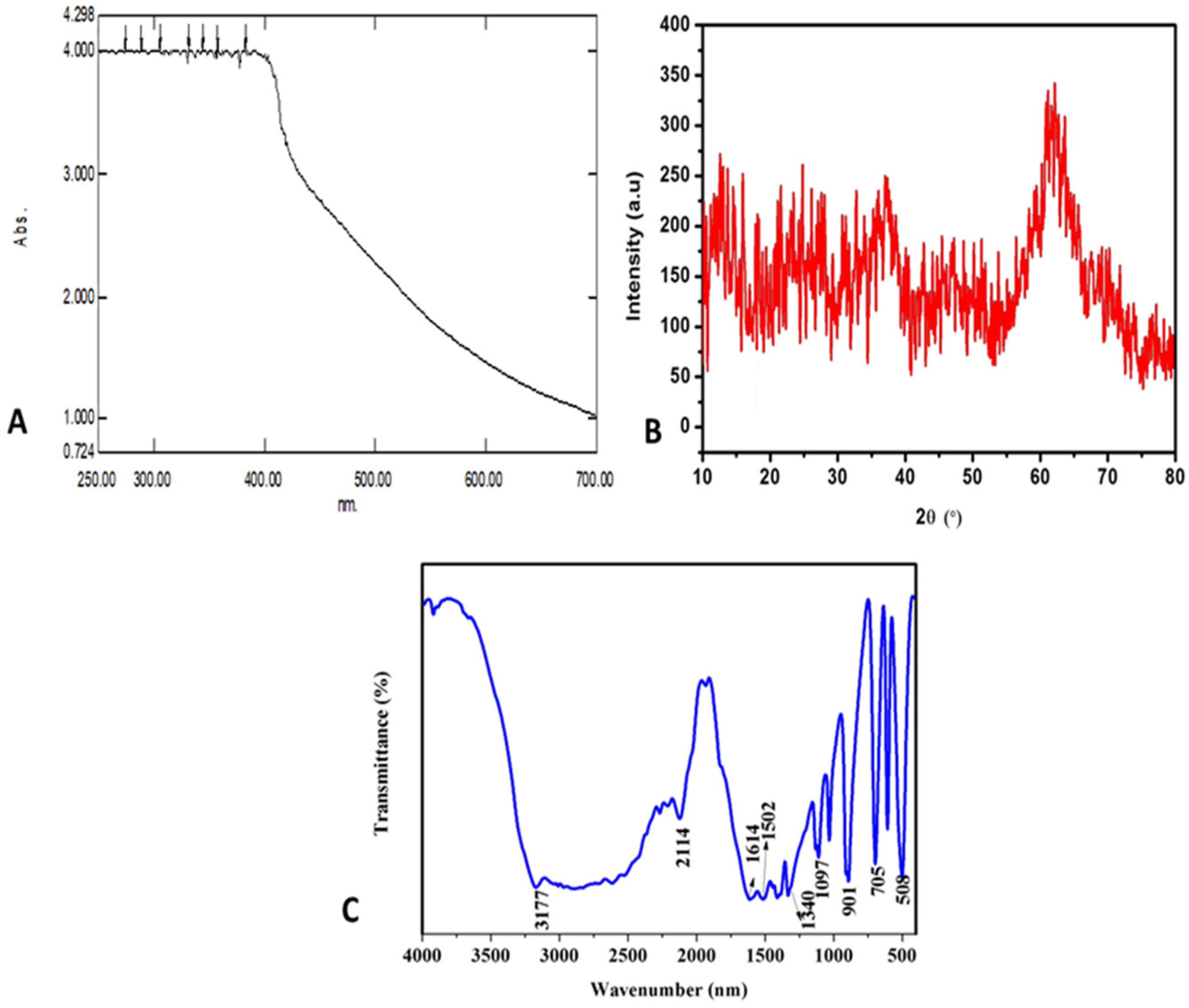
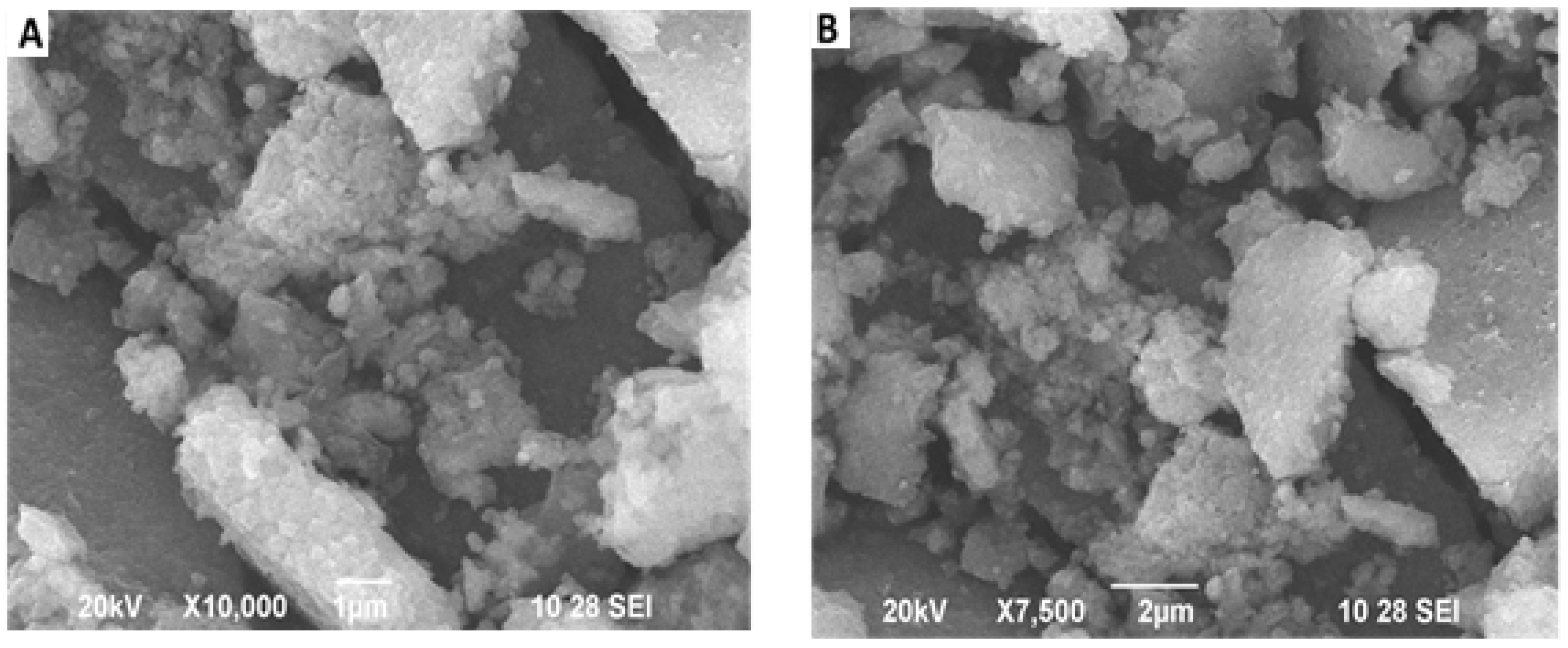
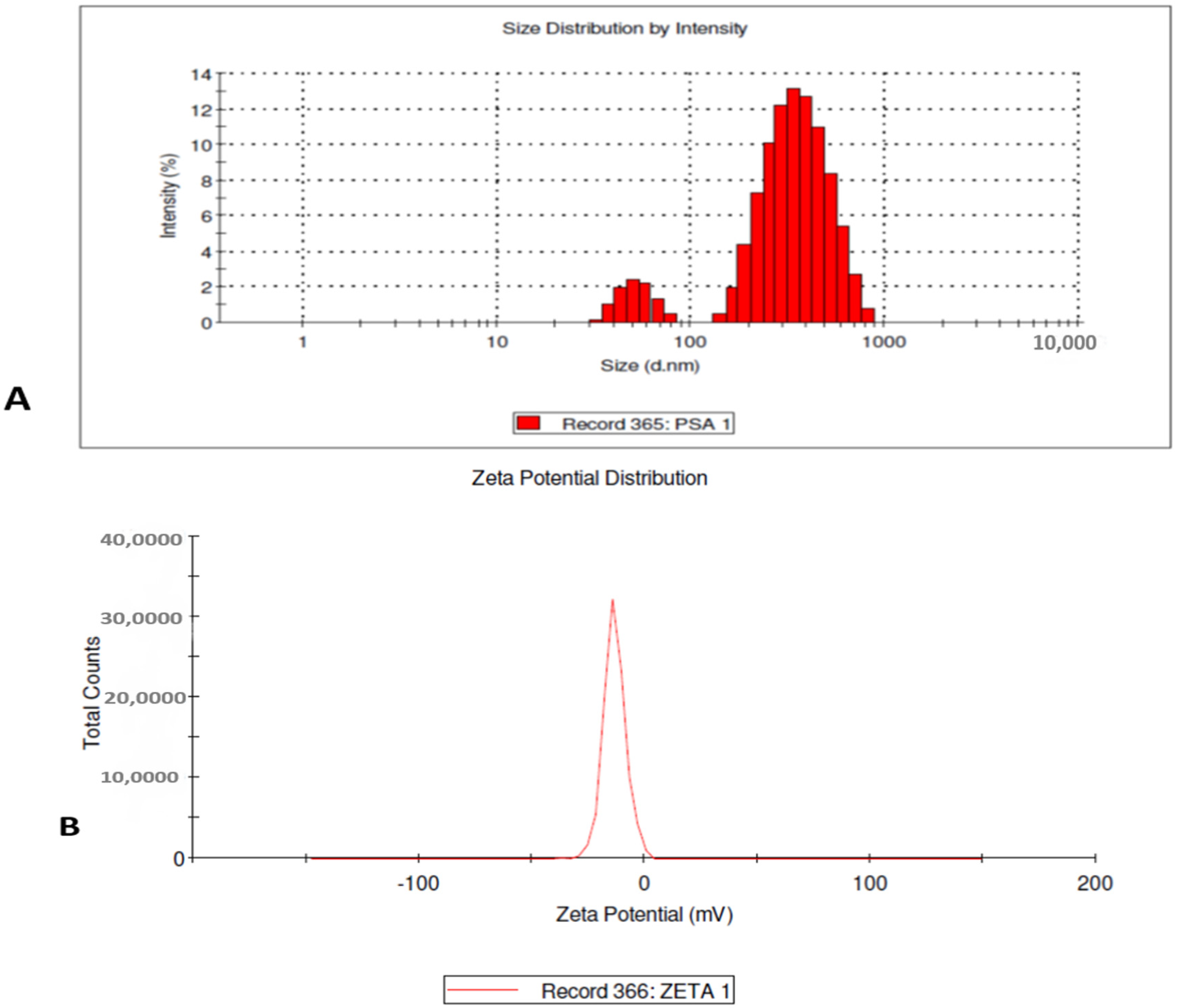
2.3. Characterization of Selenium Nanoparticles

A UV-vis spectrum was recorded at a resolution of 1 nm on a UV-3600 Shimadzu spectrophotometer (Kyoto, Japan). After freeze drying of the purified Se particles, the structure and composition were analyzed by scanning electron microscope (FEI QUANTA-200 SEM, Hillsborough, OR, USA) and energy dispersive X-ray spectroscopy. The crystalline structure of the selenium nanoparticles was determined using X-ray diffraction using Cu kα radiation (PAN anlyticalX’pert Pro MPD diffractometer). The particles’ size distribution was evaluated using dynamic light scattering (DLS) measurement and the zeta potential was studied to estimate the stability of the synthesized nanoparticles with a Malvem Zetasizer Nano (Cambridge, UK) series compact scattering spectrometer. Data obtained were analyzed using Zetasizer software (v3.30)

2.4. Collection of Mosquito Eggs

The eggs of A. aegypti and egg rafts of C. quinquefasciatus were collected from water stored containers and stagnant drains respectively from the Salem districts for bioassay. These eggs were carried to the laboratory and transferred to enamel trays containing 500 mL of water for larval hatching.

2.5. Maintenance of Larvae

Larvae of A. aegypti and C. quinquefasciatus were cultured and maintained in our laboratory at 27 ± 2 °C, 75% to 85% RH and under 14:10 (L:D) photoperiod cycles. Larvae were fed 5 g ground dog biscuit and brewers yeast daily in a 3:1 ratio. The feeding was continued till the larvae were grown to pupae.

2.6. Maintenance of Pupae and Adult

The pupae were collected from the culture trays mentioned above and were transferred to plastic containers (12 cm × 12 cm) containing 500 mL of water with the help of a dipper. The plastic jars were kept in mosquito cage (45 cm × 45 cm × 45 cm) for adult emergence. The adults were fed with 10% sugar solution for a period of three days before they were provided an animal for blood feeding.

2.7. Blood Feeding of Adult A. aegypti and C. quinquefasciatus

The adult female mosquitoes were allowed to feed on the blood of a mouse (exposed on the dorsal side) for two days. After blood feeding, enamel trays with water from the culture trays were placed in the cage for the adult to lay eggs. Both females and males were provided with 10% glucose solution on cotton wicks. The cotton was always kept moist with the solution and changed every day.

2.8. Larval Toxicity Test

F1 colonies of mosquito larvae were used for larvicidal and pupacidal activity. The selenium nanoparticles were evaluated at 10, 50 and 100 ppm mg/L concentrations. Untreated distilled water served as a control. Each treatment was set in triplicate. Twenty-five actively swimming A. aegypti and C. quinquefasciatus larvae and pupae were sieved out from different rearing trays to maintain uniformity of the batches of larvae and pupae exposed to each concentration of selenium nanoparticles and untreated control, held in separate (250 mL) capacity plastic containers [54]. Larval mortality was assessed after 24 and 48 h of exposure by probing the larvae with a needle, and moribund larvae and pupae were counted as dead [17]. The control mortalities were corrected by using Abbott’s formula [55].
Corrected mortality = observed mortality in treatment–observed mortality in control × 100
100–Control mortality.
Percentage mortality = number of dead larvae/pupae × 100
Number of larvae/pupae introduced

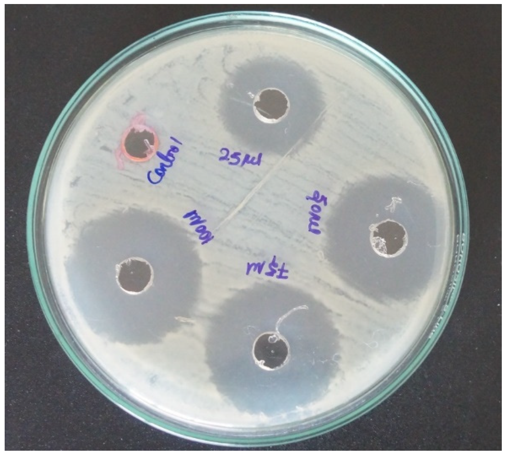
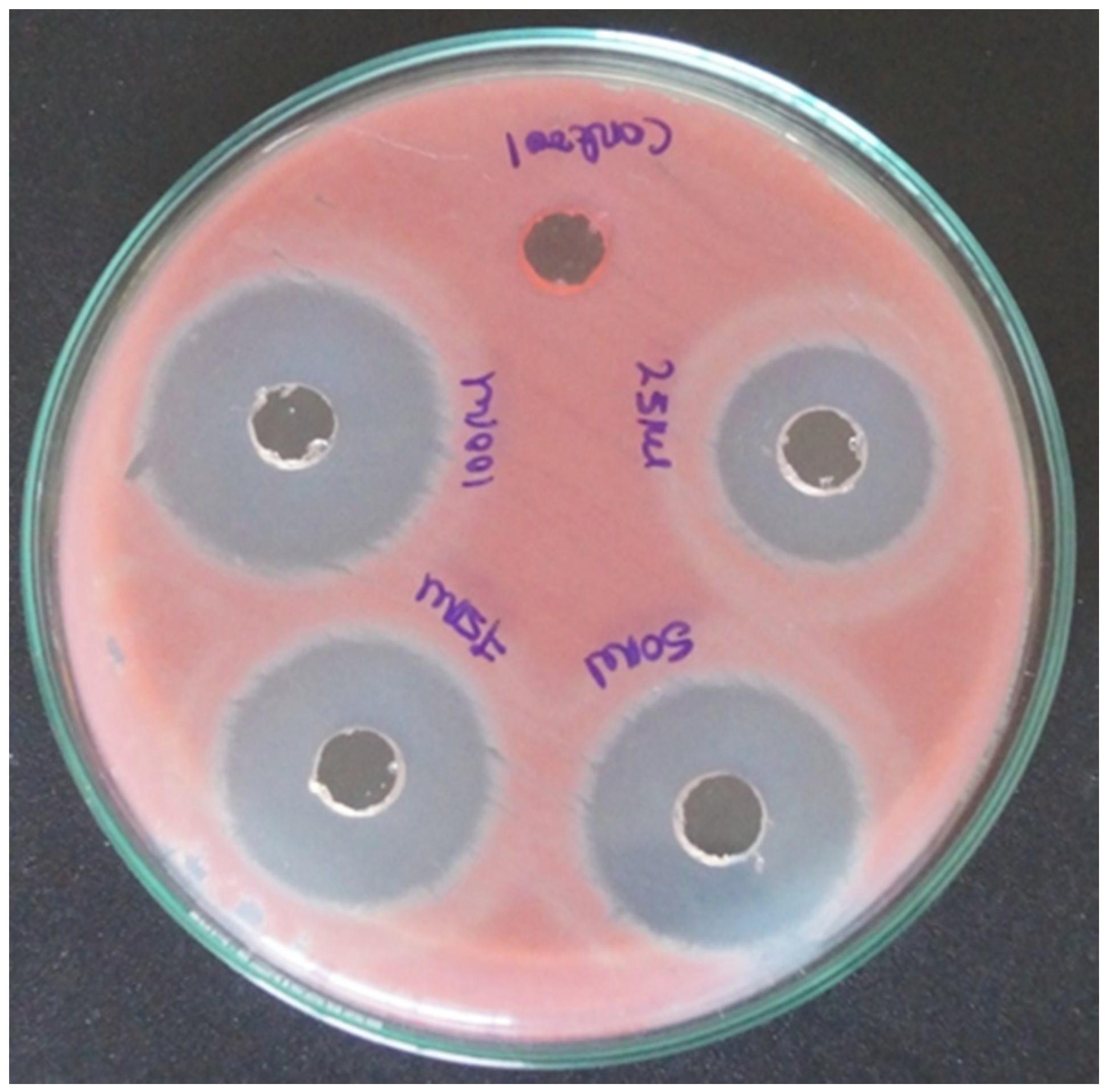
2.9. Anti-Bacterial Activity

The antibacterial activity of Se-NPs against St. aureus (MTCC96), and Se. marcescens (MTCC4822), was experimented by the standard well diffusion method [56]. The Gram-positive bacteria, St. aureus (MTCC96), and Gram-negative bacteria, Se. marcescens (MTCC4822), procured from the Department of Microbiology, Periyar University, Salem, India was cultured using agar agar type I (AA) and nutrient broth (NB). Wells of 6 mm size have been made on Muller Hilton agar plates using gel puncture. Using a micropipette, 25, 50, 75 and 100 µL of the sample of the nanoparticles solution and distilled water as the control were poured into the wells. The experiments were setup in triplicate. After incubation at 37 °C for 24 h, the different levels of the zones of inhibition were measured. The diameters of the zones of inhibition around each well of size were then recorded.

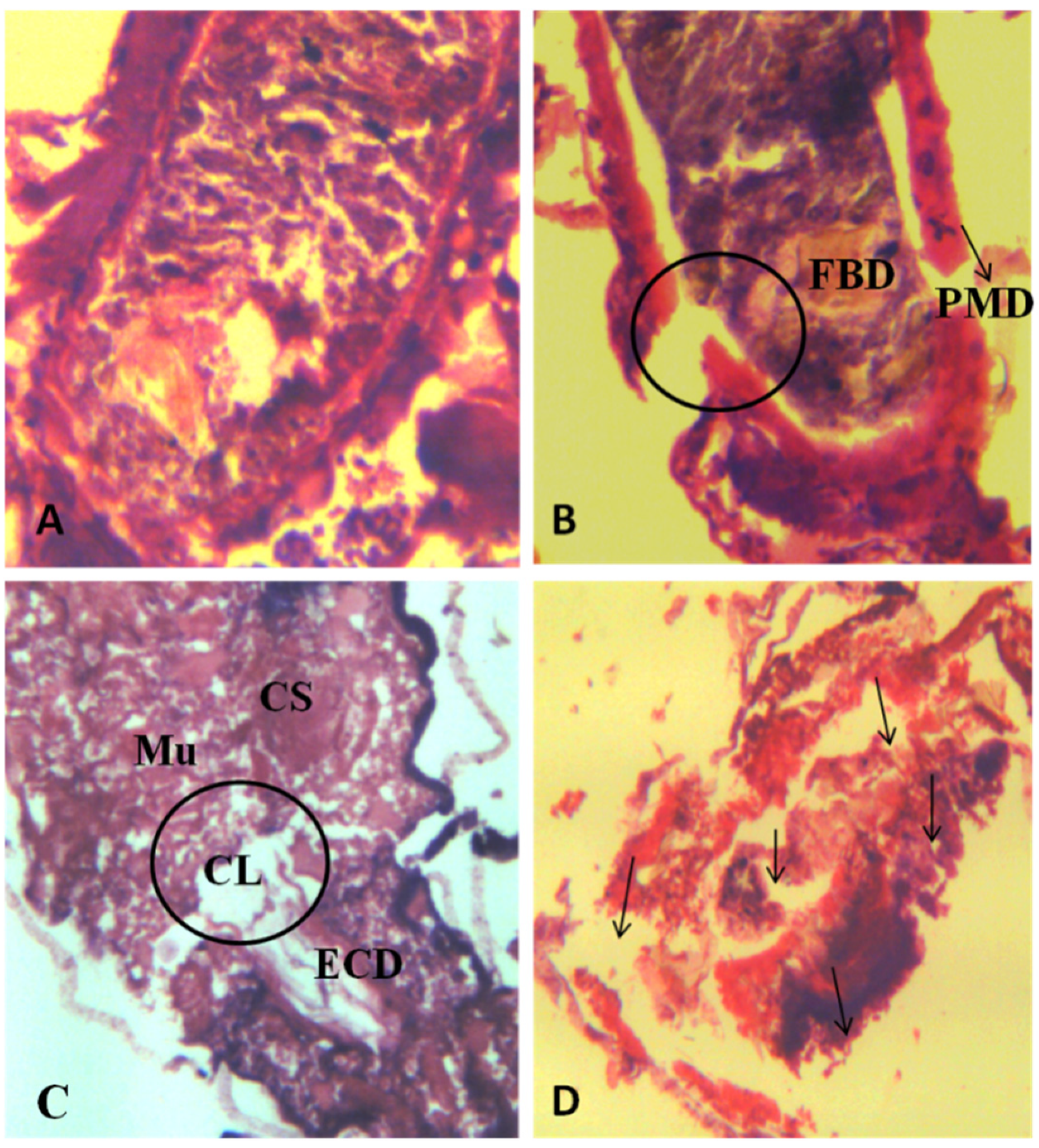
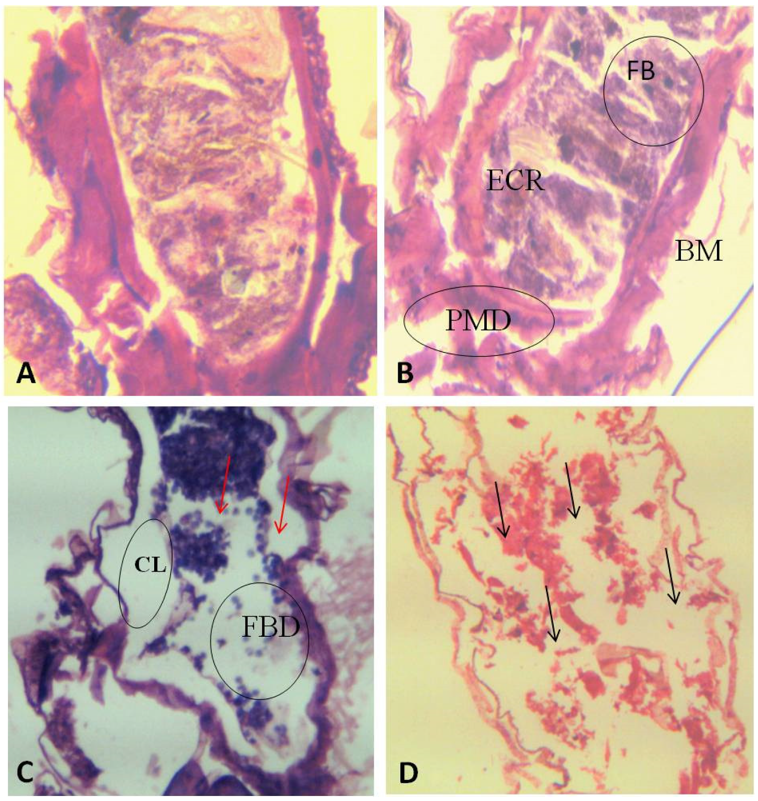
2.10. Histopathological Analysis

The control and SeNps treated fourth instar A. aegypti, C. quinquefasciatus larvae were fixed in 10% buffered formaldehyde for 24 h, and mounted in paraffin blocks for Histopathological analysis. Sections of larval tissue blocks that were 5 μm thick were cut with glass knives in a rotary microtome and mounted on glass slides. The sections were stained with haematoxylin and eosin for histopathological observation of toxicity effects on the treated A. aegypti and C. quinquefasciatus larvae, using a bright field light microscope [57,58].

2.11. Statistical Analysis

All data were subjected to analysis of variance (ANOVA). LC50 and LC90 values and their 95% confidence limits were estimated by getting a profit regressing model to the observed relationship between percentage of mortality of the larvae and pupae and the logarithmic concentration of the substance. In case of significant departure, a heterogeneity factor was used to calculate the 90% confidence limit for LC50 and LC90. All analyses were carried out using SPSS Software version 16.0.

3. Results

The selenium nanoparticles mediated by D. indica leaf extracts were visually confirmed by the color change from orange to red. The manifestation of ruby red is measured as a visual technique that shows the presence of selenium nanoparticles. The visual consideration is confirmed by UV absorption spectroscopy. The UV absorption peak of the synthesized nanoparticles recorded from the reaction medium at 80 °C using 5% D. indica leaf extract with 50 Mm selenious acid exposed to 24 h of reaction time clearly confirms the presence of selenium nanoparticles (Figure 1A). The UV absorption peak observed due to the surface plasmon resonance of the selenium nanoparticle synthesized using D. indica leaf extracts was recorded at 383 nm which was the original range of selenium nanoparticles.
The lyophilized selenium nanoparticles were used for verification under XRD. The diffraction of X-rays travelling through the selenium nanoparticles synthesized using D. indica leaf extract was recorded as diffraction peaks at 23.3 (100), 29.6 (101), 43.5 (012), and 50.05 (201), which signified the crystalline phase structure of the SeNPs (Figure 1B). The prominent peaks in the spectrum reveal the hexagonal structures which confirm the high degree of nanoparticle crystallization in the selenium particles. The diffraction peaks were in correspondence with the facets of the Joint Committee on Power Diffraction Standard (JCPDS no.73–0465) values.
The FTIR spectrum of the selenium nanoparticles were analyzed, to identify the possible bio molecules responsible for the reduction of selenious acid to selenium nanoparticles and the capping of the bio reduced selenium nanoparticles synthesised by D. indica leaf extract (Figure 1C). The FTIR spectrum recorded major peaks positioned at 3177, 2114, 1614, 1502, 1340, 1097, 901, 705, and 508 cm−1. The peaks are due to the stretching, bending and lagging of the bonds between elements of an organic compound. The sharp peak at 1340 cm−1 is attributed to the C–H bending in alkanes and the 1097 cm−1 corresponds to the C–N stretching of the amines. The band at 901 cm−1 is due to the O–H bend of carboxylic acids. The peaks that occurred around 705 cm−1 in the selenium nanoparticles mediated by D. indica leaf extract were due to the C–H medium bending and the weak band at 508 cm−1; from the FTIR it was inferred that the bio-organics such as the flavonoids, alkaloids and steroids phytoconstituents from the leaves served as a strong capping agent on the NPs. SEM images enabled us to visualize the size and shape of the selenium nanoparticles (Figure 2) obtained when the precursors treated with D. indica leaf extract at 90 °C for 120 min. The morphology of biosynthesized selenium nanoparticles studied under scanning electron micrographs revealed that they are oval in shape, with a smooth surface.
The dynamic light scattering (DLS) technique was used in this study to determine the size distribution of the selenium nanoparticles synthesized using D. indica leaf extract (Figure 3). The sizes of the selenium nanoparticles were not even, and ranged between 50 and 900 nm with a Z-average value of 248.0 nm. The zeta sizer’s report on the zeta potential of the biologically synthesized selenium nanoparticles revealed that the metal nanoparticles are highly stable in the aqueous medium (Figure 3). The zeta potential of the D. indica leaf extract mediated selenium nanoparticles was −13.2 mV, which confirms the strong repulsion among the particles and there by increases the stability of the nanoparticles.
Selenium nanoparticles synthesized using D. indica leaf extract was much effective against the dengue vector, A. aegypti in laboratory. Lower concentrations of selenium nanoparticles were capable for killing the mosquito immatures at all developmental stages. At 24 h of treatment of different concentrations of selenium nanoparticles the LC50 for the 1st to 4th larval instars and pupae of A. aegypti were 7.20, 14.78, 72.10, 113.41 and 440.99 ppm respectively (Table S1). At 48 h of treatment of different concentrations of selenium nanoparticles the LC50 for the 1st to 4th larval instars and pupae of A. aegypti were much reduced when compared to that of 24 h treatment (Table 1). The acquired chi-square values for toxicity assay on A. aegypti (1.98, 0.38, 0.10, 0.15 and 0.39 mg/L for I, II, III, IV instars pupae) proves that the observed mortality is on a par with the expected mortality. Selenium nanoparticles synthesized using D. indica leaf extract treatment on different stages of C. quinquefasciatus also proved its efficacy on this species. At 24 h treatment the LC50 for the 1st to 4th larval instars and pupae of C. quinquefasciatus were 2.53, 3.33, 7.95, 17.30 and 39.88 ppm, respectively (Table S2). At 48 h treatment the LC50 for the 1st to 4th larval instars and pupae of C. quinquefasciatus were highly reduced when compared to that of 24 h treatment. The acquired chi-square values for toxicity assay on C. quinquefasciatus (7.93, 1.66, 3.20, 0.98 and 4.68 mg/L for I, II, III, IV instars pupae respectively) proves that the observed mortality is on a par with the expected mortality (Table 2).
The antibacterial activity of selenium nanoparticles synthesized using D. indica leaf extract was investigated against the human pathogen, St. aureus (MTCC 96) and Se. marcescens (MTCC 4822) by well diffusion method (Table S3). The antibacterial activity of the synthesized selenium nanoparticles in 25, 50, 75 and 100 µL concentrations were quantitatively measured by the formation of zones of inhibition. Selenium nanoparticles synthesized using D. indica leaf extract developed inhibitory zones measuring 0.50, 0.63, 0.75 and 1.03 cm in diameter on the live culture plates of St. aureus (MTCC 96) after 24 h of treatment with 25, 50, 75 and 100 µL concentrations respectively (Figure 4). In another experiment the inhibitory zones measured 0.47, 0.66, 0.76 and 1.08 cm diameter on the live culture plates of Se. marcescens (MTCC 4822) when treated with 25, 50, 75 and 100 µL concentrations of selenium nanoparticles respectively (Figure 5).
The cellular and tissue damage caused by the SeNps to A. aegypti and C. quinquefasciatus was visualized using light microscope by mounting cross sections of SeNps treated A. aegypti and C. quinquefasciatus (Figure 6 and Figure 7). The control and treated samples clearly show the difference and damages in treatment when compared with the control. Both A. aegypti and C. quinquefasciatus larva treated with 10 ppm SeNps showed microscopically visual damages such as cell lysis, breakage of peritrophic membrane and pre-rupturing stages of epithelial cells. The pre-rupture stages were clearly visible and differentiable with the ruptured epithelial cells seen in the larvae treated with 50 ppm SeNps. The tissues of larva treated with 100 ppm SeNps showed completely disorganized and broken epithelial cells. These tissues also showed damages in the internal organs like broken midgut, caeca and totally collapsed larva.

4. Discussion

Mosquito borne diseases are one of the important causes of decline in health and economy in many countries. Among them, the species A. aegypti takes a lead in vectoring several human diseases like yellow fever, dengue, chikungunya [58], etc. C. quinquefasciatus is another species which vectors diseases like human lymphatic filariasis, Japanese encephalitis, St. Louis encephalitis etc. [18]. Nosocomial infection is a serious problem worldwide that mainly affects immune compromised patients [59]. There is a dramatic increase in St. aureus (MTCC 96) and Se. marcescens (MTCC 4822) infections, both with community-associated and hospital-acquired types. Development of resistance in the vectors as well as the pathogens is of serious concern and creates a great challenge in control program [60]. One of the most important challenges faced by the human population in the 21st century is disease and vector control [61]. Hence, much importance is given to novel eco-friendly nanotechnology tools to control the vectors and pathogens which were carried out in the present study.
In the present study we used D. indica leaf extract to synthesis selenium nanoparticles, which was achieved due to the presence of various secondary metabolites in the plant extract. These compounds are the sole reason for the reduction, formation and capping of nanoparticles. The formation of selenium nanoparticles was confirmed by the characteristic red color, due to surface plasmon excitation of the selenium at nanoscale level [62]. The absorbance peak observed in the present study that was due to surface plasmon resonance was very similar to the report of Malhotra et al. [63], who observed an absorbance peak between 320 and 550 nm, with a maximum at 390 nm. In another study, selenium colloidal solution prepared using citrus leaf extract showed the absorption peak at 395 nm which was in concordance to the present study [64]. The X-Ray diffraction studies proved that the selenium nanoparticles synthesized using D. indica leaf extract is crystalline in nature. In concordance to the present study the diffraction peaks of Allium sativum mediated selenium nanoparticles observed similar peaks corresponding to the facets of selenium showing the crystalline nature [65]. The report of Singh et al. [66] was also in support of the present results, showing the characteristic peak at 2θ value of 23.680, 29.788 and 43.9.
The functional group or the organic compound which is main cause for the stability of nanoparticles by capping them was confirmed using FTIR. The peak at 1326 cm−1 was assigned to lipid content, C–H band vibrations, or syringyl ring breathing with C=O stretching, as similar to the reports of Singh et al. [67]. Sarkar et al. [68] confirms that the sharp peak at 1340 cm−1 is attributed to the C–H bending in alkanes and the 1097 cm−1 corresponds to the C–N stretching of the amines. The absorption peak at 1762 cm−1 corresponds to the C=O stretching of the lactone of ascorbic acid and peak at 1674 cm−1 is corresponding with the C=C stretching vibrations [69] The zeta potential measurements (−13.48 mV) show that the synthesized selenium nanoparticles are highly stable in the liquid medium, which was similar to that of the results reported by Sarkar et al. [68].
The selenium nanoparticles synthesized using plant extracts have good biologic activity and adsorptive capacity due to interaction between the selenium at the nano range and NH, C=O, COO–, and C–N functional groups of proteins [70]. A recent study reported that the selenium nanoparticles synthesized using Penicillium corylophilum are a potential agent in controlling mosquitos, cell lines and Gram-positive and Gram-negative bacteria [71]. In another study, selenium nanoparticles synthesized using Clausena dentate were effective against different mosquito species viz., A. stephensi, A. aegypti, and C. quinquefasciatus at 240.714, 104.13 and 99.602 mg/L concentrations respectively [72]. Whereas, in the present study selenium nanoparticles synthesized using D. indica are more effective by controlling early instar A. aegypti and C. quinquefasciatus at 0.39 and 1.11 mg/L respectively. The toxicity of biologically synthesized selenium nanoparticles proves equally good as that of silver and gold nanoparticles [73] in controlling both A. aegypti and C. quinquefasciatus.
The toxicity of selenium nanoparticles synthesized using D. indica leaf extract on larvae and pupae of the two mosquito species may be due to the intracellular toxic effects of nano scale particle inside cuticular and other peripheral cells. The toxicity of the selenium nanoparticles is may also be due to the denaturation of organelles and enzymes which reduce the membrane permeability which further affects the ATP synthesis and finally blocks the cellular function, leading to death [74]. The toxicity of selenium nanoparticles synthesized using D. indica leaf extract is significant on St. aureus (MTCC 96), a Gram-positive bacterium. A similar study on selenium nanoparticles using a different plant extract also proved toxic against the same bacterial species [75] The antibacterial effect of selenium nanoparticles was proved on both Gram-positive and Gram-negative bacteria [76]. Tran and Webster [77] reported that the selenium nanoparticles prepared using colloidal synthesis method highly inhibits the growth of St. aureus (MTCC 96) by up to 60 times compared to control. The mechanism in the antibacterial activity of selenium nanoparticles is not exactly proven. The mode of action exerted by the selenium nanoparticles can be the adherence of positive surface charge on their own surface to the bacteria with negative surface charges, resulting in enhancement of bactericidal effects [78]. It is also believed that the selenium nanoparticles can damage the bacterial cell wall by interfering with the peptidoglycan layer [79]. Consequently, the penetration of selenium nanoparticles into the St. aureus is (MTCC 96) is made easier by chemisorption, where the lipoproteins involved are of the diacyl and triacyl forms [80].
The inhibitory effect of selenium nanoparticles synthesized using D. indica leaf extract is significant on Se. marcescens (MTCC 4822), a Gram-negative bacterium. The toxicity of selenium nanoparticles on Se. marcescens (MTCC 4822) can be due to the penetration of metal particles into the membranes which have different proteins and phospholipids that interact with each other and disturb their function [81,82,83]. It is also believed that the selenium nanoparticles can interact with thiol groups present in enzymes such as NADH dehydrogenases and affect the energy production [84].

5. Conclusions

Environmentally safer drugs and pesticides are of current interest to researchers, as they are needed for the well-being of humans. We synthesized SeNps in an eco-friendly protocol in which D. indica leaf extract acted as a reducing and capping agent. The eco-friendly SeNps were significantly effective against the disease transmitting mosquitoes A. aegypti and C. quinquefasciatus, as well as the microbial pathogens St. aureus (MTCC 96) and Se. marcescens (MTCC4822). Thus, the D. indica leaf-extract-mediated selenium nanoparticles amplify the remedial source against the pathogenic microbes and vector mosquitoes that claim many human lives every year. The proposal maintains a green protocol, as the synthesized nanoparticles are nontoxic to both the environment and non-target organisms. Future studies are needed to reveal the mechanism underlying mosquitocidal and antibacterial activities.

Supplementary Materials

The following are available online at https://www.mdpi.com/2079-6412/10/7/626/s1, Table S1: Effect of Selenium Nanoparticles on developmental stages of Aedes aegypti at 24 h exposure. Table S2: Effect of Selenium Nanoparticles on developmental stages of C. quinquefasciatus at 24 h exposure. Table S3: Antibacterial effect of Selenium nanoparticles against Staphylococcus aureus and Serratia marcescens.

Author Contributions

Conceptualization, N.A.; methodology, M.K., K.R., and P.M.; validation, N.A., S.K. and P.T.; formal analysis, N.A., S.K. and P.T.; investigation, N.A.; writing—original draft preparation, M.K.; writing—review and editing, N.A. and S.K.; visualization, N.A.; supervision, N.A.; project administration, N.A.; funding acquisition, M.K. and N.A. All authors have read and agreed to the published version of the manuscript.

Funding

This research was funded by University Grants Commission (UGC), New Delhi, India for the award of Rajiv Gandhi National Fellowship, grant number F1-17.1/ 2015-16/ RGNF2015-17 –SC-TAM-10313”.

Acknowledgments

We sincerely acknowledge and thank Periyar University, Salem, and Tamil Nadu for providing all required laboratory facilities in relation to this research work.

Conflicts of Interest

The authors declare no conflict of interest.

References

  1. Talukdar, A.; Talukdar, N.; Deka, S.; Jyoti Sahariah, B. Dillenia Indica (Outenga) as anti-diabetic herb found in Assam: A review. Int. J. Pharm. Sci. Res. 2018, 3, 2482–2486. [Google Scholar]
  2. Janick, J.; Paull, R.E. The Encyclopedia of Fruit and Nuts, 1st ed.; CABI: London, UK, 2008. [Google Scholar]
  3. Dipal, G.; Priti, M. Dillenia indica Linn. and Dillenia pentagyna Roxb.: Pharmacognostic, Phytochemical and Therapeutic aspects. J. Appl. Pharm. Sci. 2013, 3, 134–142. [Google Scholar]
  4. Yazan, L.S.; Armania, N. Dillenia species: A review of the traditional uses, active constituents and pharmacological properties from pre-clinical studies. Pharm. Biol. 2014, 52, 890–897. [Google Scholar] [CrossRef] [PubMed]
  5. Abdille, M.H.; Singh, R.P.; Jayaprakasha, G.K.; Jena, B.S. Antioxidant activity of the extracts from Dillenia indica fruits. Food Chem. 2005, 90, 891–896. [Google Scholar] [CrossRef]
  6. Biswas, S.; Pandita, N. Phytochemical analysis and chromatographic evaluation of alcoholic extract of Dillenia indica linn Leaves. IJPSR 2015, 7, 2799–2812. [Google Scholar]
  7. Govindarajan, M.; Rajeswary, M.; Veerakumar, K.; Muthukumaran, U.; Hoti, S.L.; Benelli, G. Green synthesis and characterization of silver nanoparticles fabricated using Anisomeles indica: Mosquitocidal potential against malaria, dengue and Japanese encephalitis vectors. Exp. Parasitol. 2016, 161, 40–47. [Google Scholar] [CrossRef]
  8. Veerakumar, K.; Govindarajan, M. Adulticidal properties of synthesized silver nanoparticles using leaf extracts of Feronia elephantum (Rutaceae) against filariasis, malaria, and dengue vector mosquitoes. J. Ecobiotechnol. 2014, 113, 40–85. [Google Scholar] [CrossRef]
  9. Benelli, G. Plant-borne ovicides in the fight against mosquito vectors of medical and veterinary importance: A systematic review. Parasitol. Res. 2015, 114, 3201–3212. [Google Scholar] [CrossRef]
  10. Benelli, G.; Mehlhorn, H. Declining malaria, rising dengue and Zika virus: Insights for mosquito vector control. Parasitol. Res. 2016, 115, 1747–1754. [Google Scholar] [CrossRef]
  11. Muthukumaran, U.; Govindarajan, M.; Rajeswary, M. Synthesis and characterization of silver nanoparticles using Gmelinaasiatica leaf extract against filariasis, dengue, and malaria vector mosquitoes. Parasitol. Res. 2015, 114, 1817–1827. [Google Scholar] [CrossRef]
  12. Murugan, K.; Panneerselvam, C.; Subramaniam, J.; Madhiyazhagan, P.; Hwang, J.S.; Wang, L.; Dinesh, D.; Suresh, U.; Roni, M.; Higuchi, A.; et al. Eco-friendly drugs from the marine environment: Spongeweed-synthesized silver nanoparticles are highly effective on Plasmodium falciparum and its vector Anopheles stephensi, with little non-target effects on predatory copepods. Environ. Sci. Pollut. Res. 2016, 23, 16671–16685. [Google Scholar] [CrossRef] [PubMed]
  13. Kovendan, K.; Murugan, K.; Shanthakumar, S.P.; Vincent, S. Evaluation of larvicidal and pupicidal activity of Morinda citrifolia L. (Noni) (Family: Rubiaceae) against three mosquito vectors. Asian Pac. J. Trop. Med. 2012, 2, S362–S369. [Google Scholar] [CrossRef]
  14. Pavela, R.; Benelli, G. Ethnobotanical knowledge on botanical repellents employed in the African region against mosquito vectors a review. Plant Prot. Sci. 2016, 167, 103–108. [Google Scholar] [CrossRef] [PubMed]
  15. Murugan, K.; Sanoopa, C.P.; Madhiyazhagan, P.; Dinesh, D.; Subramaniam, J.; Panneerselvam, C.; Roni, M.; Suresh, U.; Nicolettib, M.; Alarfajc, A.A.; et al. Rapid biosynthesis of silver nanoparticles using Crotalaria verrucosa leaves against the dengue vector Aedes aegypti: What happens around. An analysis of dragonfly predatory behaviour after exposure at ultra-low doses. Parasitol Res. 2015, 114, 2243–2253. [Google Scholar] [CrossRef] [PubMed]
  16. Alyahya, I.A.; Almohsen, H.A.; AlSaleem, I.A.; Al-Hamid, M.M.; Arafah, A.M.; Al Turki, Y.A. Assessment of knowledge, attitude, and practice about first aid among male school teachers and administrators in Riyadh, Saudi Arabia. J. Fam. Med. Prim. Care 2019, 8, 684–688. [Google Scholar] [CrossRef]
  17. World Health Organization. Dengue and Severe Dengue Fact Sheet. 2015. Available online: www.elsevier.com/locate/parepi (accessed on 26 February 2020).
  18. Sutthanont, N.; Siriluck, A.; Surang, N. Larvicidal Activity of Synthesized Silver Nanoparticles from Curcuma zedoaria Essential Oil against Culex Quinquefasciatus. Insects 2019, 10, 27. [Google Scholar] [CrossRef] [Green Version]
  19. Subra, R.; Service, M.W.; Mosha, F.W. The effect of domestic detergents on the population dynamics of the immature stages of two competitor mosquitoes, Culex cinereus Theobald and Culex quinquefasciatus Say (Diptera, Culicidae) in Kenya. Acta Trop. 1984, 41, 69–75. [Google Scholar]
  20. Ajayi, M.B.; Adeleke, M.A.; Idowu, E.T.; Awolola, T.S. Surveillance of mosquitoes vectors in Ajumoni Estate Ogun State, Nigeria. Ann. Biol. Res. 2010, 1, 16–19. [Google Scholar]
  21. Levin, N.; Zahavi, J. Predictive modeling using segmentation. J. Interact. Mark. 2001, 15, 2–22. [Google Scholar] [CrossRef]
  22. Rizzello, L.; Pompa, P.P. Nanosilver-based antibacterial drugs and devices: Mechanisms, methodological drawbacks, and guidelines. Chem. Soc. Rev. 2014, 43, 1501–1518. [Google Scholar] [CrossRef]
  23. Lin, P.L.; Oram, R.J.; Lauderdale, D.S. Requirement of NAD and SIR2 for life-span extension by calorie restriction in Saccharomyces cerevisiae. Science 2000, 289, 2126–2128. [Google Scholar] [CrossRef] [PubMed] [Green Version]
  24. Ghalehnoo, Z.R. Diseases caused by Staphylococcus Aureus. Int. J. Med. Health Res. 2018, 4, 65–67. [Google Scholar]
  25. Okuda, T.; Endo, N.; Osada, Y.; ZenYoji, H. Outbreak of nosocomial urinary tract infections caused by Serratia marcescens. Enzyme 1984, 20, 691–695. [Google Scholar] [CrossRef] [Green Version]
  26. Buckle, J. Infection, Clinical Aromatherapy, 3rd ed.; Elsevier: Amsterdam, The Netherlands, 2015; Volume 7, pp. 130–167. [Google Scholar]
  27. Santhosh, S.B.; Ragavendran, C.; Natarajan, D. Spectral and HRTEM analyses of Annona muricata leaf extract mediated silver nanoparticles and its Larvicidal efficacy against three mosquito vectors Anopheles stephensi, Culex quinquefasciatus, and Aedes aegypti. J. Photochem. Photobiol. 2015, 153, 184–190. [Google Scholar] [CrossRef] [PubMed]
  28. Veerakumar, K.; Govindarajan, M.; Rajeswary, M. Green synthesis of silver nanoparticles using Sidaacuta (Malvaceae) leaf extract against Culex quinquefasciatus, Anopheles stephensi, and Aedes aegypti (Diptera: Culicidae). Parasitol. Res. 2013, 112, 4073–4085. [Google Scholar] [CrossRef] [PubMed]
  29. Nathiya, S.; Santhi, N.; Kalaiselvi, S. A comparative study on ontogenic expression of antioxidants and secondary metabolites in Withania somnifera. Int. J. Curr. Sci. 2012, 3, 210–215. [Google Scholar]
  30. Singh, S.; Singh, B.K.; Yadav, S.M.; Gupta, A.K. Applications of Nanotechnology in Agricultural and their Role in Disease Management. Res. J. Nanosci. Nanotechnol. 2015, 5, 1–5. [Google Scholar] [CrossRef]
  31. Sosa, I.O.; Noguez, C.; Barrera, R.G. Optical properties of metal nanoparticles with arbitrary shapes. J. Phys. Chem. B 2003, 107, 6269–6975. [Google Scholar] [CrossRef] [Green Version]
  32. Sunanda, K.; Kikuchi, Y.; Hashimoto, K.; Fujishima, A. Bactericidal and detoxification effects of TiO2 thin film photocatalysts. Environ. Sci. Technol. 1998, 32, 726–728. [Google Scholar] [CrossRef]
  33. Taylor, P.L.; Ussher, A.L.; Burrell, R.E. Impact of heat on nanocrystalline silver dressings. Part I: Chemical and biological properties. Biomaterials 2005, 26, 7221–7229. [Google Scholar] [CrossRef]
  34. Ingle, A.; Gade, A.; Pierrat, S.; Sonnichsen, C.; Rai, M. Mycosynthesis of silver nanoparticles using the fungus Fusarium acuminatum and its activity against some human pathogenic bacteria. Curr. Nanosci. 2008, 4, 141–144. [Google Scholar] [CrossRef]
  35. Duran, N.; Marcato, P.D.; Alves, O.L.; Souza, G. Mechanistic aspects of biosynthesis of silver nanoparticles by several Fusariumoxy sporum strains. J. Nanotechnol. 2005, 3, 1–7. [Google Scholar]
  36. Morejon, B.; Pilaquinga, F.; Domenech, F.; Ganchala, D.; Debut, A.; Neira, M. Larvicidal Activity of Silver Nanoparticles Synthesized Using Extracts of Ambrosia arborescens (Asteraceae) to Control Aedes aegypti L. (Diptera: Culicidae). J. Nanotechnol. 2018, 2018, 6917938. [Google Scholar] [CrossRef] [Green Version]
  37. Barik, T.K.; Kamaraju, R. Silica nanoparticle: A potential new insecticide for mosquito vector control. Parasitol. Res. 2012, 111, 1075–1083. [Google Scholar] [CrossRef] [PubMed]
  38. Deepak, P.; Amutha, V.; Birundha, R.; Sowmiya, R.; Kamaraj, C.; Balasubramanian, V.; Balasubramani, G.; Aiswarya, D.; Arul, D.; Perumal, P. Facile green synthesis of nanoparticles from brown seaweed Sargassum wightii and its biological application potential. Adv. Nat. Sci. Nanosci. Nanotechnol. 2018, 9, 035019. [Google Scholar] [CrossRef] [Green Version]
  39. Murugan, K.; Dinesh, D.; Nataraj, D.; Subramaniam, J.; Amuthavalli, P.; Madhavan, J.; Rajasekar, A.; Rajan, M.; Palani Thiruppathi, K.; Suresh, K.; et al. Iron and iron oxide nanoparticles are highly toxic to Culex quinquefasciatus with little non-target effects on larvivorous fishes. Environ. Sci. Pollut. Res. 2017. [Google Scholar] [CrossRef] [PubMed]
  40. Benelli, G.; Pavela, R.; Canale, A.; Cianfaglione, K.; Ciaschetti, G.; Conti, F.; Nicoletti, M.; Senthil-Nathan, S.; Mehlhorn, H.; Maggi, F. Acute Larvicidal Toxicity of Five Essential Oils (Pinus nigra, Hyssopus officinalis, Satureja montana, Aloysia citrodora and Pelargonium graveolens) Against the Filariasis Vector Culex Quinquefasciatus: Synergistic and Antagonistic Effects. Parasitol. Int. 2017, 66, 166–171. [Google Scholar] [CrossRef]
  41. Bai, X.; Mou, C.; Xu, L.; Wang, S.; Pu, S.; Zeng, X. Passively Q-switched erbium-doped fiber laser using Fe3O4-nanoparticle saturable absorber. Jpn. Soc. Appl. Phys. 2016, 9, 042701. [Google Scholar] [CrossRef] [Green Version]
  42. Rayman, M.; Thompson, A.; Warren-Perry, M.; Galassini, R.; Catterick, J.; Hall, E.; Lawrence, D.; Bliss, J. Impact of selenium on mood and quality of life: A randomized, controlled trial. Biol. Psychiatry 2006, 59, 147–154. [Google Scholar] [CrossRef] [Green Version]
  43. Schomburg, L. Dietary selenium and human health. Nutrients 2017, 9, 22. [Google Scholar] [CrossRef] [Green Version]
  44. Bozena, H.; Marta, K.; Sylvie, S.; Carlos, F.; Branislav, R.N.; Qiuming, P.; Mojmir, B.; Magdalena, M.; Radka, O.; Jarmila, Z.; et al. Nano-selenium and its nanomedicine applications: A critical review. Int. J. Nanomed. 2018, 13, 2107–2128. [Google Scholar]
  45. Ajitha, B.; Reddy, Y.A.; Reddy, P.S. Green synthesis and characterization of silver nanoparticles using Lantana camara leaf extract. Mater. Sci. Eng. C 2015, 49, 373–381. [Google Scholar] [CrossRef] [PubMed]
  46. Guo, M.; Li, W.; Yang, F.; Liu, H. Controllable biosynthesis of gold nanoparticles from a Eucommia ulmoides bark aqueous extract. Spectrochim. Acta Part A Mol. Biomol. Spectrosc. 2015, 142, 73–79. [Google Scholar] [CrossRef] [PubMed]
  47. Dhanjal, S.; Singh, C. Aerobic biogenesis of selenium nanospheres by Bacillus cereus isolated from coalmine soil. Microb. Cell Factories 2010, 52, 1–11. [Google Scholar] [CrossRef] [PubMed] [Green Version]
  48. Venkatesan, S.; Sujatha, K. Characterization of Barnyard Millet Cultivars using Seed Image Analysis. Seed Res. J. 2018, 45, 1–3. [Google Scholar]
  49. Gunti, M.; Ramachandran, D.; Bharat, P. Design and Preparation of Guar Gum Nano Particles Based on Emulsification Technique. Int. J. Chemtech. Res. 2019, 12, 189–193. [Google Scholar]
  50. Ramamurthy, C.H.; Sampath, K.S.; Arunkumar, P.; Suresh Kumar, M.; Sujatha, V.; Premkumar, K.; Thirunavukkarasu, C. Green Synthesis and Characterization of Selenium Nanoparticles and Its Augmented Cytotoxicity with Doxorubicin on Cancer Cells. Bioprocess Biosyst. Eng. 2013, 36, 1131–1139. [Google Scholar] [CrossRef]
  51. Cui, D.; Liang, T.; Sun, L.; Meng, L.; Yang, C.; Wang, L.; Liang, T.; Li, Q. Green synthesis of selenium nanoparticles with extract of hawthorn fruit induced HepG2 cells apoptosis. Pharm. Biol. 2018, 56, 528–534. [Google Scholar] [CrossRef] [Green Version]
  52. Fardsadegh, B.; Jafarizadeh-Malmiri, H. Aloe vera leaf extract mediated green synthesis of selenium nanoparticles and assessment of their In vitro antimicrobial activity against spoilage fungi and pathogenic bacteria strains. Green Process. Synth. 2019, 8, 399–407. [Google Scholar] [CrossRef]
  53. Sharma, G.; Sharma, A.R.; Bhavesh, R.; Jongbong, P.; Ganbold, B.; Ju-Suk, N.; Sang-Soo, L. Biomolecule-Mediated Synthesis of Selenium Nanoparticles using Dried Vitis vinifera (Raisin) Extract. Molecules 2014, 19, 2761–2770. [Google Scholar] [CrossRef]
  54. Tan, D.J.; Dvinge, H.; Christoforou, A.; Bertone, P.; Martinez Arias, A.; Lilley, K.S. Supporting Information: Table S5. Localizations of proteins predicted by LOPIT. J. Proteome Res. 2009, 8, 2667–2678. [Google Scholar] [CrossRef]
  55. Azmi, W.; Sani, R.K.; Banerjee, U.C. Biodegradation of triphenylmethane dyes. Enzym. Microb. Technol. 1998, 22, 185–191. [Google Scholar] [CrossRef]
  56. Abbott, W.S. A method of computing the effectiveness of an insecticide. J. Econ. Entomol 1925, 18, 265–267. [Google Scholar] [CrossRef]
  57. Kalagatur, N.K.; Mudili, V.; Siddaiah, C.; Gupta, V.K.; Natarajan, G.; Sreepathi, M.H.; Vardhan, B.H.; Putcha, V.L.R. Antagonistic activity of Ocimum sanctum L. essential oil on growth and zearalen one production by Fusarium graminearum in maize grains. Front. Microbiol. 2015, 6, 892. [Google Scholar] [CrossRef]
  58. Fallatah, S.A.; Khater, E.I. Potential of medicinal plants in mosquito control. J. Egypt Soc. Parasitol. 2010, 40, 1–26. [Google Scholar]
  59. Suganya, G.; Karthi, S.; Shivakumar, M.S. Larvicidal potential of silver nanoparticles synthesized from Leucas aspera leaf extracts against dengue vector Aedes Aegypti. Parasitol. Res. 2014, 113, 875–880. [Google Scholar] [CrossRef] [PubMed]
  60. Gilbert, P.; Allison, D.G.; McBain, A.J. Biofilms in vitro and in vivo: Do singular mechanisms imply cross-resistance. Symp. Ser. Soc. Appl. Microbiol. 2002, 92, 98S–110S. [Google Scholar] [CrossRef]
  61. Trinh, T.D.; Zasowski, E.J.; Claeys, K.C.; Casapao, A.M.; Compton, M.; Lagnf, A. Role of vancomycin minimum inhibitory concentrations by modifed population analysis profle method and clinical outcomes in high inoculum methicillin-resistant Staphylococcus Aureus Infections. Infect. Dis. Ther. 2018, 7, 161–169. [Google Scholar] [CrossRef] [Green Version]
  62. Roni, M.; Murugan, K.; Panneerselvam, C.; Subramaniam, J.; Nicoletti, M.; Madhiyazhagan, P.; Dinesh, D.; Udaiyan, S.U.; Hanem, F.; Wei, H.; et al. Characterization and biotoxicity of Hypnea musciformis synthesized silver nanoparticles as potential eco-friendly control tool against Aedes aegypti and Plutella Xylostella. Ecotoxicol. Environ. Saf. 2015, 121, 31–38. [Google Scholar] [CrossRef]
  63. Wang, T.; Yang, L.; Zhang, B.; Liu, J. Extracellular biosynthesis and transformation of selenium nanoparticles and application in H2O2 biosensor. Colloid Surf. B 2010, 80, 94–102. [Google Scholar] [CrossRef]
  64. Malhotra, S.; Jha, N.; Desai, K.A. Superficial synthesis of selenium nanospheres using wet chemical approach. Int. J. Nanotechnol. 2014, 3, 7–14. [Google Scholar]
  65. Prasad, R.; Kumar, V.; Prasad, K.S. Nanotechnology in sustainable agriculture: Present concerns and future aspects. Afr. J. Biotechnol. 2014, 13, 705–713. [Google Scholar]
  66. Ezhuthupurakkal, P.B.; Polaki, L.R.; Suyavaran, A.; Subastri, A.; Sujatha, V.; Thirunavukkarasu, C. Selenium nanoparticles synthesized in aqueous extract of Allium sativum perturbs the structural integrity of Calf thymus DNA through intercalation and groove binding. Mater. Sci. Eng. C 2017, 74, 597–608. [Google Scholar] [CrossRef] [PubMed]
  67. Singh, N.; Saha, P.; Rajkumar, K.; Abraham, J. Biosynthesis of silver and selenium nanoparticles by Bacillus sp. JAPSK2 and evaluation of antimicrobial activity. Der Pharm. Lett. 2014, 6, 175–181. [Google Scholar]
  68. Wang, S.; McGuirk, M.; Andrea, D.; AquinoJarad, C.; MasonChad, A.; Mirkin, A. Metal–Organic Framework Nanoparticles. Adv. Mater. 2018. [Google Scholar] [CrossRef] [PubMed]
  69. Salem, S.S.; Fouda, M.M.G.; Fouda, A. Antibacterial, Cytotoxicity and Larvicidal Activity of Green Synthesized Selenium Nanoparticles Using Penicillium corylophilum. J. Clust. Sci. 2020. [Google Scholar] [CrossRef]
  70. Sowndarya, P.; Ramkumar, G.; Shivakumar, M.S. Green synthesis of selenium nanoparticles conjugated Clausenadentata plant leaf extract and their insecticidal potential against mosquito vectors. Artif. Cells 2016, 45, 1–6. [Google Scholar] [CrossRef] [Green Version]
  71. Sarkar, J.; Dey, P.; Saha, S.; Acharya, K. Mycosynthesis of selenium nanoparticles. Micro Nano Lett. 2011, 6, 599–602. [Google Scholar] [CrossRef]
  72. Alam, H.; Khatoon, N.; Raza, M.; Ghosh, P.C.; Sardar, M. Synthesis and characterization of nano selenium using plant biomolecules and their potential applications. BioNanoScience 2018, 9, 96–104. [Google Scholar] [CrossRef]
  73. Zhang, J.; Wang, W.; Yongping, B.; Zhang, L. Nano red elemental selenium has no size effect in the induction of seleno-enzymes in both cultured cells and mice. Life Sci. 2004, 75, 237–244. [Google Scholar] [CrossRef]
  74. Soni, N.; Prakash, S. Green Nanoparticles for Mosquito Control. Sci. World J. 2014, 2014, 496362. [Google Scholar] [CrossRef] [PubMed] [Green Version]
  75. Benelli, G.; Maggi, F.; Pavela, R.; Murugan, K.; Govindarajan, M.; Vaseeharan, B.; Petrelli, R.; Cappellacci, L.; Kumar, S.; Hofer, A.; et al. Mosquito control with green nanopesticides: Towards the One Health approach? A review of non-target effects. Environ. Sci. Pollut. 2017. [Google Scholar] [CrossRef] [PubMed]
  76. Tran, P.A.; Brien Simpson, N.; Reynolds, E.C.; Pantarat, N.; Biswas, D.P.; Connor, A.J. Low cytotoxic trace element selenium nanoparticles and their differential antimicrobial properties against S. aureus and E. coli. Nanotechnology 2015, 27, 045101. [Google Scholar] [CrossRef] [PubMed]
  77. Tran, P.A.; Webster, T.J. Selenium nanoparticles inhibit Staphylococcus aureus growth. Int. J. Nanomed. 2011, 6, 1553–1558. [Google Scholar] [CrossRef] [Green Version]
  78. Seil, J.T.; Webster, T.J. Antimicrobial applications of nanotechnology: Methods and literature. Int. J. Nanomed. 2012, 7, 27–67. [Google Scholar]
  79. Sonkusre, P.; Nanduri, R.; Gupta, P.; Cameotra, S.S. Improved extraction of intracellular biogenic selenium nanoparticles and their specificity for cancer chemoprevention. Sci. Signal. 2014, 7, 333–366. [Google Scholar] [CrossRef] [Green Version]
  80. Nakayama, H.; Kurokawa, K.; Lee, B.L. Lipoproteins in bacteria: Structures and biosynthetic pathways. FEBS J. 2012, 279, 4247–4268. [Google Scholar] [CrossRef]
  81. Rehman, M.A.; Ferraris, S.; Goldmann, W.H.; Perero, S.; Bastan, F.E.; Nawaz, Q.; Confiengo, G.G.D.; Ferraris, M.; Boccaccini, A.R. Antibacterial and Bioactive Coatings Based on Radio Frequency Co-Sputtering of Silver Nanocluster-Silica Coatings on PEEK/Bioactive Glass Layers Obtained by Electrophoretic Deposition. J. Biosoc. Sci. 2017, 9, 32489–32497. [Google Scholar] [CrossRef]
  82. Seuss, S.; Heinloth, M.; Boccaccini, A.R. Development of bioactive composite coatings based on combination of PEEK, bioactive glass and Ag nanoparticles with antibacterial properties. Surf. Coat. Technol. 2016, 301, 100–105. [Google Scholar] [CrossRef]
  83. Garcia, C.P.; Burchardt, A.D.; Carvalho, R.N.; Gilliland, D.; Antonio, D.C.; Rossi, F.; Lettieri, T. Detection of Silver Nanoparticles inside Marine Diatom Thalassiosira pseudonana by Electron Microscopy and Focused Ion Beam. PLoS ONE 2014, 9, e96078. [Google Scholar]
  84. Matsumura, Y.; Yoshikata, K.; Kunisaki, S.; Tsuchido, T. Mode of bactericidal action of silver zeolite and its comparison with that of silver nitrate. Appl. Environ. Microbiol. 2003, 69, 4278–4281. [Google Scholar] [CrossRef] [PubMed] [Green Version]
Figure 1. (A) Ultraviolet-visible spectra recorded, (B) XRD recorded and (C) FT-IR recorded of selenium nanoparticles synthesized using D. indica leaf broth.
Coatings 10 00626 g001
Figure 2. SEM images of selenium nanoparticles synthesized using D. indica plant broth. (A) 1 µm view, (B) 2 µm view.
Coatings 10 00626 g002
Figure 3. Dynamic light scattering (DLS) size distribution (A) and zeta potential analysis (B) of selenium nanoparticles synthesized using D. indica.
Coatings 10 00626 g003
Figure 4. Antibacterial activity of selenium nanoparticles synthesized using D. indica on St. aureus (MTCC96).
Coatings 10 00626 g004
Figure 5. Antibacterial activity of selenium nanoparticles synthesized using D. indica on Se. marcescens (MTCC4822).
Coatings 10 00626 g005
Figure 6. Longitudinal sections in the posterior region of the midgut of an A. aegypti 4th instar larvae, magnification: 10×. (A) control; (B) a larva under treatment with 10 ppm selenium nanoparticles; (C) a larva under treatment with 50 ppm selenium nanoparticles; (D) a larva under treatment with 100 ppm selenium nanoparticles. The arrow indicates the disordered and broken epithelial cell layer, complete breakup of mid-gut and caeca and collapsed larval structure, respectively. Mu: muscles; FB: food bolus; CL: cell lysis; PMD: peritrophic membrane damage; FBD: food bolus damage; ECD: epithelial cells damage; CS: separated cells.
Coatings 10 00626 g006
Figure 7. Longitudinal sections in the posterior region of the midgut of C. quinquefasciatus 4th instar larvae, magnification: 10×. (A) control; (B) a larva under treatment with 10 ppm zinc oxide nanoparticles; (C) a larva under treatment with 50 ppm selenium nanoparticles; (D) a larva under treatment with 100 ppm selenium nanoparticles. EC: epithelial cells; BM: basement membrane; PM: peritrophic membrane FB: food bolus; PMD: peritrophic membrane damage; FBD: food bolus damage; ECR: epithelial cells ruptured; ECD: epithelial cells damage.
Coatings 10 00626 g007
Table 1. Effect of selenium nanoparticles on developmental stages of A. aegypti at 48 h exposure.
S. No.Larval StagesLarvae
Introduced
Mortality/Concentration (mg/L)LC50LC90X2
Control10 ppm/uL50 ppm/uL100 ppm/uL
1.I instar1000 ± 076.00 ± 2.3080.00 ± 0.0090.00 ± 0.000.39199.581.98 *
2.II instar1000 ± 062.66 ± 14.4278.66 ± 2.3080.00 ± 2.302.42507.170.38 *
3.III instar1000 ± 053.33 ± 6.1170.66 ± 2.3077.33 ± 2.307.50637.080.10 *
4.IV instar1000 ± 044.00 ± 4.0058.66 ± 2.3068.00 ± 4.0018.632517.460.15 *
5.Pupa1000 ± 040.00 ± 4.0052.00 ± 4.0062.66 ± 2.3031.256644.220.39 *
* Significant at p = 0.005 (heterogeneity factor used in calculation of confidence limits).
Table 2. Effect of selenium nanoparticles on developmental stages of C. quinquefasciatus at 48 h exposure.
S. No.Larval StagesLarvae
Introduced
Mortality/Concentration (Mg/L)LC50LC90X2
Control10 ppm/uL50 ppm/uL100 ppm/uL
1.I instar1000 ± 089.33 ± 2.3097.33 ± 2.30100.00 ± 0.001.1111.091.19 *
2.II instar1000 ± 080.00 ± 0.0093.33 ± 2.30100.00 ± 0.002.5022.643.40 *
3.III instar1000 ± 073.33 ± 2.3082.66 ± 2.30100.00 ± 0.002.8746.8212.87 *
4.IV instar1000 ± 074.66 ± 2.3088.00 ± 4.0098.66 ± 2.303.2538.214.27 *
5.Pupa1000 ± 046.66 ± 2.3062.66 ± 2.3089.33 ± 2.3014.09203.268.76 *
* Significant at p = 0.005 (heterogeneity factor used in calculation of confidence limits).

