Article

Corrosion Protection of A36 Steel with SnO2 Nanoparticles Integrated into SiO2 Coatings

by
Ana Karen Acero-Gutiérrez
1,
Ana Lilia Pérez-Flores
2,
Jesús Gilberto Godínez-Salcedo
2,
Joel Moreno-Palmerin
3 and
Ángel de Jesús Morales-Ramírez
1,2,*
1
Centro de Investigación e Innovación Tecnológica, Instituto Politécnico Nacional, Ciudad de Mexico 07738, Mexico
2
Escuela Superior de Ingeniería Química e Industrias Extractivas, Instituto Politécnico Nacional, Ciudad de Mexico 07738, Mexico
3
Metalurgia y Geología, Minas Depto., Universidad de Guanajuato, Guanajuato 36000, Mexico
*
Author to whom correspondence should be addressed.
Coatings 2020, 10(4), 385; https://doi.org/10.3390/coatings10040385
Submission received: 20 February 2020 / Revised: 28 March 2020 / Accepted: 30 March 2020 / Published: 14 April 2020
(This article belongs to the Section Corrosion, Wear and Erosion)

Abstract

:
Tin oxide (SnO2) nanoparticles were successfully added to silicon oxide (SiO2) coatings deposited on A36 steel by the sol-gel and dip-coating methods. These coatings were developed to improve the performance of corrosion protection of steel in a 3 wt % NaCl solution. The effects of modifying the SnO2 particle concentration from 0–7.5 vol % were investigated by polarization resistance, Tafel linear polarization, and electrochemical impedance spectroscopy (EIS). The formation of protective barriers and their corrosion inhibition abilities were demonstrated. It was found by electrochemical studies that all of the coated samples presented higher corrosion resistances compared with an uncoated sample, indicating a generally beneficial effect from the incorporation of the nanoparticles. Furthermore, it was established that the relationship between the SnO2 content and the corrosion inhibition had parabolic behaviour, with an optimum SnO2 concentration of 2.5 vol %. EIS showed that the modified coatings improved barrier properties. The resistance for all of the samples was increased compared with the bare steel. The corrosion rate measurements highlighted the corrosion inhibition effect of SnO2 nanoparticles, and the Tafel polarization curves demonstrated a decrease in system dissolution reactions at the optimal nanoparticle concentration.

1. Introduction

In order to protect metallic devices from corrosive environments, a relatively simple approach is to avoid direct contact between the environment and the exposed surface. Therefore, ceramic barriers have been developed from oxides due to their excellent thermal and chemical stability [1,2]. However, most of the analysed coatings have been deposited on stainless steel (SS), despite their natural tendency to passivate. For example, TiO2 [3,4], ZnO [5], SnO2 [6,7], SiO2 [8], ZrO2 [9,10], CuAlO2 [11], and NiO [12] ceramics have been studied and in all cases improved the corrosion resistance. The properties of these coatings have been studied by electrochemical techniques in aggressive environments, such as simulated body fluid and solutions of NaCl and H2SO4. Despite these promising results, there is little information on the use of these ceramics as protection on carbon steels, which are used more frequently and present an onerous industrial problem due to failures caused by their generally very low resistance. For this type of steel, only ZnO [13], ZrO2 [14], and TiO2 composites [15,16] have been analysed and shown to yield an improvement in its anticorrosion properties.
For preparing coatings, one of the most promising corrosion prevention approaches has been to use sol-gel films prepared by a dip-coating technique, which produces materials with significant chemical stability, oxidation control and good adhesion [17,18,19]. This process offers potential advantages because it modifies the properties of surfaces through low-temperature treatment without altering the original strength and toughness of the substrates while achieving good homogeneity and allowing the use of compounds that do not introduce impurities into the final product [8,20].
Furthermore, recent evidence suggests that the incorporation of nanoparticles into sol-gel systems increases the corrosion protection because they act as fillers and decrease the porosity, modifying the layer and improving its barrier properties [17,21,22]. Therefore, particles such as TiO2, SiO2, Fe2O3, and CeO2 have been used and were found to improve wear, corrosion resistance, and mechanical properties [23,24,25].
Of all analysed ceramics, SiO2 emerged as one of the most promising coatings since it presents excellent properties of hardness and resistance to wear and corrosion when applied in various kinds of materials [26,27]. Likewise, tin oxide (SnO2) is a versatile material which has found applications in different fields, including coatings [28,29], sensors [30], and fuel cells [31], among others. The application of SnO2 for corrosion protection has been shown to achieve good resistance when deposited on 304 stainless steel, demonstrating that the corrosion current density for SnO2-coated 304 SS was significantly decreased in a corrosive environment [7].
The purpose of this study was to produce SiO2 coatings with dispersed SnO2 nanoparticles using the sol-gel and dip-coating techniques for the protection of a common carbon steel and to study the effect of the SnO2 concentration on the corrosion resistance of the system when it is in contact with an aggressive solution of NaCl.

2. Materials and Methods

2.1. SnO2 Nanoparticles Preparation

For the preparation of the tin oxide particles, the following materials were used: stannous oxalate 0.35 M (SnC2O4) was used as the precursor, citric acid 1.16 M (C6H8O7·H2O) was used as the catalyst, triethanolamine 1.08 M (TEA, N(CH2CH2OH)3) was added dropwise as the dispersion agent and ethanol (C2H5OH) was used as the solvent. Using the sol-gel method and heat treatment, tin oxide nanoparticles were obtained as a powder. Five samples were prepared. Each was heat-treated for 1 h at different temperatures (300, 450, 600, 800, and 1000 °C) in order to analyse the structural evolution of SnO2 with the aim of obtaining impurity-free nanoparticles.

2.2. SiO2 Sol-Gel Synthesis

The coating solution was obtained using the sol-gel method. Tetraethyl orthosilicate 0.35 M (TEOS) was used as the chemical precursor and water and ethanol were used as solvents, while NH4OH 0.038 M was added to the solution as a catalyst. The solution was stirred for 12 h to allow hydrolysis and condensation reactions to occur, in accordance with the method of Domínguez-Crespo [32]. Later, the SnO2 particles were added at 0, 1%, 2.5%, 5%, and 7.5% by volume and the sol was stirred for 30 min in an ultrasonic bath for dispersion.
Typically, the addition of catalysts increases the hydrolysis rate. The mechanism of hydrolysis-condensation of TEOS is directly affected by the category of the catalyst present. TEOS condenses rapidly and hydrolyses slowly in acidic conditions, while it experiences rapid hydrolysis and slow condensation in alkaline conditions. Therefore, the reaction time was shortened by the NH4OH catalyst [33].

2.3. Steel Substrate Preparation

A36 carbon steel with a nominal chemical composition of 0.24 wt % Si, 0.5 wt % Mn, 0.05 wt % P, 0.05 wt % S and 0.18 wt % C was used as the substrate. The samples were machined to dimensions of 25.4 mm × 50.8 mm × 1.6 mm. They were ground using SiC abrasive paper with grit sizes from 320 to 600 in order to increase their rugosity. Then, all specimens were cleaned using an ultrasonic bath over three rinsing steps (detergent solution, distilled water, and acetone) to remove residual grease.

2.4. Coatings

The deposition of coatings was realized by the dip-coating technique with a withdrawal rate of 2.5 cm·min−1. Each layer of the deposition was followed by a thermal treatment of 15 min at 120 °C and 20 min at 450 °C to form a dense film and remove organic compounds. The thickness was increased to 1.13 µm (thickness was measured by the perfilometer technique using a Bruker contour GT Inmotion 3D perfilometer, Billerica, MA, USA) by performing a total of five depositions, since thin layers might have cracked during drying, which would have resulted in low corrosion resistance performance. In contrast, multilayer coatings had the advantage of producing even coverage and providing high corrosion resistance [33].

2.5. Characterization Methods

2.5.1. X-ray Diffraction and Scanning Electron Microscopy

X-ray diffraction (XRD) powder patterns were obtained using a Bruker D8 ADVANCE diffractometer (Bruker Corporation, Billika, MA, USA) using Cu Kα radiation over a 2θ range from 20° to 80°. Scanning electron microscopy (SEM) was carried out on the coatings and powders using a JEOL JXA-8530F microscope (JEOL Ltd., Tokyo, Japan).

2.5.2. Electrochemical Measurements

Electrochemical techniques, including electrical impedance spectroscopy (EIS), polarization curves, and Tafel analysis were used to study the protection characteristics of the coatings.
The electrochemical measurements were carried out in a Potentiostat/Galvanostat Model 263A (EG&G Instruments, Champaign, IL, USA) using a three-electrode cell consisting of a working electrode (A36 carbon steel, surface area 1 cm2), a platinum auxiliary electrode, and a reference electrode (saturated calomel electrode). Prior to the electrochemical measurements, the samples were kept in the solution for 60 min in order to stabilize the free corrosion potential (Ecorr). Potentiodynamic polarization was undertaken from Ecorr ± 250 mV at a scan rate of 1 mV/s. Electrochemical Impedance Spectroscopy (EIS) measurements were made in the range 0.1 to 10 Hz. The analysis was performed at room temperature (20 °C) and 3 wt % NaCl solution with pH 5.5 was used as electrolyte.

3. Results and Discussion

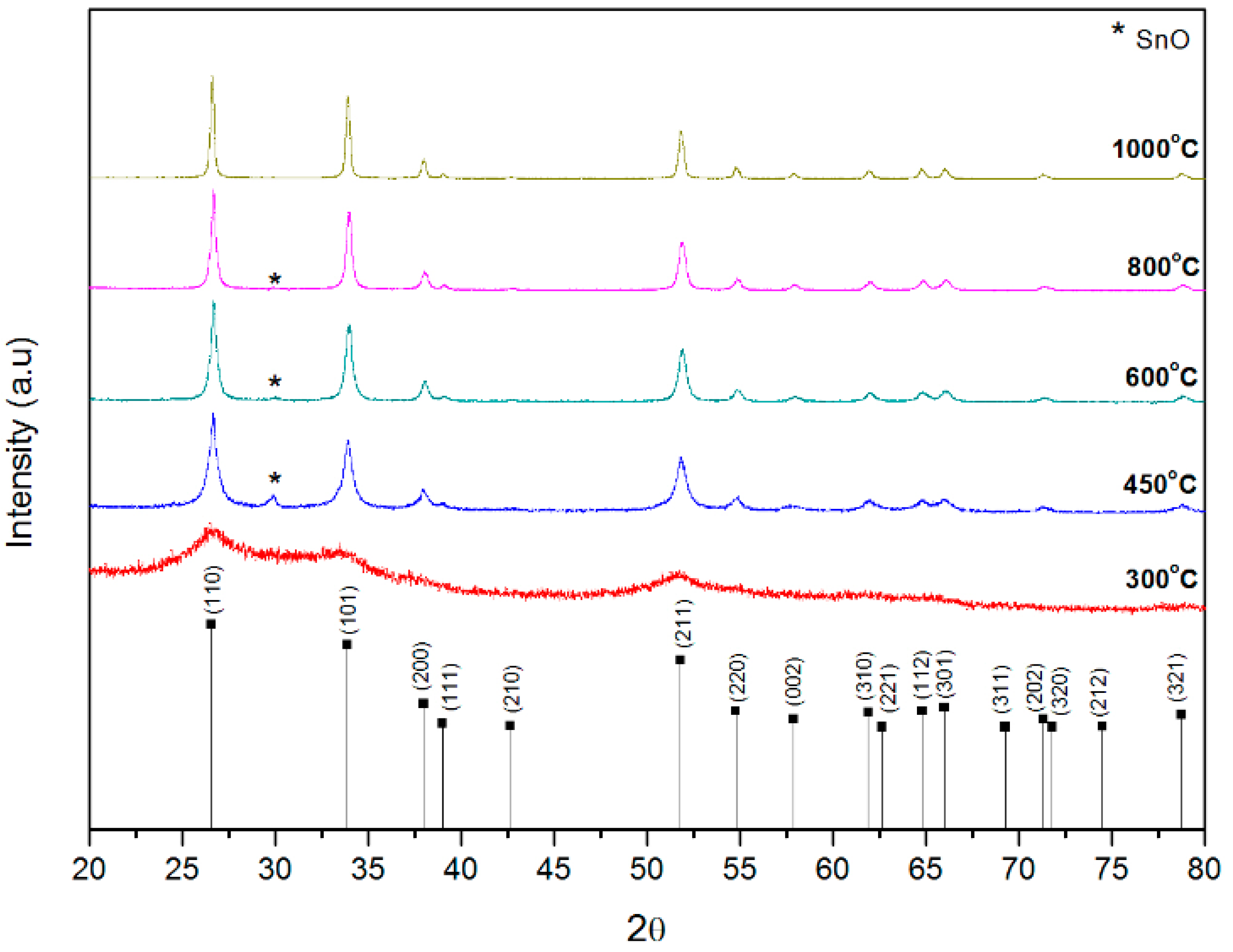
3.1. Structure of SnO2 Particles

Figure 1 presents the X-ray diffraction patterns of SnO2 particles annealed at different temperatures. The XRD pattern at 300 °C shows that the crystallization process has begun, as the presence of SnO2 characteristic peaks is observed. From 450 °C, it is clear that the SnO2 phase is obtained, however, at 29.87° (*) a peak corresponding to a romarchite (tetragonal) structure (SnO JCPDS 96-901-2141) was detected. SnO is a metastable phase due to its oxygen deficiency [34], so it transforms into the more stable SnO2 phase at temperatures above 200 °C [35,36]. The SnO disappeared completely after 800 °C, as shown in Table 1. The results regarding the SnO content in the samples were obtained by X’Pert High Score Plus software (version 4.8) using the XRD patterns. These results suggest that the oxidation of tin atoms was probably incomplete during heat treatment, leading to the coexistence of the second phase (SnO) in the powder [37]. All the other diffraction peaks are in good accordance with the standard sample of cassiterite (tetragonal) SnO2 (JCPDS 96-900-9083). To ensure the purity and presence of single-phase SnO2, a powder treated at 1000 °C was used to prepare the coatings.
The grain sizes were estimated by Scherrer’s equation, as listed in Table 1. The results correspond with those of the study by Agrahari [38], who also observed that the grain size of SnO2 particles increased with increasing calcination temperatures. In the current study, a grain size of 11.5 nm was obtained at 450 °C, increasing up to 25.7 nm at 1000 °C. The calcination temperatures play a significant role in determining the size of the SnO2 nanoparticles synthesized via the sol-gel method [39]. Previous studies indicate that the grain size distribution has an important role in determining the corrosion behaviour of materials. Improved corrosion resistance is exhibited with decreasing grain size in NaCl media [40]. However, in H2SO4 solutions, having a higher grain boundary density accelerates corrosion due to the absence of passive processes, and the corrosion resistance decreases with decreasing grain size. The difference in experimental results in corrosion behaviour can be reasonably explained by the positive or negative effect of high-density grain boundary densities in different corrosive media, so that the nano-crystal deposited coating has a high density of nucleation sites for passive films, leading to a high fraction of passive layers and low corrosion rates [40,41,42].

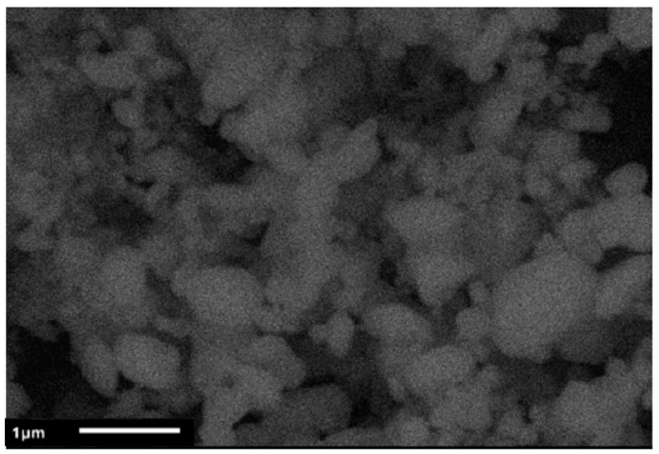
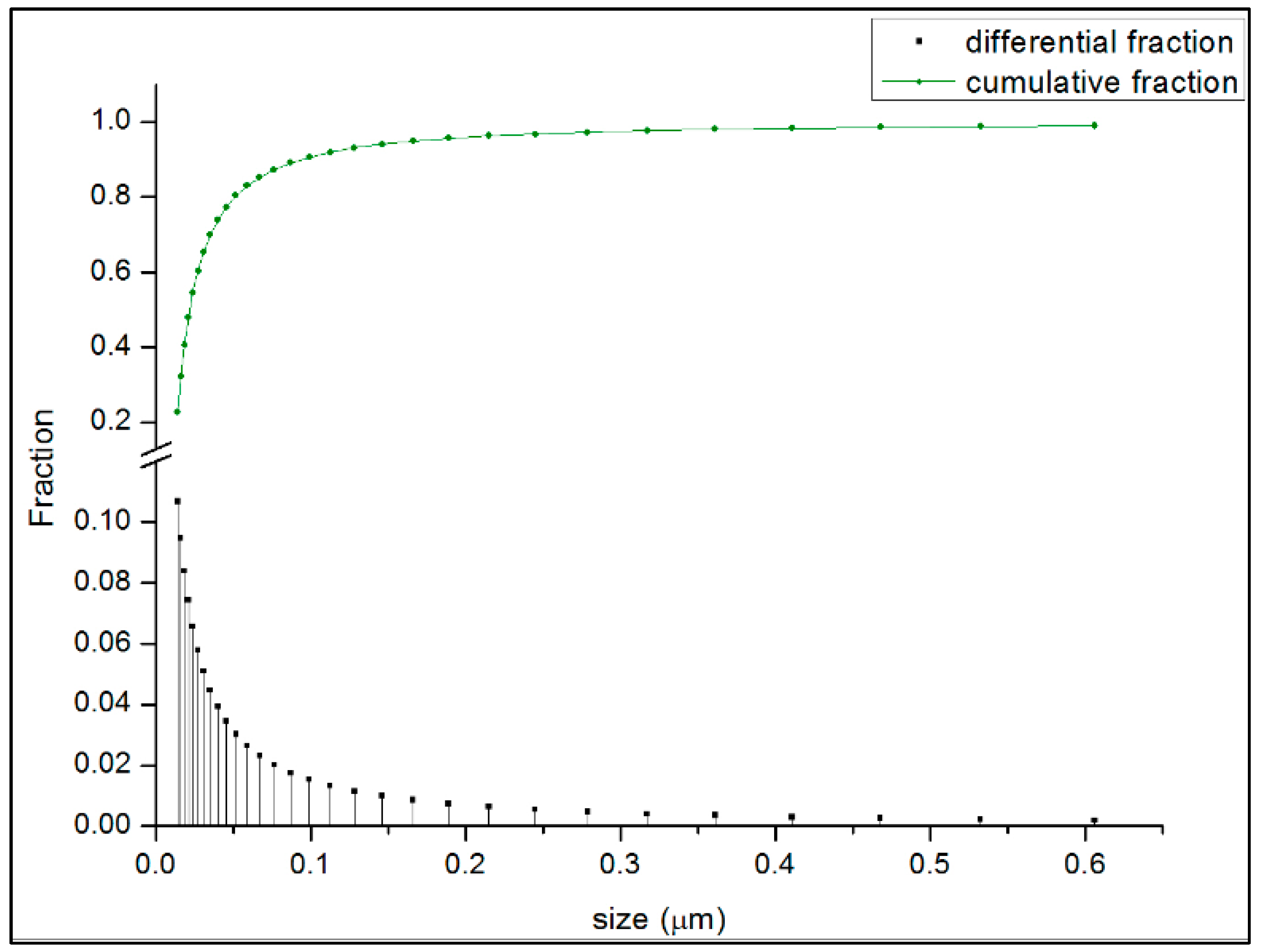
3.2. Morphological Study of SnO2 Particles

According to the XRD results, the grain size increases proportionally with the temperature. Meanwhile, the agglomeration also increases, as has been shown in some previous studies [38,43]. In the SEM micrograph shown in Figure 2, particle agglomeration is clearly observed, with aggregate diameters in the range of 100 to 800 nm. This behaviour occurs because the sol-gel and calcination processes are prone to nanoparticle clustering, resulting in poor dispersion. The use of a solvent is beneficial for the dispersion of nanoparticles [24]. In this work, to determine the particle size, SnO2 was dispersed in ethanol and measurements were obtained in the range of 14 to 69 nm with d50 = 23 nm using the dynamic light scattering (DLS) technique. The distribution of the particle sizes is shown in Figure 3, which presents a mono-modal distribution with a strong tendency toward smaller-sized particles (11–13 nm). On the other hand, with respect to morphology, spherical forms are observed, which is considered an advantage because they have better fluidity and fewer contact interfaces than more irregular particles. When vibrated, smaller particles can occupy vacancies, and thus spherical particles are more easily close-packed [39]. Therefore, if the particles are nanometric in size, it is expected that nucleation occurs and more zones exist that prevent the passage of the current applied to the system for the corrosion test.

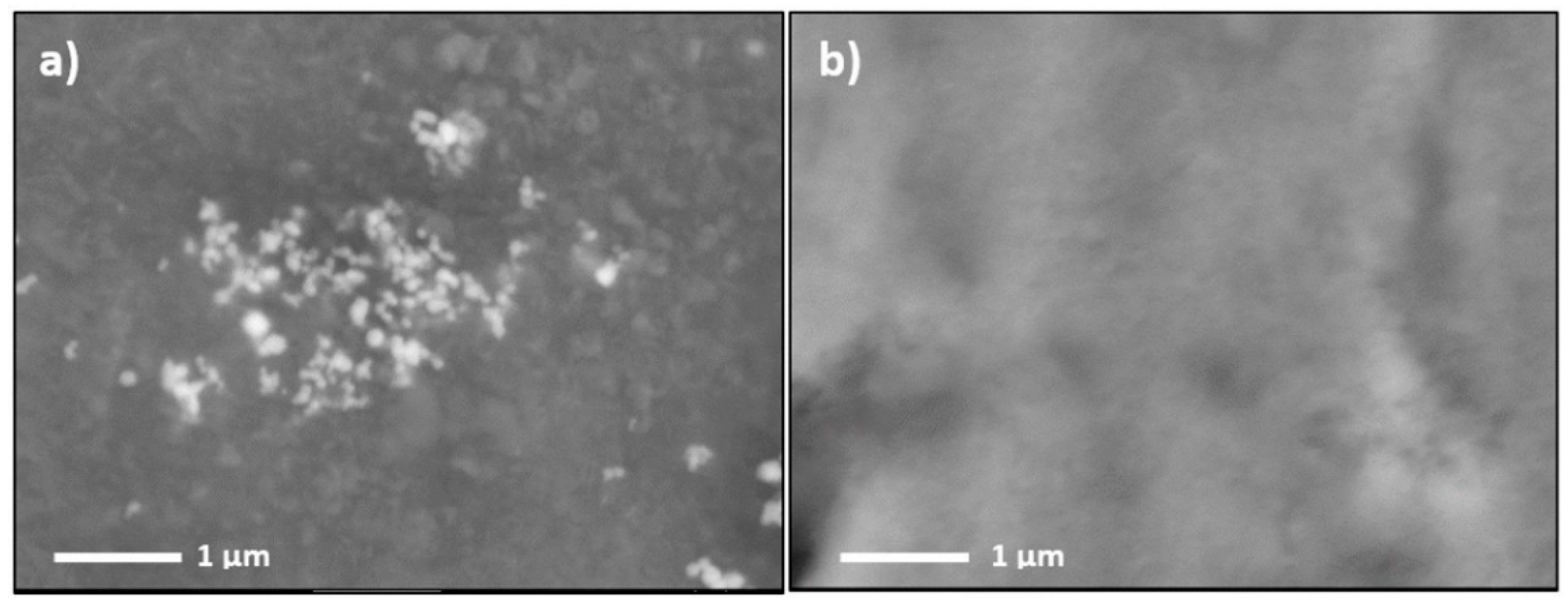
3.3. Surface Morphology of SiO2 Coating with SnO2 Nanoparticles

SEM imaging was conducted to obtain qualitative information about the coating’s surface morphology. Figure 4a shows the image of the SiO2 coating modified with SnO2 nanoparticles. For comparison, the SEM image of an unmodified SiO2 coating (Figure 4b) is also included. The micrographs show that the film is homogeneous, continuous, and free of cracks. The clearer points in Figure 4a correspond to SnO2 nanoparticles.

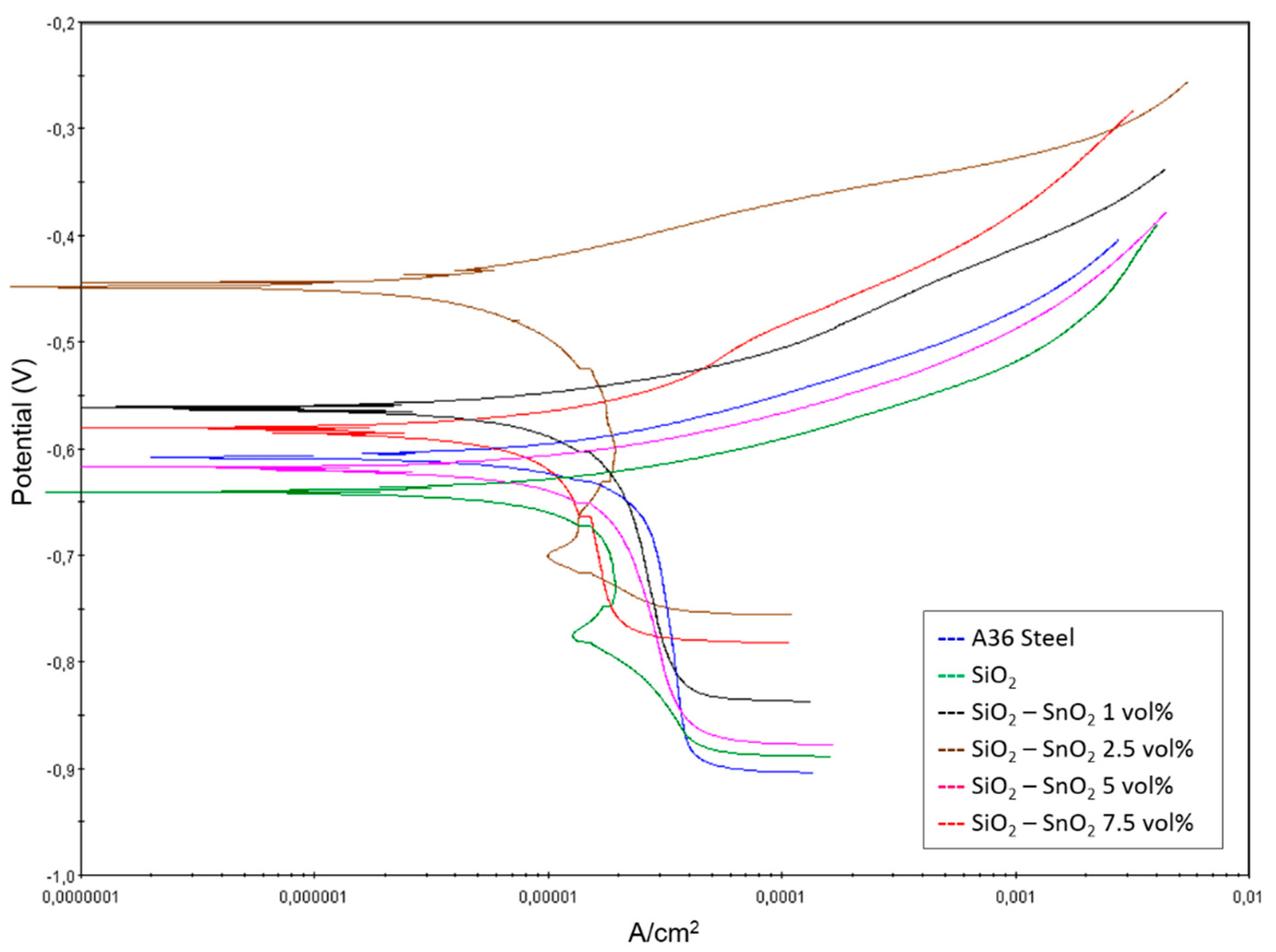
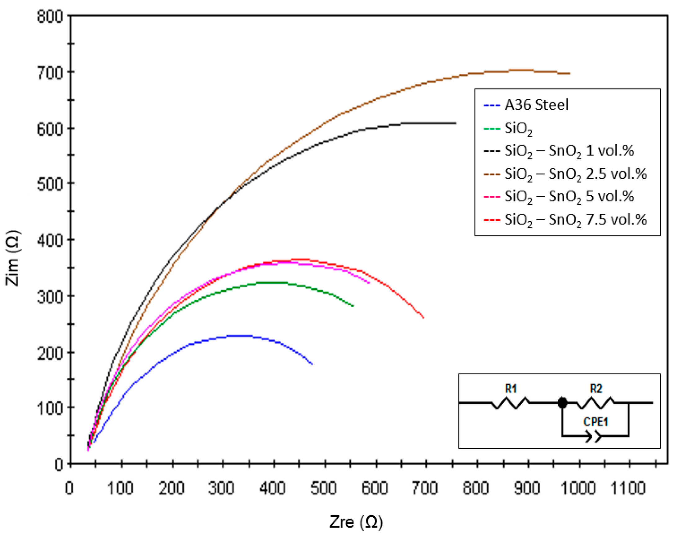
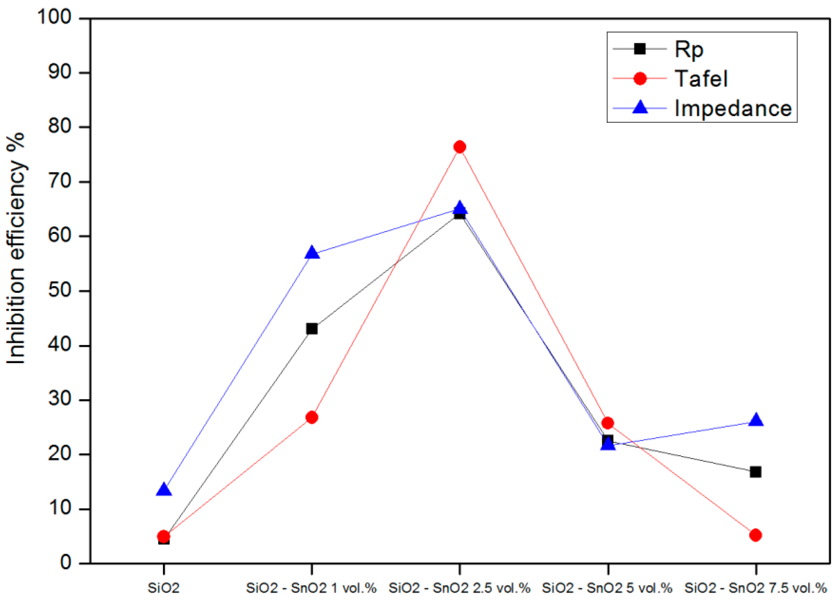
3.4. Corrosion Behaviour of SiO2 Coatings with SnO2 Nanoparticles

The efficiency of the coatings was analysed by different electrochemical tests in a 3 wt % NaCl solution at room temperature. First, the results of polarization resistance for all the samples are presented. In previous research [21,33,44], it was reported that silane coatings provided very good barrier properties and improved corrosion protection compared to bare steel substrates. Previous results also suggested that the coatings provided a protective layer for defects formed in the steel. Increased protection was observed when particles were added to the coating because this promoted self-healing through the partial recovery of protective properties of the coated system when damaged [45]. The most suitable way to evaluate self-healing is by forming an artificial defect on the surface and monitoring the electrochemical behaviour using the polarization resistance technique. Each electrochemical measurement was repeated with three independent samples under the same conditions in order to demonstrate repeatability and reproducibility; therefore, the mean values and standard deviations of the results are reported. Tafel analysis and electrochemical impedance were used to complement the polarization resistance tests and determine the corrosion resistance of the coatings in the presence and absence of SnO2 nanoparticles in the NaCl solution at room temperature.

3.5. Polarization Resistance

Polarization resistance (Rp) is defined as the slope of the potential current density plot at the corrosion potential [24]. Then, Icorr was used to calculate the inhibition efficiency (IE%) using the following relationship, where Io and Ii are the current densities in the absence and presence of the coating, respectively [46].
I E % = I o I i I o × 100
The protective properties are also dependent on the coating composition [47]. According to the corrosion potential data, the bare A36 steel (Ecorr = −702.83 ± 0.17 mV) presented a high tendency to react compared with the coated samples, and a relation was found with the corrosion resistance due to the Icorr values obtained. For the coated samples, the corrosion resistance initially increased up to 64.2% and then decreased as the concentration of SnO2 nanoparticles went up. However, all coated samples presented better corrosion resistance than the uncoated substrate. This effect can be attributed to the fact that when modifying the surface with a coating, the ions of the corrosive solution must first diffuse through this barrier. This behaviour has been observed in similar systems, such as silica coatings on galvanized steel with different concentrations of Ce and CeO2 nanoparticles during polarization in a 3.5% NaCl solution. Ceria nanoparticles have the ability to change the silane solution chemistry, promoting the formation of reactive silanol groups, and of more condensed species in a more stable and protective layer of zinc oxides and hydroxides formed in the alkaline environment [21,25].
For the samples prepared with different amounts of SnO2 particles, better corrosion resistance was obtained at a SnO2 concentration of 2.5 vol % compared with the non-coated substrate. The electrochemical parameters obtained in this analysis and IE% calculated are presented in Table 2.

3.6. Tafel Analysis

The polarization Tafel method was applied to analyse the inhibitor efficiency of the coatings. Figure 5 represents typical anodic and cathodic potentiodynamic polarization curves for carbon steel carried out in the absence and presence of SiO2 coatings with various concentrations of SnO2 particles. The figure shows that in saline solutions, the presence of SnO2 in the coating on the metal’s surface shifts both the anodic and cathodic sections of the Tafel plots to lower values of current density at all investigated SnO2 particle contents. The corrosion current density decreased from 25.43 µA·cm−2 for the bare A36 steel to 6.01 µA·cm−2 for the most effective coating (2.5 vol %). These results indicated that the coatings displayed better barrier properties in the NaCl environment and inhibited the reactions associated with corrosion processes [23]. The same trends found in the resistance polarization test results were also observed in this analysis. In this case, the plots show that the curve corresponding to 2.5 vol % of SnO2 nanoparticles had a smaller slope than for other samples and therefore had a decreased current density.
The electrochemical parameters, including the corrosion potential (Ecorr), current density (Icorr), polarization resistance (Rp), and inhibition efficiency (IE%) are listed in Table 3.

3.7. Impedance

Figure 6 shows Nyquist plots of A36 steel in a 3 wt % NaCl solution modified with SiO2 coatings in the absence and presence of different concentrations of SnO2 nanoparticles. When the steel is immersed in a solution, EIS will detect the NaCl solution and form a resistive solution [46]. The Nyquist plots of all samples show only one capacitive loop in the entire frequency range. According to the EIS results and visual inspection, the plots are composed of large, unfinished semicircular arcs. This shape is attributed to the charge transfer process in the electrode/electrolyte interface. Charge transfer resistance is the resistance offered by the metal atom to become ionized when in contact with the electrolyte (R1), and the coating resistance is denoted by R2. In addition, a constant phase element (CPE1) is used as a substitute for a double-layer capacitor to fit the impedance data of the interface between an electrode and the surrounding electrolyte [40].
Silica coatings improve the corrosion resistance due to the formation of protective oxides that act as a barrier to oxygen diffusion to the metal surface [27]. As shown in Figure 6, the Nyquist plots of the coatings in the NaCl solution exhibit an increasingly large semicircle with additional SnO2 nanoparticles. The corresponding R2 values also increase evidently, however, the same behaviour was obtained, so the test indicates that there is an ideal concentration of SnO2 for corrosion protection.
The R2 value of the 2.5 vol % sample is higher than the R2 value of the bare substrate. Therefore, the curve corresponding to SiO2–2.5 vol % SnO2 should exhibit better corrosion protection performance in the NaCl solution. An equivalent electric circuit is generally used to interpret EIS data. The data of impedance was analysed by ZView software to calculate the equivalent circuit parameter values, which are listed in Table 4. The calculated adjustment values confirm the results obtained by Rp and indicate the positive influence that can be achieved with the precise SnO2 concentration. The simplified equivalent circuit is shown in the inset of Figure 6. The presence of SnO2 particles in the SiO2 coating promotes an increase in the low-frequency impedance, resulting in the inhibition of corrosion efficiency compared to the bare A36.
In the study by Yang [7], a modified sample exhibited high stability in the test solution when the SnO2 was applied as a film on stainless steel, increasing the impedance values with respect to the bare 304 stainless steel. They demonstrated that SnO2 is an effective barrier to inward penetration and that it reduced the corrosion of the substrate [7].
In order to get a better understanding of the corrosion behaviour of the coated samples, the inhibition efficiencies obtained from the three methods were compared (Figure 7). It is clear that for all three electrochemical techniques, the coating efficiency follows the same trend for each concentration compared with the substrate. All the coatings present protective corrosion properties for the A36 steel. Apparently, the coating layer on the surface of the carbon steel prevented iron from reacting with the corrosive media in all of the study cases. The results also indicated that the SiO2 coating material should completely isolate the substrate from the corrosive media so that no electrochemical reactions occur at the coating-substrate interface. From this viewpoint, a good protective coating should be as dense as possible so that the corrosive medium cannot penetrate through it. With this basic knowledge, we can conclude that the poor anticorrosion ability in some coatings may have been due to the presence of pores which allowed the corrosive media to penetrate through.
On the other hand, it is known that the electrode potential of Fe is more negative than that of SnO2. The nanoscale pores in the coating allow seawater to penetrate through, leading to severe corrosion at the coating-substrate interface via the formation of an SnO2–Fe galvanic cell. A possible mechanism for the above significant improvement in anticorrosion ability of the composite coating might be related to the satisfactory sealing of the pores in the coating by SnO2 nanoparticles, as suggested by Dongyun Yu in previous work using TiO2 and/or ZnO nanoparticles in composite coatings for improved corrosion protection on carbon steel [15].
In addition, it is demonstrated here that the optimal efficiency occurred at an SnO2 nanoparticle concentration of 2.5 wt %. When the concentration increases or decreases about this value, the IE% decreased as well. Although the value of the inhibitor efficiency depends on the electrochemical test used, it is clear that the addition of SnO2 particles had a beneficial effect on the corrosion protection of A36 steel.

4. Conclusions

In order to improve its corrosion resistance, carbon steel was successfully coated with SiO2 by the sol-gel method. It was found that adding SnO2 nanoparticles to the coating had a great influence on the sol-gel film formation and thus on the stability of the coated substrates in corrosive media. The investigation showed that the incorporation of tin oxide (IV) in sol-gel SiO2 coatings helped protect A36 steel from corrosion. All of the coatings tested provided a barrier that inhibited corrosion. Very low and very high SnO2 concentrations led to a reduction in inhibition, and the optimum concentration of SnO2 nanoparticles was found to be 2.5 vol %.

Author Contributions

A.K.A.-G., A.G.M.-R. and J.M.-P. conceived of the presented idea; A.K.A.-G. developed the theory and methodology; Á.d.J.M.-R., J.M.-P., J.G.G.-S. and A.L.P.-F. verified the analytical methods and supervised the findings of this work. All authors discussed the results and contributed to the final manuscript. All authors have read and agreed to the published version of the manuscript.

Funding

This research was funded by Instituto Politécnico Nacional.

Acknowledgments

The authors would like to acknowledge the facilities granted in the use of the LIDTRA and LICAMM infrastructure, as well as the technical assistance of Daniela Moncada and Adair Jimenez.

Conflicts of Interest

The authors declare no conflict of interest.

References

  1. Li, M.; Jin, Z.-X.; Zhang, W.; Cao, Y.-Q.; Li, W.-M.; Wu, D.; Li, A.-D. Comparison of chemical stability and corrosion resistance of group IV metal oxide films formed by thermal and plasma-enhanced atomic layer deposition. Sci. Rep. 2019, 9, 10438–10449. [Google Scholar] [CrossRef] [PubMed] [Green Version]
  2. Gregory, O.J.; Luo, Q.; Crisman, E.E. High temperature stability of indium tin oxide thin films. Thin Solid Film 2002, 406, 286–293. [Google Scholar] [CrossRef]
  3. Curkovic, L.; Curkovic, H.O.; Salopek, S.; Renjo, M.M.; Segota, S. Enhancement of corrosion protection of AISI 304 stainless steel by nanostructured sol-gel TiO2 films. Corros. Sci. 2013, 77, 176–184. [Google Scholar] [CrossRef] [Green Version]
  4. Li, S.; Wang, Q.; Chen, T.; Zhou, Z.; Wang, Y.; Fu, J. Study on cerium-doped nano-TiO2 coatings for corrosion protection of 316 L stainless Steel. Nanoscale Res. Lett. 2012, 7, 1–9. [Google Scholar] [CrossRef] [Green Version]
  5. Xu, H.; Liu, W.; Cao, L.; Su, G.; Duan, R. Preparation of porous TiO2/ZnO composite film and its photocathodic protection properties for 304 stainless Steel. Appl. Surf. Sci. 2014, 301, 508–514. [Google Scholar] [CrossRef]
  6. Subasri, R.; Shinohara, T. The applicability of SnO2 coatings for corrosion protection of metals. Electrochem. Solid State Lett. 2004, 7, B17–B20. [Google Scholar] [CrossRef]
  7. Yang, L.; Qin, Z.; Pan, H.; Yun, H.; Min, Y.; Xu, Q. Corrosion protection of 304 stainless steel bipolar plates of PEMFC by coatung SnO2 film. Electrochem. Sci. 2017, 12, 10946–10957. [Google Scholar] [CrossRef]
  8. Atik, M.; De Lima Neto, P.; Avaca, L.A.; Aegerter, M.A.; Zarzycki, J. Protection of 316L stainless steel against corrosion by SiO2 coatings. Mater. Sci. Lett. 1994, 13, 1081–1085. [Google Scholar] [CrossRef]
  9. Nouri, E.; Shahmiri, M.; Rezaie, H.R.; Talayian, F. Investigation of structural evolution and electrochemical behaviour of zirconia thin films on the 316L stainless steel substrate formed via sol-gel process. Surf. Coat. Technol. 2011, 205, 5109–5115. [Google Scholar] [CrossRef]
  10. Norouzi, M.; Garekani, A.A. Corrosion protection by zirconia-based thin films deposited by a sol-gel spin coating method. Ceram. Int. 2014, 40, 2857–2861. [Google Scholar] [CrossRef]
  11. Chang, S.-H.; Chen, J.-Z.; Hsiao, S.-H.; Lin, G.-W. Nanohardness, corrosion and protein adsorption properties of CuAlO2 films deposited on 316L stainless steel for biomedical applications. Appl. Surf. Sci. 2014, 289, 455–461. [Google Scholar] [CrossRef]
  12. Magaña, C.R.; Ángeles, M.E.; Rodríguez, F.J. Comportamiento de películas delgadas de níquel y óxido de níquel en NaCl al 3%. Revista de Metalurgia 2006, 1, 49–55. [Google Scholar]
  13. Dhoke, S.K.; Khanna, A.; Sinha, T.J.M. Effect of nano-ZnO particles on the corrosion behaviorof alkyd-based waterborne coatings. Prog. Org. Coat. 2009, 64, 371–382. [Google Scholar] [CrossRef]
  14. Li, H.; Liang, K.; Mei, L.; Gu, S.; Wang, S. Corrosion protection of mild steel by zirconia sol-gel coatings. Mater. Sci. Lett. 2001, 20, 1081–1083. [Google Scholar] [CrossRef]
  15. Yu, D.; Tian, J.; Dai, J.; Wang, X. Corrosion resistance of three-layer superhydrophobic composite coating on carbon steel in seawater. Electrochim. Acta 2013, 97, 409–419. [Google Scholar] [CrossRef]
  16. I-Lateef, H.M.A.E.; Khalaf, M.M. Corrosion resistance of ZrO2-TiO2 nanocomposite multilayer thin films coated on carbon steel in hydrochloric acid solution. Mater. Charact. 2015, 108, 29–41. [Google Scholar] [CrossRef]
  17. Zheludkecich, M.L.; Salavado, I.M.; Ferreira, M.G.S. Sol-gel coatings for corrosion protection of metals. J. Mater. Chem. 2005, 15, 5099–5111. [Google Scholar] [CrossRef]
  18. Li, Q. Sol-gel coatings to improve the corrosion resistance of magnesium (Mg) alloys. In Corrosion Prevention of Magnesium Alloys, 1st ed.; Woodhead Publishing: Cambridge, UK, 2013; pp. 469–485. [Google Scholar]
  19. Wang, D.; Bierwagen, G.P. Sol-gel coatings on metals for corrosion protection. Prog. Org. Coat. 2009, 64, 327–338. [Google Scholar] [CrossRef]
  20. Stambolova, I.; Yordanov, S.; Lakov, L.; Vassilev, S.; Blaskov, V.; Jivov, B. Preparation of sol-gel SiO2 coatings on steel and their corrosion resistance. In National Congress on Theoretical and Applied Mechanics 2017; Institute of Mechanics: Sofia, Bulgaria, 2018. [Google Scholar]
  21. Montemor, M.; Pinto, R.; Ferreira, M.G.S. Chemmical composition and corrosion protection of silane films modified with CeO2 nanoparticles. Electrochim. Acta 2009, 54, 5179–5189. [Google Scholar] [CrossRef]
  22. Venila, R.; Kamaraj, P.; Arthanareeswari, M.; Devikala, S. Surface modification of mild steel using Ag doped SnO2 nanoparticles for corrosion inhibition. Int. J. Adv. Chem. Sci. Appl. 2014, 1, 16–20. [Google Scholar]
  23. Deyab, M.A.; Keera, S.T. Effect of nano- TiO2 particles size on the corrosion resistance of alkyd coating. Mater. Chem. Phys. 2014, 146, 406–411. [Google Scholar] [CrossRef]
  24. Shi, X.; Nguyen, T.A.; Suo, Z.; Liu, Y.; Avci, R.A. Effect of nanoparticles on the anticorrosion and mechanical properties of epoxy coating. Surf. Coat. Technol. 2009, 204, 237–245. [Google Scholar] [CrossRef]
  25. Zand, R.Z.; Verbeken, K.; Adriaens, A. Influence of the cerium concentration on the corrosion performance of Ce-doped silica hybrid coatings on hot dip galvanized steel substrates. Int. J. Electrochem. Sci. 2013, 8, 548–563. [Google Scholar]
  26. Garcia-Heras, M.; Jimenez-Morales, A.; Casal, B.; Galvan, J.C.; Radzki, S.; Villegas, M.A. Preparation and electrochemical study of cerium–silica sol–gel thin films. Alloy Compd. 2004, 380, 219–224. [Google Scholar] [CrossRef] [Green Version]
  27. Hamdy, A.S.; Butt, D.P.; Ismail, A.A. Electrochemical impedance studies of sol–gel based ceramic coatings systems in 3.5% NaCl solution. Electrochim. Acta 2007, 52, 3310–3316. [Google Scholar] [CrossRef]
  28. Liu, T.J.; Jin, Z.G.; Feng, L.R.; Wang, T. Conducting antimony-doped tin oxide films derived from stannous oxalate by aqueous sol-gel method. Appl. Surf. Sci. 2008, 254, 6547–6553. [Google Scholar] [CrossRef]
  29. Benrabah, B.; Bouaza, A.; Kadari, A.; Maaref, M.A. Impedance studies of Sb doped SnO2 thin film prepared by sol-gel process. Superlattices Microstruct. 2011, 50, 591–600. [Google Scholar] [CrossRef]
  30. Dong, C.; Liu, X.; Xiao, X.; Chen, G.; Wang, Y.; Djerdj, I. Combustion synthesis of porous Pt-functionalized SnO2 sheets for isopropanol gas detection with a significant enhancement in response. Mater. Chem. A 2014, 2, 20089–20095. [Google Scholar] [CrossRef]
  31. Andersen, S.M.; Nørgaard, C.F.; Larsen, M.J.; Skou, E. Tin Dioxide as an effective antioxidant for proton exchange membrane fuel cells. J. Power Sources 2015, 273, 158–161. [Google Scholar] [CrossRef]
  32. Crespo, M.D.; Meneses, E.R.; Huerta, A.T.; Murillo, A.G.; Estrada, E.M.A.; Hernández, O.C. Synthesis by Sol-gel route and characterization of ceria doped silica coatings on commercial carbon steel. Portugaliae Electrochim. Acta 2009, 3, 257–267. [Google Scholar] [CrossRef]
  33. Wang, H.; Huang, S.L.; Zuo, Y.J.; Zhou, T.; Zhang, L.R. Corrosion resistance of lamellar aluminium pigments coated by SiO2 by sol–gel method. Corros. Sci. 2011, 53, 161–167. [Google Scholar] [CrossRef]
  34. Giefers, H.; Porsch, F.; Wortmann, G. Kinetics of the Disproportionation of son. Solid State Ion. 2005, 176, 199–207. [Google Scholar] [CrossRef]
  35. Dai, Z.R.; Pan, Z.W.; Wang, Z.L. Growth and structure evolution of novel Tin Oxide Diskettes. J. Am. Chem. Soc. 2002, 124, 8673–8680. [Google Scholar] [CrossRef] [PubMed]
  36. Zhang, F.; Lian, Y.; Gu, M.; Yu, J.; Tang, T.B. Static and dynamic disorder in metastable phases of Tin Oxide. J. Phys. Chem. 2017, 121, 16006–16011. [Google Scholar] [CrossRef]
  37. Akgul, F.A.; Gumus, C.; Er, A.O.; Farha, A.H.; Akgul, G.; Ufuktepe, Y.; Liu, Z. Structural and electronic properties of SnO2. J. Alloy Compd. 2013, 579, 50–56. [Google Scholar] [CrossRef]
  38. Agrahari, V.; Mathpal, M.C.; Kumar, M.; Agarwal, A. Investigations of optoelctronic properties in DMS SnO2 nanoparticles. J. Alloy Compd. 2015, 622, 48–53. [Google Scholar] [CrossRef]
  39. Aziz, M.; Abbas, S.S.; Baharom, W.R.W. Size-controlled synthesis of SnO2 nanoparticles by sol-gel method. Mater. Lett. 2013, 91, 31–34. [Google Scholar] [CrossRef]
  40. Qin, L.Y.; Lian, J.S.; Jiang, Q. Effect of grain size on corrosion behavior of electrodeposited bulk nanocrystalline Ni. Trans. Nonferrous Met. Soc. China 2010, 20, 82–89. [Google Scholar] [CrossRef]
  41. Gollapudi, S. Grain size distribution effects on the corrosion behaviour of materials. Corros. Sci. 2012, 62, 90–94. [Google Scholar] [CrossRef]
  42. Afshari, V.; Dehghanian, C. Effects of grain size on the electrochemical corrosion behaviourof electrodeposited nanocrystalline Fe coatings in alkaline solution. Corros. Sci. 2009, 51, 1844–1849. [Google Scholar] [CrossRef]
  43. Martinez, J.A.; Esneider-Alcala, M.; Hernández, M.; Canul, M.P.; Shaji, S. Optimal parameters for synthesizing single phase spinel-type Co2SnO4 by sol–gel technique: Structure determination and microstructure evolution. J. Alloy Compd. 2013, 574, 278–282. [Google Scholar] [CrossRef]
  44. Wang, J.; Wu, L.K.; Zhou, J.H.; Hu, J.M.; Zhang, J.Q.; Cao, C.N. Construction of a novel painting system using electrodeposited SiO2 film as the pretreatment layer. Corros. Sci. 2013, 68, 57–65. [Google Scholar] [CrossRef]
  45. Rosero-Navarro, N.C.; Pellice, S.A.; Durán, A.; Aparicio, M. Effects of Ce-containing sol–gel coatings reinforced with SiO2 nanoparticles on the protection of AA2024. Corros. Sci. 2008, 50, 1283–1291. [Google Scholar] [CrossRef]
  46. Pradityana, A.; Shahab, A.; Noerochim, L.; Susanti, D. Inhibition of corrosion of carbon steel in 3.5% NaCl solution by Myrmecodia Pendans extract. Int. J. Corros. 2016. [Google Scholar] [CrossRef] [Green Version]
  47. Feil, F.; Furberth, W.; Schutze, M. Nanoparticle based inorganic coatings for corrosion protection of magnesium alloys. Surf. Eng. 2008, 24, 198–204. [Google Scholar] [CrossRef]
Figure 1. X-ray diffraction patterns for samples of tin oxide (SnO2) annealed at 300, 450, 600, 800, and 1000 °C for 1 h.
Coatings 10 00385 g001
Figure 2. SEM image of tin oxide (SnO2) nanoparticles annealed at 1000 °C.
Coatings 10 00385 g002
Figure 3. Particle size distribution obtained by dynamic light scattering.
Coatings 10 00385 g003
Figure 4. SEM images of SiO2 coatings (a) with SnO2 nanoparticles and (b) absence of SnO2 nanoparticles.
Coatings 10 00385 g004
Figure 5. Polarization curves obtained in a 3 wt % NaCl solution for A36 steel with SiO2 coatings in the absence and presence of SnO2 nanoparticles.
Coatings 10 00385 g005
Figure 6. Nyquist plots of the composite coatings in the absence and presence of different concentrations of SnO2 particles from EIS measurements in NaCl media.
Coatings 10 00385 g006
Figure 7. Comparison of IE% plots produced from the results for each corrosion test.
Coatings 10 00385 g007
Table 1. Grain sizes of SnO2 particles.
SamplesGrain Size (nm)% SnO
SnO2–450 °C11.5 ± 1.0210
SnO2–600 °C15.8 ± 2.025
SnO2–800 °C18.9 ± 2.201
SnO2–1000 °C25.7 ± 3.170
Table 2. The corrosion parameters obtained from polarization resistance measurements.
SnO2 Concentration (vol %)Ecorr (mV)Icorr (µA·cm−2)Rp (Ω)IE%
A36 Steel (Bare)−702.83 ± 2.0924.24 ± 0.17896.66 ± 6.36-
0−639.52 ± 3.0123.15 ± 0.36939.38 ± 14.494.5
1−597.81 ± 0.7613.82 ± 0.221572.54 ± 25.0943
2.5−521.20 ± 8.668.68 ± 0.352507.92 ± 102.2464.2
5−636.78 ± 0.4218.79 ± 0.191156.73 ± 11.6122.5
7−656.46 ± 2.0620.16 ± 0.021078.06 ± 1.2816.8
Table 3. The corrosion parameters obtained from the polarization Tafel method.
SnO2 Concentration (vol %)Ecorr (mV)Icorr (µA cm−2)IE%
A36 Steel (Bare)−606.6425.43-
0−640.2424.184.9
1−561.5218.6226.8
2.5−447.496.0176.4
5−619.7218.9025.7
7−518.2224.105.2
Table 4. The corrosion parameters obtained from EIS.
SnO2 Concentration (vol %)R1 (Ω)R2 (Ω)CPE1 (µF/cm2)IE%
A36 Steel (bare)28.36675.50.0012-
029.72779.90.001313.4
127.715630.001156.8
2.530.7819350.000865.1
529.42862.90.001321.7
729.17913.90.000926.1

