Article

Work Function Tuning of Zinc–Tin Oxide Thin Films Using High-Density O2 Plasma Treatment

1
School of Electrical and Electronics Engineering, Chung-Ang University, Seoul 06974, Korea
2
Electronics and Telecommunications Research Institute (ETRI), Daejeon 34129, Korea
3
Department of Advanced Device Technology, Korea University of Science and Technology (UST), Daejeon 34113, Korea
4
School of Materials Science and Engineering, Georgia Institute of Technology (GIT), Atlanta, GA 30332, USA
5
Department of Electrical Engineering, Sejong University, Seoul 05006, Korea
*
Authors to whom correspondence should be addressed.
Coatings 2020, 10(11), 1026; https://doi.org/10.3390/coatings10111026
Submission received: 15 September 2020 / Revised: 10 October 2020 / Accepted: 20 October 2020 / Published: 25 October 2020
(This article belongs to the Special Issue Plasma Surface Engineering)

Abstract

:
Work function tuning has a significant influence on the performance of semiconductor devices, owing to the formation of potential barriers at the interface between metal-semiconductor junctions. In this work, we introduce a technique for tuning the work function of ZnSnO thin films using high-density O2 plasma treatment. The work function and chemical composition of the ZnSnO thin film surfaces were investigated with regards to plasma treatment time through UPS/XPS systems. The optical band gap was estimated using Tauc’s relationship from transmittance data. The work function of Zn0.6Sn0.4O thin film increased from 4.16 eV to 4.64 eV, and the optical band gap increased from 3.17 to 3.23 eV. The surface of Zn0.6Sn0.4O thin films showed a smooth morphology with an average of 0.65 nm after O2 plasma treatment. The O2 plasma treatment technique exhibits significant potential for application in high-performance displays in optical devices, such as thin-film transistors (TFTs), light-emitting diodes (LEDs), and solar cells.

1. Introduction

Particular attention needs to be focused on the device-structure design to improve the efficiencies of electrical and optical devices. Notably, the work function of thin films is directly related to the performances of these devices [1,2]. For example, the difference in the work function between the metal-semiconductor junction at the source (or drain) leads to an increase in contact resistance, resulting in performance degradation of the device [3,4]. Meanwhile, the operation of Schottky diodes, metal–semiconductor–metal photodetectors, and Schottky junction solar cells is based on the difference in work function between the metal-semiconductor junctions [5,6,7]. It is evident that the characteristics of the electrical and optical devices are closely related to the work function of the materials [8]. Therefore, considering work functions while designing devices is crucial to improve performance. Kang et al. reported an increase in carrier mobility of InGaZnO thin films through Ar plasma treatment [9]. Furthermore, Fang et al. proposed tuning the work function of the InSnO (ITO) thin film through Ar plasma treatment by employing a capacitively coupled plasma system [10]. Plasma treatment induces oxygen vacancy on the thin film surface and modulates the Sn doping concentration, resulting in the tuning of the work function and improvement of the carrier mobility [11,12].
Zinc–tin oxide (Zn1−xSnxO) thin films are transparent oxide semiconductors with wide bandgaps (Eg ~ 3.6 eV) and have been studied as potential materials for channel layers in thin-film transistors (TFTs) [13,14], emitters for photovoltaic devices [15], as well as transparent conducting electrodes [16,17]. ZnSnO TFTs exhibit lower carrier mobility than InGaZnO or InZnSnO TFTs, and ZnSnO as transparent electrodes exhibit lower performance than ITO-based electrodes. However, they boast low manufacturing cost and wide functionality as indium-free oxide-based thin films [18,19,20].
In this work, we present a technique for tuning the work function and electrical properties of ZnSnO thin films through high-density O2 plasma treatment in an inductively coupled plasma (ICP) system. Depending on the O2 plasma treatment time, the work function changed from 4.16 to a maximum value of 4.64 eV, while the optical band gap remained steady. Moreover, while the electrical properties were altered during the plasma treatment, there was no significant change observed in surface roughness.

2. Materials and Methods

A 100 nm Zn0.6Sn0.4O thin film was deposited on soda-lime glass (SLG) by an RF co-sputtering system (SNETEK Co., LTD., Suwon, Korea) with the pure ZnO (99.99%) and Sn (99.99%) metal targets under 100 and 40 W RF power, respectively. The substrate temperature was 200 °C, while an Ar/O2 mixed gas (90/10%) was injected with a total gas flow rate of 100 sccm. The base and working pressures were maintained at 5.0 × 10⁻⁷ Torr and 20 mTorr, respectively. The Sn/(Sn + Zn) ratio was controlled by varying the oxygen partial pressure with fixed sputter power. The deposited-Zn0.6Sn0.4O (60%Zn:40%Sn atomic ratio) thin films were treated with O2 plasma in the inductively coupled plasma (ICP) system (Vacuum Science, Yangju, Korea) for tuning the work function [21]. During O2 plasma treatment, the total flow rate of the O2 gas was fixed at 20 sccm. The RF power, DC bias voltage, process pressure, and substrate temperature were fixed at 500 W, −200 V, 15 mTorr and room temperature, respectively. After O2 plasma treatment, the surface chemical state and the work function of the Zn0.6Sn0.4O thin films were investigated using X-ray/ultraviolet photoelectron spectroscopy (XPS/UPS). The XPS (K-alpha, Thermo VG, UK) measurements employed a monochromatic Al Kα source (1486.6 eV). The X-ray power, current, and base pressure were 12 kV, 3 mA, and 2.2 × 10⁻⁷ Torr, respectively. Here, binding energies were referenced to the neutral C 1s peak at 284.6 eV. The UPS (AXIS Ultra DLD, Kratos, Inc., Manchester, UK) measurements were carried out using an He I (21.2 eV) gas discharge lamp. The base pressure and dwell time were 4.0 × 10⁻⁸ Torr and 100 ms, respectively. A UV-Vis-NIR spectrophotometer (Perkin elmer, Waltham, MA, US) was used to measure the transmittance and optical band gap (Eg) of the Zn0.6Sn0.4O thin films. A Hall measurement system (Accent Optical Technologies, Bend, OR, US)) was used to measure the resistance, mobility, and carrier concentration with van der Pauw geometry. Atomic force microscopy (AFM) (XE-100, PSIA Inc., Suwon, Korea) was used to determine surface morphology. Topographic images were taken in non-contact mode with X-Y scanner and Z scanner of 1 × 1 μm2 and 2 nm, respectively.

3. Results and Discussion

The changes in the work function according to the O2 plasma treatment time were studied through UPS measurements. Figure 1 shows the UPS spectra of the Zn0.6Sn0.4O thin films, depending on the O2 plasma treatment time. The binding energy cutoff for secondary electrons and the near-Fermi edge on the Zn0.6Sn0.4O thin films are expressed in Figure 1a,b, respectively. The work function (ø) was calculated using the following equation:
ø = hvEB (secondary cutoff)
where hv is the photon energy (He I, 21.2 eV) and EB is the binding energy. The as-deposited Zn0.6Sn0.4O thin film exhibited a work function of 4.16 eV, whereas the Zn0.6Sn0.4O thin films treated with O2 plasma for 60, 90, 120 and 150 s displayed work functions of 4.35, 4.64, 4.38, and 4.56 eV, respectively. As the O2 plasma treatment time increased, the binding energy of the valence structure at the surface of Zn0.6Sn0.4O thin films changed, owing to the change in the ionization potential. In other words, during plasma treatment, the SnO component is easily etched away, and oxygen penetrates into the oxygen vacancy on the surface, resulting in an increase in the oxygen concentration on the Zn0.6Sn0.4O surface. This results in a change in the work function and valence band offset (VBO) [22]. Furthermore, the change in the work function could be ascribed to the difference in the chemical state or elemental ratio [23,24]. The irregular trend in the work function for O2 plasma treatment time between 90 and 120 s can be attributed to the difference in the heat of formation of ZnO (−348.0), SnO (−285), and SnO2 (−577.63). In other words, up to 90 s of O2 plasma treatment affects the SnO component while ZnO is mainly affected between 90 and 120 s [25]. Therefore, it was observed that the work function of Zn0.6Sn0.4O thin films with O2 plasma treatment for 90 s is exceptionally higher than the reference values.
The work function of semiconducting thin films is related to their band gap. We calculated the optical band gap of each Zn0.6Sn0.4O thin film from transmission spectra using, Tauc’s relation,
(αhv)n = B(hvEg)
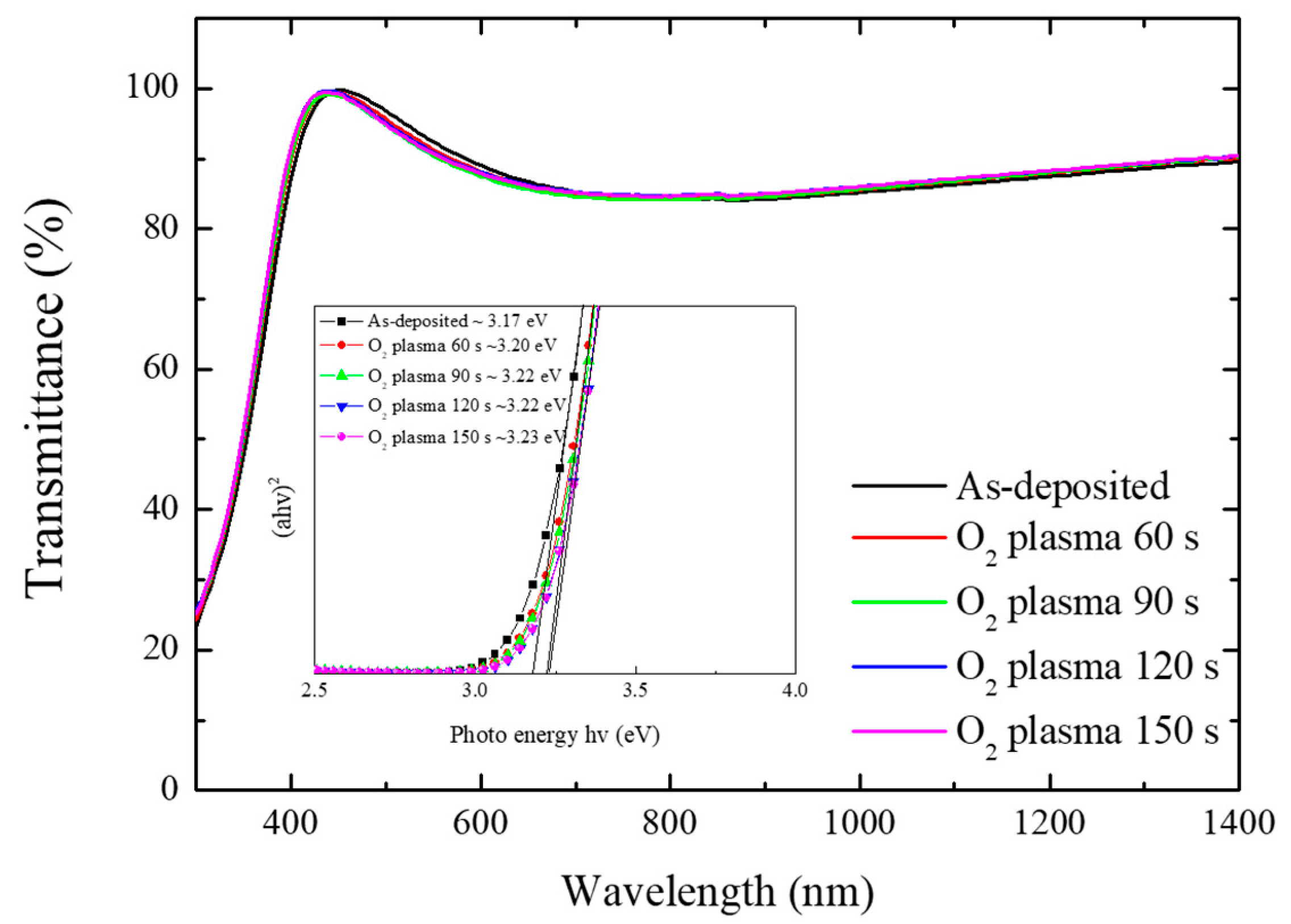
where α, B, and Eg are the absorption coefficient, the band tailing parameter, and the optical band gap, respectively. Figure 2 presents the transmission spectra of Zn0.6Sn0.4O thin films depending on the O2 plasma treatment time. The average transmittance of O2 plasma-treated Zn0.6Sn0.4O thin films was 92% in the visible region (from 400 to 700 nm). The optical band-gap of the Zn0.6Sn0.4O thin films was calculated to be 3.17 + Δ0.06 eV, based on transmittance data from the Tauc plot (inset image of Figure 2). The band gap was relatively steady with O2 plasma treatment, while the work function changes were significant.
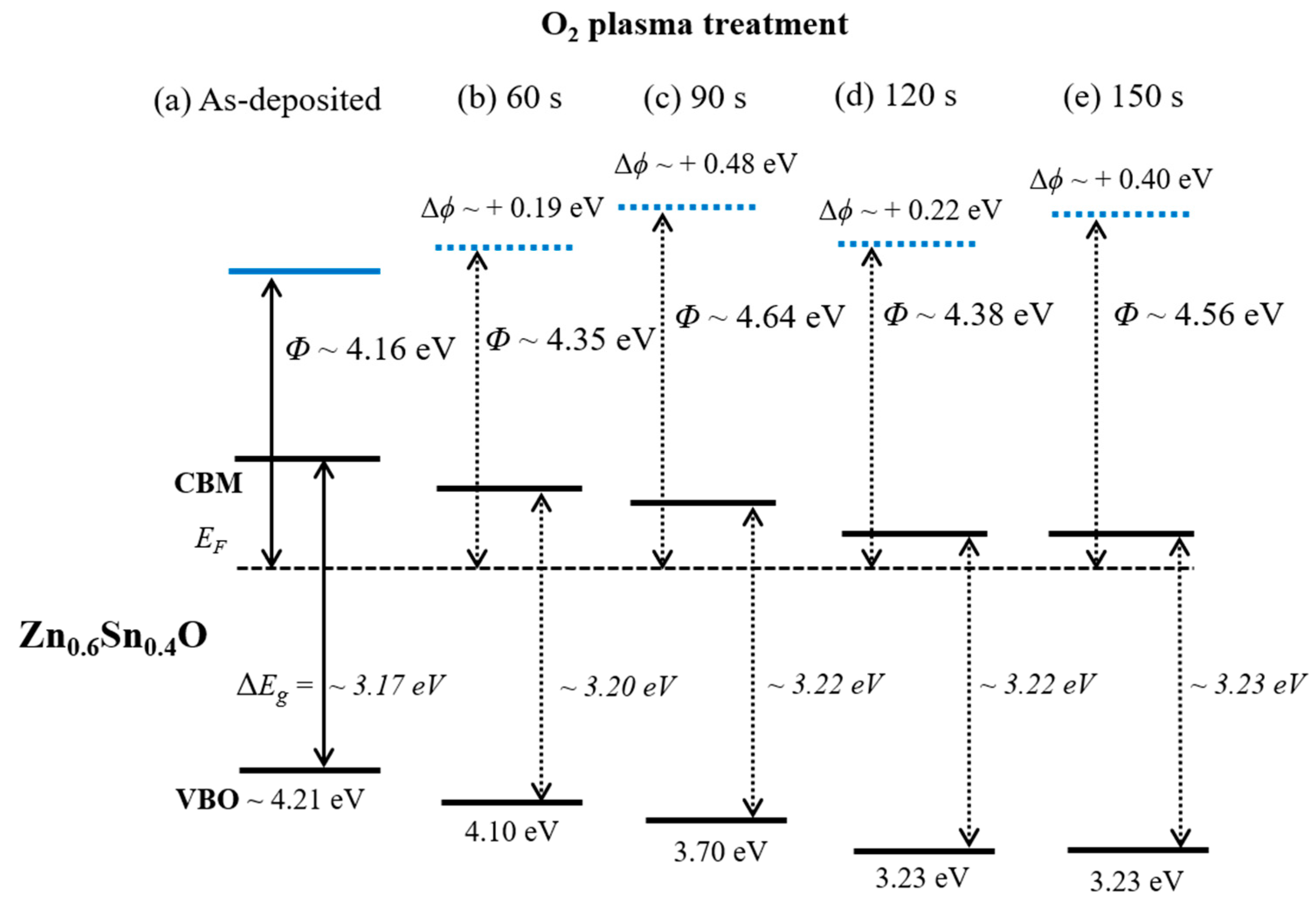
Figure 3 shows a schematic of a modified Zn0.6Sn0.4O band position, including work function and band gaps of the O2 plasma-treated materials. The substituted metal in the ZnO results regarding the movement of the Fermi level toward the conduction band edge with the work function changed [26]. Therefore, the Zn0.6Sn0.4O thin film with the O2 plasma treatment is comparable with other films for potential applications in high-performance devices, as shown in Figure 3.
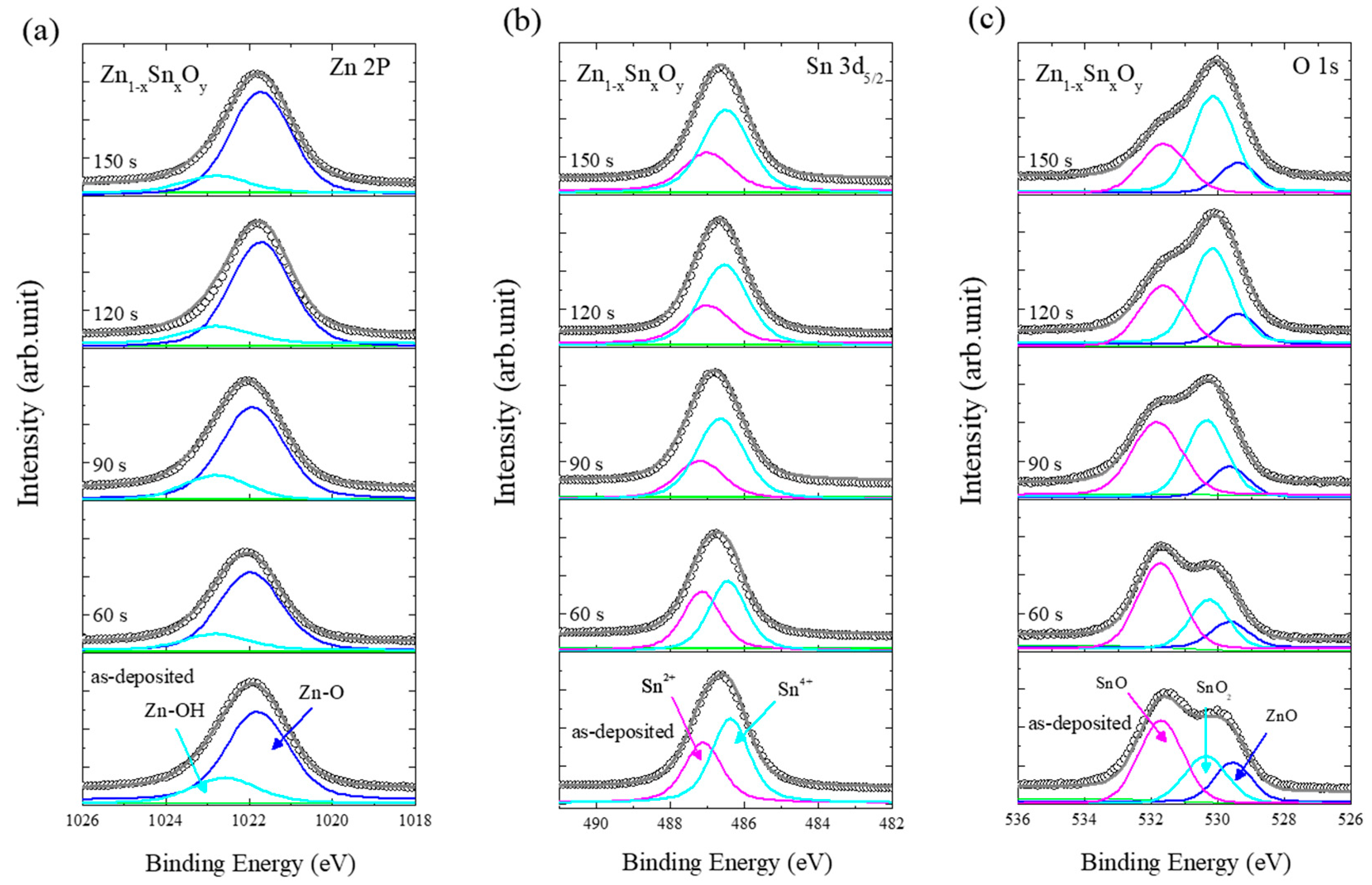
Figure 4 shows the changes in the XPS peaks of the Zn0.6Sn0.4O thin films with respect to the O2 plasma treatment time. The XPS peaks (Zn 2p3/2, Sn 3d5/2, and O 1s) were deconvoluted for the identification of elements and observation of changes in the composition of the Zn0.6Sn0.4O thin films. In the as-deposited samples, Zn 2p3/2 core-level spectra were deconvoluted into two main Gaussian components, Zn–OH (1022.58 eV) and Zn–O (1021.78 eV) (Figure 4a) [27,28,29]. The Sn 3d core-level spectra were also deconvoluted into two Gaussian components, Sn2+ (487.08 eV) and Sn4+ (486.38 eV), displayed in Figure 4b. The Sn2+ and Sn4+ oxidation states can lead to p-type SnO and n-type SnO2 behavior, respectively [30,31]. The O 1s sub-peaks corresponding to ZnO (529.6 eV), SnO (531.7 eV), and SnO2 (530.4 eV) can be attributed to oxidation by reactive sputtering (Figure 4c). The presence of SnO derived components could be attributed to lattice oxygen deficiency and reduced oxygen concentration at the surface [31].
There was no shift of the Zn 2p peak. However, a slight shift of the Sn 3d peak was observed, while the O 1s peak shifted towards the lower binding energy with increasing O2 plasma treatment time. Deconvolution analysis of the O2 plasma treatment time explains the reaction of the O2 plasma on the surface of the ZnSnO thin film. The Zn–OH related peaks decreased after O2 plasma treatment from 60 to 150 s (Figure 4a). The O+ and O2+ ions bombard and break the OH–Zn–O or OH–Sn–O bonds during the plasma treatment, resulting in the formation of O2 or H2O molecules [22]. The intensity of the Sn2+ state decreased dramatically, whereas that of the Sn4+ oxidation state remained stable (Figure 4b). This result is attributed to the difference in the Gibbs free energy associated with the formation of SnO and SnO2fGm(Sn2+, 298.15 K) = −(27.87 ± 0.08) kJ·mol−1, ΔfGm (Sn4+, 298.15 K) = (46.7 ± 3.9) kJ·mol−1) [32]. Therefore, SnO bonds are weaker than SnO2 bonds. The intensity of the ZnO bonding peak remained constant, while the intensities of the SnO2 and SnO bonding peaks exhibited a consistent increase and decrease, respectively (Figure 4c). This result indicates that ZnO has more ionic characteristics than SnO because Zn is significantly more electropositive than Sn. The equilibrium constant (Kp) of formation for ZnO is higher than that of SnO, because the electrons in Zn are more readily given up than those of Sn [33]. Therefore, the energy required to convert SnO into Sn and O (g) should be lesser than the energy required to convert ZnO into Zn and O (g). The reduction in the Sn2+ oxidation state with O2 plasma treatment (Figure 4b) results in the formation of more n-type SnO2 in the Zn0.6Sn0.4O thin film, as indicated in Figure 4c.
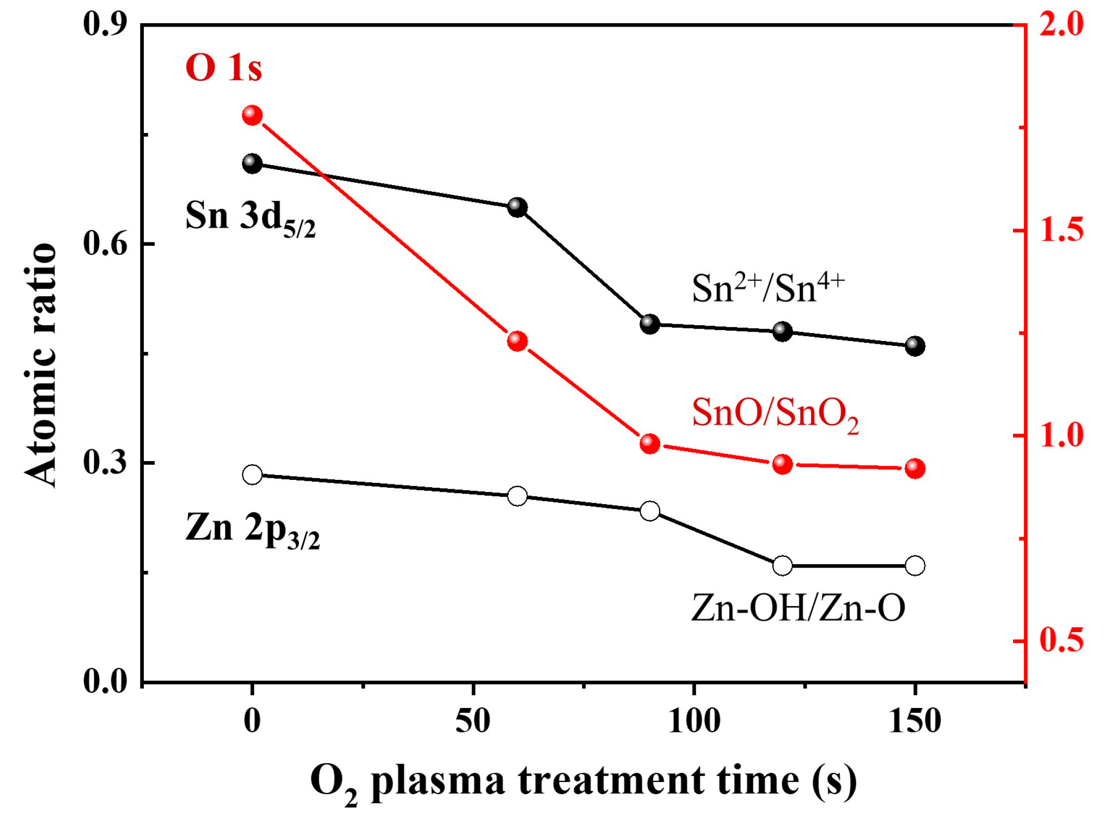
Figure 5 shows the atomic ratios of the deconvoluted components for Sn2+/Sn4+, SnO/SnO2, and Zn–OH/Zn–O as a function of O2 plasma treatment times based on the results of the XPS analysis in Figure 4. The atomic ratios of Sn2+/Sn4+ and SnO/SnO2 sharply decrease as the O2 plasma treatment time increases up to 90 s, which then shows a gradual decrease. However, the atomic ratio of Zn–OH/Zn–O shows a sharp decline in the O2 plasma treatment time between 90 and 120 s.
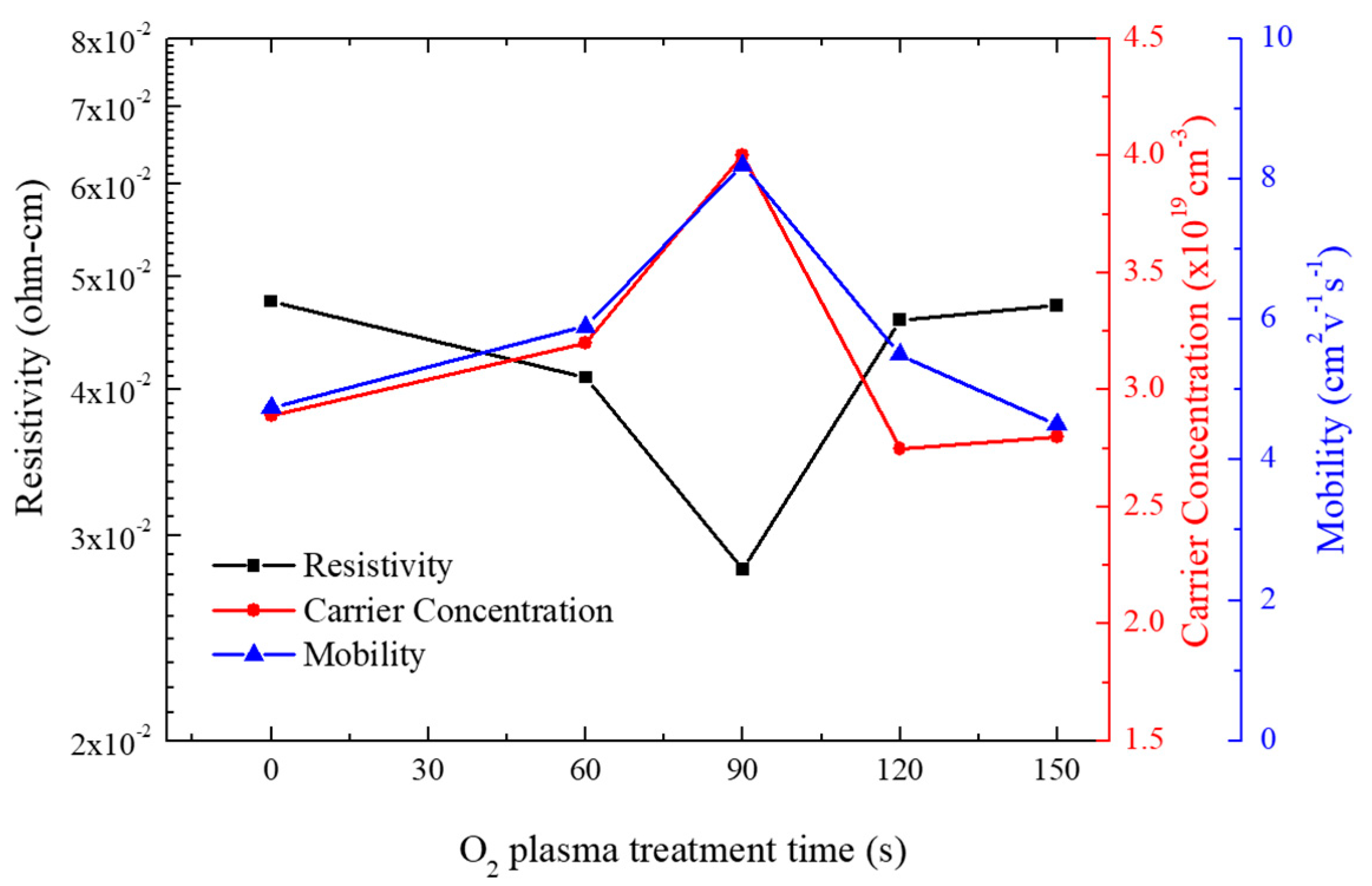
Figure 6 shows the resistivity, carrier concentration, and mobility for the Zn0.6Sn0.4O films as a function of O2 plasma treatment times. As the O2 plasma treatment time increased up to 90 s, a decrease in resistivity was observed. Meanwhile, the mobility and carrier concentration increased. The lowest resistivity of 2.80 × 10⁻² Ω-cm and the highest carrier concentration and mobility of 4.0 × 1019 cm−3 and 8.15 cm2·V−1·s−1, respectively, were achieved at 90 s. However, the resistivity increased and the mobility and carrier concentration decreased as the plasma treatment time increased above 90 s. The O2 plasma treatment causes a reduction in the Sn/Zn ratio on the Zn0.6Sn0.4O surface. The SnO component in the Zn0.6Sn0.4O film is easily affected until the O2 plasma treatment reaches 90 s, which increases the carrier concentration at the surface of the film. However, the ZnO component is more easily affected than the SnO component during O2 plasma treatment between 90 and 120 s (Figure 5), resulting in the increase in the Sn/Zn ratio. In addition, as described for the change in the work function, this sequential effect can be explained by the “heat of formation”.
Surface morphology affects the contact resistance at the metal-semiconductor and/or P-N junction [10,34]. Therefore, we employed AFM to investigate the surface properties of Zn0.6Sn0.4O thin films as the O2 plasma treatment time increased, as shown in Figure 7. The surface roughness (root-mean square, RMS) of the as-deposited Zn0.6Sn0.4O thin film was 0.684 nm and that of the Zn0.6Sn0.4O thin films treated by O2 plasma for 90 and 150 s were 0.626 and 0.624 nm, respectively. It is evident from these results that the O2 plasma treatment has no significant effect on the surface roughness.

4. Conclusions

In this study, we observed changes in the work function and surface state of Zn0.6Sn0.4O thin film under the influence of high-density O2 plasma treatment. The work function increased by approximately 0.5 eV (from 4.16 to up to 4.64 eV). However, the change in optical band gap was exceedingly small (3.17 + Δ0.06 eV). The change in work function was ascribed to the change in the surface morphology of the Zn0.6Sn0.4O thin films, inferred from the XPS analysis. In addition, the O2 plasma treatment resulted in changes in the electrical properties of the thin film, including its resistivity, carrier concentration, and mobility. The mobility significantly increased from 4.75 to 8.2 cm2·V−1·s−1. Meanwhile, there was no significant change in the surface roughness of the Zn0.6Sn0.4O thin films. In conclusion, we confirmed that the work function and mobility of Zn0.6Sn0.4O thin films can be tuned and improved through O2 plasma treatment. The O2 plasma treatment technique exhibits significant potential for application in high-performance displays in optical devices, such as thin-film transistors (TFT), light-emitting diodes (LEDs), and solar cells.

Author Contributions

Conceptualization, Y.-H.J. and J.-H.W.; Methodology, Y.-H.J. and J.-H.W.; Validation, Y.-H.J., J.-H.W., W.-J.L., Y.-D.C., D.-H.C. and S.K.; Data Curation, Y.-H.J., J.-H.W., W.-J.L., Y.-D.C., D.-H.C., S.K. and D.-S.U.; Writing-Original Draft Preparation, Y.-H.J. and J.-H.W.; Writing-Review & Editing, D.-S.U.; Visualization, Y.-H.J. and D.-S.U.; Supervision, D.-S.U. and C.-I.K.; Project Administration, C.-I.K.; Funding Acquisition, C.-I.K. All authors have read and agreed to the published version of the manuscript.

Funding

This research was funded by the National Research Foundation of Korea (NRF) grant funded by the Korea government (MSIT) (No. 2018R1D1A1B07051429).

Acknowledgments

This work supported by the Chung-Ang University Research Scholarship Grants in 2017.

Conflicts of Interest

The authors declare no conflict of interest.

References

  1. Ravi, S.K.; Sun, W.X.; Nandakumar, D.K.; Zhang, Y.X.; Tan, S.C. Optical manipulation of work function contrasts on metal thin films. Sci. Adv. 2018, 4, eaao6050. [Google Scholar] [CrossRef] [Green Version]
  2. Chuang, S.; Battaglia, C.; Azcatl, A.; McDonnell, S.; Kang, J.S.; Yin, X.T.; Tosun, M.; Kapadia, R.; Fang, H.; Wallace, R.M.; et al. MoS2 P-type transistors and diodes enabled by high work function MoOx contacts. Nano Lett. 2014, 14, 1337–1342. [Google Scholar] [CrossRef] [Green Version]
  3. Burgi, L.; Richards, T.J.; Friend, R.H.; Sirringhaus, H. Close look at charge carrier injection in polymer field-effect transistors. J. Appl. Phys. 2003, 94, 6129–6137. [Google Scholar] [CrossRef]
  4. Dominguez, M.A.; Alcantara, S.; Soto, S. Physically-based simulation of zinc oxide thin-film transistors: Contact resistance contribution on density of states. Solid-State Electron. 2016, 120, 41–46. [Google Scholar] [CrossRef]
  5. Liang, S.; Sheng, H.; Liu, Y.; Huo, Z.; Lu, Y.; Shen, H. ZnO Schottky ultraviolet photodetectors. J. Cryst. Growth 2001, 225, 110–113. [Google Scholar] [CrossRef]
  6. Goykhman, I.; Sassi, U.; Desiatov, B.; Mazurski, N.; Milana, S.; de Fazio, D.; Eiden, A.; Khurgin, J.; Shappir, J.; Levy, U.; et al. On-chip integrated, silicon–graphene plasmonic schottky photodetector with high responsivity and avalanche photogain. Nano Lett. 2016, 16, 3005–3013. [Google Scholar] [CrossRef]
  7. Alnuaimi, A.; Almansouri, I.; Saadat, I.; Nayfeh, A. High performance graphene-silicon Schottky junction solar cells with HfO2 interfacial layer grown by atomic layer deposition. Sol. Energy 2018, 164, 174–179. [Google Scholar] [CrossRef]
  8. Lee, K.H.; Jang, H.W.; Kim, K.B.; Tak, Y.H.; Lee, J.L. Mechanism for the increase of indium-tin-oxide work function by O2 inductively coupled plasma treatment. J. Appl. Phys. 2004, 95, 586–590. [Google Scholar] [CrossRef] [Green Version]
  9. Kang, J.H.; Cho, E.N.; Kim, C.E.; Lee, M.J.; Lee, S.J.; Myoung, J.M.; Yun, I. Mobility enhancement in amorphous InGaZnO thin-film transistors by Ar plasma treatment. Appl. Phys. Lett. 2013, 102, 222103. [Google Scholar] [CrossRef] [Green Version]
  10. Fang, M.; Zhang, C.M.; Chen, Q. Tuning the ITO work function by capacitively coupled plasma and its application in inverted organic solar cells. Appl. Surf. Sci. 2016, 385, 28–33. [Google Scholar] [CrossRef]
  11. Jain, V.K.; Kumar, P.; Kumar, M.; Jain, P.; Bhandari, D.; Vijay, Y.K. Study of post annealing influence on structural, chemical and electrical properties of ZTO thin films. J. Alloy. Compd. 2011, 509, 3541–3546. [Google Scholar] [CrossRef]
  12. Chandra, R.D.; Rao, M.; Zhang, K.K.; Prabhakar, R.R.; Shi, C.; Zhang, J.; Mhaisalkar, S.G.; Mathews, N. Tuning electrical properties in amorphous zinc tin oxide thin films for solution processed electronics. ACS Appl. Mater. Interfaces 2014, 6, 773–777. [Google Scholar] [CrossRef]
  13. Chiang, H.Q.; Wager, J.F.; Hoffman, R.L.; Jeong, J.; Keszler, D.A. High mobility transparent thin-film transistors with amorphous zinc tin oxide channel layer. Appl. Phys. Lett. 2005, 86, 013503. [Google Scholar] [CrossRef]
  14. Heineck, D.P.; McFarlane, B.R.; Wager, J.F. Zinc tin oxide thin-film-transistor enhancement/depletion inverter. IEEE Electron Device Lett. 2009, 30, 514–516. [Google Scholar] [CrossRef]
  15. Lindahl, J.; Watjen, J.T.; Hultqvist, A.; Ericson, T.; Edoff, M.; Torndahl, T. The effect of Zn1−xSnxOy buffer layer thickness in 18.0% efficient Cd-free Cu(In,Ga)Se2 solar cells. Prog. Photovolt. 2014, 21, 1588–1597. [Google Scholar] [CrossRef]
  16. Choi, K.H.; Koo, H.W.; Kim, T.W.; Kim, H.K. Antireflective ZnSnO/Ag bilayer-based transparent source and drain electrodes for transparent thin film transistors. Appl. Phys. Lett. 2012, 100, 263505. [Google Scholar] [CrossRef]
  17. Moriga, T.; Hayashi, Y.; Kondo, K.; Nishimura, Y.; Murai, K.; Nakabayashi, I.; Fukumoto, H.; Tominaga, K. Transparent conducting amorphous Zn–Sn–O films deposited by simultaneous dc sputtering. J. Vac. Sci. Technol. A 2004, 22, 1705–1710. [Google Scholar] [CrossRef]
  18. Huang, G.; Duan, L.; Dong, G.; Zhang, D.; Qiu, Y. High-mobility solution-processed tin oxide thin-film transistors with high-κ alumina dielectric working in enhancement mode. ACS Appl. Mater. Interfaces 2014, 6, 20786–20794. [Google Scholar] [CrossRef]
  19. Alshammari, F.H.; Hota, M.K.; Wang, Z.; Al-jawhari, H.; Alshareef, H.N. Atomic-Layer-Deposited SnO2 as Gate Electrode for Indium-Free Transparent Electronics. Adv. Electron. Mater. 2017, 3, 1700155. [Google Scholar] [CrossRef]
  20. Morales-Masis, M.; Dauzou, F.; Jeangros, Q.; Dabirian, A.; Lifka, H.; Gierth, R.; Ruske, M.; Moet, D.; Hessler-Wyser, A.; Ballif, C. An indium-free anode for large-area flexible OLEDs: Defect-free transparent conductive zinc tin oxide. Adv. Funct. Mater. 2016, 26, 384–392. [Google Scholar] [CrossRef]
  21. Um, D.S.; Woo, J.C.; Kim, C.I. Etching characteristics of TaN thin film using an inductively coupled plasma. Surf. Coat. Technol. 2010, 205, S333–S336. [Google Scholar] [CrossRef]
  22. Ratcliff, E.L.; Sigdel, A.K.; Macech, M.R.; Nebesny, K.; Lee, P.A.; Ginley, D.S.; Armstrong, N.R.; Berry, J.J. Surface composition, work function, and electrochemical characteristics of gallium-doped zinc oxide. Thin Solid Films 2012, 520, 5652–5663. [Google Scholar] [CrossRef]
  23. Sugiyama, K.; Ishii, H.; Ouchi, Y.; Seki, K. Dependence of indium–tin–oxide work function on surface cleaning method as studied by ultraviolet and x-ray photoemission spectroscopies. J. Appl. Phys. 2000, 87, 295–298. [Google Scholar] [CrossRef]
  24. Shin, H.; Kim, C.; Bae, C.; Lee, J.S.; Lee, J.; Kim, S. Effects of ion damage on the surface of ITO films during plasma treatment. Appl. Surf. Sci. 2007, 253, 8928–8932. [Google Scholar] [CrossRef]
  25. Masterton, W.L.; Slowinski, E.J.; Stanitski, C.L. Chemical Principles: Alternative Edition with a Qualitative Analysis Supplement; Saunders College Pub: Philadelphia, PA, USA, 1983. [Google Scholar]
  26. D’Agostino, D.; Giorgio, C.D.; Bobba, F.; Trolio, A.D.; Alippi, P.; Cucolo, A.M.; Bonapasta, A.A. Effects of cobalt substitution on ZnO surface reactivity and electronic structure. J. Mater. Chem. C 2019, 7, 8364–8373. [Google Scholar] [CrossRef]
  27. Gutmann, S.; Conrad, M.; Wolak, M.A.; Beerbom, M.M.; Schlaf, R. Work function measurements on nano-crystalline zinc oxide surfaces. J. Appl. Phys. 2012, 111, 123710. [Google Scholar] [CrossRef]
  28. Zhang, X.; Qin, J.; Xue, Y.; Yu, P.; Zhang, B.; Wang, L.; Liu, R. Effect of aspect ratio and surface defects on the photocatalytic activity of ZnO nanorods. Sci. Rep. 2014, 4, 4596. [Google Scholar] [CrossRef] [Green Version]
  29. Hu, X.; Masuda, Y.; Ohji, T.; Kato, K. Fabrication of Zn(OH)2/ZnO nanosheet-ZnO nanoarray hybrid structured films by a dissolution-recrystallization route. J. Am. Ceram. Soc. 2010, 93, 881–886. [Google Scholar] [CrossRef]
  30. Hwang, S.; Kim, Y.Y.; Lee, J.H.; Seo, D.K.; Lee, J.Y.; Cho, H.K. Irregular electrical conduction types in tin oxide thin films induced by nanoscale phase separation. J. Am. Ceram. Soc. 2012, 95, 324–327. [Google Scholar] [CrossRef]
  31. Kover, L.; Kovacs, Z.; Sanjines, R.; Moretti, G.; Cserny, I.; Margaritondo, G.; Palinkas, J.; Adachi, H. Electronic structure of tin oxides: High-resolution study of XPS and Auger spectra. Surf. Interface Anal. 1995, 23, 461–466. [Google Scholar] [CrossRef]
  32. Gamsjäger, H.; Gajda, T.; Sangster, J.; Saxena, S.K.; Voigt, W.; Perrone, J. Chemical Thermodynamics of TIN; OECD Publishing: Paris, France, 2012; Volume 12. [Google Scholar]
  33. Reisman, A.; Berkenblit, M.; Ghez, R.; Chan, S.A. The equilibrium constant for the reaction of ZnO + H2 and the chemical vapor transport of ZnO via the Zn + H2O reaction. J. Electron. Mater. 1972, 1, 395–419. [Google Scholar] [CrossRef]
  34. Liang, Z.; Zhang, Q.; Wiranwetchayan, O.; Xi, J.; Yang, Z.; Park, K.; Li, C.; Cao, G. Effects of the morphology of a ZnO buffer layer on the photovoltaic performance of inverted polymer solar cells. Adv. Funct. Mater. 2012, 22, 2194–2201. [Google Scholar] [CrossRef]
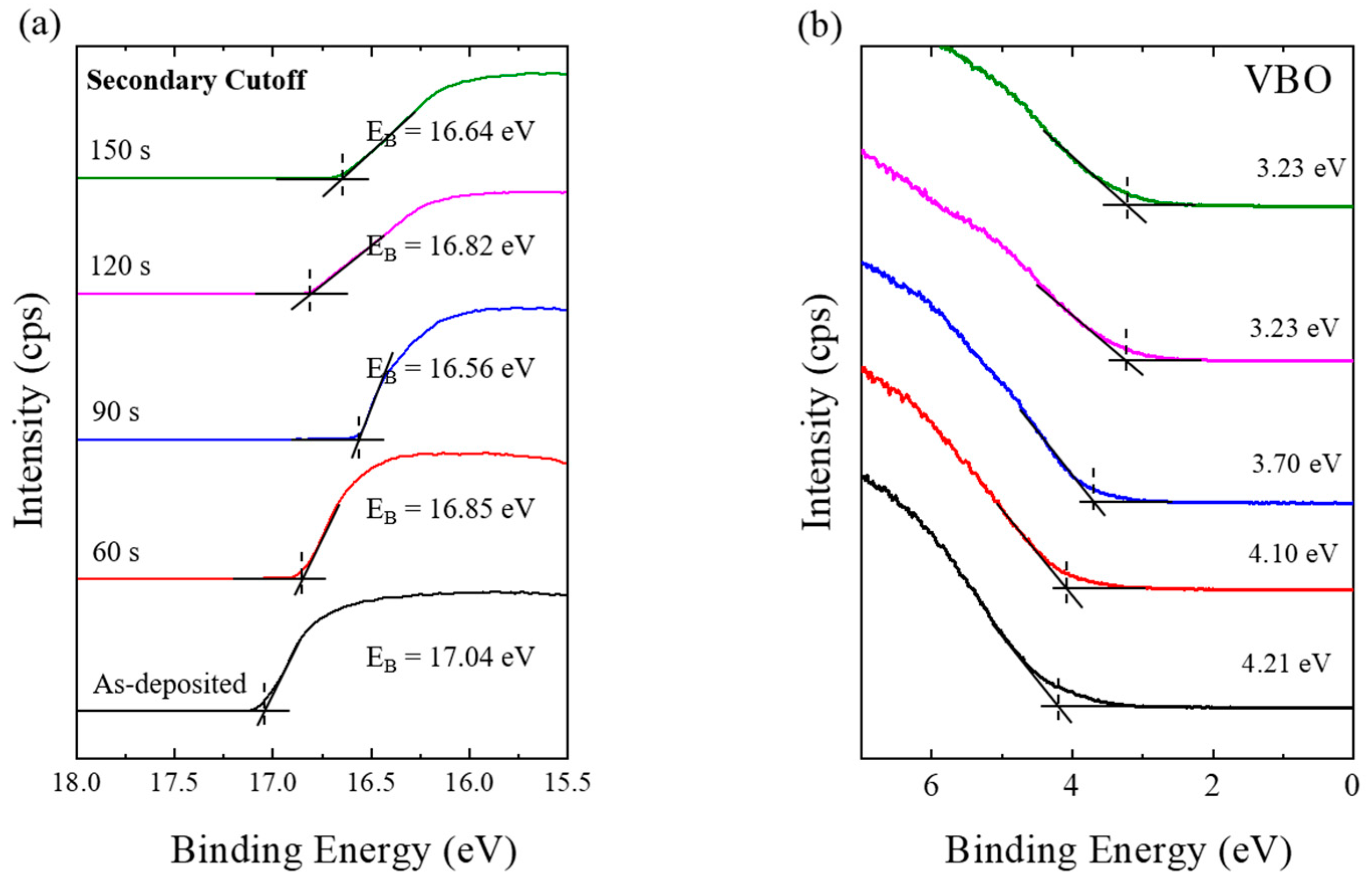
Figure 1. UPS measurements of Zn0.6Sn0.4O thin films exposed to different O2 plasma treatment times. (a) the higher binding energy edge of the secondary electrons and (b) the valence structure.
Coatings 10 01026 g001
Figure 2. The optical transmittance spectra as a function of O2 plasma treatment time for the Zn0.6Sn0.4O thin films. In the insert, the magnified spectra of the Zn0.6Sn0.4O thin films and the bandgap of Zn0.6Sn0.4O.
Coatings 10 01026 g002
Figure 3. Schematic of modified Zn0.6Sn0.4O band alignment including work functions and band gaps of all of the O2 plasma treated materials.
Coatings 10 01026 g003
Figure 4. XPS results for Zn 2p, Sn 3d and O 1s core levels as a function of O2 plasma treatment time for the Zn0.6Sn0.4Oy thin films. (a) The Zn 2p3/2 was deconvoluted with two components for Zn–O and Zn–OH bonds, (b) the Sn 3d5/2 was deconvoluted with two components for Sn4+, and Sn2+, (c) the O 1s was deconvoluted with three components for SnO, SnO2, and ZnO bonds.
Coatings 10 01026 g004
Figure 5. The atomic ratios of the deconvoluted components in the XPS peaks for Sn2+/Sn4+, SnO/SnO2, and Zn–OH/Zn–O as a function of O2 plasma treatment time.
Coatings 10 01026 g005
Figure 6. The electrical resistivity, carrier concentration and mobility as a function of O2 plasma treatment time for the Zn0.6Sn0.4O thin films.
Coatings 10 01026 g006
Figure 7. AFM images of the Zn0.6Sn0.4O thin films post O2 plasma treatment. (a) as-deposited, (b) 90 s and (c) 150 s.
Coatings 10 01026 g007
Publisher’s Note: MDPI stays neutral with regard to jurisdictional claims in published maps and institutional affiliations.