UPLC-MS-ESI-QTOF Analysis and Antifungal Activity of the Spondias tuberosa Arruda Leaf and Root Hydroalcoholic Extracts


 ,
,  ,
,
Abstract
1. Introduction
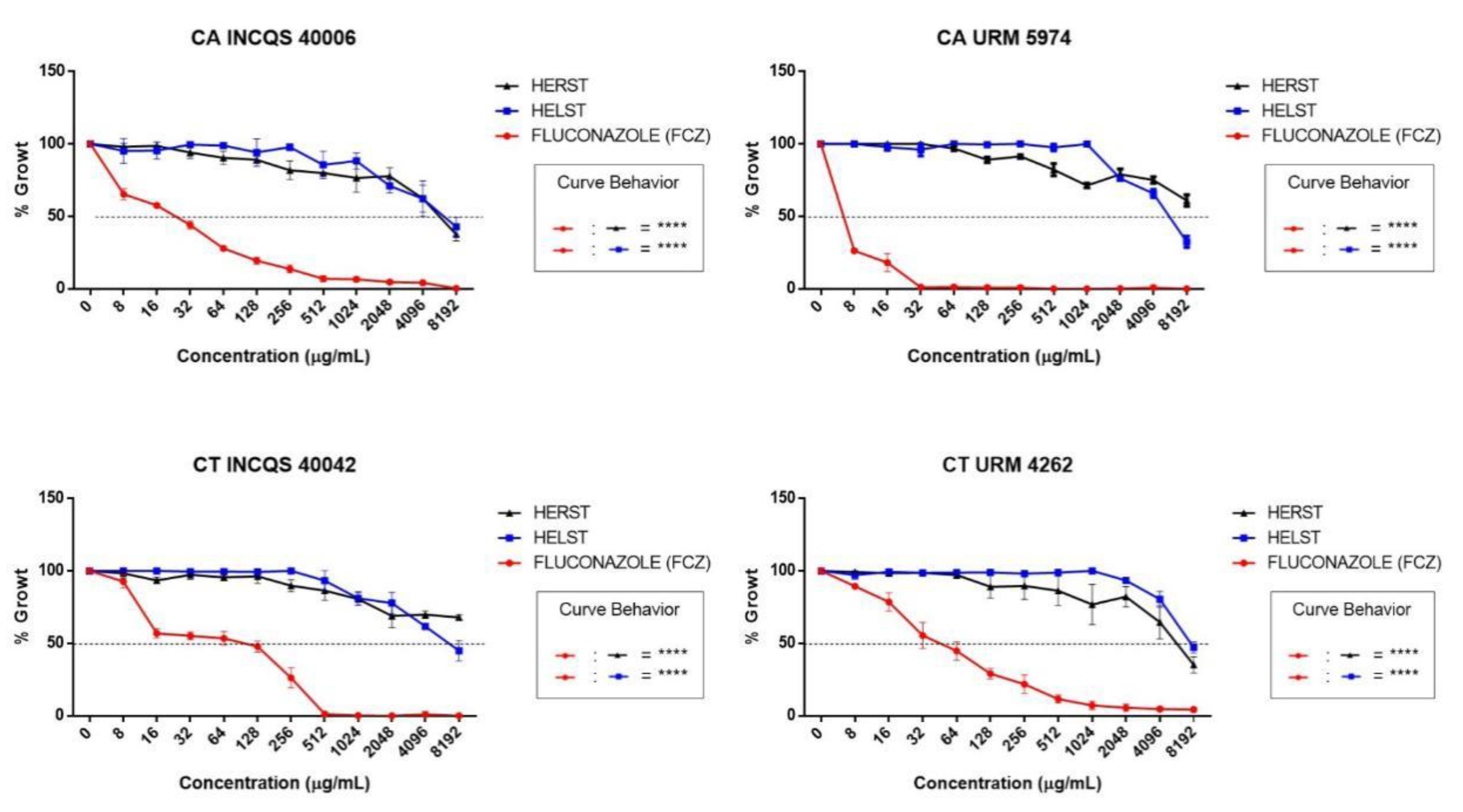
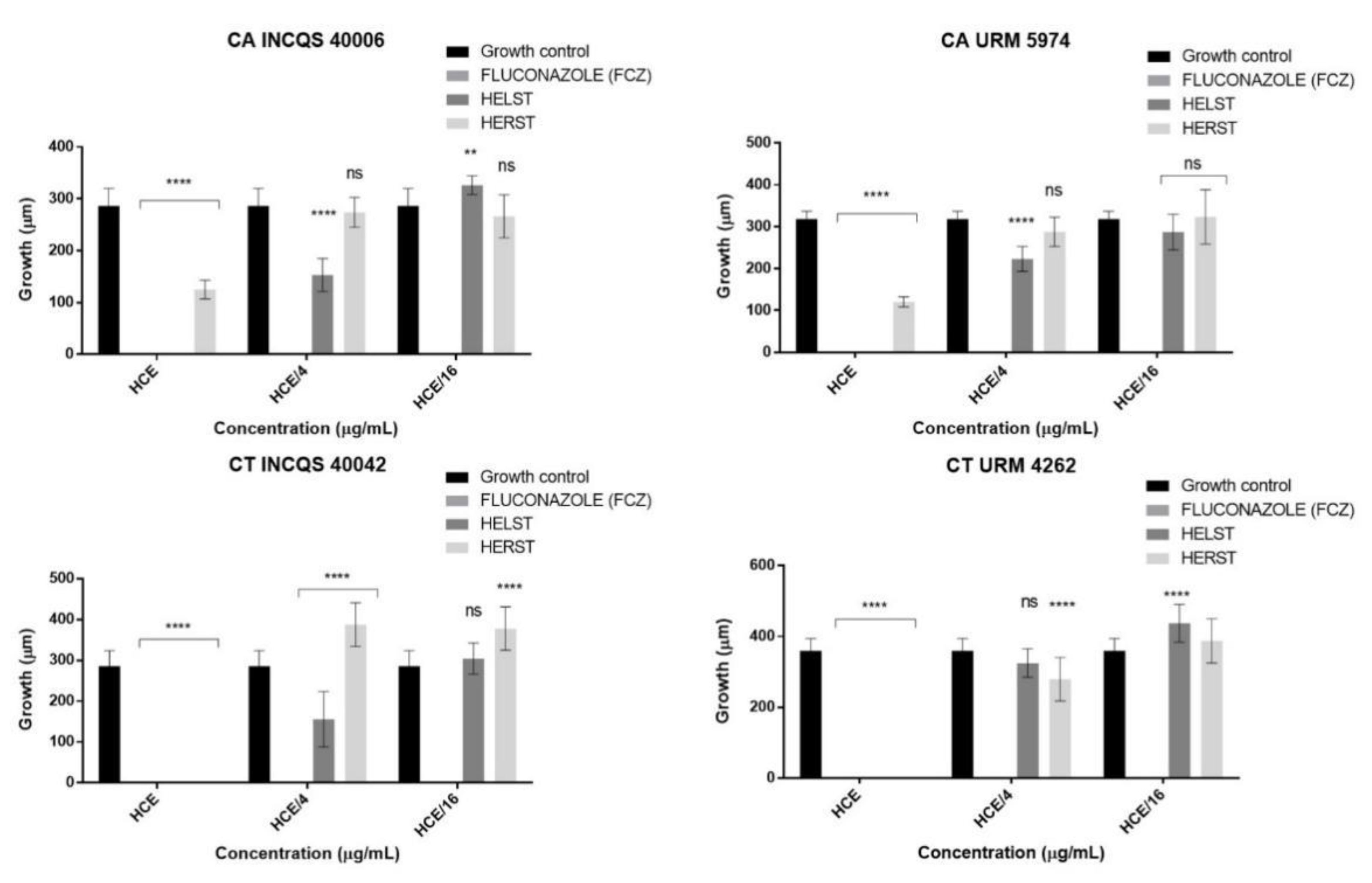
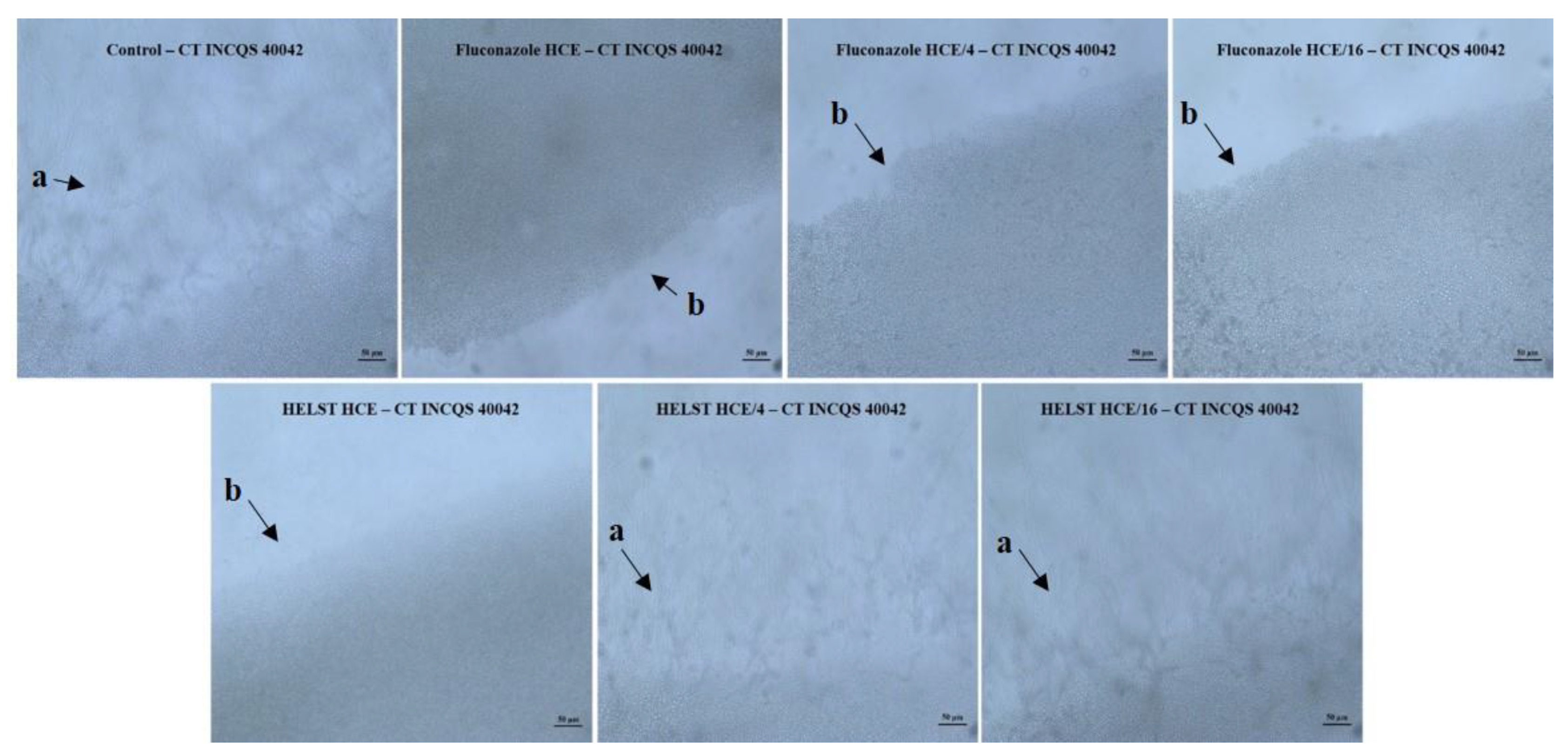
2. Results
3. Discussion
4. Materials and Methods
4.1. Botanical Material Collection and Identification
4.2. Extract Acquisition
4.3. Chemical Analysis
4.3.1. Preliminary Phytochemical Analysis
4.3.2. Compound Identification by Ultra-Performance Liquid Chromatography Coupled to Quadrupole/Time of Flight System (UPLC-MS-ESI-QTOF)
4.4. Antifungal Assays
4.4.1. Microorganisms, Culture Media, Inoculum Preparation, and Drugs and Reagents Used
4.4.2. IC50 Determination and Cell Viability Curve
4.4.3. Minimum Fungicidal Concentration (MFC) Determination
4.4.4. Evaluation of the Modifying Effect of Fluconazole Action
4.4.5. Effect of the Extracts and Fluconazole on Candida Morphological Transition
4.5. Statistical Analysis
5. Conclusions
Author Contributions
Funding
Conflicts of Interest
References
- Zarrinfar, H.; Kaboli, S.; Dolatabadi, S.; Mohammadi, R. Rapid detection of Candida species in bronchoalveolar lavage fluid from patients with pulmonary symptoms. Braz. J. Microbiol. 2016, 47, 172–176. [Google Scholar] [CrossRef] [PubMed]
- Navarro-Arias, M.J.; Hernández-Chávez, M.J.; García-Carnero, L.C.; Amezcua-Hernández, D.G.; Lozoya-Pérez, N.E.; Estrada-Mata, E.; Martínez-Duncker, I.; Franco, B.; Mora-Montes, H.M. Differential recognition of Candida tropicalis, Candida guilliermondii, Candida krusei, and Candida auris by human innate immune cells. Infect. Drug Resist. 2019, 12, 783–794. [Google Scholar] [CrossRef] [PubMed]
- Sharma, J.; Rosiana, S.; Razzaq, I.; Shapiro, R.S. Linking cellular morphogenesis with antifungal treatment and susceptibility in candida pathogens. J. Fungi 2019, 5, 17. [Google Scholar] [CrossRef] [PubMed]
- Vieira, A.J.H.; Santos, J.I. Mecanismos de resistência de Candida albicans aos antifúngicos anfotericina B, fluconazol e caspofungina. RBAC 2017, 49, 235–239. [Google Scholar] [CrossRef]
- Murugesh, J.; Annigeri, R.G.; Mangala, G.K.; Mythily, P.H.; Chandrakala, J. Evaluation of the antifungal efficacy of different concentrations of Curcuma longa on Candida albicans: An in vitro study. J. Oral Maxillofac. Pathol. 2019, 23, 305. [Google Scholar] [CrossRef]
- De Faria Goes, T.Z.; Gonçalves, A.P.P.; Cunha, P.N.A.; de Deus Vieira, G.; Nicolete, R.; Hernandez, A.E.F.; Teles, C.B.G. Prospecção fitoquímica e antimicrobiana dos extratos de Lantana Camara L. e Lantana Trifolia L.(Prospecção fitoquímica e antimicrobiana de L. Camara e L. Trifolia). Saber Cient. 2016, 5, 1–11. [Google Scholar]
- Albuquerque, H.; Dias, J.; Dantas, R.; Cabral, I.; Medeiros, A.; Santos, V. POTENCIAL ATIVIDADE ANTIULCEROGÊNICA DA ESPÉCIE Spondias mombin. J. Biol. Pharm. Agric. Manag. 2015, 10, 26–29. [Google Scholar]
- Flora do Brasil 2020 em construção. Jardim Botânico do Rio de Janeiro. Available online: http://floradobrasil.jbrj.gov.br/reflora/floradobrasil/FB4405 (accessed on 23 September 2019).
- Da Silva Siqueira, E.M.; Félix-Silva, J.; de Araújo, L.M.L.; Fernandes, J.M.; Cabral, B.; Gomes, J.A.D.S.; de Araújo Roque, A.; Tomaz, J.C.; Lopes, N.P.; de Freitas Fernandes-Pedrosa, M.; et al. Spondias tuberosa (Anacardiaceae) leaves: Profiling phenolic compounds by HPLC-DAD and LC–MS/MS and in vivo anti-inflammatory activity. Biomed. Chromatogr. 2016, 30, 1656–1665. [Google Scholar] [CrossRef]
- Siqueira, E.M.D.S. Spondias Tuberosa Arr. (UMBU): Estudo Fitoquímico e Avaliação do Potencial Anti-Inflamatório; Universidade Federal do Rio Grande do Norte: Natal, Brasil, 2015. [Google Scholar]
- Zeraik, M.L.; Queiroz, E.F.; Marcourt, L.; Ciclet, O.; Castro-Gamboa, I.; Silva, D.H.S.; Cuendet, M.; da Silva Bolzani, V.; Wolfender, J.L. Antioxidants, quinone reductase inducers and acetylcholinesterase inhibitors from Spondias tuberosa fruits. J. Funct. Foods 2016, 21, 396–405. [Google Scholar] [CrossRef]
- Da Silva, A.R.A.; De Morais, S.M.; Mendes Marques, M.M.; De Oliveira, D.F.; Barros, C.C.; De Almeida, R.R.; Vieira, Í.G.P.; Guedes, M.I.F. Chemical composition, antioxidant and antibacterial activities of two Spondias species from Northeastern Brazil. Pharm. Biol. 2012, 50, 740–746. [Google Scholar] [CrossRef]
- Almeida, A.L.S.; Albuquerque, U.P.; Castro, C.C. Reproductive biology of Spondias tuberosa Arruda (Anacardiaceae), an endemic fructiferous species of the caatinga (dry forest), under different management conditions in northeastern Brazil. J. Arid Environ. 2011, 75, 330–337. [Google Scholar] [CrossRef]
- Silva, A.R.A.; Morais, S.M.; Marques, M.M.M.; Lima, D.M.; Santos, S.C.C.; Almeida, R.R.; Vieira, I.G.P.; Guedes, M.I.F. Antiviral activities of extracts and phenolic components of two spondias species against dengue virus. J. Venom. Anim. Toxins Incl. Trop. Dis. 2011, 17, 406–413. [Google Scholar]
- Lasano, N.F.; Hamid, A.H.; Karim, R.; Dek, M.S.P.; Shukri, R.; Shazini Ramli, N. Nutritional composition, anti-diabetic properties and identification of active compounds using UHPLC-ESI-orbitrap-MS/MS in Mangifera odorata L. peel and seed kernel. Molecules 2019, 24, 320. [Google Scholar] [CrossRef] [PubMed]
- Cunha, A.G.; Brito, E.S.; Moura, C.F.; Ribeiro, P.R.; Miranda, M.R.A. UPLC–qTOF-MS/MS-based phenolic profile and their biosynthetic enzyme activity used to discriminate between cashew apple (Anacardium occidentale L.) maturation stages. J. Chromatogr. B 2017, 1051, 24–32. [Google Scholar] [CrossRef] [PubMed]
- Shoko, T.; Maharaj, V.J.; Naidoo, D.; Tselanyane, M.; Nthambeleni, R.; Khorombi, E.; Apostolides, Z. Anti-aging potential of extracts from Sclerocarya birrea (A. Rich.) Hochst and its chemical profiling by UPLC-Q-TOF-MS. BMC Complement. Altern. Med. 2018, 18, 54. [Google Scholar] [CrossRef] [PubMed]
- Dos Reis Luz, L.; Porto, D.D.; Castro, C.B.; Silva, M.F.S.; de Godoy Alves Filho, E.; Canuto, K.M.; de Brito, E.S.; Becker, H.; do Ó Pessoa, C.; Zocolo, G.J. Metabolomic profile of Schinopsis brasiliensis via UPLC-QTOF-MS for identification of biomarkers and evaluation of its cytotoxic potential. J. Chromatogr. B 2018, 1099, 97–109. [Google Scholar] [CrossRef]
- Ramirez, J.; Zambrano, R.; Sepúlveda, B.; Simirgiotis, M. Antioxidant properties and hyphenated HPLC-PDA-MS profiling of chilean Pica mango fruits (Mangifera indica L. cv. piqueño). Molecules 2014, 19, 438–458. [Google Scholar] [CrossRef]
- Da Rocha, P.D.S.; de Araújo Boleti, A.P.; do Carmo Vieira, M.; Carollo, C.A.; da Silva, D.B.; Estevinho, L.M.; dos Santos, E.L.; de Picoli Souza, K. Microbiological quality, chemical profile as well as antioxidant and antidiabetic activities of Schinus terebinthifolius Raddi. Comp. Biochem. Phys. C 2019, 220, 36–46. [Google Scholar]
- Sayed, E.A.; Martiskainen, O.; Sinkkonen, J.; Pihlaja, K.; Ayoub, N.; Singab, A.E.N.; El-Azizi, M. Chemical composition and bioactivity of Pleiogynium timorense (Anacardiaceae). Nat. Prod. Commun. 2010, 5, 1934578X1000500410. [Google Scholar] [CrossRef]
- Cádiz-Gurrea, M.D.L.L.; Lozano-Sánchez, J.; Fernández-Ochoa, Á.; Segura-Carretero, A. Enhancing the Yield of Bioactive Compounds from Sclerocarya birrea Bark by Green Extraction Approaches. Molecules 2019, 24, 966. [Google Scholar]
- Galvão, W.A.; Braz Filho, R.; Canuto, K.M.; Ribeiro, P.R.V.; Campos, A.R.; Moreira, A.C.O.M.; Silva, S.O.; Mesquita Filho, F.A.; Santos, S.A.A.R.; Junior, J.M.; et al. Gastroprotective and anti-inflammatory activities integrated to chemical composition of Myracrodruon urundeuva Allemão-A conservationist proposal for the species. J. Ethnopharmacol. 2018, 222, 177–189. [Google Scholar] [CrossRef]
- Uchôa, A.D.A.; Oliveira, W.F.; Pereira, A.P.C.; Silva, A.G.; Cordeiro, B.M.P.C.; Malafaia, C.B.; Almeida, C.M.A.; Silva, N.H.; Albuquerque, J.F.C.; Silva, M.V.; et al. Antioxidant Activity and Phytochemical Profile of Spondias tuberosa Arruda Leaves Extracts. Am. J. Plant Sci. 2015, 6, 3038–3044. [Google Scholar] [CrossRef]
- Chaves, T.P.; Clementino, E.L.C.; de Castro Felismino, D.; Silva, H.; Santos, J.S.; de Medeiros, A.C.D. Phytochemical composition and antimicrobial and toxicological activity of Spondias mombin L.(jobo). Rev. Cuba. Plantas Med. 2018, 23, 1–5. [Google Scholar]
- Da Costa Cordeiro, B.M.P.; De Lima Santos, N.D.; Ferreira, M.R.A.; De Araújo, L.C.C.; Junior, A.R.C.; Da Conceição Santos, A.D.; De Oliveira, A.P.; Da Silva, A.G.; Da Silva Falcão, E.P.; Dos Santos Correia, M.T.; et al. Hexane extract from Spondias tuberosa (Anacardiaceae) leaves has antioxidant activity and is an anti-Candida agent by causing mitochondrial and lysosomal damages. BMC Complement. Altern. Med. 2018, 18, 1–10. [Google Scholar] [CrossRef] [PubMed]
- De Brito Costa, E.M.M.; Barbosa, A.S.; Florentino, V.G.B.; da Silva, J.D.F.; Trovão, D.M.D.B.M.; de Medeiros, A.C.D. In vitro antimicrobial activity of plant extracts of semi-arid region of Paraíba, PB, Brazil. Rev. Odonto Ciência 2013, 28, 101–104. [Google Scholar]
- Simões, C.M.O.; Schenkel, E.P.; Gosmann, G.; Mello, J.C.P.; Mentz, L.A.; Petrovick, P.R. Farmacognosia: Da planta ao medicamento; UFRGS: Porto Alegre, Brasil; UFSC: Florianópolis, Brasil, 2002. [Google Scholar]
- Stoilova, I.; Gargova, S.; Stoyanova, A.; Ho, I. Antimicrobial and antioxidant activity of the polyphenol mangiferin. Herba Pol. 2005, 1, 2706–2716. [Google Scholar]
- Muthamil, S.; Balasubramaniam, B.; Balamurugan, K.; Pandian, S.K. Synergistic effect of quinic acid derived from syzygium cumini and undecanoic acid against candida spp. Biofilm and virulence. Front. Microbiol. 2018, 9, 2835. [Google Scholar] [CrossRef]
- Espino, M.; Solari, M.; de los Ángeles Fernández, M.; Boiteux, J.; Gómez, M.R.; Silva, M.F. NADES-mediated folk plant extracts as novel antifungal agents against Candida albicans. J. Pharm. Biomed. Anal. 2019, 167, 15–20. [Google Scholar] [CrossRef]
- Sung, W.S.; Lee, D.G. Antifungal action of chlorogenic acid against pathogenic fungi, mediated by membrane disruption. Pure Appl. Chem. 2010, 82, 219–226. [Google Scholar] [CrossRef]
- Martínez, G.; Regente, M.; Jacobi, S.; Del Rio, M.; Pinedo, M.; de la Canal, L. Chlorogenic acid is a fungicide active against phytopathogenic fungi. Pestic. Biochem. Physiol. 2017, 140, 30–35. [Google Scholar] [CrossRef]
- Temitope, O.O.; Ogunmodede, A.F.; Fasusi, O.A.; Thonda, A.O.; Odufunwa, A.E. Synergistic Antibacterial and Antifungal Activities of Spondias mombin Extracts and Conventional Antibiotic and Antifungal Agents on Selected Clinical Microorganisms. Sch. J. Appl. Med. Sci. 2017, 5, 307–318. [Google Scholar]
- Cristofoli, N.L.; Lima, C.A.R.; Vieira, M.M.C.; Andrade, K.S.; Ferreira, S.R.S. Antioxidant and antimicrobial potential of cajazeira leaves (Spondias mombin) extracts. Sep. Sci. Technol. 2019, 54, 580–590. [Google Scholar] [CrossRef]
- Anand, G.; Ravinanthan, M.; Basaviah, R.; Shetty, A. In vitro antimicrobial and cytotoxic effects of Anacardium occidentale and Mangifera indica in oral care. J. Pharm. Bioallied Sci. 2015, 7, 69–74. [Google Scholar] [PubMed]
- Da Silva, R.A.; Liberio, S.A.; do Amaral, F.M.M.; do Nascimento, F.R.F.; Torres, L.M.B.; Neto, V.M.; Guerra, R.N.M. Antimicrobial and Antioxidant Activity of Anacardium occidentale L. Flowers in Comparison to Bark and Leaves Extracts. J. Biosci. Med. 2016, 4, 87–99. [Google Scholar]
- Houghton, P.J.; Howes, M.J.; Lee, C.C.; Steventon, G. Uses and abuses of in vitro tests in ethnopharmacology: Visualizing an elephant. J. Ethnopharmacol. 2007, 110, 391–400. [Google Scholar] [CrossRef]
- Andrade, J.C.; da Silva, A.R.P.; dos Santos, A.T.L.; Freitas, M.A.; de Matos, Y.M.L.S.; Braga, M.F.B.M.; Bezerra, C.F.; Gonçalo, M.I.P.; Gomez, M.C.V.; Rolóm, M. Chemical composition, antiparasitic and cytotoxic activities of aqueous extracts of Ziziphus joazeiro Mart. Asian Pac. J. Trop. Biomed. 2019, 9, 222. [Google Scholar]
- Matos, F.J.A. Farmácias Vivas, 4th ed.; Editora UFC: Fortaleza, Brasil, 2020; pp. 36–40. [Google Scholar]
- Masters, K. Spray Drying Handbook, 5th ed.; Longman Scientific & Technical: New York, NY, USA, 1991. [Google Scholar]
- Sousa, E.O.; Miranda, C.M.B.A.; Nobre, C.B.; Boligon, A.A.; Athayde, M.L.; Costa, J.G.M. Phytochemical analysis and antioxidant activities of lantana camara and lantana montevidensis extracts. Ind. Crop. Prod. 2015, 70, 7–15. [Google Scholar] [CrossRef]
- Souza, E.L.; Stamford, T.L.M.; Lima, E.O.; Trajano, V.N. Effectiveness of Origanum vulgare L. essential oil to inhibit the growth of food spoiling yeasts. Food Control 2007, 18, 409–413. [Google Scholar] [CrossRef]
- Javadpour, M.M.; Juban, M.M.; Lo, W.C.J.; Bishop, S.M.; Alberty, J.B.; Cowell, S.M.; Alberty, J.B.; Cowell, S.M.; Becker, C.L.; McLaughlin, M.L. De novo antimicrobial peptides with low mammalian cell toxicity. J. Med. Chem. 1996, 39, 3107–3113. [Google Scholar] [CrossRef]
- Morais-Braga, M.F.B.; Sales, D.L.; Carneiro, J.N.P.; Machado, A.J.T.; dos Santos, A.T.L.; de Freitas, M.A.; Martins, G.M.D.A.B.; Leite, N.F.; de Matos, Y.M.L.; Tintino, S.R.; et al. Psidium guajava L. and Psidium brownianum Mart ex DC.: Chemical composition and anti—Candida effect in association with fluconazole. Microb. Pathog. 2016, 95, 200–207. [Google Scholar] [CrossRef]
- Ernst, E.J.; Klepser, M.E.; Ernst, M.E.; Messer, S.A.; Pfaller, M.A. In vitro pharmacodynamic properties of MK-0991 determined by time-kill methods. Diagn. Microbiol. Infect. Dis. 1999, 33, 75–80. [Google Scholar] [CrossRef]
- Coutinho, H.D.M.; Costa, J.G.M.; Lima, E.O.; Falcão-Silva, V.S.; Siqueira, J.P. Enhancement of the antibiotic activity against a multiresistant Escherichia coli by Mentha arvensis L. and chlorpromazine. Chemotherapy 2008, 54, 328–330. [Google Scholar] [CrossRef] [PubMed]
- Sidrim, J.J.C.; Rocha, M.F.G. Micologia Médica à Luz de Autores Contemporâneos; Rio de Janeiro: Guanabara Koogan, Brazil, 2010; p. 388. [Google Scholar]
- Mendes, J.M. Investigação da Atividade Antifúngica do Óleo Essencial de Eugenia Caryophyllata Thunb. Sobre Cepas de Candida Tropicalis; UFPB: João Pessoa, Brazisil, 2011; p. 74. [Google Scholar]
- Pereira Carneiro, J.N.; da Cruz, R.P.; da Silva, J.C.P.; Rocha, J.E.; de Freitas, T.S.; Sales, D.L.; Bezerra, C.F.; de Oliveira Almeida, W.; da Costa, J.G.M.; da Silva, L.E.; et al. Piper diospyrifolium Kunth.: Chemical analysis and antimicrobial (intrinsic and combined) activities. Microb. Pathog. 2019, 136, 103700. [Google Scholar] [CrossRef] [PubMed]


| Special Metabolite Classes (SMC) | ||||||||||
|---|---|---|---|---|---|---|---|---|---|---|
| SMC 1 | SMC 2 | SMC 3 | SMC 4 | SCM 5 | SMC 6 | SMC 7 | SMC 8 | SMC 9 | SMC 10 | |
| HELST | + | - | + | - | - | + | - | + | - | + |
| HERST | + | - | - | + | - | + | - | - | + | - |
| Peak no. | Rt min | [M-H]- Observed | [M-H]- Calculated | Product Ions (MS/MS) | Empirical Formula | Ppm (error) | Putative Name | References |
|---|---|---|---|---|---|---|---|---|
| 1 | 2.82 | 174.9525 | 174.9502 | 175.9599 | C6H6O6 | Dehydroascorbic acid | [15] | |
| 2 | 3.28 | 443.1294 | 443.1283 | 381.1805, 281.1358, 119.0361 | C21H32O10 | 2.5 | Dehydrophaseic acid hexose | [16] |
| 3 | 3.40 | 189.0021 | 189.0035 | 207,0115, 188.9965, 126.9987 | C6H5O7 | −7.4 | None identified | - |
| 4 | 3.65 | 188.9986 | 188.9977 | 207.0199, 188.9996, 127.0019 | C13HO2 | 4.8 | None identified | - |
| 5 | 4.53 | 191.0109 | 191.0133 | 173.0243, 127.0278, 85.0283 | C7H12O6 | −12.6 | Quinic acid (Organic acid) | [17,18] |
| 6 | 4.65 | 343.0833 | 343.0818 | 191.0497, 169.0125, 125.0265 | C14H16O10 | 4.4 | Galloyl quinic acid isomer Ia | [18] |
| 7 | 4.67 | 343.0508 | 343.0513 | 191.0523, 169.0112, 125.0259 | C14H16O10 | −1.5 | Galloyl quinic acid isomer IIa | [18] |
| 8 | 4.74 | 343.1045 | 343.1029 | 191.0196, 169.0011, 125.9942 | C14H16O10 | 4.9 | Galloyl quinic acid isomer III | [18]) |
| 9 | 4.89 | 343.0904 | 343.0877 | 191.0540, 169.0121, 125.0247 | C14H16O10 | 7.9 | Galloyl quinic acid isomer IV | [18] |
| 10 | 6.28 | 421.2582 | 421.2590 | 331.0801, 3010357 | C19H18O11 | −1.9 | Mangiferin | [15,19] |
| 11 | 6.48 | 939.4997 | 939.4989 | 787.4410, 277.2166, 125.0243 | C41H32O26 | 0.9 | Penta-O-galloyl hexoside | [20] |
| 12 | 6.56 | 397.1313 | 397.1287 | 502.2910, 474.2616, 277.2155 | C22H21O7 | 6.5 | None identified | - |
| 13 | 6.90 | 277.2142 | 277.2168 | 279.2315, 277.2151, 189.0038 | C18H29O2 | −9.4 | None identified | - |
| 14 | 7.00 | 339.2007 | 339.2019 | - | C22H28O3 | −3.5 | Caffeoyl-D-glucose | [16] |
| 15 | 7.50 | 483.2540 | 483.2535 | 271.0134, 169.0511, 125.9984 | C20H20O14 | 1.0 | Digalloyl glucose (Digalloyglucose) | [16,21] |
| Peak no. | Rt min | [M-H]- Observed | [M-H]- Calculated | Product Ions (MS/MS) | Empirical Formula | Ppm (error) | Putative Name | References |
|---|---|---|---|---|---|---|---|---|
| 1 | 2.83 | 272.9554 | 272.9578 | 274.9536, 273.9563, 158.9753 | C15H12O5 | −8.8 | (±)-Naringenin | [15] |
| 2 | 3.11 | 341.2104 | 341.2117 | 297.2246, 295.2048, 119.0465 | C22H30O3 | 2.1 | Anacardic acid 1 | [16] |
| 3 | 3.14 | 343.1186 | 343.1182 | 299.2344 | C22H21O3 | 1.2 | Anacardic acid 2 | [16] |
| 4 | 3.16 | 377.0822 | 377.0814 | 379.0782, 377.0770, 341.1009 | C25H13O4 | 2.1 | No identified | - |
| 5 | 3.22 | 345.0010 | 345.0035 | 301.2471 | C22H34O3 | −7.2 | Anacardic acid 3 | [16] |
| 6 | 3.52 | 355.0232 | 355.0243 | 355.0258, 163.0371 | C16H18O9 | −3.1 | Chlorogenic acid | [9] |
| 7 | 4.39 | 411.0173 | 411.0213 | 411.0232, 240.9987, 169.0123 | C17H7N4O9 | −9.7 | None identified | - |
| 8 | 4.50 | 461.1219 | 461.1236 | 257.0848, 229.8624, 151.0033 | C22H21O11 | −3.7 | Kaempferol-7-Oglucuronide | [16] |
| 9 | 4.62 | 197.0424 | 197.0450 | 199.06, 198.05, 182.0187 | C9H10O5 | −13.2 | 2-Hydroxy-3,4-dimethoxybenzoic acid | [15] |
| 10 | 4.90 | 433.0583 | 433.0560 | 300.9944, 271.0716 | C20H18O11 | 5.3 | Quercetin O-pentoside | [20] |
| 11 | 4.99 | 315.0096 | 315.0082 | 394.9633, 315.0095, 299.9882 | C16H12O7 | 4.4 | Rhamnetin | [22] |
| 12 | 5.05 | 463.1048 | 463.1029 | 316.0255, 271.0625 | C21H20O12 | 4.1 | Myricetin O-deoxyhexoside | [20] |
| 13 | 5.08 | 449.1247 | 449.1236 | 316.0205 | C20H18O12 | 2.4 | Myricetin O-pentoside | [20] |
| 14 | 5.35 | 331.2458 | 331.2484 | 271.0428, 241.0313, 125.0252 | C9H16O13 | −7.8 | Monogalloyl-glucose | [16] |
| 15 | 5.52 | 461.2616 | 461.2598 | 315.0131 | C21H17O12 | 3.9 | Isorhamnetin Orhamnoside | [23] |
| 16 | 6.15 | 833.5256 | 833.5262 | 833.5175, 507.2970, 175.0390 | C43H77O15 | 0.7 | None identified | - |
| CA INCQS 40006 | CA URM 5974 | CT INCQS 40042 | CT URM 4262 | |
|---|---|---|---|---|
| HELST | 6211.1 * | 5716.3 * | 7166.8 * | 7805.8 * |
| HERST | 6264.8 * | 8919.9 * | 51070.9 * | 6175.4 * |
| FLUCONAZOLE | 22.79 | 3.97 | 88.08 | 47.30 |
| HELST + FCZ | 13.60 | 2.65 * | 44.86 * | 43.75 |
| HERST + FCZ | 25.81 | 4.38 | 278.41 * | 26.15 * |
| CA INCQS 40006 | CA URM 5974 | CT INCQS 40042 | CT URM 4262 | |
|---|---|---|---|---|
| HELST | ≥16,384 | ≥16,384 | ≥16,384 | ≥16,384 |
| HERST | ≥16,384 | ≥16,384 | ≥16,384 | ≥16,384 |
| FLUCONAZOLE | 8192 | 8192 | ≥16,384 | ≥16,384 |
| HELST + FCZ | 8192 | 2048 | ≥16,384 | 8192 |
| HERST + FCZ | 8192 | 4096 | ≥16,384 | ≥16,384 |
© 2019 by the authors. Licensee MDPI, Basel, Switzerland. This article is an open access article distributed under the terms and conditions of the Creative Commons Attribution (CC BY) license (http://creativecommons.org/licenses/by/4.0/).
Share and Cite
Thassya Lucas dos Santos, A.; Pereira Carneiro, J.N.; Pereira da Cruz, R.; Lima Sales, D.; Cosmo Andrade, J.; de Oliveira Almeida, W.; Martins da Costa, J.G.; Riceli Vasconcelos Ribeiro, P.; Sousa de Brito, E.; Alves Batista, F.L.; et al. UPLC-MS-ESI-QTOF Analysis and Antifungal Activity of the Spondias tuberosa Arruda Leaf and Root Hydroalcoholic Extracts. Antibiotics 2019, 8, 240. https://doi.org/10.3390/antibiotics8040240
Thassya Lucas dos Santos A, Pereira Carneiro JN, Pereira da Cruz R, Lima Sales D, Cosmo Andrade J, de Oliveira Almeida W, Martins da Costa JG, Riceli Vasconcelos Ribeiro P, Sousa de Brito E, Alves Batista FL, et al. UPLC-MS-ESI-QTOF Analysis and Antifungal Activity of the Spondias tuberosa Arruda Leaf and Root Hydroalcoholic Extracts. Antibiotics. 2019; 8(4):240. https://doi.org/10.3390/antibiotics8040240
Chicago/Turabian StyleThassya Lucas dos Santos, Antonia, Joara Nályda Pereira Carneiro, Rafael Pereira da Cruz, Débora Lima Sales, Jacqueline Cosmo Andrade, Waltécio de Oliveira Almeida, José Galberto Martins da Costa, Paulo Riceli Vasconcelos Ribeiro, Edy Sousa de Brito, Francisco Lucas Alves Batista, and et al. 2019. "UPLC-MS-ESI-QTOF Analysis and Antifungal Activity of the Spondias tuberosa Arruda Leaf and Root Hydroalcoholic Extracts" Antibiotics 8, no. 4: 240. https://doi.org/10.3390/antibiotics8040240
APA StyleThassya Lucas dos Santos, A., Pereira Carneiro, J. N., Pereira da Cruz, R., Lima Sales, D., Cosmo Andrade, J., de Oliveira Almeida, W., Martins da Costa, J. G., Riceli Vasconcelos Ribeiro, P., Sousa de Brito, E., Alves Batista, F. L., Alves Magalhães, F. E., Iriti, M., Morais-Braga, M. F. B., & Coutinho, H. D. M. (2019). UPLC-MS-ESI-QTOF Analysis and Antifungal Activity of the Spondias tuberosa Arruda Leaf and Root Hydroalcoholic Extracts. Antibiotics, 8(4), 240. https://doi.org/10.3390/antibiotics8040240