Article

Synthesis and Biological Evaluation of Lipophilic Nucleoside Analogues as Inhibitors of Aminoacyl-tRNA Synthetases

1
KU Leuven, Rega Institute for Medical Research, Medicinal Chemistry, Herestraat 49 BOX 1030, 3000 Leuven, Belgium
2
KU Leuven, Department of Pharmaceutical and Pharmacological Sciences, Biocrystallography, Herestraat 49 BOX 822, 3000 Leuven, Belgium
*
Author to whom correspondence should be addressed.
Antibiotics 2019, 8(4), 180; https://doi.org/10.3390/antibiotics8040180
Submission received: 10 September 2019 / Revised: 2 October 2019 / Accepted: 4 October 2019 / Published: 9 October 2019
(This article belongs to the Special Issue Chemical Tools for Antibiotics Research)

Abstract

:
Emerging antibiotic resistance in pathogenic bacteria and reduction of compounds in the existing antibiotics discovery pipeline is the most critical concern for healthcare professionals. A potential solution aims to explore new or existing targets/compounds. Inhibition of bacterial aminoacyl-tRNA synthetase (aaRSs) could be one such target for the development of antibiotics. The aaRSs are a group of enzymes that catalyze the transfer of an amino acid to their cognate tRNA and therefore play a pivotal role in translation. Thus, selective inhibition of these enzymes could be detrimental to microbes. The 5′-O-(N-(L-aminoacyl)) sulfamoyladenosines (aaSAs) are potent inhibitors of the respective aaRSs, however due to their polarity and charged nature they cannot cross the bacterial membranes. In this work, we increased the lipophilicity of these existing aaSAs in an effort to promote their penetration through the bacterial membrane. Two strategies were followed, either attaching a (permanent) alkyl moiety at the adenine ring via alkylation of the N6-position or introducing a lipophilic biodegradable prodrug moiety at the alpha-terminal amine, totaling eight new aaSA analogues. All synthesized compounds were evaluated in vitro using either a purified Escherichia coli aaRS enzyme or in presence of total cellular extract obtained from E. coli. The prodrugs showed comparable inhibitory activity to the parent aaSA analogues, indicating metabolic activation in cellular extracts, but had little effect on bacteria. During evaluation of the N6-alkylated compounds against different microbes, the N6-octyl containing congener 6b showed minimum inhibitory concentration (MIC) of 12.5 µM against Sarcina lutea while the dodecyl analogue 6c displayed MIC of 6.25 µM against Candida albicans.

Graphical Abstract

1. Introduction

The surge of antimicrobial-resistant strains of pathogenic bacteria is a significant concern to human health worldwide and has a profound impact on hospital born infections, chemotherapy, tuberculosis, and surgical procedures. This resistance crisis is estimated to cause 300 million cumulative premature deaths by 2050, with a loss of up to $100 trillion to the global economy [1]. A recent report from Public Health England stated that antibiotic resistance could make three million surgical procedures deadly in England alone [2]. The situation becomes more critical as there are very few antibiotics in the drug discovery pipeline [3]. To overcome antimicrobial resistance, various government organizations, academic institutions, and industry specialists have made different suggestions ranging from prevention of infection [4] to rational use of antibiotics, over the use of alternatives (e.g., vaccines, bacteriophages, etc.) [5], for the development of faster diagnostic methods or new techniques of diagnosis. In addition, the combination of antibiotics or the modification of existing antibiotics, and finally, finding completely new antibiotics directed towards novel targets for which development of resistance will be less straightforward [6,7] are alternative strategies in our continuous war against microbes. As medicinal chemists, our focus is on the development of new classes of antibiotics against new and existing targets, which must be essential for bacterial cell survival.
The major problem with the discovery of target-based antibiotics is the failure of compounds in an early phase of development due to their inability to permeate through the bacterial membrane. The bacterial membranes are complex and vary in different microbes. Based on the differences in the composition of cell membrane bacteria are divided into two classes, Gram-positive and Gram-negative. The former has a thick cell wall, composed of multiple layers of peptidoglycan and anionic glycopolymers called teichoic acids, which allow passage of nutrients and small molecules. However, Gram-negative bacteria have comparatively thin cell walls, composed of only a few layers of peptidoglycans surrounded by an outer membrane containing lipopolysaccharides. The cell-envelope permeability barrier is particularly strong in pathogens like Pseudomonas aeruginosa, Burkholderia cepacia, and some other Gram-negative bacteria [8]. These species are mainly associated with pneumonia, bacteremia, and lung infection in cystic fibrosis. Many antimicrobials proved ineffective against Gram-negative bacteria, presumably because they could not cross the cell membrane and did not reach the site of action. Due to this varying difference in bacterial membranes compared to eukaryotic cells, it can be inferred that structural and physicochemical properties that govern compound permeability (i.e., the Lipinski rule of five [9]) in eukaryotic cells will not be applicable in antibiotics. The high molecular weight and increased polarity in antibiotics are the most notable physicochemical property differences in comparison to other drug classes [10]. However, due to significant differences in the bacterial membrane between the different microbial groups, it is difficult to envision the set of rules governing the passage of compounds through a bacterial membrane.
In the current work, we aim for the development of in vivo active 5′-O-(N-(L-aminoacyl)) sulfamoyladenosines (aaSAs) based antibiotics. aaSAs are the most potent inhibitors of the corresponding aminoacyl-tRNA synthetases (aaRSs). aaRSs catalyze ligation of the correct amino acid to their cognate tRNA and therefore play a vital role in determining fidelity and accuracy of protein synthesis [11]. aaSAs are mimics of the reaction intermediate aminoacyl-adenylate (aa-AMP) (Figure 1, Structure a) and act by competitively inhibiting aaRSs, thus preventing the translation, and ultimately causing cell death. Although aaSAs are nanomolar bisubstrate inhibitors of aaRSs, they are inactive against bacteria due to their poor permeability. Recently Davis et al. analyzed the various physicochemical properties of aaSA (targeting adenylation enzymes), which are responsible for the permeation through bacterial membranes [12]. From this study, it was observed that aaSA analogues having high logP values (high lipophilicity) accumulated more in bacteria as compared to analogues having low logP values. Therefore, while the authors proved many more physicochemical parameters are influencing uptake at varying extent across different bacteria, the inactivity of aaSAs (targeting aaRSs) can be correlated with their high hydrophilicity [12].
There are several strategies to increase the antimicrobial activity of compounds suffering from limited permeability through the bacterial membrane. One of them is the combination of the antibiotic (inhibitor) with a compound acting on the outer membrane of the bacterial cell wall altering its permeability, and leading to increased concentration of the antibiotic (inhibitor) in the bacterial cells via increased diffusion.
Examples of compounds acting on the outer membrane include pore-forming antimicrobial lipopeptides like daptomycin [13]. An alternative approach could be the conjugation of the inhibitor with a transport vector like siderophores or peptide uptake signals and make use of the associated transport system, i.e., the “Trojan-horse” strategy, as seen for instance with microcin C [14]. and albomycin [15,16]. In the past, our group has tried such a Trojan-horse approach to promote the uptake of aaSA analogues and provide antibacterial activity without success [17,18,19,20,21]. An alternative strategy to increase the permeability of compounds is based on increasing the lipophilicity, especially for polar compounds. This could be accomplished by the addition of a lipophilic moiety to the lead compound at a non-interacting or least interacting site, or by introducing a lipophilic cleavable moiety, to release the active warhead following uptake of the “disguised” compound.

2. Design Rationale

In this work, we tried both the above-mentioned approaches to increase the lipophilicity of aaSAs, which should lead to an increased influx of compounds and antibacterial activity. The first approach involved the coupling of different lipophilic groups to the N6-amino group of aaSA (Figure 1). As observed from the crystal structures of Thermus thermophilus (Tt) isoleucine tRNA synthetase (IleRS) in complex with isoleucylsulfamoyl adenosine (ISA) or with mupirocin (ileRS inhibitor; unpublished work), there is sufficient space available around the adenine base to accommodate some chemical modifications [22,23]. In addition, the N6-amine is only making one hydrogen bond interaction with the backbone of the protein chain. Likewise, the aliphatic chain of mupirocin is interacting with the adenine binding domain [24]. strengthening our choice for longer alkyl moieties. Therefore, we alkylated ISA at the N6-position with alkyl moieties of various chain length to analyze for the best fit within the cavity and to achieve the maximum inhibitory effect. Thus, various chain lengths from octyl, dodecyl to octadecyl, and a phenyl group were introduced at the N6-amino moiety of the adenosine part (6b–e). Additionally, the 6-NH-CH3 (6a) and 6-O-methyl moieties (6f) were introduced to evaluate the effect of oxygen vs nitrogen regarding enzymatic affinity, as well as the orientation of the methyl substituent in the active site of ileRS.
The second approach deals with the attachment of a promoiety at the α-amine of aaSA (Figure 1, structure b). Two carbamate promoieties which have been shown to be cleaved intracellularly following uptake, are the 4-nitrobenzyloxycarbonyl [25,26] and 4-acetoxybenzyloxycarbonyl carbamate [27,28]. The functional group at the para-position (either nitro or acetoxy here) on activation by nitroreductase or esterase respectively, should lead to cleavage of the prodrug and release of the active aaSA. The mentioned functionalities therefore are attached at the α-amine of leucylsulfamoyl adenosine (LSA) providing compounds 14 and 18 as the desired prodrugs. Following synthesis, their biological activity will be measured against different microorganisms while their intrinsic activity will be tested in a cellular extract using the aminoacylation assay. For this prodrug synthesis, we opted for leucylsulfamoyl adenosine, which already showed to be strongly active in vitro against leucine tRNA synthetase (LeuRS).
The first mentioned activation calls for the nitroreductase (NTR) [20], which are not found in human for activation of 14, and hence could provide a selectivity handle. In fact, the nitrobenzyl moiety has been used, as well as a selectivity handle, in cancer treatments in generating directed prodrugs with a metabolic trigger, but then requires the NTR to be introduced as well by transfection methodologies or other means [29]. The second principle makes use of various esterases to release LSA from 18. Both enzyme types are present in several bacteria such as E. coli. Following exposure to the enzyme, the inactive prodrug should be metabolized to produce the active compound. In a typical prodrug strategy, the 4-nitrobenzyl group is attached to a leaving moiety such as a phosphoramide or a carboxylate. We here opted for the 4-nitrobenzyloxycarbonyl which on reduction by NTR produces the p-hydroxylamino-benzyl carbamate. The latter is prone to hydrolytic cleavage (Scheme 1) as the transformation converts an electron-withdrawing nitro group into an electron donating group [25,26]. Likewise, esterases upon hydrolysis of 18 will provide the hydrolytically cleavable p-hydroxybenzylcarbamate leading to the active compound [27] (Scheme 1).

3. Results

3.1. Chemistry

For the synthesis of N6-alkylated compounds, initially, we attempted to introduce various N6-alkyl groups at the pre-final protected stage by nucleophilic aromatic substitution on the C6-position. However, we observed no reaction or degradation of the protected 6-chloropurine ISA analogue. Later we attempted to modify the 6-chloropurine analogue of sulfamoyl adenosine; however, when trying to substitute 6-Cl with different alkylamines on 3f, cleavage of the sulfamoyl group was observed, as of the presence of the base N,N-diisopropylethylamine (DIPEA) and quite harsh microwave conditions (Supplementary reaction Scheme S1). We finally opted for introduction of the various alkylamines on the protected 6-chloropurine riboside (2), followed by sulfamoylation and amino acid coupling to obtain the respective coupled products 5af which on deprotection gave compounds 6af.

3.1.1. Synthesis of N6–alkylated Analogues of 5′-O-(N-(L-isoleucyl)) Sulfamoyl Adenosine

As shown in Scheme 2, previously reported strategies were followed to obtain the desired N6-alkylated derivatives. Their synthesis started via acetonide protection of commercially available 6-chloropurine riboside (1) utilizing dimethoxypropane using para-toluene sulfonic acid as the catalyst and acetone as solvent. The acetonide-protected 2 on microwave-assisted nucleophilic aromatic substitution with a series of alkylamines and aniline afforded compounds 3ae [30].
In the next reaction, these compounds 3(a–e) were sulfamoylated at the 5′-O-position using in situ prepared sulfamoyl chloride, which is synthesized by reacting formic acid with chlorosulfonylisocyanate [20], to obtain 4(a–e). These were then coupled with the N-hydroxysuccinimide active ester of Boc-Ile (Boc-Ile-OSu) in the presence of DBU, leading to the formation of the coupled products 5(a–e). Further deprotection of boc and acetonide functionality by the action of 60% TFA:water mixture lead to the formation of the desired compounds 6(a–e). For the synthesis of the O6-methylated analogue, 5′-O sulfamoylation of acetonide-protected 6-chloropurine riboside 2, was performed generating compound 3f. Next, this compound on nucleophilic aromatic substitution by sodium methoxide in methanol [31]. yielded compound 4f, which on further coupling with Boc-Ile-OSu and deprotection using 60% TFA: H2O mixture led to the desired 6f.

3.1.2. Synthesis of α-amine Promoiety Analogues

The synthesis of the intended prodrugs was carried out based on reported methodologies [30] and are described in Scheme 3 and Scheme 4. Synthesis started from the commercially available adenosine 7, which was first persilylated to generate compound 8 after which the 5′-position was liberated using a mixture of TFA:THF:H2O. The obtained 9 on reaction with the in situ generated sulfamoyl chloride led to the formation of 10. The sulfamoylated adenosine 10 was coupled with Boc-Leu-OSu in the presence of DBU to yield 11. The coupled product 11 was then treated with a 50:50 mixture of TFA and DCM to yield 12. The 4-nitrobenzyl chloroformate was reacted by using DIPEA as base and DMF as the solvent. The promoiety coupled compound 13 is finally treated with HF in TEA to cleave the silyl protection which lead to the desired prodrug 14.
For the synthesis of 4-acetoxybenzyloxycarbonyl protected α-amine, the 4-acetoxybenzyl chloroformate 16 was generated in situ by reacting triphosgene with 4-acetoxybenzyl alcohol 15 in the presence of DIPEA [32]. The 4-acetoxybenzyl chloroformate is used as such for further coupling with compound 12 which led to the silyl protected intermediate 17 that on further treatment with HF in TEA yielded the desired compound 18.

3.2. Biology

All synthesized compounds were evaluated in vitro against either purified enzyme (IleRS) in a buffer or in a cellular extract (S30), followed by determination of MIC values against different microbes.

3.2.1. Measurement of in vitro Inhibitory Activity with Purified E. coli IleRS

Following successful synthesis of compounds (6a–f), inhibitory activity was determined using radiolabel transfer assay. IleRS and total tRNA isolated from E. coli were used. In this assay the quantity of C14 labelled isoleucine transferred to tRNAIle was determined by precipitating the [C14] Ile-tRNAIle complex using a 10% TCA solution. Out of six tested derivatives, four showed IC50 in the nanomolar range in the radiolabel transfer assay (6a,b and 6e,f; Figure 2A and Table 1). However, for all four a 2- or 3-fold decrease in inhibitory activity was observed versus ISA in analogy with which was reported in the past [20,33]. A general trend of decrease in inhibitory activity was observed with increasing alkyl chain length.
Indeed, the octadecyl compound 6d was found to be active only in the micromolar range, while the dodecyl derivative 6c was active in submicromolar range (Figure 2B); in view of their lower inhibitory activity, their dose-response curves were not determined. Remarkably, the O6-methyl derivative 6f showed about 3-fold better inhibitory activity compared to the N6-methylated ISA 6a and matched the activity of ISA (Table 1), while the phenyl substituted ISA (6e) showed activity similar to methylamine substituted ISA (6a).

3.2.2. Time-dependent in vitro Inhibitory Activity with E. coli Cellular Extract

The compounds with either a 4-nitrobenzyloxycarbonyl (14) or 4-acetoxybenzyloxycarbonyl moiety (18) attached to the alpha-amino group of LSA were tested in E. coli S30 cellular extract to determine the time required for metabolic activation of the mentioned promoieties. Therefore, the compounds were incubated with the cellular extract at 37 °C for different time periods and inhibitory activities were measured in comparison with LSA. The cellular lysate of E. coli appeared to be enriched with the nitroreductases and esterases responsible for activation to the parent compound. Both prodrugs showed equal inhibitory effect on LeuRS compared to LSA, irrespective of the time of incubation. Hence, in view of using only 250 nM of prodrug equivalent to the concentration of the parent inhibitor LSA, the (partial) early release of the LSA warhead must be concluded (Figure 3).

3.2.3. Antimicrobial Assay

All the synthesized analogues were tested against six different microbes to cover the spectrum of activity against gram-positive—Staphylococcus aureus ATCC 6538P, Staphylococcus epidermidis RP62A, Sarcina lutea ATCC9341; gram-negative—Escherichia coli NCIB 8743, P. aeruginosa PAO1; and fungi—Candida albicans CO11. The results are described in Table 2. The antimicrobial activities were obtained by measuring the optical density at 600 nM of the individual wells of a microtiter plate reached by the microbial culture in the presence of different concentrations of respective inhibitors.
The aaSA derivatives having a dodecyl or octyl substitution at the N6-position proved to be active against some microbes. The octyl derivative (6b) showed the best MIC against S. lutea of 12.5 µM and similarly, dodecyl derivative (6c) showed the best MIC of 6.25 µM against C. albicans. This proves an amelioration of the uptake properties via increase of hydrophobic-hydrophilic balance, but less than was hoped for. Potentially, the distance between hydrophobic and hydrophilic portions of the molecule or so called “amphiphilic moment”, likewise contributes to improved uptake properties [35]. Unexpectedly, both compounds with a prodrug moiety were found to be inactive under the applied assay conditions. Possibly the promoieties are released too rapidly to secure sufficient uptake of these prodrugs. The compound with octadecyl (6d) substitution on the other hand, might be too lipophilic to dissolve thoroughly in the culture media (providing aggregates) or proved unable to cross the bacterial membrane to show its activity.

3.2.4. Computational Analysis of Molecular Properties

For understanding the change in physicochemical properties of the synthesized compounds with respect to the parent analogues, computational prediction of molecular properties was attempted. The different parameters were determined using the online physicochemical predictor toolkit (www.molinspiration.com/cgi-bin/properties). Both modification strategies as expected appeared to be increasing the compounds’ partition coefficient (logP). The analogues carrying either a phenyl, octyl, or dodecyl substitution at the N6–position and both synthesized prodrugs more or less satisfied the Lipinski rules (Table 3). Obviously, there is no change in the number of hydrogen bond donors or acceptors and in total polar surface area for C6-modified compounds. However, the number of hydrogen bond acceptors and polar surface area slightly increases for the synthesized prodrugs in comparison to LSA (not included in Table 3).

4. Discussion

The primary reason for the failure of antibacterial lead molecules discovered after a rational design approach by SAR, is their limited permeability through the bacterial membrane. In the past, trying to overcome the permeability issue of aaSA and their derivatives, our group already synthesized several conjugates comprising of aaSA analogues coupled with various transporter peptide [17,18,19,20,21] or siderophores [21]. However, we achieved limited success due to tedious synthesis, purification, and stability of peptide and siderophore coupled compounds.
There is widespread belief that the Lipinski “rule-of-five” guidelines for oral uptake of classical drugs should not be extrapolated to antibiotics and indeed many (natural) antibiotics do not comply with this guideline. A more recent report however, made a clear distinction between compounds targeting riboproteins or those targeting regular bacterial protein targets, where the latter upon analysis mostly comply with the Lipinski rules [36]. In addition, it has been observed that increase in lipophilicity of sulfamoyladenosine derivatives leads to increased permeability of the latter through the bacterial membrane [12]. Following our abovementioned approach, we were able to synthesize six compounds having lopP values from one to five obeying the Lipinski rule (Table 3), except for the compounds having either a methoxy or octadecylamine substitution at the C6-position resulting in a too polar or too lipophilic compound, respectively.
Therefore, in aim to improve the in vivo efficacy of aaSAs, we used 2 different strategies utilizing either one of two amino functionalities present in aaSAs. As one can observe from crystal structures that there is considerable space around the adenine binding region [24], we decided to attach lipophilic moieties to the adenine heterocycle. Thus, we synthesized six lipophilic N6-modified analogues of ISA with the intention to find a compound which can cross the bacterial membrane while retaining the inhibitory activity. As expected, four compounds (6a, 6b, 6e, and 6f) out of six were found to have quite similar inhibitory activity against IleRS as compared to the parent analogue ISA. The gradually decreasing inhibitory activity with increasing chain length (6a–d) could be due to increasing entropic losses of the longer alkyl moieties, insufficiently compensated by increased hydrophobic interaction. Compounds 6b and 6c showed some improved antimicrobial activity against different microbes, but less than was hoped for. The longer octadecyl chain presumably leads to aggregates, leading again to reduced uptake. These results also show that our approach of substituting the N6-position was the correct one, as only a limited reduction of the enzyme inhibitory activity was observed. This is in stark contrast to our previous efforts of methylating the alpha-amine, which was accompanied with a dramatic loss in inhibitory activity [37]. The adenine moiety seems not to be essential to generate high affinity molecules against class I aaRS, as it can be readily substituted with other nucleobases or even tetrazole moieties while still showing good inhibitory activity [11,38].
In the second approach, we used prodrug moieties to enhance the permeability and also increase the bacterial selectivity towards these compounds. Therefore, we opted for 4-acetoxybenzyloxycarbonyl and 4-nitrobenzyloxycarbonyl moieties, which can be cleaved by esterases or nitroreductases, respectively. Following coupling of the promoieties to the α-amine of the LSA, the compounds were evaluated in a cellular lysate. We hypothesized that compounds would take some time for activation and therefore planned a time-point study from 2 to 120 min. However, the compounds appeared to be sufficiently metabolized already within 2 min to retain the full activity of the parent molecules. On further testing of these prodrugs against different microbes, no significant inhibitory activity could be observed, which might hint to preliminary degradation in the LB media, or alternatively, still no uptake in bacterial cells is accomplished. Nitrobenzyl carbamates (NBC) of a variety of cytotoxic amines are metabolized efficiently by nitroreductases to the hydroxylamines, which fragment to release the amines [29,39,40]. Preliminary chemical hydrolysis of our prodrugs is unlikely at the physiological pH buffer conditions used (see experimental). This type of prodrug has been used before at many occasions, as a means of directed prodrugs for cancer treatment. NBC derivatives of doxorubicine with the carbamate attached to the aminated sugar daunosamine [29], resemble the closest our alpha-amine carbamate as in 14, and still show 36% remaining carbamate following 24 h incubation in minimum essential medium (Eagle) supplemented with 5% fetal calf serum. Obviously however, selectivity in inhibiting the bacterial aaRS is another issue in development of the aaSA compounds, only dealt with in this work in using the nitrobenzylated prodrug. But the feasibility of selective targeting has been shown previously by the availability of two marketed drugs inhibiting an aaRS, with mupirocin (likewise equipped with a long aliphatic tail) and tavaborole, and in using heterocyclic sulfonamide scaffolds [41,42]. Our extensive 3D structural work [33] on various aaRS in complex with inhibitory ligands (including unpublished work) will further pave the way for improved selectivity.

5. Conclusions and Future Perspective

A challenging and lengthy synthesis of C6-purine substituted analogues of ISA and of two LSA based prodrugs was performed to increase the lipophilicity of their parent compounds. The C6-purine substituted compounds showed potent inhibitory activity versus purified IleRS in a radiolabel transfer assay, albeit at a slightly higher concentration than the parent compound. The synthesized prodrugs appeared very effective against LeuRS in an E. coli cellular extract, showing rapid conversion to the parent compound without apparent loss in efficacy. The compounds 6b and 6c proved most effective displaying MIC in the micromolar range against few microbes indicating a positive effect of octyl and dodecyl substitution on permeation through the bacterial membrane and subsequent IleRS inhibitory activity. In general, although we met some difficulties, we still believe these novel approaches for altering the physicochemical properties of potent but too polar lead molecules could be utilized for their further development towards a novel antibiotic targeting an aminoacyl-tRNA synthetase. Detailed crystallographic studies on the interaction of the N6-modified compound with IleRS will further guide the rational design of future compounds against aaRSs.

6. Experimental Section

6.1. Materials and Methods

Reagents and solvents were purchased from commercial suppliers and used as provided unless indicated otherwise. DMF and THF were of analytical grade and were stored over 4 Å molecular sieves. All other solvents used for reactions were analytical grade and used as provided. Reactions were carried out in oven-dried glassware under a nitrogen atmosphere with stirring at room temperature unless indicated otherwise. All microwave irradiation experiments were carried out in a dedicated CEM-Discover mono-mode microwave apparatus. C14-radiolabeled amino acids and scintillation liquid were purchased from Perkin Elmer.
1H and 13C NMR spectra of the compounds dissolved in CDCl3, CD3OD, DMSO-d6 or D2O have recorded on a Bruker Ultra Shield Avance 300 MHz, 500 MHz or when needed at 600 MHz spectrometers. The chemical shifts are expressed as δ values in parts per million (ppm), using the residual solvent peaks (CDCl3: 1H 7.26 ppm; 13C, 77.16 ppm; DMSO: 1H, 2.50 ppm; 13C, 39.52 ppm; HOD: 1H, 4.79 ppm; CD3OD: 1H, 3.31 ppm; 13C, 49.00 ppm) as a reference. Coupling constants are given in Hertz (Hz). The peak patterns are indicated by the following abbreviations: bs = broad singlet, d = doublet, m = multiplet, q = quadruplet, s = singlet and t = triplet. High-resolution mass spectra were recorded on a quadrupole time-of-flight mass spectrometer (Q-Tof-2, Micromass, Manchester, UK) equipped with a standard ESI interface; samples were infused in 2-propanol/H2O (1:1) at 3 µL.min−1.
For TLC, precoated aluminium sheets were used (Merck, Silica gel 60 F254). The spots were visualized by UV light at 254 nm. Column chromatography was performed on ICN silica gel 60A 60–200 µm. Final products were purified using a C-18 110 Å column connected to a Shimadzu SPD-20A HPLC and Shimadzu SPD-20A detector. Eluent compositions are expressed as v/v. Recordings were performed at 254 nm and 214 nm. Analytical data are only provided for all new compounds.

6.2. Chemical synthesis of the Intermediates and Final Compounds

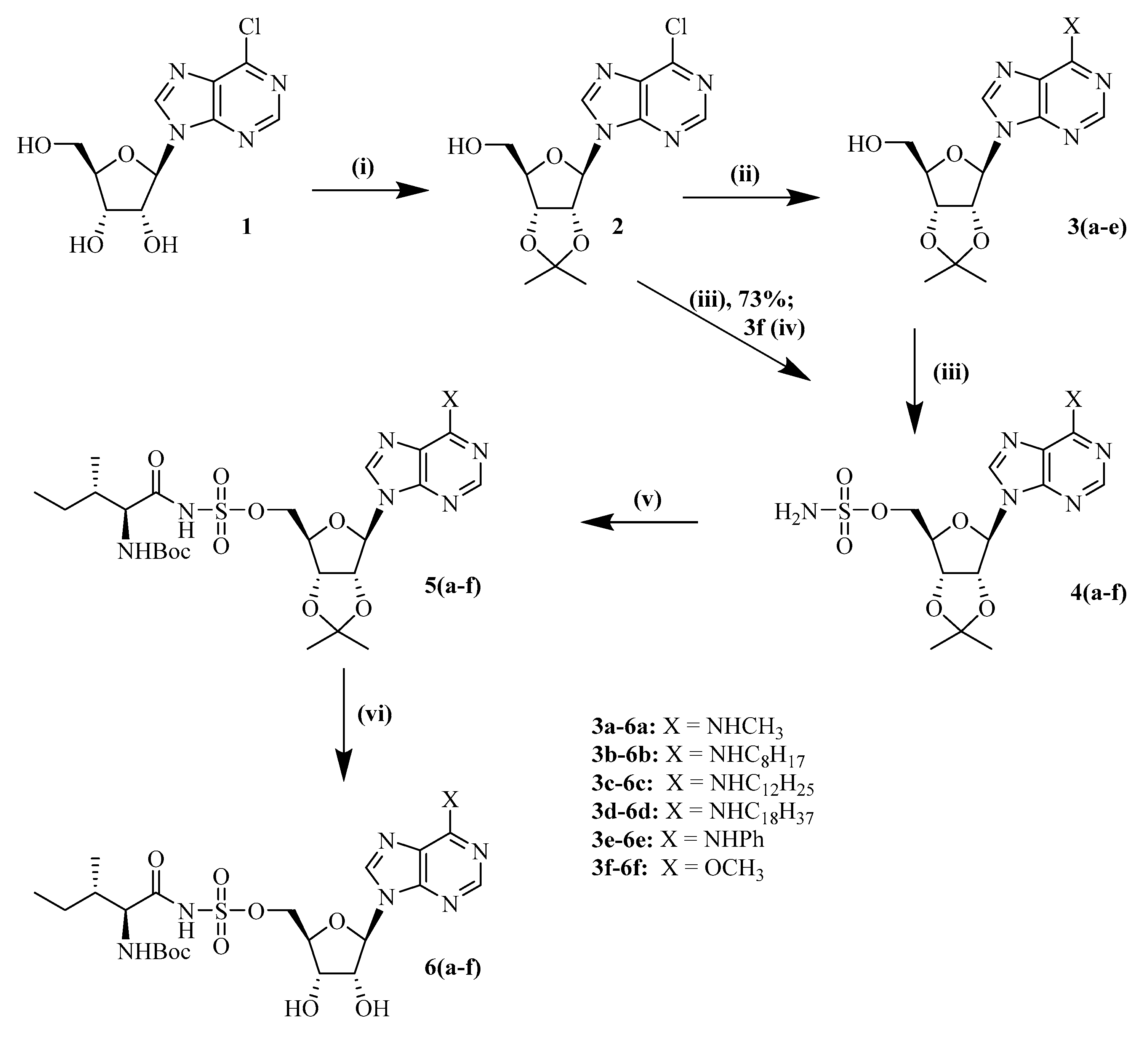
6.2.1. 2,3′-isopropylidene 6-chloropurine Riboside (2)

As reported in literature [43], compound 1 (6-chloropurine riboside, 8 g, 0.029 mol) was stirred with a mixture of dimethoxypropane (DMP) (34.31 mL, 0.29 mol) and paratoluenesulfonic acid (PTSA) (2.66 g, 0.014 mol) in dry acetone (80 mL) at room temperature for overnight. Thin layer chromatography TLC (developed at 10% methanol in dichloromethane (DCM)) was used to monitor the reaction. Saturated sodium bicarbonate was added to quench the reaction. Afterwards, the solvent was evaporated under reduced pressure. The crude product was dissolved in DCM, and the organic layer was washed two times with saturated sodium bicarbonate and one time with brine. Column chromatography was performed with a gradient of 5% methanol in DCM to obtain the desired compound 2 at 86% yield. 1H NMR (300 MHz, CDCl3) δ 1.37 (s, 3H, C–CH3), 1.64 (s, 3H, C–CH3), 3.81 (m, 1H, H-5′a), 3.97 (m, 1H, H-5′b), 4.54 (m, 1H, H-4′), 5.00 (dd, J = 10.0, 2.6 Hz, 1H, H-3′), 5.10 (dd, J = 6.0, 1.6 Hz, 1H, H-2′), 5.19 (dd, J = 6.0, 4.5 Hz, 1H, H-1′), 6.00 (d, J = 4.5 Hz, 1H, 5′OH ), 8.28 (s, 1H, H-2), 8.75 (s, 1H, H-8). 13C NMR (75 MHz, CDCl3) δ 25.5 (C–CH3), 27.8 (C–CH3), 63.4 (C-5′), 81.8 (C-4′), 83.7 (C-3′), 86.8 (C-2′), 94.2 (C-1′), 114.8 (C–(CH3)2), 145.0 (C-5), 152.0 (C-4), 152.5 (C-2). HRMS [ESI] m/z: calcd. for C13H16ClN4O4 ([M+H]+) 327.0854, found: 327.0835.

6.2.2. 2′,3′-isopropylidene-N6-(methyl)-adenosine (3a)

Compound 2 (200 mg, 0.613 mmol) was dissolved in 6 mL of DMSO in a microwave vial. Methylamine (40% solution in water) (1.5 equiv, 0.102 mL) and DIPEA (3 equiv, 0.32 mL, 1.84 mmol) were added, and the mixture was put in a microwave for 45 min, at 110 °C utilizing 150W power. The reaction was monitored by doing TLC in an acetone/hexane gradient (50:50 v/v). To perform the TLC, take 100 microliters of the crude reaction mixture and add 500 microliters of ethyl acetate and 500 microliters of water to an Eppendorf tube. Extract the organic layer and use this for TLC. After complete conversion, 350 mL of DCM was added, and the organic layer was washed with 100 mL potassium hydrogen sulfate (10% m/v) and 100 mL of brine. The organic layer was evaporated under vacuum, and column chromatography was performed with a gradient of 40% acetone in hexane to obtain 3a. Yield: 97% (0.19 g). 1H NMR (300 MHz, CDCl3) δ 1.31 (s, 3H, C–CH3), 1.58 (s, 3H, C–CH3), 3.10 (s, 3H, N6–CH3), 3.74 (d, J = 12.8 Hz, 1H, H-5′a), 3.91 (dd, J = 12.9, 1.6 Hz, 1H, H-5′b), 4.48 (s, 1H, H-4′), 5.06 (dd, J = 5.7, 1.3 Hz, 1H, H-3′), 5.17 (t, 1H, H-2′), 5.81 (d, J = 4.8 Hz, 1H, H-1′), 6.53 (s, 1H, N6-H), 6.75 (bs, 1H, 5′OH), 7.74 (s, 1H, H-2), 8.28 (s, 1H, H-8). 13C NMR (75 MHz, CDCl3) δ 25.5 (C-CH3), 27.9 (C–CH3), 29.6 (N6-CH3), 63.6 (C-5′), 82.0 (C-4′), 83.4 (C-3′), 86.5 (C-2′), 94.4 (C-1′), 114.2 (C-5), 121.6 (C–(CH3)2), 139.6 (C-8), 153.1 (C-2), 156.1 (C-6). HRMS [ESI] m/z: calcd. for C14H20N5O4 ([M+H]+) 322.1510, found: 322.1512.

6.2.3. 2′,3′-isopropylidene-N6-(octyl)-adenosine (3b)

Compound 2 (200 mg, 0.613 mmol) was added to a microwave vial containing octylamine (0.152 mL, 0.92 mmol) and DIPEA (0.32 mL, 1.84 mmol) in DMSO (6 mL). The reaction condition, TLC and purification were similar as 3a. Yield: 90% (0.23 g). 1H NMR (300 MHz, CDCl3) δ 1.21–1.64 (m, 23H, C–(CH3)2, H3C–(CH2)7), 3.56 (bs, 2H, N6–CH2), 3.74 (dd, J = 12.7, 1.6 Hz, 1H, H-5′a), 4.90–3.95 (m, 1H, H-5′b), 5.06 (dd, J = 5.9, 1.2 Hz, 1H, H-4′), 5.17 (t, 1H, H-3′), 5.81 (d, J = 4.8 Hz, 1H, H-2′), 6.11 (t, 1H, H-1′), 6.78 (d, J = 11.0 Hz, 1H, 5′OH), 7.73 (s, 1H, H-2), 8.28 (s, 1H, H-8). 13C NMR (75 MHz, CDCl3) δ 14.1- 40.9 (C–(CH3)2, H3C–(CH2)7), 63.6 (C-5′), 82.0 (C-4′), 83.4 (C-3′), 86.5 (C-2′), 94.5 (C-1′), 114.2 (C-5), 121.4 (C–(CH3)2), 139.6 (C-8), 153.1 (C-2), 155.6 (C-6). HRMS [ESI] m/z: calcd. for C21H34N5O4 ([M+H]+) 420.2605, found: 420.2597.

6.2.4. 2′,3′-isopropylidene-N6-(dodeceyl)-adenosine (3c)

Compound 2 (200 mg, 0.613 mmol) was added to a microwave vial containing dodecylamine (170.43 mg, 0.92 mmol) and DIPEA (0.32 mL, 1.84 mmol) in DMSO (6 mL). The reaction condition, TLC and purification were similar as 3a. Yield: 71% (0.21 g). 1H NMR (300 MHz, CDCl3) δ 1.24–1.70 (m, 31H, C–(CH3)2, H3C–(CH2)11), 3.2 (m, 1H, N6-H), 3.7 (d, J = 47.9 Hz, 3H, H-5′, H-4′, H-3′), 5.2 (d, J = 3.6 Hz, 2H, H-2′, H-1′), 5.78–5.83 (m, 1H, 5′OH), 7.90 (d, 1H, H-2), 8.39 (s, 1H, H-8). 13C NMR (75 MHz, CDCl3) δ 14.2–46.1 (m, C–(CH3)2, H3C–(CH2)11), 63.6 (C-5′), 81.9 (C-4′), 83.9 (C-3′), 86.7 (C-2′), 95.3 (C-1′), 114.3 (C-5), 121.4 (C–(CH3)2), 139.3 (C-8), 153.6 (C-2). HRMS [ESI] m/z: calcd. for C25H42N5O4 ([M+H]+) 476.3231, found: 476.3227.

6.2.5. 2′,3′-isopropylidene-N6-(octadecyl)-adenosine (3d)

Compound 2 (200 mg, 0.613 mmol) was added to a microwave vial containing octadecylamine (248 mg, 0.92 mmol) and DIPEA (0.32 mL, 1.84 mmol) in DMSO (6 mL). The reaction condition, TLC and purification were similar as 3a. Yield: 57% (0.20 g). HRMS [ESI] m/z: calcd. for C31H54N5O4 ([M+H]+) 560.4170, found: 560.4174.

6.2.6. 2′,3′-isopropylidene-N6-(phenyl)-adenosine (3e)

Compound 2 (200 mg, 0.613 mmol) was added to a microwave vial containing aniline (0.083 mL, 0.92 mmol) and DIPEA (0.32 mL, 1.84 mmol) in DMSO (6 mL). The reaction condition, TLC and purification were similar as 3a. Yield: 62% (0.15 g). 1H NMR (300 MHz, CDCl3) δ 1.3–1.6 (m, 6H, C–(CH3)2), 3.7–3.9 (m, 1H, N6-H), 3.9–4.0 (m, 2H, H-5′a, H-5′b), 4.5 (m, 1H, C-4′), 4.9–5.1 (m, 1H, C-3′), 5.2 (m, 1H, C-2′), 5.9 (d, J = 4.8 Hz, 1H, C-1′), 6.0 (d, J = 4.2 Hz, 1H, 5′OH), 6.4–7.6 (m, 5H, N6–C6H5), 8.4 (s, 1H, H-2), 8.7 (s, 1H, H-8). 13C NMR (75 MHz, CDCl3) δ 25.5 (C–(CH3)2), 27.9 (C–(CH3)2), 63.6 (C-5′), 81.8 (C-4′), 82.0 (C-3′), 83.5 (C-2′), 94.5 (C-1′), 114.3 (C-5), 114.7 (p-C-aniline), 121.0 (m-C-aniline), 122.0 (m-C-aniline), 124.3 (C-(CH3)2), 129.3 (o-C-aniline), 133.2 (o-C-aniline), 138.5 (N6-C-aniline), 140.5 (C-8), 145.0 (C-6), 148.3 (C-4), 152.3 (C-2). HRMS [ESI] m/z: calcd. for C19H22N5O4 ([M+H]+) 384.1661, found: 384.1664.

6.2.7. 2′,3′-isopropylidene-5′-O-sulfamoyl-6-chloropurine riboside20 (3f)

Formic acid (1.73 mL, 46 mmol) was added dropwise to CSI (4.0 mL, 46 mmol) in an ice bath for 10 min. After several minutes, the formation of a white solid was observed. Afterwards, acetonitrile (20 mL) was added and the solution was stirred for four hours at room temperature. Following stirring, the solution was added to compound 2 (5 g, 15 mmol) in DMA (20 mL) and was reacted overnight at room temperature. Column chromatography was performed with a gradient of 15–25% acetone in hexane to obtain desired compound at 73% (4.53 g) yield.1H NMR (300 MHz, CDCl3) δ 1.36 (s, 3H, C–CH3), 1.60 (s, 3H, C–CH3), 4.29–4.44 (m, 2H, C’5–H2), 4.60 (m, 1H, H-4′), 5.06 (dd, J = 6.2, 2.7 Hz, 1H, H-3′), 5.34 (dd, J = 6.2, 2.6 Hz, 1H, H-2′), 6.25 (d, J = 2.6 Hz, 1H, H-1′), 6.31 (d, J = 5.1 Hz, 2H, SO2-NH2), 8.39 (s, 1H, H-2), 8.73 (s, 1H, H-8). 13C NMR (75 MHz, CDCl3) δ 25.4 (C–CH3), 27.2 (C-CH3), 69.4 (C-5′), 81.3 (C-4′), 84.4 (C-3′), 84.7 (C-2′), 91.5 (C-1′), 115.1 (C–(CH3)2), 132.0 (C-5), 144.4 (C-8), 151.2 (C-6), 152.4 (C-2). HRMS [ESI] m/z: calcd. for C13H17ClN5O6S ([M+H]+) 406.0582, found: 406.0574.

6.2.8. 2′,3′-isopropylidene-5′-O-sulfamoyl-N6-methyl-adenosine (4a)

Formic acid (0.62 mL) was added dropwise to CSI (1.42 mL) in an RBF kept in an ice bath for 10 min. After a few minutes, the formation of a white solid was observed. Afterwards, acetonitrile (20 mL) was added, and the solution was stirred for four hours at room temperature. The obtained solution of sulfamoyl chloride is used in the next reaction as such. Sulfamoyl chloride solution (2.65 mL, 1.95 mmol) was added to a solution of compound 3a (208 mg, 0.65 mmol) in DMA (10 mL) and was stirred overnight. TLC (50:50 acetone/hexane) was used to monitor the reaction. After overnight stirring, the solvent was evaporated under reduced pressure. Column chromatography was performed with a gradient of 40–50% acetone in hexane to obtain 4a. Yield: 25% (0.065 g). 1H NMR (300 MHz, CDCl3) δ 1.3 (s, 3H, C–CH3), 1.6 (s, 3H, C–CH3), 3.0 (d, J = 7.0 Hz, 3H, NH-CH3), 4.4 (s, 2H, H-5′a, H-5′b), 4.5 (s, 1H, H-4′), 5.0 (d, J = 6.8 Hz, 1H, H-3′), 5.3 (d, J = 6.2 Hz, 1H, H-2′), 6.1 (d, J = 2.5 Hz, 1H, H-1′), 6.4 (s, 1H, N6-H), 7.9 (s, 1H, H-2), 8.3 (s, 1H, H-8). 13C NMR (75 MHz, CDCl3) δ 25.5 (C–CH3), 27.3 (C–CH3), 29.6 (N6CH3), 69.6 (C-5′), 81.4 (C-4′), 84.4 (C-3′), 84.6 (C-2′), 90.9 (C-1′), 115.0 (C-5), 120.0 (C–(CH3)2), 139.1 (C-8), 153.6 (C-2), 155.5 (C-6). HRMS [ESI] m/z: calcd. for C14H21N6O6S ([M+H]+) 401.1238, found: 401.1236.

6.2.9. 2′,3′-isopropylidene-5′-O-sulfamoyl-N6-octyl-adenosine (4b)

Sulfamoyl chloride solution (2.8 mL, 2.04 mmol) was added to compound 3b (284 mg, 0.68 mmol, 0.33 equivalent) in DMA (10 mL). Similar reaction conditions and purification methods as described for the synthesis of compound 4a were used to obtain 4b. Yield: 97% (0.33 g). 1H NMR (300 MHz, CDCl3) δ 0.7–3.1 (m, 26H, C–(CH3)2, H3C–(CH2)7, NH–CH3), 3.5 (m, 2H, N6-H, H-5′a), 4.2–4.6 (m, 3H, SO2-NH2, H-5′b), 5.1 (dd, J = 6.3, 3.0 Hz, 1H, H-4′), 5.3 (dd, J = 6.5, 2.5 Hz, 1H, H-3′), 5.9–6.2 (m, 2H, H-2′, H-1′), 7.9 (d, J = 2.4 Hz, 1H, H-2), 8.3 (s, 1H, H-8). 13C NMR (75 MHz, CDCl3) δ 14.1–40.9 (C–(CH3)2, H3C–(CH2)7), 69.2 (C-5′), 81.2 (C-4′), 84.2 (C-3′), 84.4 (C-2′), 90.8 (C-1′), 114.8 (C-5), 119.9 (C-(CH3)2), 138.8 (C-8), 153.6 (C-2), 155.0 (C-6). HRMS [ESI] m/z: calcd. for C21H35N6O6S ([M+H]+) 499.2333, found: 499.2329.

6.2.10. 2′,3′-isopropylidene-5′-O-sulfamoyl-N6-dodecyl-adenosine (4c)

Sulfamoyl chloride solution (2.4 mL, 1.76 mmol) was added to compound 3c (210 mg, 0.44 mmol) in DMA (10 mL). Similar reaction conditions and purification methods as described for the synthesis of compound 4a were used to obtain 4c. Yield: 69% (0.17 g). 1H NMR (300 MHz, CDCl3) δ 0.7–3.6 (m, 33H, C–(CH3)2, H3C–(CH2)11, N6-H), 4.3–4.6 (m, 2H, SO2–NH2), 5.1 (m, 3H, H-5′a, H-5′b, H-4′), 5.4 (m, 2H, H-3′, H-2′), 5.9–6.1 (m, 1H, H-1′), 7.9 (s, 1H, H-2), 8.3 (s, 1H, H-8). HRMS [ESI] m/z: calcd. for C25H43N6O6S ([M+H]+) 555.2959, found: 555.2958.

6.2.11. 2′,3′-isopropylidene-5′-O-sulfamoyl-N6-octadecyl-adenosine (4d)

Sulfamoyl chloride solution (1.95 mL, 1.44 mmol) was added to compound 3d (200 mg, 0.36 mmol) in DMA (10 mL). Similar reaction conditions and purification methods as described for the synthesis of compound 4a were used to obtain 4d. Yield: 56% (0.13 g). 1H NMR (300 MHz, CDCl3) δ 0.9–1.3 (m, 44H, C–(CH3)2, H3C–(CH2)17, N6-H), 4.3–4.6 (m, 3H, SO2–NH2, H-5′a), 5.1 (dd, J = 6.4, 3.0 Hz, 1H, H-5′b), 5.3 (dd, J = 6.3, 2.6 Hz, 1H, H-4′), 5.8–6.2 (m, 3H, H-3′, H-2′, H-1′), 7.9 (s, 1H, H-2), 8.3 (s, 1H, H-8). 13C NMR (75 MHz, CDCl3) δ 14.4–32.2 (C–(CH3)2, H3C–(CH2)17), 69.7 (C-5′), 81.3 (C-4′), 84.2 (C-3′), 84.6 (C-2′), 91.1 (C-1′), 115.1 (C-(CH3)2), 139.2 (C-8), 153.8 (C-2). HRMS [ESI] m/z: calcd. for C25H43N6O6S ([M+H]+) 555.2959, found: 555.2958.

6.2.12. 2′,3′-isopropylidene-5′-O-sulfamoyl-N6-phenyl-adenosine (4e)

Sulfamoyl chloride solution (2 mL, 1.47 mmol) was added to compound 3e (188 mg, 0.68 mmol) in DMA (10 mL). Reaction conditions and purification methods were as described for 4a to obtain 4e. Yield: 76% (0.24 g).1H NMR (300 MHz, CDCl3) δ 1.4–1.6 (m, 6H, C-(CH3)2), 2.1–3.0 (m, 3H, N6-H, SO2NH2), 4.4 (m, 2H, H-5′a, H-5′b), 4.5–4.7 (m, 1H, H-4′), 5.1 (m, 1H, H-3′), 5.3–5.4 (m, 1H, H-2′), 6.2 (m, 1H, H-1′), 6.5 (s, 1H, p-CH-aniline), 7.4 (m, 2H, 2x m-CH-aniline), 7.7–7.8 (m, 1H, o-H-aniline), 8.1 (d, J = 3.2 Hz, 1H), 8.4 (d, J = 18.6 Hz, 1H, H-2), 8.7 (d, J = 2.1 Hz, 1H, H-8). 13C NMR (75 MHz, CDCl3) δ 25.5 (C–(CH3)2), 27.4 (C–(CH3)2), 69.6 (C-5′), 81.5 (C-4′), 84.6 (C-3′), 84.8 (C-2′), 91.2 (C-1′), 115.0 (C-5), 115.2 (p-C-aniline), 120.9 (m-C-aniline), 124.0 (m-C-aniline), 129.2 (o-C-aniline), 138.8 (N6-C-aniline), 139.9 (o-C-aniline), 144.5 (C-8), 149.2 (C-6), 151.3 (C-4), 152.5 (C-2). HRMS [ESI] m/z: calcd. for C19H22N6O6S ([M+H]+) 463.1394, found: 463.1385.

6.2.13. 2′, 3′-isopropylidene-5′-O-sulfamoyl-6-O-methyl-purine riboside (4f)

A 20% solution of sodium methoxide in methanol (0.27 mL, 0.98 mmol) was added to a cooled solution of 3f in dry methanol. The reaction was performed at 0 °C. After two hours, a few drops of glacial acetic acid were added to quench the reaction. The reaction mixture was evaporated under reduced pressure. Column chromatography was performed with a gradient of 40% acetone in hexane to yield 4f. Yield: 39% (0.15 g). 1H NMR (300 MHz, CDCl3) δ 1.3 (s, 3H, C–CH3), 1.4 (s, 3H, C–CH3), 4.1 (s, 3H, O–CH3), 4.3–4.5 (m, 2H, H-5′a, H-5′b), 4.6 (s, 1H, H-4′), 5.1 (dd, J = 6.4, 2.8 Hz, 1H, H-3′), 5.4 (dd, J = 6.2, 2.4 Hz, 1H, H-2′), 6.2 (d, J = 2.4 Hz, 1H, H-1′), 8.1 (s, 1H, H-2), 8.5 (s, 1H, H-8). 13C NMR (75 MHz, CDCl3) δ 25.5 (C-CH3), 27.4 (C–CH3), 54.6 (O-CH3), 69.8 (C-5′), 81.5 (C-4′), 84.5 (C-3′), 84.8 (C-2′), 91.6 (C-1′), 115.2 (C-5), 122.1 (C– (CH3)2), 141.5 (C-8), 151.4 (C-2), 152.8 (C-6).

6.2.14. 2′,3′-isopropylidene-5′-O-(N-(Nα-Boc-L-isoleucyl))-sulfamoyl-N6-methyl-adenosine (5a)

Compound 4a (100 mg, 0.27 mmol) was added to a solution of Boc-Ile-Osu (133 mg, 0.405 mmol) and DBU (0.06 mL, 0.405 mmol) in DMF (12 mL). The solution was stirred overnight at room temperature. TLC was developed with a methanol/ethyl acetate mixture (10:90) to monitor the reaction. After stirring overnight, the solvent was evaporated under reduced pressure. Column chromatography was performed with a mixture of 5% methanol in ethyl acetate to obtain compound 5a. Yield: 85% (0.14 g). 1H NMR (300 MHz, CDCl3) δ 0.8–3.4 (m, 30H, Boc-Ile protons, C-(CH3)2, NH-CH3), 3.9 (s, 1H, N6-H), 4.1 (m, 1H, H-5′a), 4.2 (m, 3H, H-5′b, H-4′, H-3′), 4.5 (s, 1H, H-2′), 6.2 (d, J = 2.5 Hz, 1H, H-1′), 8.3 (d, J = 12.1 Hz, 2H, H-2, H-8). 13C NMR (75 MHz, CDCl3) δ 11.6–62.2 (C-(CH3)2), Boc-Ile carbons), 69.1 (C-5′), 81.5 (C-4′), 85.1 (C-3′), 90.7 (C-1′), 114.3 (C-5), 119.5 (C–(CH3)2), 139.6 (C-8), 148.6 (C-4), 153.7 (C-6), 155.3 (-C(O)-tBu). HRMS [ESI] m/z: calcd. for C25H39N7O9S ([M-H]-) 612.2457, found: 612.2462.

6.2.15. 2′,3′-isopropylidene-5′-O-(N-(Nα-Boc-L-isoleucyl))-sulfamoyl-N6-octyl-adenosine (5b)

Compound 4b (350 mg, 0.763 mmol) was added to a solution of Boc-Ile-Osu (376 mg, 1.145 mmol) and DBU (0.171 mL, 1.145 mmol) in DMF (12 mL). Reaction conditions and purification were similar as described for the synthesis of 5a, except that TLC and column chromatography were performed with a mixture of 30% acetone in hexane, to obtain 5b. Yield: 87% (0.47 g). 1H NMR (300 MHz, CDCl3) δ 0.8 - 3.6 (m, 44H, Boc-Ile protons, C–(CH3)2, NH(CH2)7CH3), 3.7 (d, J = 11.8 Hz, 1H, N6–H), 4.3 (m, 2H, H-5′a, H-5′b), 4.5 (d, J = 5.8 Hz, 1H, H-4′), 4.5 (s, 1H, H-3′), 4.9 (s, 1H, H-2′), 6.2 (s, 1H, H-1′), 8.2 (d, J = 15.9 Hz, 2H, SO2NH, Nα-H), 8.4 (s, 1H, H-2), 8.6 (s, 1H, H-8). HRMS [ESI] m/z: calcd. for C32H53N7O9S ([M-H]-) 710.3524, found: 710.3397.

6.2.16. 2′,3′-isopropylidene-5′-O-(N-(Nα-Boc-L-isoleucyl))-sulfamoyl-N6-dodecyl-adenosine (5c)

Compound 4c (200 mg, 0.36 mmol) was added to a solution of Boc-Ile-OSu (177 mg, 0.54 mmol) and DBU (0.08 mL, 0.54 mmol) in DMF (12 mL). Reaction conditions and purification were similar as described for the synthesis of 5a, except that TLC and column chromatography were carried out with a mixture of 35% acetone in hexane, to obtain 5c. Yield: 51% (0.14 g). 1H NMR (300 MHz, CDCl3) δ 1.4 –4.0 (m, 52H, Boc-Ile protons, C–(CH3)2, NH(CH2)11CH3), 4.2 (s, 1H, N6–H), 4.6 (s, 1H, H-5′a), 4.9 (s, 1H, H-5′b), 5.2 (s, 1H, H-4′), 5.7 (s, 1H, H-3′), 5.8 (s, 1H, H-2′), 8.9 (s, 1H, H-8). 13C NMR (75 MHz, CDCl3) δ 11.8–44.7 (C-(CH3)2), Boc-Ile carbons), 68.7 (C-5′), 80.5 (C-4′), 85.1 (C-3′), 85.9 (C-2′), 92.5 (C-1′), 114.3 (C-5), 153.7 (C-6), 155.0 (-C(O)-tBu). HRMS [ESI] m/z: calcd. for C36H61N7O9S ([M-H]-) 766.41784, observed 766.4161.

6.2.17. 2′,3′-isopropylidene-5′-O-(N-(Nα-Boc-L-isoleucyl))-sulfamoyl-N6-octadecyl-adenosine (5d)

Compound 4d (150 mg, 0.234 mmol) was added to a solution of Boc-Ile-Osu (115 mg, 0.35 mmol) and DBU (0.05 mL, 0.35 mmol) in DMF (12 mL). Reaction conditions and were similar as described for 5a, except that TLC and column chromatography were performed with a mixture of 35% acetone in hexane, to obtain 5d. Yield: 43% (0.12 g). 1H NMR (300 MHz, CDCl3) δ 0.8 - 3.6 (m, 52H, Boc-H, C-(CH3)2, NH(CH2)17CH3), 4.0 (s, 1H, N6-H), 4.3 (s, 2H, H-5′a, H-5′b), 4.6 (s, 1H, H-4′), 5.0–5.1 (m, 1H, H-3′), 5.2 (s, 1H, H-2′), 5.4 (s, 1H, H-1′), 6.3 (s, 1H, Nα-H), 6.6 (s, 1H, SO2NH), 8.4 (s, 1H, H-2). HRMS [ESI] m/z: calcd. for C42H73N7O9S ([M-H]-) 850.5117, found: 850.5115.

6.2.18. 2′,3′-isopropylidene-5′-O-(N-(Nα-Boc-L-isoleucyl))-sulfamoyl-N6-phenyl-adenosine (5e)

Compound 4e (250 mg, 0.60 mmol) was added to a solution of Boc-Ile-Osu (295 mg, 0.90 mmol) and DBU (0.134 mL, 0.90 mmol) in DMF (12 mL). Reaction conditions and purification were similar as described for 5a, except that TLC and column chromatography were carried out with a mixture of 35% acetone in hexane to obtain 5e. Yield: 49% (0.20 g). 1H NMR (300 MHz, CDCl3) δ 1.4–2.1 (m, 22H, Boc-Ile-protons, C–(CH3)2), 4.5 (s, 1H, H-Cα), 4.6 (s, 1H, H-5′a), 4.9 (s, 1H, H-5′b), 5.2 (s, 1H, H-4′), 5.5 (d, J = 14.3 Hz, 1H, H-3′), 5.7 (s, 1H, H-2′), 6.9 (d, J = 7.7 Hz, 1H, H-1′), 7.7 (m, 1H, p-CH-aniline), 7.8 –8.0 (m, 2H, m-CH-aniline), 8.3 (d, J = 8.0 Hz, 2H, o-CH-aniline), 9.0 (s, 1H, H-2), 9.1 (s, 1H, H-8). 13C NMR (75 MHz, CDCl3) δ 11.8–62.2 (C–(CH3)2), Boc-Ile carbons), 69.2 (C-5′), 80.5 (C-4′), 85.3 (C-3′), 85.8 (C-2′), 91.3 (C-1′), 114.5 (C-5), 120.3 (p-C-aniline), 121.2 (m-C-aniline), 121.8 (m-C-aniline), 123.2 (C–(CH3)2), 123.8 (o-C-aniline), 126.9 (o-C-aniline), 139.0 (N6-C-aniline), 139.5 (C-8), 149.5 (C-6), 152.3 (C-4), 152.8 (C-2), 156.7 (-C(O)-tBu). HRMS [ESI] m/z: calcd. for C30H41N7O9S ([M-H]-) 674.2613, found: 674.2592.

6.2.19. Synthesis of 2′, 3′-isopropylidene-5′-O-sulfamoyl-(N-(Nα-Boc-L-isoleucyl)-6-O-methyl-purine riboside (5f)

Compound 4f (150 mg, 0.37 mmol) was added to a solution of Boc-Ile-Osu (184 mg, 0.54 mmol) and DBU (0.08 mL, 0.54 mmol) in DMF (12 mL). The reaction was stirred overnight at room temperature. The solvent was evaporated under reduced pressure. Column chromatography of the obtained residue was carried out with a gradient of 10–20% acetone in hexane to yield 5f. Yield: 92% (0.21 g).

6.2.20. Synthesis of 5′-O-(N-L-isoleucyl)-sulfamoyl-N6-methyl-adenosine (6a)

Compound 5a (120 mg, 0.20 mmol) was dissolved in a mixture of TFA, water and DCM (50:25:25 v/v/v) and stirred for 3 h at room temperature. TLC was performed in a methanol/DCM mixture (10:90 v/v) to check the reaction progression. After 3h, the mixture was evaporated at reduced pressure at 25 °C. RP-HPLC was performed with a C18 column with gradient elution using water/acetonitrile as the mobile phase to purify and obtain 6a. Yield: 37% (0.035 g). 1H NMR (300 MHz, D2O) δ 0.90–1.02 (m, 6H, Ile-δ-CH3, Ile-γ-CH3), 1.20-1.26 (m, 1H, Ile-γ-CH2 Ha), 1.56–1.61 (m, 1H, Ile-γ-CH2 Hb), 1.95-1.99 (m, 1H, Ile-β-CH), 3.11 (bs, 3H, N–CH3), 3.56 (d, J = 4.1 Hz, 1H, Ile-α-CH), 4.29–4.39 (m, 4H, H-5′, H-5″, H-4′, H-3′), 4.62 (t, 1H, J = 5.0 Hz, H-2′), 6.06 (s, 1H, J = 5.3 Hz, H-1′), 8.24 (s, 1H, H-2), 8.45 (s, 1H, H-8). 13C NMR (75 MHz, D2O) δ 12.1 (Ile- δ -CH3), 15.5 (Ile-γ-CH3), 25.7 (Ile- δ-CH2), 38.2 (Ile-β-CH), 61.5 (Ile-α-CH), 69.0 (C-5′), 72.0 (C-4′), 76.2 (C-3′), 84.2 (C-2′), 89.4 (C-1′), 120.6 (C-5), 140.6 (C-8), 153.9 (C-2), 156.7 (C-6), 174.9 (-C(C=O, Ile). HRMS [ESI] m/z: calcd. for C17H27N7O7S ([M-H]-) 472.1620, found: 472.1633.

6.2.21. Synthesis of 5′-O-(N-L-isoleucyl)-sulfamoyl-N6-octyl-adenosine (6b)

Compound 5b (300 mg, 0.42 mmol) was treated like compound 5a regarding reaction condition and purification to obtain 6b. Yield: 21% (0.051 g). 1H NMR (300 MHz, MeOD) δ 0.90–0.95 (m, 6H, CH3-Octyl chain and Ile-δ-CH3), 1.03 (d, J = 7.0 Hz, 3H, Ile-γ-CH3), 1.32-1.46 (m, 11H, Octyl chain and Ile-γ-CH2 Ha), 1.58–1.62 (m, 1H, Ile-γ-CH2 Hb), 1.70–172 (m, 2H CH2-Octyl chain), 1.97–2.01 (m, 1H, Ile-β-CH), 3.56 (d, J = 3.7 Hz, 1H, Ile-α-CH), 3.58–3.62 (m, 2H, NH-CH2 octyl chain), 4.31–4.42 (m, 4H, H-5′a, H-5′b, H-4′, H-3′), 4.64 (t, J = 5.1 Hz, 1H, H-2′), 6.10 (d, J = 5.4 Hz, 1H, H-1′), 8.25 (s, 1H, H-2), 8.48 (s, 1H, H-8). 13C NMR (75 MHz, D2O) δ 12.1 (Ile- δ -CH3), 14.4 (C8 alkyl chain), 15.5 (Ile-γ-CH3), 23.7(C8 alkyl chain), 25.6 (Ile- δ-CH2), 28.0–33.0 (C8 alkyl chain), 38.2 (Ile-β-CH), 41.7 (C8 alkyl chain), 61.5 (Ile-α-CH), 69.1 (C-5′), 72.0 (C-4′), 76.2 (C-3′), 84.3 (C-2′), 89.3 (C-1′), 120.4 (C-5), 140.4 (C-8), 149.7 (C-6), 154.0 (C-4), 156.1 (C-2), 175.0 (C=O, Ile). HRMS [ESI] m/z: calcd. for C24H41N7O7S ([M+H]+) 572.2861, found 572.2864.

6.2.22. 5′-O-(N-L-isoleucyl)-sulfamoyl-N6-dodecyl-adenosine (6c)

Compound 5c (90 mg, 0.12 mmol) was treated in the same way as compound 5a regarding reaction condition and purification to obtain 6c. Yield: 11% (0.008 g). 1H NMR (300 MHz, MeOD) δ 0.87–0.90 (m, 6H, CH3-dodecyl chain and Ile-δ-CH3), 1.04 (d, J = 7.0 Hz, 3H,), 1.28–1.30 (m, 31H, dodecyl chain, Ile-γ-CH3), 1.68–1.73 (m, 4H, Ile-β-CH, dodecyl chain), 3.37–3.39 (m, 2H, NH-CH2 dodecyl chain), 3.71–3.77 (m, 3H, Ile-α-CH), 4.51–4.66 (m, 5H, H-5′a, H-5′b, H-3′, H-4′), 4.89–4.90 (s, 1H, H-2′), 6.55 (s, 1H, H-1′), 8.31 (d, J = 4.8 Hz, 1H, H-2), 8.56 (s, 1H, H-8). HRMS [ESI] m/z: calcd. for C28H49N7O7S ([M-H]-): 626.3341, found: 626.3338.

6.2.23. 5′-O-(N-L-isoleucyl)-sulfamoyl-N6-octadecyl-adenosine (6d)

Compound 5d (80 mg, 0.09 mmol) was treated in the same way as compound 5a to obtain 6d. Yield: 19% (0.012 g). 1H NMR (300 MHz, MeOD) δ 0.89-0.98 (m, 8H, CH3-octadecyl chain and Ile-δ-CH3), 1.04 (d, J = 7.0 Hz, 3H, Ile-γ-CH3), 1.30 (s, 31H, octadecyl chain), 1.55–1.61 (m, 1H, Ile-γ-CH2 Hb), 1.69-171 (m, 2H CH2-Octadecyl chain), 1.97-2.01 (m, 1H, Ile-β-CH), 3.56-362 (m, 3H, Ile-α-CH and NH-CH2 octadecyl chain), 4.31–4.41 (m, 5H, H-5′a, H-5′b, H-3′, H-2′, H-4′), 6.09 (d, J = 5.4 Hz, H-1′), 8.25 (d, J = 4.8 Hz, 1H, H-2), 8.48 (s, 1H, H-8). HRMS [ESI] m/z: calcd. for C34H61N7O7S ([M+H]+) 712.4426, found: 712.4470.

6.2.24. Synthesis of 5′-O-(N-L-isoleucyl)-sulfamoyl-N6-phenyl-adenosine (6e)

Compound 5e (150 mg, 0.22 mmol) was treated as for 5a to obtain 6e. Yield: 18% (0.021 g). 1H NMR (300 MHz, MeOD) δ 0.90–0.95 (m, 3H, Ile-δ-CH3,), 1.03 (d, J = 6.9 Hz, 3H, Ile-γ-CH3), 1.20–1.33 (m, 1H, Ile-γ-CH2 Ha), 1.56–1.60 (m, 1H, Ile-γ-CH2 Hb), 1.97–2.01 (m, 1H, Ile-β-CH), 3.61 (d, J = 3.9 Hz, 1H, Ile-α-CH), 4.37–4.45 (m, 5H, H-5′, H-5″, H-4′, H-3′, H-2′), 6.14 (d, J = 5.1 Hz, 1H, H-1′), 7.14 (t, J = 7.1 Hz, 1H, p-CH-aniline), 7.39 (t, J = 7.5 Hz, 2H, m-CH-aniline), 7.76 (d, J = 8.1 Hz, 2H, o-CH-aniline), 8.38 (s, 1H, H-2), 8.57 (s, 1H, H-8). 13C NMR (75 MHz, D2O) δ 10.5 (Ile- δ -CH3), 13.9 (Ile-γ-CH3), 23.9 (Ile- δ-CH2), 36.5 (Ile-β-CH), 59.8 (Ile-α-CH), 67.5 (C-5′), 70.2 (C-4′), 74.4 (C-3′), 82.5 (C-2′), 87.7 (C-1′), 119.4 (C-5), 120.7 (o-CH-aniline), 123.4 (p-CH-aniline), 128.3 (m-CH-aniline), 138.3 (NH-CH-aniline), 139.8 (C-8), 142.6 (C-6), 149.0 (C-4), 152.0 (C-2), 173.7 (C=O, Ile). HRMS [ESI] m/z: calcd. for C22H29N7O7S ([M-H]-): 534.1776, found 534.1777.

6.2.25. Synthesis of 5′-O-(N-L-isoleucyl)-sulfamoyl-6-O-methyl-purine riboside (6f)

Compound 5f (100 mg, 0.16 mmol) was added to a fresh solution of TFA/water/DCM (50:25:25 v/v), and the reaction was stirred for 3 h at room temperature. TLC, with an acetone/hexane gradient, was used to monitor the reaction. The reaction mixture was evaporated under reduced pressure at 25 °C. The product was purified with RP-HPLC using a C18 column and gradient elution, with water/acetonitrile as the mobile phase, to obtain 6f. Yield: 55% (0.042 g). 1H NMR (300 MHz, MeOD) δ 0.91–1.03 (m, 6H, Ile-δ-CH3, Ile-γ-CH3), 1.28–1.29 (m, 1H, Ile-γ-CH2 Ha), 1.54–1.56 (m, 1H, Ile-γ-CH2 Hb), 1.95–1.97 (m, 1H, Ile-β-CH), 3.58 (d, J = 4.0 Hz, 1H, Ile-α-CH), 4.18 (s, 3H, O-CH3), 4.32–4.43 (m, 4H, H-5′a, H-5′b, H-4′, H-3′), 4.66–4.69 (m, 1H, H-2′), 6.17 (d, J = 5.2 Hz, 1H, H-1′), 8.53 (s, 1H, H-2), 8.68 (s, 1H, H-8). 13C NMR (75 MHz, D2O) δ 12.1 (Ile- δ -CH3), 15.4 (Ile-γ-CH3), 25.7 (Ile- δ-CH2), 38.2 (Ile-β-CH), 54.8 (O-Me), 61.4 (Ile-α-CH), 69.0 (C-5′), 72.0 (C-4′), 76.2 (C-3′), 84.4 (C-2′), 89.8 (C-1′), 143.5 (C-2), 153.5 (C-2). HRMS [ESI] m/z: calcd. for C17H26N6O8S ([M-H]-) 473.1460, found: 473.1471.
The synthesis of compound 2′,3′,5′-tri-O-TBDMS-adenosine (8), 2′,3′-di-O-TBDMS adenosine (9), and 2′,3′-di-O-TBDMS-5′-O-sulfamoyl adenosine (10) was performed by following reported procedure20 and obtained at 88, 84 and 97% yields respectively.

6.2.26. 5′-O-[N-(N-Boc)leucyl]sulfamoyl adenosine (11)

Compound 10 (300 mg, 0.52 mmol), Boc-Leu-OSu (1.2 equivalent, 205.85 mg, 0.63 mmol), DBU (1 equivalent, 0.08 mL, 0.52 mmol) were added together using DMF (10 mL) as solvent. The reaction, which after a short period of time turned pink, was let to react at room temperature and overnight. A small sample was work-upped with EtOAc and 10% KHSO4 for TLC analysis which was later developed with 1% MeOH in EtOAc and sprayed with ammonium molybdate (Rf = 0.73). The solvents were evaporated after completion of the reaction and the obtained wine-red dense liquid was partitioned between water and EtOAc. A small amount of 10% KHSO4 was added in the first wash to assure that the pH from the aqueous layer had pH 5-6. The organic layers were collected, dried over MgSO4, filtered and, lastly, the solvent was evaporated. The residue was purified using silica gel column chromatography with elution at 1.5% MeOH:EtOAc. The UV-active fractions were collected and further dried. Yield: 77%. 1H NMR (300 MHz, MeOD) δ -0.42 (3H, s, CH3-Si), -0.18 (3H, m, CH3-Si), 0.11–0.12 (6H, m, CH3-Si), 0.66 (9H, s, tBu CH3), 0.84–0.86 (6H, m, Leu-δ’-CH3, Leu-δ″), 0.92 (9H, s, tBu CH3), 1.36 (10H, s, Boc-tBu CH3 Leu-γ-CH), 1.47–1.63 (2H, m, Leu-ß-CH2), 3.77-3.78 (1H, m, Leu-α-CH), 4.02–4.14 (3H, m, H5″, H5’, H3’), 4.32 (1H, d, J = 7.4 Hz, H2’), 4.89–4.93 (1H, m, H4’), 5.95 (1H, d, J = 7.4 Hz, H1’), 7.26 (2H, bs, NH2), 8.13 (1H, s, H8), 8.47 (1H, s, H2). 13C NMR (300 MHz, MeOD), δ -5.6 (CH3-Si), -5.7 (CH3-Si), -4.6 (CH3-Si), 17.5 (tBu C(CH3)3), 17.9 (tBu C(CH3)3), 22.1 (Leu-δ’-CH3), 23.4 (Leu-δ″-CH3), 24.5 (Leu-γ-CH), 25.6 (tBu C(CH3)3), 25.9 (tBu C(CH3)3), 28.4 (Boc-tBu CH3), 43.0 (Leu-ß-CH2), 54.9 (Leu-α-CH), 66.9 (C-5′), 73.4 (C-3′), 74.7 (C-2′), 77.5 (C-4′), 84.1 (Boc-tertiary C), 86.1 (C-1′), 119.0 (C-5), 139.7 (C-8)150.0 (C-4), 152.8 (C-2), 155.1 (Boc- carbonyl C), 156.1(C-6), 177.0 (CONH). HRMS [ESI] m/z: calcd. for C33H60N7O9S1Si2 ([M-H]-) 786.3717, found: 786.3720.

6.2.27. 2′,3′-di-O-TBDMS-5′-O-(N-leucyl)sulfamoyl adenosine (12)

Selective Boc deprotection was performed by adding a 10 mL solution of TFA:DCM:water (2:1:1) to the Boc and TBDMS protected compound 9 (273 mg). The reaction was first started at 0 °C and then held at room temperature for 2 h. For TLC (20% MeOH in EtOAc plus a few drops of TEA was used as mobile phase; Rf = 0.3), the sample was first evaporated in high-vacuum, then the dried compound was dissolved and evaporated two times after dissolving with EtOH to assure that there was no TFA left, as it could interfere with TLC analysis. Upon completion of the reaction, TFA and DCM were evaporated, and the resulting residue was purified by silica gel column chromatography. 1H NMR (300 MHz, MeOD) δ -0.36 (3H, s, CH3-Si), 0.06 (3H, m, CH3-Si), 0.11–0.14 (6H, m, CH3-Si), 0.69 (9H, s, tBu CH3), 0.88–0.93 (15H, m, tBu CH3, Leu-δ’-CH3, Leu-δ″), 1.47–1.53 (1H, m, Leu-γ-CH), 1.69–1.77 (2H, m, Leu-ß-CH2), 3.48–3.53 (1H, m, Leu-α-CH), 4.24–4.39 (4H, m, H5″, H5’, H3’, H2’), 4.76–4.80 (1H, m, H4’), 6.06 (1H, d, J = 7.1 Hz, H1’), 8.14 (1H, s, H8), 8.53 (1H, s, H2). 13C NMR (300 MHz, MeOD), δ -5.2 (CH3–Si), -4.3 (CH3–Si), -4.2 (CH3–Si), 18.7 (tBu C(CH3)3), 18.9 (tBu C(CH3)3), 22.3 (Leu-δ’-CH3), 23.4 (Leu-δ″-CH3), 25.8 (Leu-γ-CH), 26.2 (tBu C(CH3)3), 26.4 (tBu C(CH3)3), 43.3 (Leu-ß-CH2), 55.9 (Leu-α-CH), 69.1 (C-5′), 74.6 (C-3′), 77.6 (C-2′), 85.9 (C-4′), 88.5 (C-1′), 120.1 (C-5), 141.6 (C-8), 151.0 (C-4), 153.9 (C-2), 157.3(C-6), 178.6 (CONH). HRMS [ESI] m/z: calcd. for C28H52N7O7S1Si2 ([M-H]-) 686.3193, found 686.3195.

6.2.28. 2′,3′-di-O-TBDMS-5′-O-[Nα-(p-nitrobenzyloxycarbonyl)leucyl]sulfamoyl adenosine (13)

For coupling of the leucyladenosine derivative and the promoiety, the dried compound 12 (116 mg, 0.17 mmol) was mixed with 4-nitrobenzyl chloroformate (1.2 equivalent, 43.62 mg, 0.204 mmol) and DIPEA (3 equivalent, 0.09 mL, 0.51 mmol) and dissolved in DFM. The reaction was left at room temperature overnight. A predeveloped TLC was eluted at 5% MeOH in DCM in the presence of a few drops of TEA (Rf = 0.42). The solvent was evaporated in order to obtain a residue and silica gel column chromatography was used to purify the compound. Yield: 62%. 1H NMR (300 MHz, MeOD) δ -0.21–0.20 (3H, s, CH3–Si), 0.11–0.14 (3H, m, CH3–Si), 0.29–0.31 (6H, m, CH3–Si), 0.86–0.92 (9H, m, Leu-δ’-CH3, Leu-δ″-CH3, Leu-γ-CH, Leu-ß-CH2), 1.01–1.11 (18H, m, 2·tBu CH3), 4.43–4.57 (6H, m, H5″, H5’, H4’, H3’, H2’, Leu-α-CH), 5.34 (2H, s, Bn CH2), 6.21–6.26 (1H, m, H1’), 7.69-7.71 (2H, d, o-2CH), 8.31-8.33 (3H, d, H8, m-2H), 8.70 (1H, m, H2). 13C NMR (300 MHz, MeOD), δ -5.2 (CH3–Si), -4.3 (CH3–Si), -4.2 (CH3–Si), 22.1 (Leu-δ’-CH3), 23.4 (Leu-δ″-CH3), 23.8 (Leu-γ-CH), 26.2 (tBu C(CH3)3), 26.4 (tBu C(CH3)3), 42.5 (Leu-ß-CH2), 55.2 (Leu-α-CH), 61.1 (C-5′), 66.1 (Bn), 74.6 (C-3′), 77.4 (C-2′), 85.9 (C-4′), 88.3 (C-1′), 120.1 (C-5), 124.5 (o-CH), 130.0 (m-CH),141.5 (C), 145.5 (p-CH), 148.8 (C-4), 151.0 (C-2), 153.9 (C-6) 157.3 (COO-Bn), 177.95 (CONH). HRMS [ESI] m/z: calcd. for C36H58N8O11S1Si2 ([M-H]-) 865.3411, found: 865.3431.

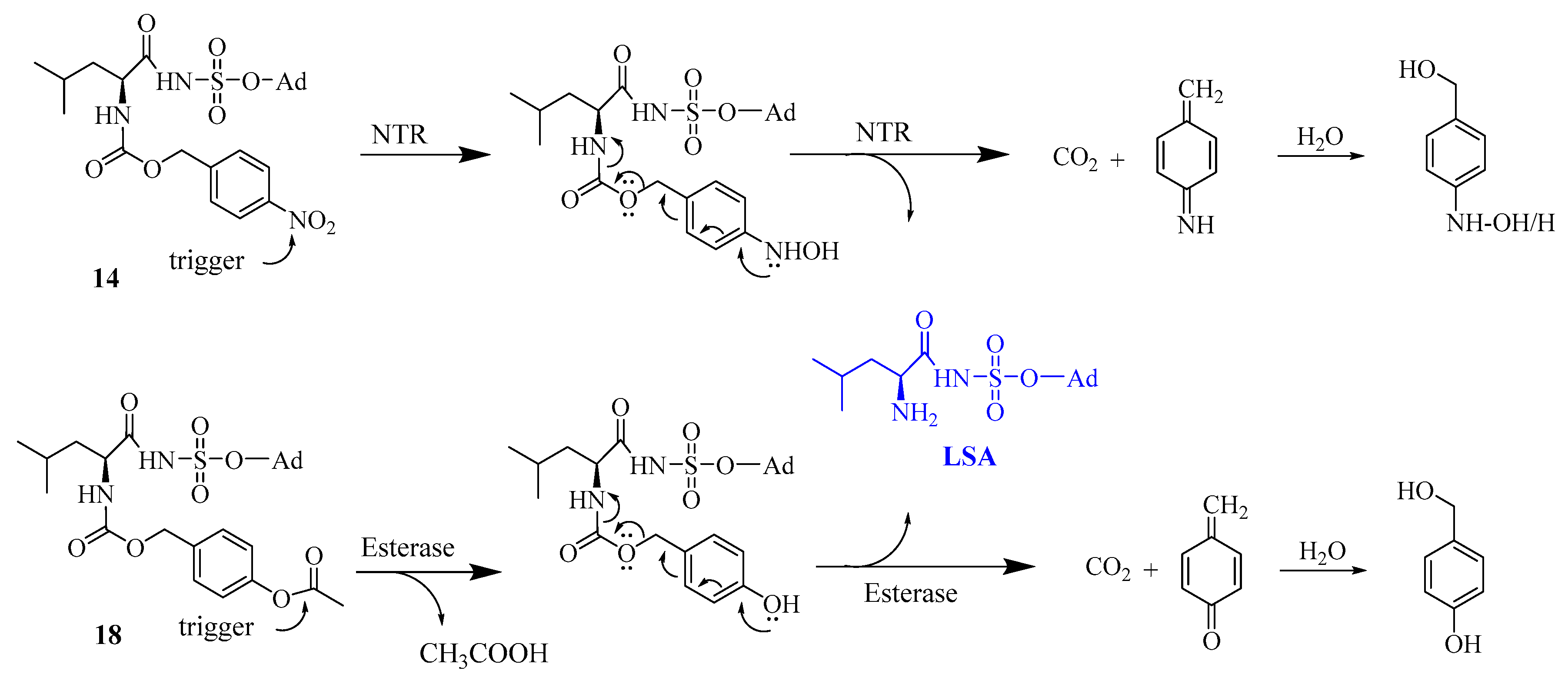
6.2.29. 5′-O-[Nα-(p-nitrobenzyloxycarbonyl)leucyl]sulfamoyl adenosine (14)

First, compound 13 (160 mg, 0.19 mmol) was dissolved in THF (5 mL) and Et3N·3HF (2 mL) was added to start the reaction at room temperature for overnight. The reaction progress was checked with TLC which was predeveloped in 10% MeOH: DCM in the presence of TEA (Rf = 0.48). The reaction mixture was purified using silica gel chromatography. Fractions corresponding to compound 14 were collected, evaporated and further purified using reverse phase HPLC. 1H NMR (300 MHz, MeOD) δ 0.89–0.95 (6H, m, Leu-δ’-CH3, Leu-δ″-CH3) 1.70–1.78 (1H, m, Leu-γ-CH), 1.90–1.92 (2H, m, Leu-ß-CH2), 4.19–4.38 (5H, m, H5″, H5’, H4’, H3’, Leu-α-CH), 4.63–4.66 (1H, m, H2’), 5.18–5.22 (2H, m, Bn-CH2), 6.06–6.08 (1H, d, H1’, J = 5.73), 7.55–7.57 (2H, d, o-2CH, J = 8.70), 8.17–8.20 (3H, m, H8, m-2CH), 8.49 (1H, s, H2). 13C NMR (300 MHz, MeOD), 20.3 (Leu-δ’-CH3), 21.6 (Leu-δ″-CH3), 24.3 (Leu-γ-CH), 40.7 (Leu-ß-CH2), 59.4 (Leu-α-CH), 64.5 (C-5′), 67.7 (Bn), 70.5 (C-3′), 74.3 (C-2′), 82.7 (C-4′), 87.4 (C-1′), 118.4 (C-5), 122.8 (m-CH), 127.4 (o-CH), 139.4 (ipso-CH), 144.3 (C-8), 147.1 (C-4), 149.2 (p-CH), 152.2 (C-2), 155.5 (C-6), 171.2 (CO–NαH), 178.02 (CONH). HRMS [ESI] m/z: calcd. for C24H30N8O11S1 ([M-H]-) 637.1682 found: 637.1689.

6.2.30. p-Acetoxybenzylchloroformate (16)

Take triphosgene (134mg, 0.45 mmol) in a round bottom flask and dry using high vacuum; then at 0 °C add DIPEA (0.16 mL, 0.9 mmol, 2 equiv.) and 5 mL THF. In another round bottom flask weigh 75 mg of p-acetoxybenzylalcohol and add 5 mL THF, then add this solution to triphosgene at 0 °C. Let the reaction continue at 0 °C for four hours. Use the reaction mixture without purification for the next reaction. The formation of the compound was confirmed by NMR. 1H NMR (300 MHz, CDCl3) δ 2.29 (s, 3H, acetyl CH3), 5.09(s, 2H, benzyl CH2), 7.35–7.38 (m, 2H, meta to acetyl), 7.04–7.09 (m, 2H, ortho to acetyl); 13C NMR (75 MHz, CDCl3) δ 21.1 (acetyl CH3), 66.5 (benzyl CH2), 121.7 (ortho C to acetyl), 128.7 (meta C to acetyl), 133.9 (para C to acetyl) 150.6 (Acetoxy-C), 156.3 (carbonyl C chloroformate), 171.2 (acetyl carbonyl C).

6.2.31. 2′,3′-di-O-TBDMS-5′-O-[Nα-(p-acetoxybenzyloxycarbonyl)leucyl]sulfamoyl adenosine (17)

The synthesis was performed like compound 13, instead of p-nitrobenzyl chloroformate we used 16 as a reactant. The compound was obtained as a white solid in 62% yield. HRMS [ESI] m/z: calcd. for C38H60N7O11S1Si2 ([M-H]-) 878.3615, found: 878.3627.

6.2.32. 5′-O-[Nα-(p-acetyloxybenzyloxycarbonyl)leucyl]sulfamoyl adenosine (18)

The synthesis was performed like compound 14, and the compound was obtained as a white solid in 28% yield. 1H NMR (300 MHz, MeOD) δ 0.92-1.29 (9H, m, Leu-δ’-CH3, Leu-δ″-CH3, Leu-γ-CH, Leu-ß-CH2), 2.25 (3H, s, CH3-Acetyl), 4.07–4.57 (6H, m, H5″, H5’, H4’, H3’, H2’, Leu-α-CH), 5.02–5.05 (2H, m, Bn CH2), 6.07-6.08 (1H, d, H1’, J = 6.67), 7.02-7.03 (2H, d, o-2CH, J = 8.10), 7.35–7.36 (2H, d, m-2CH, J = 9.06), 8.19 (1H, s, H8), 8.51 (1H, s, H2). 13C NMR (300 MHz, MeOD), 20.9 (CH3-Ac), 22.00 (Leu-δ’-CH3), 23.7 (Leu-δ″-CH3), 26.1 (Leu-γ-CH), 43.1 (Leu-ß-CH2), 57.2 (Leu-α-CH), 66.7 (C-5′), 69.2 (Bn), 72.3 (C-3′), 76.1 (C-2′), 84.5 (C-4′), 89.0 (C-1′), 120.1 (C-5), 122.7 (m-CH), 129.8 (o-CH), 136.1 (ipso-CH), 141.2 (C-8), 150.9 (C-4), 151.8 (p-CH), 153.8 (C-2), 157.2 (C-6), 158.5 (NH-COO) 171.2 (CO-Ac), 181.4 (CONH). HRMS [ESI] m/z: calcd. for C26H34N7O11S1 ([M+H]+) 652.2031, found: 652.2043.

6.3. In Vitro Inhibitory Activity Determination with Purified E. coli IleRS

The cloning, expression, and purification of E. coli IleRS was performed as described before33. To examine the inhibitory effect of the various compounds, the purified E. coli IleRS was used. Briefly, 10 nM IleRS, in 20 mM Tris, 100 mM KCl, 10 mM MgCl2, 5 mM β-mercaptoethanol, pH 7.5 was preincubated with the compound, at different concentrations, at 37 °C in the presence of 50 µM of the 14C-labeled isoleucine (Perkin–Elmer), 2 mg/mL tRNA (Sigma) and 0.5 mg/mL inorganic pyrophosphatase. After 10 min, pre-warmed ATP was added to the mixture at a final concentration of 500 µM. The reaction was quenched by the addition of 4 µL of quenching buffer containing 0.2 M sodium acetate pH 4, 0.1% N-lauroylsarcosine and 5 mM unlabeled isoleucine. 20 µL was spotted on 3MM Whatman paper. After thorough washing with cold 10% TCA, the filters were washed twice with acetone and air dried. Addition of scintillation liquid was followed by measurement of the radioactivity using the scintillation counter. The linear zone of enzyme activity was determined for each aaRS. The quenching time was picked within this zone at which approximately 50% of total RNA is aminoacylated. The quenching time of six minutes was used.

6.4. Time-Dependent in vitro Inhibitory Activity with E. coli Cellular Extract

To determine the time-dependent inhibitory activity of prodrugs (compound 14 and 18), a mixture of inhibitor (at a stock concentration of 5 µM): S30 extract (1:4) was incubated at 37 °C for the specified period of time. The S30 extract was prepared as disclosed before20. The addition of inhibitor to cellular extract was done at time point zero and after 2, 15, 60 and 120 min, respectively, 5 µL of this mixture was added to 15 µL of the aminoacylation mixture which was kept at 37 °C and which contains phosphate (50 mM, pH 7.5), DTT (1 mM), E. coli MRE 600 tRNA (5 g/L purchased from Sigma), ATP (3 mM), magnesium acetate (10mM), potassium acetate (100mM), and 28.6 µM of 14C-radiolabeled leucine. The aminoacylation reaction was quenched after one minute by addition of 4 µL mixture of 0.2 M sodium acetate pH 4, 0.1% N-lauroylsarcosine, and 5 mM leucine. Then 10 µL of the reaction mixture was spotted on 3MM Whatman paper and this was transferred to 10% cold TCA solution. The papers were washed thoroughly with 10% cold TCA (twice), then the papers were washed twice with acetone and later dried in air. Dried papers were transferred to scintillation vial followed by the addition of scintillation liquid (12 mL), the amount of radionuclide incorporation was determined using a Tri-card 2300 TR liquid scintillation counter.

6.5. Antimicrobial Testing

The respective microbes were inoculated overnight in LB medium (5 mL) and cultured again in the next morning in fresh LB medium (5 mL) till it reached the OD600 of approximately 0.9. An amount of 10 µL of compounds made up in 50:50 DMSO:H2O solution was used for testing. Compounds were serially diluted using 50:50 DMSO:H2O mixture in a 96-well plate and 50:50 DMSO:H2O solution was used as control. Next, 90 mL of bacterial cell culture grown to a OD600 of 0.05 was added. The cultures were next placed in an incubator at 37 °C, and subsequently, the OD600 was determined after 20–24 h. Bacterial strains and fungi used for the evaluations are S. aureus ATCC 6538P, S. epidermidis RP62A, E. coli NCIB 8743, P. aeruginosa PAO1, S. lutea ATCC9341 and C. albicans CO11. All experiments were performed in triplicate.

Supplementary Materials

The following are available online at https://www.mdpi.com/2079-6382/8/4/180/s1, Scheme S1: The synthesis of alkylated compounds.

Author Contributions

M.N. designed and performed the chemistry experiments, carried out biological evaluations, analyzed the data; B.G. helped in the broth dilution assay and chemical supervision; S.D.G. provided the E.coli ileRS; L.P. helped in evaluating the O6-methylated analogue with the purified enzyme; M.K. and Y.X. as master students assisted in the synthesis and purification of compounds; J.R. provided MS analysis; A.V.A. designed and supervised the overall work; M.N. and A.V.A. wrote the manuscript.

Funding

This work was supported by the Research Foundation Flanders [Fonds voor Wetenschappelijk Onderzoek, grant G077814N to A.V.A., G0A4616N to A.V.A., 12A3916N to B.G., 1S53516N to S.D.G.], the KU Leuven Research Fund [grant 3M14022 to A.V.A.]; and the Chinese Scholarship Council to L.P.

Acknowledgments

The authors thank the Belgian FWO for providing a SB Fellowship of the Research Foundation—Flanders to Steff De Graef, the China Scholarship Council for providing scholarships to Luping Pang and KU Leuven for financial support. Mass spectrometry was made possible by the support of the Hercules Foundation of the Flemish Government [20100225E7]. We are also indebted to Luc Baudemprez and Eveline Lescrinier for help with NMR spectra measurements.

Conflicts of Interest

No conflicts of interest are declared.

References

  1. O’Neill, J.C. Antimicrobial Resistance: Tackling a Crisis for the Health and Wealth of nations. Rev. Antimicrob. Resist. 2014, 20, 1–16. [Google Scholar]
  2. England, P.H. English Surveillance Programme for Antimicrobial Utilisation and Resistance (ESPAUR) Report; Public Health England: London, UK, 2018. [Google Scholar]
  3. Trust, P.C. Antibiotics Currently in Global Clinical Development. 2018. Available online: https://www. pewtrusts. org/-/media/assets/2018/09/antibiotics_currently_in_global_clinical_development_sept2018. pdf (accessed on 7 October 2019).
  4. Organization, W.H. Guidelines on Core Components of Infection Prevention and Control Programmes at the National and Acute Health Care Facility level; World Health Organization: Geneva, Switzerland, 2016. [Google Scholar]
  5. Czaplewski, L.; Bax, R.; Clokie, M.; Dawson, M.; Fairhead, H.; Fischetti, V.A.; Foster, S.; Gilmore, B.F.; Hancock, R.E.W.; Harper, D.; et al. Alternatives to antibiotics—A pipeline portfolio review. Lancet Infect. Dis. 2016, 16, 239–251. [Google Scholar] [CrossRef]
  6. Martens, E.; Demain, A.L. The antibiotic resistance crisis, with a focus on the United States. J. Antibiot. 2017, 70, 520–526. [Google Scholar] [CrossRef] [PubMed] [Green Version]
  7. Brown, E.D.; Wright, G.D. Antibacterial drug discovery in the resistance era. Nature 2016, 529, 336–343. [Google Scholar] [CrossRef] [PubMed]
  8. Bockstael, K.; Aerschot, A. Antimicrobial resistance in bacteria. Open Med. 2009, 4, 141–155. [Google Scholar] [CrossRef]
  9. Lipinski, C.A.; Lombardo, F.; Dominy, B.W.; Feeney, P.J. Experimental and computational approaches to estimate solubility and permeability in drug discovery and development settings. Adv. Drug Deliv. Rev. 1997, 23, 3–25. [Google Scholar] [CrossRef]
  10. O’Shea, R.; Moser, H.E. Physicochemical Properties of Antibacterial Compounds: Implications for Drug Discovery. J. Med. Chem. 2008, 51, 2871–2878. [Google Scholar] [CrossRef]
  11. Schimmel, P.; Tao, J.; Hill, J. Aminoacyl tRNA synthetases as targets for new anti-infectives. FASEB J. 1998, 12, 1599–1609. [Google Scholar] [CrossRef]
  12. Davis, T.D.; Gerry, C.J.; Tan, D.S. General Platform for Systematic Quantitative Evaluation of Small-Molecule Permeability in Bacteria. ACS Chem. Biol. 2014, 9, 2535–2544. [Google Scholar] [CrossRef] [Green Version]
  13. Hindler, J.A.; Wong-Beringer, A.; Charlton, C.L.; Miller, S.A.; Kelesidis, T.; Carvalho, M.; Sakoulas, G.; Nonejuie, P.; Pogliano, J.; Nizet, V.; et al. in vitro activity of daptomycin in combination with beta-lactams, gentamicin, rifampin, and tigecycline against daptomycin-nonsusceptible enterococci. Antimicrob. Agents Chemother. 2015, 59, 4279–4288. [Google Scholar] [CrossRef]
  14. Guijarro, J.I.; González-Pastor, J.E.; Baleux, F.; Millán, J.L.S.; Castilla, M.A.; Rico, M.; Moreno, F.; Delepierre, M. Chemical Structure and Translation Inhibition Studies of the Antibiotic Microcin C7. J. Biol. Chem. 1995, 270, 23520–23532. [Google Scholar] [CrossRef] [PubMed] [Green Version]
  15. Gause, G.F. Recent Studies on Albomycin, a New Antibiotic. BMJ 1955, 2, 1177–1179. [Google Scholar] [CrossRef] [PubMed] [Green Version]
  16. Bhuyan, B. Albomycin. In Antibiotics; Springer: Berlin/Heidelberg, Germany, 1967; pp. 153–155. [Google Scholar]
  17. Van De Vijver, P.; Vondenhoff, G.H.M.; Kazakov, T.S.; Semenova, E.; Kuznedelov, K.; Metlitskaya, A.; Van Aerschot, A.; Severinov, K. Synthetic Microcin C Analogs Targeting Different Aminoacyl-tRNA Synthetases. J. Bacteriol. 2009, 191, 6273–6280. [Google Scholar] [CrossRef] [PubMed] [Green Version]
  18. Vondenhoff, G.H.M.; Blanchaert, B.; Geboers, S.; Kazakov, T.; Datsenko, K.A.; Wanner, B.L.; Rozenski, J.; Severinov, K.; Van Aerschot, A. Characterization of Peptide Chain Length and Constituency Requirements for YejABEF-Mediated Uptake of Microcin C Analogues. J. Bacteriol. 2011, 193, 3618–3623. [Google Scholar] [CrossRef] [PubMed] [Green Version]
  19. Vondenhoff, G.H.; Dubiley, S.; Severinov, K.; Lescrinier, E.; Rozenski, J.; Van Aerschot, A. Extended targeting potential and improved synthesis of Microcin C analogs as antibacterials. Bioorganic Med. Chem. 2011, 19, 5462–5467. [Google Scholar] [CrossRef] [PubMed] [Green Version]
  20. Gadakh, B.; Vondenhoff, G.; Lescrinier, E.; Rozenski, J.; Froeyen, M.; Van Aerschot, A. Base substituted 5′-O-(N-isoleucyl)sulfamoyl nucleoside analogues as potential antibacterial agents. Bioorganic Med. Chem. 2014, 22, 2875–2886. [Google Scholar] [CrossRef] [PubMed]
  21. Vondenhoff, G.H.; Gadakh, B.; Severinov, K.; Van Aerschot, A. Microcin C and Albomycin Analogues with Aryl-tetrazole Substituents as Nucleobase Isosters Are Selective Inhibitors of Bacterial Aminoacyl tRNA Synthetases but Lack Efficient Uptake. ChemBioChem 2012, 13, 1959–1969. [Google Scholar] [CrossRef] [PubMed]
  22. Lee, J.; Kim, S.E.; Lee, J.Y.; Kim, S.Y.; Kang, S.U.; Seo, S.H.; Chun, M.W.; Kang, T.; Choi, S.Y.; Kim, H.O. N-Alkoxysulfamide, N-hydroxysulfamide, and sulfamate analogues of methionyl and isoleucyl adenylates as inhibitors of methionyl-tRNA and isoleucyl-tRNA synthetases. Bioorganic Med. Chem. Lett. 2003, 13, 1087–1092. [Google Scholar] [CrossRef]
  23. Zhang, B.; De Graef, S.; Nautiyal, M.; Pang, L.; Gadakh, B.; Froeyen, M.; Van Mellaert, L.; Strelkov, S.V.; Weeks, S.D.; Van Aerschot, A. Family-wide analysis of aminoacyl-sulfamoyl-3-deazaadenosine analogues as inhibitors of aminoacyl-tRNA synthetases. Eur. J. Med. Chem. 2018, 148, 384–396. [Google Scholar] [CrossRef] [PubMed]
  24. Nakama, T.; Nureki, O.; Yokoyama, S. Structural Basis for the Recognition of Isoleucyl-Adenylate and an Antibiotic, Mupirocin, by Isoleucyl-tRNA Synthetase. J. Biol. Chem. 2001, 276, 47387–47393. [Google Scholar] [CrossRef] [PubMed] [Green Version]
  25. Miller, A.F.; Park, J.T.; Ferguson, K.L.; Pitsawong, W.; Bommarius, A.S. Informing Efforts to Develop Nitroreductase for Amine Production. Molecules 2018, 23, 211. [Google Scholar] [CrossRef]
  26. Hay, M.P.; Sykes, B.M.; Denny, W.A.; O’Connor, C.J. Substituent effects on the kinetics of reductively-initiated fragmentation of nitrobenzyl carbamates designed as triggers for bioreductive prodrugs. J. Chem. Soc. Perkin Trans. 1 1999, 19, 2759–2770. [Google Scholar] [CrossRef]
  27. Larsen, E.M.; Johnson, R.J. Microbial esterases and ester prodrugs: An unlikely marriage for combating antibiotic resistance. Drug Dev. Res. 2019, 80, 33–47. [Google Scholar] [CrossRef] [PubMed]
  28. Levinn, C.M.; Steiger, A.K.; Pluth, M.D. Esterase-Triggered Self-Immolative Thiocarbamates Provide Insights into COS Cytotoxicity. ACS Chem. Biol. 2019, 14, 170–175. [Google Scholar] [CrossRef] [PubMed]
  29. Hay, M.P.; Wilson, W.R.; Denny, W.A. Nitroarylmethylcarbamate prodrugs of doxorubicin for use with nitroreductase gene-directed enzyme prodrug therapy. Bioorganic Med. Chem. 2005, 13, 4043–4055. [Google Scholar] [CrossRef] [PubMed]
  30. Lukkarila, J.L.; Da Silva, S.R.; Ali, M.; Shahani, V.M.; Xu, G.W.; Berman, J.; Roughton, A.; Dhe-Paganon, S.; Schimmer, A.D.; Gunning, P.T. Identification of NAE Inhibitors Exhibiting Potent Activity in Leukemia Cells: Exploring the Structural Determinants of NAE Specificity. ACS Med. Chem. Lett. 2011, 2, 577–582. [Google Scholar] [CrossRef] [PubMed] [Green Version]
  31. Wang, S.; Zhu, W.; Wang, X.; Li, J.; Zhang, K.; Zhang, L.; Zhao, Y.J.; Lee, H.C.; Zhang, L. Design, Synthesis and SAR Studies of NAD Analogues as Potent Inhibitors towards CD38 NADase. Molecules 2014, 19, 15754–15767. [Google Scholar] [CrossRef] [Green Version]
  32. Sarkar, A.R.; Heo, C.H.; Kim, E.; Lee, H.W.; Singh, H.; Kim, J.J.; Kang, H.; Kang, C.; Kim, H.M. A cysteamine-selective two-photon fluorescent probe for ratiometric bioimaging. Chem. Commun. 2015, 51, 2407–2410. [Google Scholar] [CrossRef]
  33. Nautiyal, M.; De Graef, S.; Pang, L.; Gadakh, B.; Strelkov, S.V.; Weeks, S.D.; Van Aerschot, A. Comparative analysis of pyrimidine substituted aminoacyl-sulfamoyl nucleosides as potential inhibitors targeting class I aminoacyl-tRNA synthetases. Eur. J. Med. Chem. 2019, 173, 154–166. [Google Scholar] [CrossRef]
  34. Copeland, R.A. Evaluation of Enzyme Inhibitors in Drug Discovery: A Guide for Medicinal Chemists and Pharmacologists, 2nd ed.; John Wiley & Sons: Hoboken, NJ, USA, 2013; pp. 245–285. [Google Scholar]
  35. Richter, M.F.; Drown, B.S.; Riley, A.P.; Garcia, A.; Shirai, T.; Svec, R.L.; Hergenrother, P.J. Predictive compound accumulation rules yield a broad-spectrum antibiotic. Nature 2017, 545, 299–304. [Google Scholar] [CrossRef] [Green Version]
  36. Mugumbate, G.; Overington, J.P. The relationship between target-class and the physicochemical properties of antibacterial drugs. Bioorganic Med. Chem. 2015, 23, 5218–5224. [Google Scholar] [CrossRef] [PubMed] [Green Version]
  37. Vondenhoff, G.H.; Pugach, K.; Gadakh, B.; Carlier, L.; Rozenski, J.; Froeyen, M.; Severinov, K.; Van Aerschot, A. N-Alkylated Aminoacyl sulfamoyladenosines as Potential Inhibitors of Aminoacylation Reactions and Microcin C Analogues Containing D-Amino Acids. PLoS ONE 2013, 8, e79234. [Google Scholar] [CrossRef] [PubMed]
  38. Yu, X.Y.; Hill, J.M.; Yu, G.; Wang, W.; Kluge, A.F.; Wendler, P.; Gallant, P. Synthesis and structure-activity relationships of a series of novel thiazoles as inhibitors of aminoacyl-tRNA synthetases. Bioorganic Med. Chem. Lett. 1999, 9, 375–380. [Google Scholar] [CrossRef]
  39. Asche, C.; Dumy, P.; Carrez, D.; Croisy, A.; Demeunynck, M. Nitrobenzylcarbamate prodrugs of cytotoxic acridines for potential use with nitroreductase gene-directed enzyme prodrug therapy. Bioorganic Med. Chem. Lett. 2006, 16, 1990–1994. [Google Scholar] [CrossRef] [PubMed]
  40. Denny, W.A. Nitroreductase-based GDEPT. Curr. Pharm. Des. 2002, 8, 1349–1361. [Google Scholar] [CrossRef] [PubMed]
  41. Teng, M.; Hilgers, M.T.; Cunningham, M.L.; Borchardt, A.; Locke, J.B.; Abraham, S.; Haley, G.; Kwan, B.P.; Hall, C.; Hough, G.W.; et al. Identification of Bacteria-Selective Threonyl-tRNA Synthetase Substrate Inhibitors by Structure-Based Design. J. Med. Chem. 2013, 56, 1748–1760. [Google Scholar] [CrossRef] [PubMed]
  42. Charlton, M.H.; Aleksis, R.; Saint-Léger, A.; Gupta, A.; Loza, E.; De Pouplana, L.R.; Kaula, I.; Gustina, D.; Madre, M.; Lola, D.; et al. N-Leucinyl Benzenesulfonamides as Structurally Simplified Leucyl-tRNA Synthetase Inhibitors. ACS Med. Chem. Lett. 2018, 9, 84–88. [Google Scholar] [CrossRef] [PubMed]
  43. An, H.; Statsyuk, A.V. Development of Activity-Based Probes for Ubiquitin and Ubiquitin-like Protein Signaling Pathways. J. Am. Chem. Soc. 2013, 135, 16948–16962. [Google Scholar] [CrossRef] [PubMed]
Figure 1. The general structure of natural reaction intermediate aminoacyl-adenylate (aa-AMP) and their 5′-O-(N-(L-aminoacyl)) sulfamoyladenosines (aaSA) analogues with the proposed site of modifications.
Antibiotics 08 00180 g001
Scheme 1. Prodrug moiety—leucylsulfamoyl adenosine (LSA) conjugates 14 and 18 and the proposed release of LSA by the action of nitroreductase or esterase, respectively. NTR = nitroreductase; Ad = adenosyl.
Antibiotics 08 00180 sch001
Scheme 2. Reagents and conditions. (i) Acetone, of dimethoxypropane (DMP), paratoluenesulfonic acid (PTSA), overnight, 86%; (ii) DIPEA, R-NH2, dimethyl sulfoxide (DMSO), 110 °C, 45 min, microwave (57–97%); (iii) (a) CSI, HCOOH, 0 °C, 15 min; (b) ACN, 4–6 h, RT; (c) nucleoside in DMF, overnight, RT, (overall 25–97%); (iv) MeOH/NaOMe, 0 °C, 2h, 39%; (v). DBU, Boc-Ile-OSu, DMF, overnight, (35–92%); (vi) TFA: H2O (60:40), 3 h, (11–55%).
Antibiotics 08 00180 sch002
Scheme 3. Synthesis of 5′-O-[N-(p-nitrobenzyloxycarbonyl)leucyl]sulfamoyl adenosine. Reagents and conditions: (i) Imidazole, TBDMSCl, DMF, 50ºC, 3 days, 88%; (ii) THF:TFA: H2O (4:1:1), 0 °C, 6 h, 84%; (iii) NH2SO2Cl, DMA, from 0 °C during 1 h to rt during 3 h, TEA 10 min, MeOH 15 min, 96%; (iv) Boc-Leu-OSu, DBU, DMF, rt, overnight, 77%; (v) TFA:DCM (1:1), rt, 2 h, quantitative; (vi) 4-nitrobenzyl chloroformate, DIPEA, DMF, rt, overnight, 62%; (vii) TEA·3HF, THF, rt, overnight, 35%.
Antibiotics 08 00180 sch003
Scheme 4. Synthesis of 5′-O-[N-(p-acetoxybenzyloxycarbonyl)leucyl]sulfamoyl adenosine. Reagents and conditions: (i) Triphosgene, DIPEA, THF, 0 °C, 1 h; (ii) DIPEA, DMF, rt, overnight, 62%; (iii) TEA·3HF, THF, rt, 24 h, 28%.
Antibiotics 08 00180 sch004
Figure 2. Inhibitory results as obtained via radiolabel aminoacyl transfer assay. (A) Dose-response curves of purified E. coli IleRS with the most potent inhibitors on a logarithmic scale. The activity of the enzyme is reported as a percentage value relative to that measured in the absence of inhibitor. The presented fit of the measured points was calculated using the Sigmoidal dose-response (variable slope) [34]. (B). Relative inhibitory activity of the dodecyl and octadecyl derivatives at higher concentration against purified E. coli IleRS. The relative activity was determined by comparing to values measured in the absence of an inhibitor and assuming its 100% enzyme activity. Average of three experiments with SD error bar.
Antibiotics 08 00180 g002
Figure 3. Relative LeuRS activity in the presence of LSA prodrugs in E. coli NCIB 8743 cellular extracts. The activity was determined by measuring the transfer of the appropriate C14-labeled amino acid to tRNA in the presence of 250 nM of each compound. The stated prodrugs were incubated at 37 °C in the cell extract for either 2, 15, 60, and 120 min, after which aliquots were taken, and the aminoacylation activity was measured. The relative activity was determined by comparing to the values of the extract measured in the absence of an inhibitor and assuming 100% enzyme activity. The results correspond to an average of three experiments, with SD presented as error bars.
Antibiotics 08 00180 g003
Table 1. IC50 of selected ISA analogues substituted with different lipophilic groups.
CompoundSubstitution at the C6-PositionIC50 (nM)
ISAa-NH-H4.3 ± 1.1
6a-NH-methyl13.9 ± 1.0
6b-NH-octyl42.8 ± 1.0
6e-NH-phenyl10.5 ± 1.0
6f-O-methyl4.9 ± 1.1
a value is taken from ref [23].
Table 2. MIC50 values of all aaSA analogues against different microorganisms.
S. No.Compound aMIC50 (µM)
S.lutea
ATCC 9341
S. aureus
ATCC 6538P
S. epidermidis
RP62A
E. coli
NCIB 8743
P. aeruginosa
PAO1
C. albicans
CO11
16a (NH-CH3)>200>200>200200>200200
26b (NH-C8)12.5>20050200>200100
36c (NH-C12)50200501002006.25
46d (NH-C18)>200>200>200>200>200200
56e (NH-Ph)>200>200>200>200>200>200
66f (O-CH3)>200>200200>200>200>200
714 (ABP)>200200ND200200200
818 (NBP)>200>200ND>20020050
a Maximum inhibitor concentration tested was 200 µM. ABP—p-acetoxybenzyloxycarbonyl LSA prodrug, NBP—p-nitrobenzyloxycarbonyl LSA prodrug, ND—not determined.
Table 3. Predicted logP values of the respected compounds.
Compound CodeM. wtlogP (o/w)Compound CodeM. wtlogP (o/w)
ISA459.49−1.296e535.581.22
6a473.51−0.926f474.50−1.53
6b571.702.54LSA459.49−1.27
6c627.814.5614638.623.00
6d711.977.5918651.652.85
Calculations were performed using Molinspiration online property calculation toolkit (http://www.molinspiration.com).

