Article

Molecular Detection of Linezolid Resistance Determinants and Identification of a Novel C2626T Mutation in Clinical Isolates of Enterococcus Species from India


by
Madhumala Shanmugasundaram
1,
MuthuLakshmi BackiaSubramanian
1,
Shanthi Mariappan
1,*,
Uma Sekar
1,
Kennedy Kumar Palraj
1,
Binesh Lal Yesudhason
2 and
Rhea Michelle J. Khodabux
1
1
Department of Microbiology, Sri Ramachandra Institute of Higher Education and Research, Chennai 600116, Tamil Nadu, India
2
Department of Clinical Microbiology, Christian Medical College, Vellore 632004, Tamil Nadu, India
*
Author to whom correspondence should be addressed.
Antibiotics 2026, 15(9), 841; https://doi.org/10.3390/antibiotics15090841
Submission received: 26 April 2026 / Revised: 25 May 2026 / Accepted: 27 May 2026 / Published: 31 August 2026
(This article belongs to the Special Issue Antibiotic Resistance Genes: Mechanisms, Evolution and Dissemination)

Abstract

Background: Enterococcus species have emerged as significant multidrug-resistant nosocomial pathogens. Linezolid remains a vital last-resort therapeutic option for the management of severe infections caused by vancomycin-resistant Enterococci. However, resistance to linezolid occurs through several mechanisms, including the presence of the cfr, cfr(D), and optrA genes, as well as mutations in domain V region of the 23S ribosomal RNA gene. This study aimed to detect and characterize linezolid resistance in clinical Enterococcus species. Methodology: A total of 266 clinical isolates belonging to the Enterococcus species were analyzed in this study. Antimicrobial susceptibility testing was performed using the disc diffusion method. The Minimum Inhibitory Concentration (MIC) of linezolid was determined by the agar dilution technique. PCR was performed to detect the presence of cfr, cfr(D), and optrA genes, along with mutations in domain V of the 23S rRNA gene. Results: Among the 266 isolates analyzed, 25 (9.4%) were found to be resistant to linezolid, with MIC values ≥ 8 µg/mL. Of these resistant isolates, the cfr gene was detected in one isolate, cfr(D) in sixteen isolates, and optrA in nine isolates. Notably, four isolates carried mutations in the domain V region of the 23S rRNA gene, including a novel C2626T mutation (in India) along with the previously reported G2592T mutation. Conclusions: This study reports the detection of cfr- and cfr(D)-mediated linezolid resistance among Enterococcus species in India. Furthermore, the presence of optrA and a novel C2626T mutation, alongside the G2592T mutation, was identified among resistant isolates. These findings underscore the urgent need for molecular surveillance to prevent further dissemination of these multidrug-resistant pathogens.

1. Introduction

Enterococci are Gram-positive cocci and gastrointestinal commensals. They are significant nosocomial pathogens leading to various infections such as urinary tract infections, skin and soft tissue infections, and intra-abdominal infections. Moreover, they are associated with conditions such as endocarditis, bacteremia, and meningitis [1]. Enterococcus faecalis and Enterococcus faecium are the foremost Enterococcus species detected in clinical isolates. Treatment options are dwindling due to their intrinsic resistance to various commonly used antimicrobial agents (particularly cephalosporins and low-level aminoglycosides) and their propensity to quickly acquire resistance [2].
Linezolid is a synthetic bacteriostatic antimicrobial within the oxazolidinone class, widely utilized in clinical settings to combat infections acquired in the hospital as well as those acquired within the community because of its high oral bioavailability [3,4]. It exerts its antibacterial activity by binding to a specific site on the 23S ribosomal RNA of the 50S ribosomal subunit, thereby inhibiting bacterial protein synthesis and preventing the formation of the 70S initiation complex. It is considered a last-resort antimicrobial treatment for multidrug-resistant Gram-positive bacteria, notably vancomycin-resistant Enterococci [4]. Among Enterococci, vancomycin-resistant Enterococci (VRE) are a major clinical concern, with reported global mortality rates ranging from 60% to 70% [5]. In 2002, an initial case of linezolid-resistant Enterococci was reported from the UK [6]. Subsequently, Kumar et al. (2014) unveiled the initial report of linezolid-resistant Enterococci in India [7]. In light of the escalating detection of linezolid-resistant Enterococci in recent years, there is an imperative need for extensive research into the mechanisms behind their resistance.
Linezolid resistance in Enterococcus is primarily attributed to mutations in the 23S ribosomal RNA, which alter the target site of linezolid and disrupt its binding efficacy [8]. Apart from these mutational mechanisms, highly efficient and easily transmissible non-mutational resistance has also been identified. One such mechanism involves the cfr gene, which encodes the cfr methyltransferase. This enzyme modifies the adenine at position 2503 in the 23S rRNA, leading to resistance against five distinct classes of antimicrobials: pleuromutilins, streptogramin A, oxazolidinones, phenicols, and lincosamides [9,10]. The presence of the cfr gene on mobile genetic elements such as plasmids and transposons facilitates its horizontal transfer among bacterial species and contributes to the dissemination of antimicrobial resistance. A cfr-like variant, the cfr(D) gene, exhibits a 64 percent similarity in amino acid sequence within the cfr gene [11,12]. Another transferable resistance mechanism is associated with the optrA gene, which encodes an ATP-binding cassette (ABC-F) protein. This protein mediates resistance to oxazolidinones, such as linezolid and tedizolid, by protecting the bacterial ribosome from the action of these antibiotics [13]. Linezolid-resistant enterococci (LZRE) are most commonly associated with urinary tract and bloodstream infections [14]. Moreover, infections caused by LZRE have been linked to a significantly increased risk of in-hospital mortality compared with linezolid-susceptible enterococcal infections (OR 9.3; 95% CI: 1.8–51.2) [15]. Only a limited number of studies from India have investigated the mechanisms of linezolid resistance in Enterococcus. Among these studies, mutations in the 23S rRNA and optrA-mediated resistance were most commonly observed [16,17]. This study aimed to characterize the distribution of optrA, cfr, cfr(D), and 23S rRNA mutations in linezolid-resistant Enterococcus isolates.

2. Results

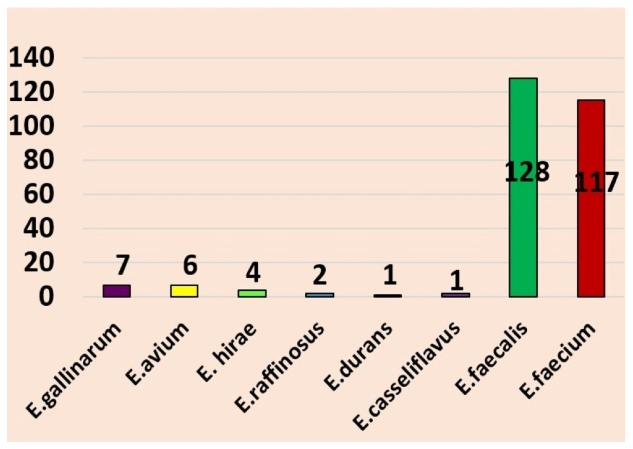
A total of 266 isolates were examined in this study. Among them, Enterococcus faecalis predominated (128; 48.1%), followed by E. faecium (117; 44.0%), E. gallinarum (7; 2.6%), E. avium (6; 2.3%), E. hirae (4; 1.5%), E. raffinosus (2; 0.7%), E. casseliflavus (1; 0.4%), and E. durans (1; 0.4%). The species-wise distribution of Enterococcus isolates is shown in Figure 1.

2.1. Antimicrobial Susceptibility Pattern

The antimicrobial resistance patterns are listed as follows: ampicillin (57%, 152/266), ciprofloxacin (84.6%, 83/98), nitrofurantoin (52%, 51/98), erythromycin (82.1%, 138/168), vancomycin (27.8%, 74/266), and linezolid (9.4%, 25/266).

2.2. Minimum Inhibitory Concentration of Linezolid-Resistant Enterococcus Species

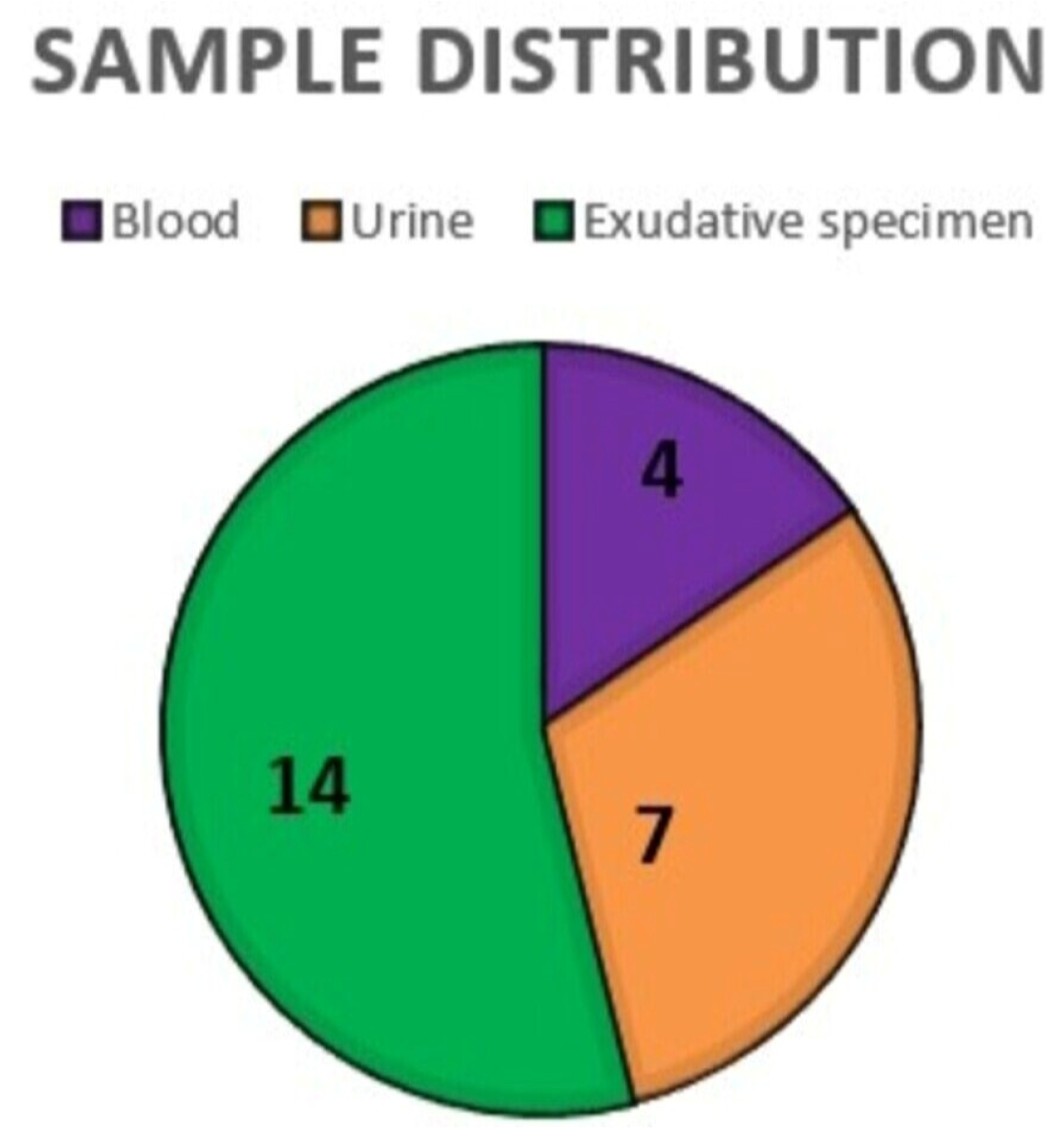
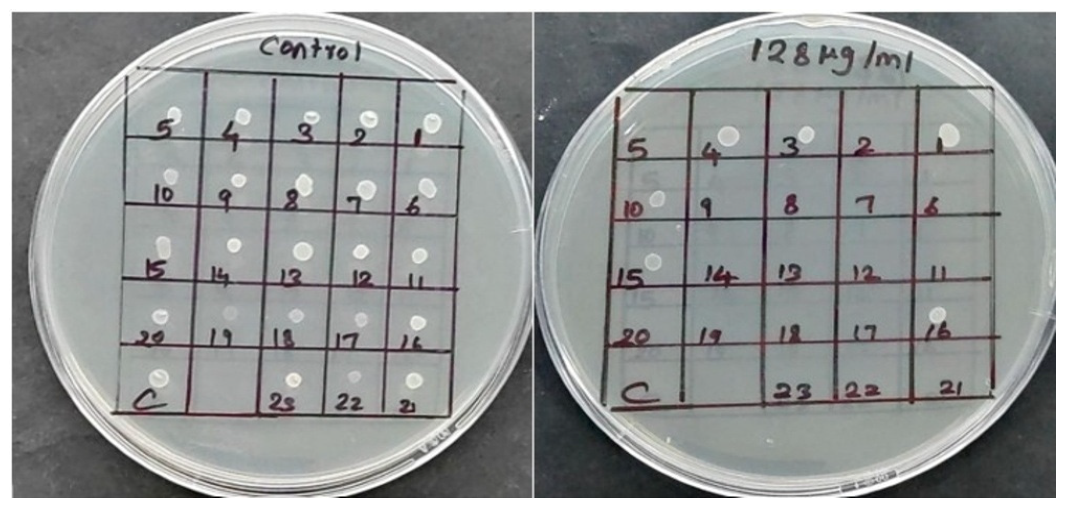
Out of 266 isolates, linezolid resistance was detected in 25 (9.4%) isolates with MIC (≥ 8 µg/mL). Of these 25 isolates, 22 (88%) were E. faecium, E. faecalis—2 (8%), and E. gallinarum—1 (4%). Among them, sixteen (64%) belonged to male patients and nine (36%) to female patients. The distribution of linezolid-resistant Enterococcus isolates by clinical source is illustrated in Figure 2. Furthermore, the Minimum Inhibitory Concentrations (MIC) range of these isolates is listed in Table 1, with a representative MIC image depicted in Figure 3.

2.3. Molecular Analysis

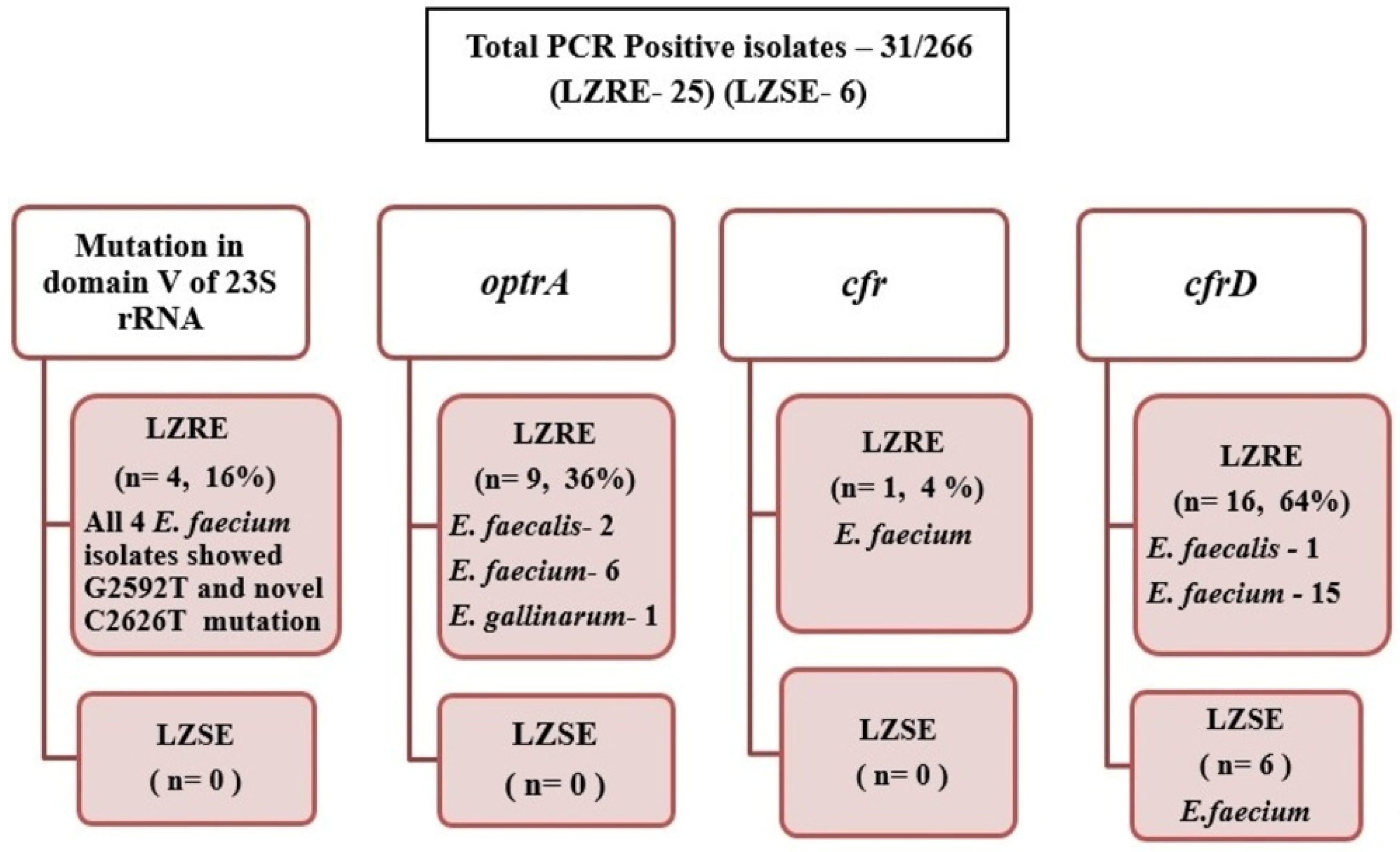
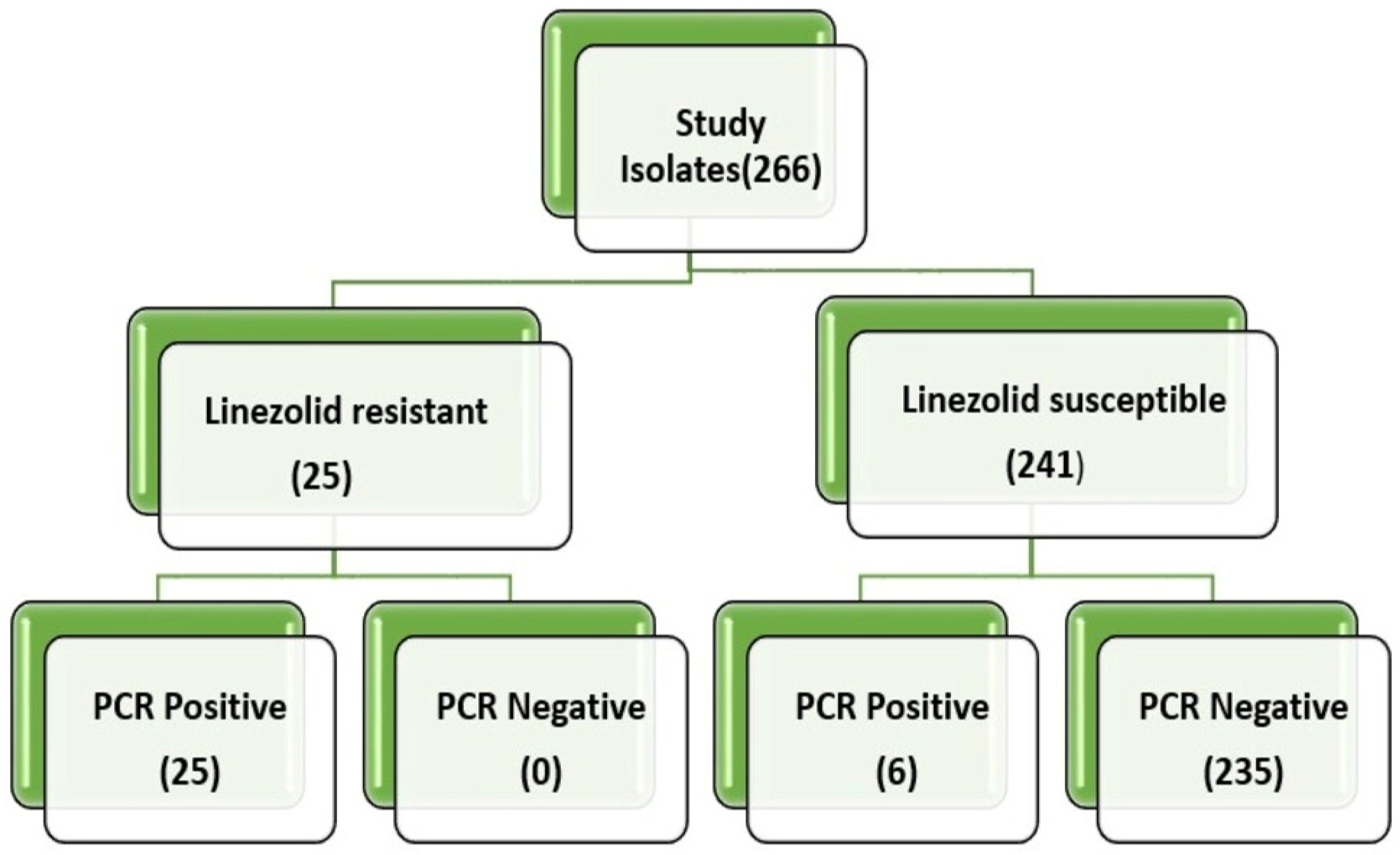
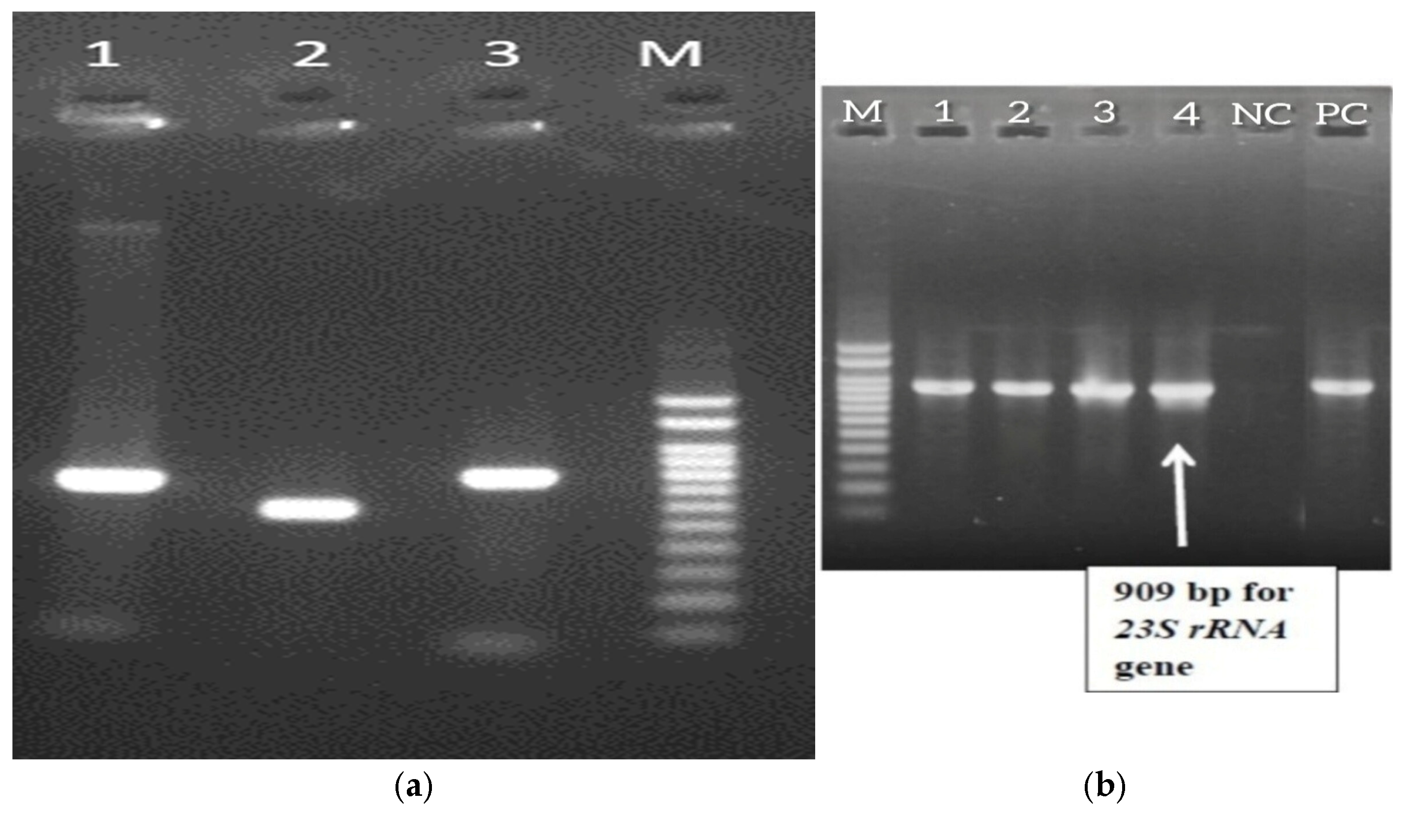
In a total of 266 isolates, 31 (11.7%) carried one or a combination of linezolid resistance genes (including optrA, cfr, cfr(D), or a mutation in domain V of 23S rRNA). Molecular analysis revealed that 9 isolates carried optrA, 1 isolate harbored cfr, and 22 isolates carried the cfr(D) gene. Of the 22 cfr(D) positive isolates, six were susceptible to linezolid. The cfr(D) gene was present in both E. faecium and E. faecalis. Furthermore, of the twenty-five linezolid-resistant Enterococci, mutations in domain V of the 23S rRNA gene were observed in four E. faecium isolates. BLAST alignment unveiled a novel C2626T mutation, which is not previously reported in India, and a G2592T mutation (already documented in India). All four isolates displayed both the mutations with MIC > 128 (µg/mL). Two isolates harbored optrA with C2626T and G2592T mutations. Both cfr(D) and optrA co-occurred in three isolates. Representative PCR amplicons of cfr, cfr(D), and optrA were sequenced and deposited in GenBank under accession numbers (PX531853—cfr, PX626519—cfr(D), PX682460—optrA) and C2626T and G2592T mutation in 23S rRNA with the IDs PX830352, PX830353, PX830354, and PX830355. Molecular detection of linezolid resistance in Enterococcus species is depicted in Figure 4, Figure 5, Figure 6a,b and Figure 7. MIC stratification by species and resistance genes is illustrated in Table 2 and Table 3.
Among the 25 patients identified with LZRE, isolates were predominantly detected in individuals with major comorbidities such as Type 2 Diabetes Mellitus, systemic hypertension, chronic kidney disease (including dialysis-dependent CKD V), coronary artery disease, and other immunocompromised states. Most patients had prolonged exposure to invasive devices, including dialysis catheters, Foley catheters, arterial/central lines, and endotracheal tubes, indicating its predominance as a healthcare-associated pathogen. Notably, five patients had prior linezolid exposure, indicating that antibiotic selection pressure may have facilitated the emergence of resistance.
The 25 LZRE isolates were isolated from patients experiencing a broad spectrum of clinical conditions, highlighting the pathogen’s ability to cause both systemic and localized infections. The most frequently observed clinical conditions were sepsis and soft tissue/wound infections (including diabetic foot, cellulitis, and Fournier’s gangrene).
Demographic details of patients with linezolid resistance harboring 23S rRNA domain V mutations are listed in Table 4. Prior days of linezolid received and the level of care at the time of linezolid-resistant Enterococcus detection are shown in Table 5.

2.4. Results of Statistical Analysis

Statistical analysis was performed only for the predominant species, E. faecalis (128) and E. faecium (117). Chi-square analysis revealed a significant association between species and linezolid resistance (χ2 = 20.56, df = 1, p < 0.0001), with E. faecium showing higher odds of resistance than E. faecalis (Odds Ratio = 14.6). The association between clinically significant Enterococcus species and linezolid resistance is depicted in Table 6.
In addition, a significant association was observed between the presence of the mutation and high-level resistance (MIC ≥ 128; Fisher’s exact test, p = 0.0012). Specifically, high-level resistance was predominantly observed in mutated strains (100%, 4/4), whereas it was rarely observed in other resistance determinants (2/21). Fisher’s exact test was performed due to the small sample size and low expected frequencies within the comparison groups. Fisher’s exact test analysis is depicted in Table 7.

3. Discussion

Enterococcus species are remarkably resilient and can endure in adverse environmental conditions, which makes them well-suited for hospital settings [18]. Vancomycin-resistant Enterococci were categorized as a high-priority pathogen on the World Health Organization’s (WHO) 2024 worldwide list of significant antibiotic-resistant microorganisms [19]. In treating infections caused by vancomycin-resistant Enterococci, linezolid serves as a last effective option. Alarmingly, the emergence and dissemination of linezolid resistance among Enterococci has sharply increased in India and across the globe in recent times [20].
In this study, eight distinct Enterococcus species were identified among 266 isolates. Among these, E. faecalis (48.1%) was the most prevalent, followed by E. faecium (44.0%). This distribution aligns with findings from other studies [21,22] where E. faecalis is typically predominant than other species.
In our study, (25/266) 9.4% of clinical Enterococcus isolates exhibited resistance to linezolid (MIC ≥ 8 µg/mL). Whereas, other studies from India reported 16% [23] in clinical Enterococcus isolates and 3.06% [24] among urinary Enterococcus isolates. Linezolid resistance was observed in E. faecium (22/25, 88%), E. faecalis (2/25, 8%), and E. gallinarum (1/25, 4%) in this study. Of these, E. faecium is predominant in exhibiting resistance to linezolid. Amongst 25 LZRE, 8 clinical isolates were from the ICU, while the remaining 17 were from non-ICU wards. The majority of isolates were obtained from exudative specimens, followed by urine and blood. Most of the linezolid resistance Enterococcus species also exhibited resistance to ampicillin (24/25) and vancomycin (13/25).
Linezolid resistance in Enterococcus is facilitated by mutations in domain V of 23S rRNA, acquiring cfr, cfr(D), and optrA resistance genes. Based on the review of the literature, the most common mutations encompass G2576T, G2447U, and G2504A [25]. Nevertheless, this study did not observe any of the aforementioned mutations. To the best of our knowledge, this study reports a novel C2626T mutation in domain V of 23S rRNA in clinical isolates of E. faecium. In addition to that, a common G2592T mutation is also observed in this study. Similarly, a South Indian study also reported the G2592T mutation [16]. Previously, mutations in 23S rRNA of domain V were thought to be the main mechanism of linezolid resistance among clinical isolates. Recent research [25,26,27], including this study, has highlighted evolving mechanisms of resistance in hospital settings.
The optrA gene was first identified in E. faecalis from human sources in China in 2015 [28]. Following this, a nationwide surveillance study across China found that 2% of enterococcal isolates possessed the optrA gene. Additionally, research has shown that optrA-positive Enterococci are more prevalent in animals and are increasingly being reported in human infections [29]. For example, a study from Italy reported optrA carrying linezolid-resistant E. gallinarum of swine origin [30]. In our study, (9/25) 36% of linezolid-resistant Enterococcus of clinical isolates (E. faecalis, E. faecium, and E. gallinarum) possessed the optrA gene. A study from India conducted whole genome sequencing and reported an Enterococcus faecium strain carrying the optrA gene with the G2592T mutation [16]. Whereas, in our study results, optrA harboring linezolid-resistant E. faecium with both C2626T and G2592T mutation was observed.
To the best of our knowledge, this study reports cfr- and cfr(D)-mediated linezolid resistance in Enterococcus species from India. Although cfr and cfr(D) genes have previously been reported in other countries, reports from India are scarce. In addition, a novel C2626T mutation was identified among the resistant isolates. A study from Thailand documented transferable plasmid-mediated resistance conferred by the cfr gene in a clinical strain of Enterococcus faecalis [31].
The cfr(D) gene was initially found in a clinical isolate of E. faecium from France in 2015 [12] and later in a blood isolate from an Australian patient in 2019 [32]. The presence of cfr(D) in a clinical isolate of E. faecalis from Spain in 2020 [12]. In our study, 22 clinical isolates carried the cfr(D) gene. Among them, sixteen were resistant to linezolid, while the other six were phenotypically susceptible to linezolid. This may be due to incomplete gene expression. Both cfr(D) and optrA coexisted in three clinical isolates of linezolid-resistant Enterococcus (E. faecalis-1, E. faecium-2). In our study, cfr(D) is the predominant mechanism of Linezolid-resistant Enterococcus. The coexistence of clinical conditions with transferable resistance genes and chromosomal mutations may contribute to the persistence of resistant strains in healthcare environments. This underscores the need for continued surveillance, prudent antimicrobial use, and effective infection control practices.

4. Materials and Methods

4.1. Ethics Approval of Research

The Institutional Ethics Committee of Sri Ramachandra Institute of Higher Education and Research approved the study protocol (REF: IEC-N1/23/AUG/88/43).

4.2. Study Design and Setting

This in vitro, laboratory-based, prospective, cross-sectional study was carried out in a 1600-bedded teaching hospital in Chennai, South India. A total of 266 clinically significant, consecutive, non-repetitive isolates collected between 2023 and 2024 were analyzed in this study. Sources of the isolates included urine (n = 98), blood (n = 19), and exudative specimens (n = 149). The significance of the isolates was determined by correlating them with clinical history, detecting intracellular organisms in Gram staining, and observing significant growth on culture media. Species were identified through conventional biochemical tests and automated methods, including matrix-assisted laser desorption ionization-time-of-flight mass spectrometry (MALDI-TOF MS) from bioMérieux Marcy l’Etoile, France.

4.3. Antimicrobial Susceptibility Testing

Antimicrobial susceptibility was tested for various antimicrobial agents, including ampicillin (10 µg), vancomycin (30 µg), and linezolid (30 µg), using the Kirby-Bauer disc diffusion method and interpreted as per Clinical and Laboratory Standards Institute 2025 guidelines. All urinary isolates were tested for ciprofloxacin (5 µg) and nitrofurantoin (300 µg). Erythromycin (15 µg) only for non-urinary isolates.

4.4. Minimum Inhibitory Concentration

The agar dilution technique was used to ascertain the MIC for linezolid. Mueller-Hinton agar plates were formulated from a stock solution using 12 distinct concentrations ranging from 0.125 µg/mL to 128 µg/mL. Three to four colonies of Enterococcus isolates were transferred into peptone broth, adjusted to a 0.5 McFarland standard, and incubated for 20 min. Afterward, the inoculum was placed onto gridded Mueller-Hinton agar plates using a sterile loop in the specified grids. Enterococcus faecalis ATCC 29212 was used as a quality control strain. The plates were then incubated at 37 °C, and results were recorded after 24 h. If the MIC was ≥8 µg/mL, isolates were classified as phenotypically resistant to linezolid, as per the Clinical and Laboratory Standards Institute 2025 guidelines.

4.5. Molecular Methods

4.5.1. DNA Extraction

Clinical Enterococcus colonies were inoculated into Luria-Bertani broth (HiMedia Laboratories, Mumbai, Maharashtra, India) and incubated at 37 °C for 16 h. Following this, the culture underwent centrifugation at 10,000 rpm for 10 min. The resulting pellet was reconstituted in 400 μL of sterile water and boiled at 100 °C for 10 min. After cooling, the suspension underwent a second centrifugation at a speed of 10,000 rpm for a span of 10 min. The resulting supernatant was utilized as the template DNA in the following Polymerase Chain Reaction (PCR) analysis [33].

4.5.2. Polymerase Chain Reaction:

The presence of genes conferring linezolid resistance (optrA, cfr, cfr(D), and a mutation in domain V of 23S rRNA) was determined by the PCR technique. For each PCR reaction, a total volume of 10 µL was used, containing 5 µL of master mix (Takara, New Delhi, India), 2 µL of sterile Milli-Q water, 0.5 µL of each primer, and 2 µL of template DNA. Amplified products were resolved on a 2% agarose gel with ethidium bromide for visualization [33]. PCR was performed with previously characterized positive control strains and sterile Milli-Q water as a negative control. Primer specifications are listed in Table 8.

4.5.3. DNA Sequencing and Analysis

DNA sequencing was performed using the Sanger sequencing method on an ABI 3730XL sequencer (Applied Biosystems, Foster City, CA, USA) with the ABI PRISM BigDye Terminator kit. The resulting nucleotide sequences were analyzed and aligned using BioEdit software version 7.0.5.3. Sequence similarity searches were conducted using the Basic Local Alignment Search Tool (BLAST) +2.17.0 available through the National Center for Biotechnology Information (NCBI) database. The finalized sequences were submitted to GenBank, and accession numbers were obtained.

4.5.4. Statistical Analysis

All statistical analyses were performed using GraphPad Prism version 11.0.2, and p-values < 0.05 were considered statistically significant.

5. Conclusions

The findings of this study highlight the concerning evolution of linezolid resistance among clinical Enterococcus species, which threatens the effectiveness of this last-resort antibiotic. The identification of a novel C2626T mutation with high MIC values, in addition to the significant prevalence of resistance genes such as cfr and cfr(D) and their concerning coexistence with optrA, underscores a shift towards multifaceted resistance mechanisms. Notably, cfr(D) was the predominant genetic determinant identified in this study. Moreover, the detection of cfr(D) in isolates lacking phenotypic resistance emphasizes the importance of molecular surveillance, as these isolates may facilitate silent dissemination and rapid acquisition of resistance under linezolid selective pressure. To mitigate the spread of these multidrug-resistant isolates, the implementation of strengthened infection control practices, rational antimicrobial use, and early detection is an essential step for effective containment and preservation of available therapeutic options in healthcare settings.

Author Contributions

M.S. contributed to writing—original draft, conceptualization, methodology, software, validation, formal analysis, investigation, data curation, and visualization. S.M. contributed to conceptualization, investigation, resources, writing—original draft, writing—review and editing, supervision, validation, and project administration. M.B. contributed to methodology, software, validation, formal analysis, writing—original draft, and visualization. U.S. contributed to conceptualization, resources, writing—review and editing, supervision, and project administration. K.K.P. contributed to conceptualization, writing—review and editing, and supervision. B.L.Y. contributed to conceptualization, writing—review and editing, and supervision. R.M.J.K. contributed to data curation, formal analysis, and methodology. All authors have read and agreed to the published version of the manuscript.

Funding

The present study was supported by funding from the Founder-Chancellor Shri N. P. V. Ramaswamy Udayar Research Fellowship, Sri Ramachandra Institute of Higher Education and Research (Ref No. Founder-Chancellor Fellowship-2022-23-17).

Institutional Review Board Statement

The Institutional Ethics Committee of Sri Ramachandra Institute of Higher Education and Research approved the study protocol (REF: IEC-N1/23/AUG/88/43). Waiver of consent obtained.

Informed Consent Statement

This was a laboratory-based in vitro study involving no patient intervention. Only limited retrospective clinical data were collected from laboratory records, and all isolates were coded to ensure patient confidentiality. Therefore, the Institutional Ethics Committee granted a waiver of informed consent.

Data Availability Statement

The data presented in this study are openly available in NCBI at https://www.ncbi.nlm.nih.gov/nuccore/PX531853 (accessed on 26 May 2026), reference number PX531853, PX626519, PX682460, PX830352, PX830353, PX830354.

Acknowledgments

We sincerely acknowledge the management of Sri Ramachandra Institute of Higher Education and Research for providing the excellent infrastructure and research facilities to carry out this study. We also thank the staff of the Central Laboratory, SRIHER, for their technical assistance and support. Co-author Rhea Michelle J. Khodabux is currently affiliated to Department of Microbiology, Vels Medical College and Hospital, Vels Institute of Science, Technology and Advanced Studies (VISTAS), Manjakaranai. We declare that Grammarly was utilized only to improve grammar and language clarity. The scientific content, data analysis, interpretation, and conclusions were entirely generated by the authors. No other artificial intelligence (AI) tools were used.

Conflicts of Interest

The authors declare no conflicts of interest.

References

  1. Kristich, C.J.; Rice, L.B.; Arias, C.A. Enterococcal Infection—Treatment and Antibiotic Resistance. In Enterococci: From Commensals to Leading Causes of Drug Resistant Infection; Gilmore, M.S., Clewell, D.B., Ike, Y., Shankar, N., Eds.; Massachusetts Eye and Ear Infirmary: Boston, MA, USA, 2014. [Google Scholar]
  2. Torres, C.; Alonso, C.A.; Ruiz-Ripa, L.; León-Sampedro, R.; Del Campo, R.; Coque, T.M. Antimicrobial Resistance in Enterococcus spp. of Animal Origin. Microbiol. Spectr. 2018, 6, ARBA-0032-2018. [Google Scholar] [CrossRef] [Scilit]
  3. Welshman, I.R.; Sisson, T.A.; Jungbluth, G.L.; Stalker, D.J.; Hopkins, N.K. Linezolid Absolute Bioavailability and the Effect of Food on Oral Bioavailability. Biopharm. Drug Dispos. 2001, 22, 91–97. [Google Scholar] [CrossRef] [Scilit]
  4. Azzouz, A.; Preuss, C.V. Linezolid. In StatPearls; StatPearls Publishing: Treasure Island, FL, USA, 2026. [Google Scholar]
  5. Iqbal, F.; Alocious, A.; Joy, S.C.; Stanly, E.A.R.; Rajesh, V.; Unnikrishnan, M.K.; Steinke, D.; Chandra, P. Vancomycin-Resistant Enterococci: A Rising Challenge to Global Health. Clin. Epidemiol. Glob. Health 2024, 28, 101663. [Google Scholar] [CrossRef] [Scilit]
  6. Auckland, C.; Teare, L.; Cooke, F.; Kaufmann, M.E.; Warner, M.; Jones, G.; Bamford, K.; Ayles, H.; Johnson, A.P. Linezolid-Resistant Enterococci: Report of the First Isolates in the United Kingdom. J. Antimicrob. Chemother. 2002, 50, 743–746. [Google Scholar] [CrossRef] [Scilit] [PubMed]
  7. Kumar, S.; Bandyopadhyay, M.; Chatterjee, M.; Mukhopadhyay, P.; Poddar, S.; Banerjee, P. The First Linezolid-Resistant Enterococcus faecium in India: High-Level Resistance in a Patient with No Previous Antibiotic Exposure. Avicenna J. Med. 2014, 4, 13–16. [Google Scholar] [CrossRef] [Scilit]
  8. Mendes, R.E.; Deshpande, L.M.; Jones, R.N. Linezolid Update: Stable in vitro Activity Following More than a Decade of Clinical Use and Summary of Associated Resistance Mechanisms. Drug Resist. Updat. 2014, 17, 1–12. [Google Scholar] [CrossRef] [Scilit]
  9. Kehrenberg, C.; Schwarz, S.; Jacobsen, L.; Hansen, L.H.; Vester, B. A New Mechanism for Chloramphenicol, Florfenicol and Clindamycin Resistance: Methylation of 23S Ribosomal RNA at A2503. Mol. Microbiol. 2005, 57, 1064–1073. [Google Scholar] [CrossRef] [Scilit]
  10. Long, K.S.; Poehlsgaard, J.; Kehrenberg, C.; Schwarz, S.; Vester, B. The cfr rRNA Methyltransferase Confers Resistance to Phenicols, Lincosamides, Oxazolidinones, Pleuromutilins, and Streptogramin A Antibiotics. Antimicrob. Agents Chemother. 2006, 50, 2500–2505. [Google Scholar] [CrossRef] [Scilit] [PubMed]
  11. Schwarz, S.; Zhang, W.; Du, X.-D.; Krüger, H.; Feßler, A.T.; Ma, S.; Zhu, Y.; Wu, C.; Shen, J.; Wang, Y. Mobile Oxazolidinone Resistance Genes in Gram-Positive and Gram-Negative Bacteria. Clin. Microbiol. Rev. 2021, 34, e00188-20. [Google Scholar] [CrossRef] [Scilit]
  12. Guerin, F.; Sassi, M.; Dejoies, L.; Zouari, A.; Schutz, S.; Potrel, S.; Auzou, M.; Collet, A.; Lecointe, D.; Auger, G.; et al. Molecular and Functional Analysis of the Novel cfr(D) Linezolid Resistance Gene Identified in Enterococcus faecium. J. Antimicrob. Chemother. 2020, 75, 1699–1703. [Google Scholar] [CrossRef] [Scilit]
  13. Sharkey, L.K.R.; O’Neill, A.J. Antibiotic Resistance ABC-F Proteins: Bringing Target Protection into the Limelight. ACS Infect. Dis. 2018, 4, 239–246. [Google Scholar] [CrossRef] [Scilit]
  14. Bi, R.; Qin, T.; Fan, W.; Ma, P.; Gu, B. The Emerging Problem of Linezolid-Resistant Enterococci. J. Glob. Antimicrob. Resist. 2018, 13, 11–19. [Google Scholar] [CrossRef] [Scilit] [PubMed]
  15. Kainer, M.A.; Devasia, R.A.; Jones, T.F.; Simmons, B.P.; Melton, K.; Chow, S.; Broyles, J.; Moore, K.L.; Craig, A.S.; Schaffner, W. Response to Emerging Infection Leading to Outbreak of Linezolid-Resistant Enterococci. Emerg. Infect. Dis. 2007, 13, 1024–1030. [Google Scholar] [CrossRef] [Scilit]
  16. Bakthavatchalam, Y.D.; Vasudevan, K.; Babu, P.; Neeravi, A.R.; Narasiman, V.; Veeraraghavan, B. Genomic Insights of optrA-Carrying Linezolid-Resistant Enterococcus faecium Using Hybrid Assembly: First Report from India. J. Glob. Antimicrob. Resist. 2021, 25, 331–336. [Google Scholar] [CrossRef] [Scilit]
  17. Sengupta, M.; Sarkar, R.; Sarkar, S.; Sengupta, M.; Ghosh, S.; Banerjee, P. Vancomycin and Linezolid-Resistant Enterococcus Isolates from a Tertiary Care Center in India. Diagnostics 2023, 13, 945. [Google Scholar] [CrossRef] [Scilit] [PubMed]
  18. García-Solache, M.; Rice, L.B. The Enterococcus: A Model of Adaptability to Its Environment. Clin. Microbiol. Rev. 2019, 32, e00058-18. [Google Scholar] [CrossRef] [Scilit]
  19. World Health Organization. WHO Updates List of Drug-Resistant Bacteria Most Threatening to Human Health. Available online: https://www.who.int/news/item/17-05-2024-who-updates-list-of-drug-resistant-bacteria-most-threatening-to-human-health (accessed on 15 May 2026).
  20. El-Sheikh, S.M.A.; Mansour, A.I.A.-E.; Ibrahim, Z.S.M. Emerging Genetic Mechanisms of Linezolid Resistance in Enterococci. J. Comput. Anal. Appl. 2024, 33, 1676–1684. [Google Scholar]
  21. Sharifzadeh Peyvasti, V.; Mohabati Mobarez, A.; Shahcheraghi, F.; Khoramabadi, N.; Razaz Rahmati, N.; Hosseini Doust, R. High-Level Aminoglycoside Resistance and Distribution of Aminoglycoside Resistance Genes among Enterococcus spp. Clinical Isolates in Tehran, Iran. J. Glob. Antimicrob. Resist. 2020, 20, 318–323. [Google Scholar] [CrossRef] [Scilit]
  22. Saini, G.; Singh, P.; Pandey, A. Are Uropathogenic Enterococci Becoming Smarter Day by Day? Changing Trends of Antimicrobial Resistance and Species Distribution among Uropathogenic Enterococcus Species. Adesh Univ. J. Med. Sci. Res. 2025, 7, 137–142. [Google Scholar] [CrossRef] [Scilit]
  23. Jain, S. Prevalence of Linezolid-Resistant Vancomycin-Resistant Enterococcus Species (LRVRE) in Clinical Isolates from Tertiary Care Hospital of North India—A Real Threat. Int. J. Infect. Dis. 2023, 130, S14. [Google Scholar] [CrossRef] [Scilit]
  24. Naruka, H.S.; Chand, A.E.; Meena, H. Prevalence of Various Enterococcus Species and Their Antibiotic Resistance Pattern among Urinary Isolates in Tertiary Care Center in South Eastern Rajasthan. IP Int. J. Med. Microbiol. Trop. Dis. 2019, 5, 18–22. [Google Scholar] [CrossRef] [Scilit]
  25. Nasir, S.A.R.; Zeeshan, M.; Ghanchi, N.; Saeed, N.; Ghayas, H.; Zaka, S.; Ashraf, J.; Jabeen, K.; Farooqi, J.; Hasan, Z.; et al. Linezolid-Resistant Enterococcus faecium Clinical Isolates from Pakistan: A Genomic Analysis. BMC Microbiol. 2024, 24, 347. [Google Scholar] [CrossRef] [Scilit]
  26. Wang, Z.; Liu, D.; Zhang, J.; Liu, L.; Zhang, Z.; Liu, C.; Hu, S.; Wu, L.; He, Z.; Sun, H. Genomic Epidemiology Reveals Multiple Mechanisms of Linezolid Resistance in Clinical Enterococci in China. Ann. Clin. Microbiol. Antimicrob. 2024, 23, 41. [Google Scholar] [CrossRef] [Scilit] [PubMed]
  27. Suo, N.; Yu, J.; Li, H.; Yi, N.; Liu, C.; Wang, W. Molecular Characterization and Resistance Mechanisms of Linezolid-Resistant Enterococci Clinical Isolates and Identification of a Clinical Enterococcus faecium Strain Co-Harboring optrA, poxtA, and cfr(D) in a Tertiary Hospital in China. J. Glob. Antimicrob. Resist. 2025, 45, 182–191. [Google Scholar] [CrossRef] [Scilit]
  28. Yi, M.; Zou, J.; Zhao, J.; Tang, Y.; Yuan, Y.; Yang, B.; Huang, J.; Xia, P.; Xia, Y. Emergence of optrA-Mediated Linezolid Resistance in Enterococcus faecium: A Molecular Investigation in a Tertiary Hospital of Southwest China from 2014–2018. Infect. Drug Resist. 2022, 15, 13–20. [Google Scholar] [CrossRef] [Scilit] [PubMed]
  29. Morroni, G.; Brenciani, A.; Simoni, S.; Vignaroli, C.; Mingoia, M.; Giovanetti, E. Commentary: Nationwide Surveillance of Novel Oxazolidinone Resistance Gene optrA in Enterococcus Isolates in China from 2004 to 2014. Front. Microbiol. 2017, 8, 1631. [Google Scholar] [CrossRef] [Scilit] [PubMed]
  30. Coccitto, S.N.; Cinthi, M.; Fioriti, S.; Morroni, G.; Simoni, S.; Vignaroli, C.; Garofalo, C.; Mingoia, M.; Brenciani, A.; Giovanetti, E. Linezolid-Resistant Enterococcus gallinarum Isolate of Swine Origin Carrying cfr, optrA and poxtA Genes. J. Antimicrob. Chemother. 2022, 77, 331–337. [Google Scholar] [CrossRef] [Scilit]
  31. Diaz, L.; Kiratisin, P.; Mendes, R.E.; Panesso, D.; Singh, K.V.; Arias, C.A.; Murray, B.E. Transferable plasmid-mediated resistance to linezolid due to cfr in a human clinical isolate of Enterococcus faecalis. Antimicrob. Agents Chemother. 2012, 56, 3917–3922. [Google Scholar] [CrossRef] [Scilit]
  32. Pang, S.; Boan, P.; Lee, T.; Gangatharan, S.; Tan, S.J.; Daley, D.; Lee, Y.T.; Coombs, G.W. Linezolid-Resistant ST872 Enterococcus faecium Harbouring optrA and cfr(D) Oxazolidinone Resistance Genes. Int. J. Antimicrob. Agents 2020, 55, 105831. [Google Scholar] [CrossRef] [Scilit]
  33. Zarzecka, U.; Zakrzewski, A.J.; Chajęcka-Wierzchowska, W.; Zadernowska, A. Linezolid-Resistant Enterococcus spp. Isolates from Foods of Animal Origin—The Genetic Basis of Acquired Resistance. Foods 2022, 11, 975. [Google Scholar] [CrossRef] [Scilit]
  34. Li, P.; Yang, Y.; Ding, L.; Xu, X.; Lin, D. Molecular Investigations of Linezolid Resistance in Enterococci optrA Variants from a Hospital in Shanghai. Infect. Drug Resist. 2020, 13, 2711–2716. [Google Scholar] [CrossRef] [Scilit] [PubMed]
  35. Khodabux, R.M.J.; Mariappan, S.; Sekar, U. Detection of a Novel G2603T Mutation in cfr-Harboring Linezolid-Resistant Staphylococcus haemolyticus: First Report from India. J. Lab. Physicians 2023, 15, 207–211. [Google Scholar] [CrossRef] [Scilit] [PubMed]
  36. Ruiz-Ripa, L.; Feßler, A.T.; Hanke, D.; Eichhorn, I.; Azcona-Gutiérrez, J.M.; Pérez-Moreno, M.O.; Seral, C.; Aspiroz, C.; Alonso, C.A.; Torres, L.; et al. Mechanisms of Linezolid Resistance among Enterococci of Clinical Origin in Spain—Detection of optrA- and cfr(D)-Carrying E. faecalis. Microorganisms 2020, 8, 1155. [Google Scholar] [CrossRef] [Scilit] [PubMed]
Figure 1. Distribution of Enterococcus species among clinical isolates.
Antibiotics 15 00841 g001
Figure 2. Source of the LZRE isolates.
Antibiotics 15 00841 g002
Figure 3. Representative MIC image of linezolid-resistant Enterococcus isolates.
Antibiotics 15 00841 g003
Figure 4. Flowchart depicting the distribution of Linezolid Resistance and PCR Detection among Enterococcus Isolates.
Antibiotics 15 00841 g004
Figure 5. Molecular basis of linezolid resistance in clinical isolates of Enterococcus species. Footnote: LZRE—Linezolid-Resistant Enterococcus, LZSE—Linezolid Sensitive Enterococcus.
Antibiotics 15 00841 g005
Figure 6. (a) Agarose gel electrophoresis of PCR products corresponding to Linezolid-resistant genes cfr, cfr(D), and optrA. Lane 1—cfr (746 bp); Lane 2—cfr(D) (595 bp); Lane 3—optrA (792 bp); M—DNA Marker. (b) Agarose gel electrophoresis of PCR products corresponding to the 23S rRNA gene. M—DNA Marker; Lanes 1–4—23S rRNA gene.
Antibiotics 15 00841 g006
Figure 7. Comparative sequence analysis of the 23S rRNA gene in wild-type (WT) and clinical isolates, highlighting G2592T and a novel C2626T point mutation.
Antibiotics 15 00841 g007
Table 1. Minimum Inhibitory Concentration of linezolid against resistant Enterococcus isolates.
MIC Range (µg/mL)0.1250.250.51248163264128>128
No. of isolates015711640183706
[Results interpreted as per CLSI guidelines 2025. Sensitive—≤2 (µg/mL), Intermediate—4 (µg/mL), Resistant—≥8 (µg/mL)]. The MIC50 and MIC90 values for all 266 Enterococcus isolates were 2 µg/mL and 2 µg/mL, respectively. Among linezolid-resistant isolates (n = 25), MIC50 and MIC90 were 64 µg/mL and >128 µg/mL, respectively.
Table 2. MIC stratification by species.
Species0.1250.250.51248163264128>128Total
E. faecalis00042840010001128
E. faecium00024710073705117
E. gallinarum0032101000007
E. casseliflavus0100000000001
E. raffinosus0010100000002
E. avium0003300000006
E. durans0010000000001
E. hirae0000400000004
Total015711640183706266
Table 3. MIC stratification according to resistant genes.
MIC Rangecfrcfr(D)OptrAMutation
0.1250000
0.250000
0.50000
10100
20500
40000
80010
160630
320300
640720
1280000
>1281034
Footnote: [Results interpreted as per CLSI guidelines 2025. Linezolid Sensitive—≤2 (µg/mL), Linezolid Intermediate—4 (µg/mL), Linezolid-Resistant—≥8 (µg/mL)].
Table 4. Summarizes the demographic characteristics of patients with linezolid-resistant isolates carrying 23S rRNA domain V mutation.
CharacteristicsPatient 1Patient 2Patient 3Patient 4
Isolate numberGE4273GE5439GE7556GE492
Age/Sex54 M63 M50 F69 M
Hospital
location
WardWardWardWard
Enterococcus speciesE. faeciumE. faeciumE. faeciumE. faecium
Admitting
specialty
Plastic SurgeryGeneral SurgeryGeneral SurgeryDermatology and venereology
Final diagnosis50% total body surface area electrical burns of the bilateral upper limb, left lower limb, and backUGI Bleed (Resolving); Septic shock (Resolved)- UTI, Grade IV Bed sore, status post debridement of bedsore Grade IVDiabetic foot ulcer, Status post ray amputation of the right fourth and fifth toesLeft leg cellulitis S/P wound debridement and fasciotomy, Bilateral lower limb DVT
Co-morbid
conditions
nilType 2 diabetes mellitus, bipolar disorderType 2 diabetes mellitusType 2 diabetes mellitus
Days in hospital61454139
Surgical
procedures
Wound debridement, escharotomy, fasciotomyWound debridementRay amputationWound debridement and fasciotomy
Antimicrobials used prior to
detection of linezolid
resistance
Linezolid--Linezolid
OutcomeRecoveredDeathRecoveredRecovered
Source specimenTissuePus clsTissuePus cls
MIC (µg/mL)>128>128>128>128
23S rRNA
mutations in
domain V
C2626T and G2592TC2626T and G2592TC2626T and G2592TC2626T and G2592T
cfr, cfr(D)NilNilNilNil
optrANilNiloptrAoptrA
Table 5. Clinical and molecular profiles of linezolid-resistant Enterococcus isolates from patients with prior Linezolid exposure.
S.NOSpeciesSample SourcePrior Days of Linezolid ReceivedLevel of Care During Linezolid ExposureLevel of Care After Linezolid DetectionGeneClinical Outcome
1E. faeciumTissue31 days ICUWardC2626T and G2592T
MIC
≥128 (µg/mL)
Recovered
2E. gallinarumBlood cls8 days WardICUoptrA
MIC
8 (µg/mL)
Death
3E. faeciumPus cls8 days ICUWardC2626T and G2592T
MIC
≥128 (µg/mL)
Death
4E. faeciumTissue cls5 days WardWardcfr
MIC
≥128 (µg/mL)
Recovered
5E. faeciumPus cls12 days ICUWardC2626T and G2592T
MIC
≥128 (µg/mL)
Recovered
Table 6. Chi-square analysis of linezolid resistance among E. faecalis and E. faecium.
SpeciesLZSELZRETotalχ2, dfp-Value
E. faecalis126212820.56,
df = 1
<0.0001
E. faecium9522117
Total22124245
Footnote: This study identified 25 linezolid-resistant Enterococcus (LZRE) isolates, including 22—E. faecium, 2—E. faecalis, and 1—E. gallinarum isolate. Table 6 presents the 2 × 2 contingency analysis comparing the two predominant species, E. faecalis and E. faecium (Total n = 24). The E. gallinarum isolate (n=1) was not included in the chi-square analysis. (p-value < 0.0001 = statistically significant).
Table 7. Contingency table showing the association between mutation status and high-level linezolid resistance (Fisher’s exact test).
GenotypeHigh Level Resistance ≥ 128 µg/mLResistance ≤ 128 µg/mLTotalp Value
Mutation404p = 0.0012
other gene21921
Total61925
Table 8. PCR primers and cycling conditions for linezolid resistance genes.
GenePrimerPCR ConditionsBase PairRef.
23S rRNAF: GTAACGATTTGGGCACTGTCG
R: CGATTAGTATTGGTCCGCTC
Initial denaturation: 94 °C for 5 min, followed by 30 cycles of
Denaturation: 94 °C for 30 s,
Annealing: 55 °C for 30 s
Extension: 72 °C for 30 s
Final Extension: 72 °C for 5 min
909[33]
optrAF: CAGGTGGTCAGCGAACTAAGA
R: AGCCAAGAGCAGTTCTGACC
Initial denaturation: 94 °C
for 5 min followed by 30 cycles of,
Denaturation: 94 °C for 30 s,
Annealing: 56 °C for 30 s,
Extension: 72 °C for 30 s,
Final Extension: 72 °C for 5 min
792[34]
CfrF: TGAAGTATAAAGCAGGTTGGGAT
R: ACCATATAATTGACCACAAGCAGC
Initial denaturation: 94 °C for 2 min,
Denaturation: 94 °C for 10 s,
Annealing: 55 °C for 30 s,
Final extension: 7 min at 72 °C
746[35]
cfr(D)F: CAGGTGGTCAGCGAACTAAGA
R: AGCCAAGAGCAGTTCTGACC
Initial denaturation: 94 °C for 7 min,
followed by 30 cycles of
Annealing: 60 °C for 1 min,
Extension: 72 °C for 1min,
Final extension: 72 °C for 10 min.
595[36]
Disclaimer/Publisher’s Note: The statements, opinions and data contained in all publications are solely those of the individual author(s) and contributor(s) and not of MDPI and/or the editor(s). MDPI and/or the editor(s) disclaim responsibility for any injury to people or property resulting from any ideas, methods, instructions or products referred to in the content.