1. Introduction
Long before an organism causes disease, it colonizes. Asymptomatic carriage of multidrug-resistant (MDR) bacteria is defined, following Magiorakos et al. [
1], as acquired non-susceptibility to at least one agent in three or more antimicrobial categories on the skin, in the nares and in the respiratory tract, which constitutes the hidden compartment of the antimicrobial resistance (AMR) problem: it precedes infection, seeds transmission, and is far less often surveyed than the clinical isolates that dominate resistance statistics. Globally, bacterial AMR was linked to more than a million deaths in 2019 and is projected to account for tens of millions of deaths by the middle of the century [
2,
3], yet most surveillance, especially in low- and middle-income countries, still samples symptomatic, hospitalized or intensive-care patients rather than the colonized ambulatory population from which many of these organisms ultimately arise [
4,
5].
First, antimicrobial resistance is not restricted to recognized pathogens. Commensal and low-virulence bacteria recovered under antimicrobial selective pressure are increasingly recognized as overlooked components of antimicrobial resistance ecology. Despite often being excluded from routine surveillance or dismissed as contaminants, evidence suggests that they may contribute to the maintenance and dissemination of antimicrobial resistance determinants [
6,
7]. Second, colonization is seldom a single-site, single-species event; the same host may carry several organisms across several anatomical niches at once, a polymicrobial, multisite pattern with direct implications for how thoroughly and in what way patients should be screened before invasive procedures. In settings such as Egypt, where the great majority of antibiotics are dispensed without prescription, this largely unmonitored community exposure intensifies the selective pressure driving both phenomena [
8,
9].
Patients with cardiovascular disease are an instructive and clinically consequential population in which to examine these questions. Repeated healthcare contact, vascular and cardiac interventions, implanted devices and frequent antibiotic courses place them at the interface between the community and the hospital, and resistant infections in this group are associated with substantial morbidity [
10,
11,
12]. How large the colonizing reservoir is in ambulatory, non-critically ill cardiovascular patients; what commensal and low-virulence flora accompanies it; and how frequently carriage spans multiple body sites in the same individual remain largely unquantified.
We therefore screened cardiovascular outpatients at three anatomical sites, with a particular focus on the colonizing and commensal flora and its relationship to community antibiotic use. Specifically, we set out to (i) quantify the overall burden of colonization by organisms with reduced antimicrobial susceptibility; (ii) characterize the co-existing resistant commensal or low-virulence flora that is usually overlooked; and (iii) measure the extent of polymicrobial, multisite carriage in this population, within the broader context of widespread, often unsupervised, community antibiotic use. By showing that resistant colonization is already established in ambulatory cardiovascular patients, we aim to inform pre-procedural screening and community antimicrobial stewardship strategies that could protect these patients before any invasive intervention.
2. Results
The study cohort consisted of 36 adults with established cardiovascular disease attending outpatient cardiology clinics. The mean age was 55.3 ± 14.4 years (range, 21 to 85 years), and 63.9% were male. Hypertension was the predominant comorbidity (75.0%), followed by diabetes mellitus (41.7%) and heart failure (27.8%). The cohort also represented a population with substantial healthcare exposure, with 55.6% reporting at least one previous hospitalization, 50.0% having undergone prior percutaneous coronary intervention (PCI), and 25.0% receiving antibiotic therapy during the month of specimen collection. These characteristics provide the clinical context for the ecological analysis of bacterial colonization presented below.
2.1. Colonization Burden and Anatomical Distribution
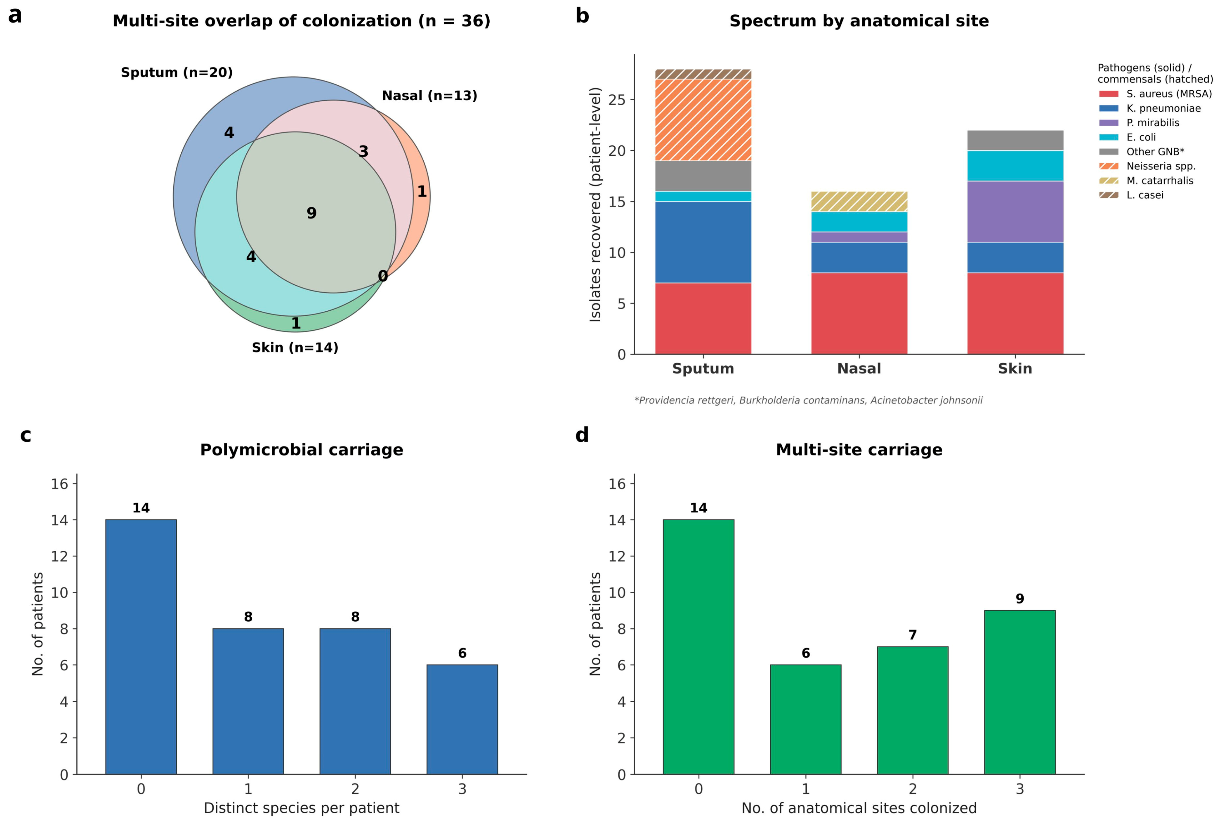
Overall, colonization by organisms with reduced antimicrobial susceptibility was detected in 22 of the 36 cardiovascular outpatients (61.1%; 95% CI, 44.9 to 75.2) using antimicrobial-supplemented selective media designed to enrich such organisms. Among these, 19 patients (52.8%; 95% CI, 37.0 to 68.0) harboured at least one validated multidrug-resistant (MDR) pathogen, whereas the remaining colonized individuals carried commensal or low-virulence organisms recovered on antimicrobial-containing media, consistent with reduced antimicrobial susceptibility. Colonization prevalence differed significantly across the three anatomical sites (Cochran’s
Q = 6.62,
p = 0.037), with sputum demonstrating the highest colonization rate (20/36, 55.6%), followed by skin (14/36, 38.9%) and nasal specimens (13/36, 36.1%) (
Table 1;
Figure 1b).
Validated MDR pathogens and resistant commensal bacteria showed distinct distribution patterns.
Klebsiella pneumoniae was recovered predominantly from sputum, whereas
Proteus mirabilis was identified primarily from skin specimens. Methicillin-resistant
Staphylococcus aureus (MRSA) was detected across all three sites with comparable frequencies. In contrast, commensal
Neisseria species were recovered exclusively from sputum, while
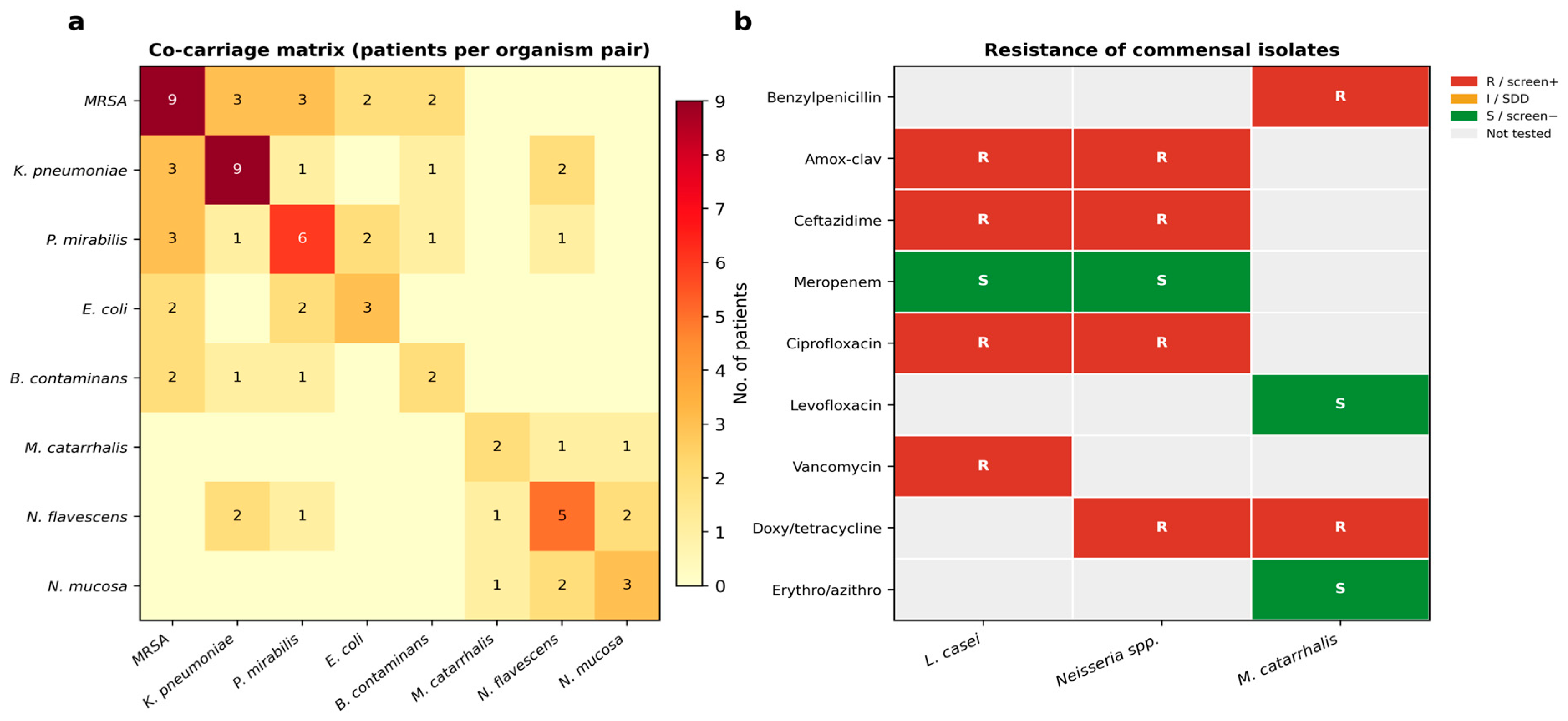
Moraxella catarrhalis was confined to nasal specimens. The complete anatomical distribution of all recovered organisms, and the co-carriage of organism pairs, are presented in
Table 1 and
Table S3 and
Figure 2a.
2.2. Clinical Correlates of Colonization
To determine whether the collected clinical variables distinguished colonized from non-colonized patients, the 22 colonized and 14 non-colonized individuals were compared across prior hospitalization, prior PCI, concurrent antibiotic therapy and major comorbidities using Fisher’s exact test (Benjamini–Hochberg FDR-adjusted). Prior hospitalization was the strongest correlate of colonization, significant for total colonization (
p < 0.001) and at every anatomical site; MDR
Klebsiella pneumoniae and MRSA were recovered exclusively from previously hospitalized patients (nine carriers each;
p = 0.002). Concurrent antibiotic therapy was also significantly associated with colonization (
p = 0.006). In contrast, prior PCI, sex, age, diabetes, hypertension, heart failure and smoking showed no significant association (
Table 2). Notably, colonization was detected even among patients with no prior hospitalization, indicating that resistant carriage in this population is not confined to those with a documented hospital history.
2.3. Multisite Colonization
Multisite colonization was common among colonized cardiovascular outpatients. Of the 22 colonized patients, 16 (72.7%) harboured bacteria at two or more anatomical sites, while 9 (40.9%) were colonized simultaneously at all three sampling sites (
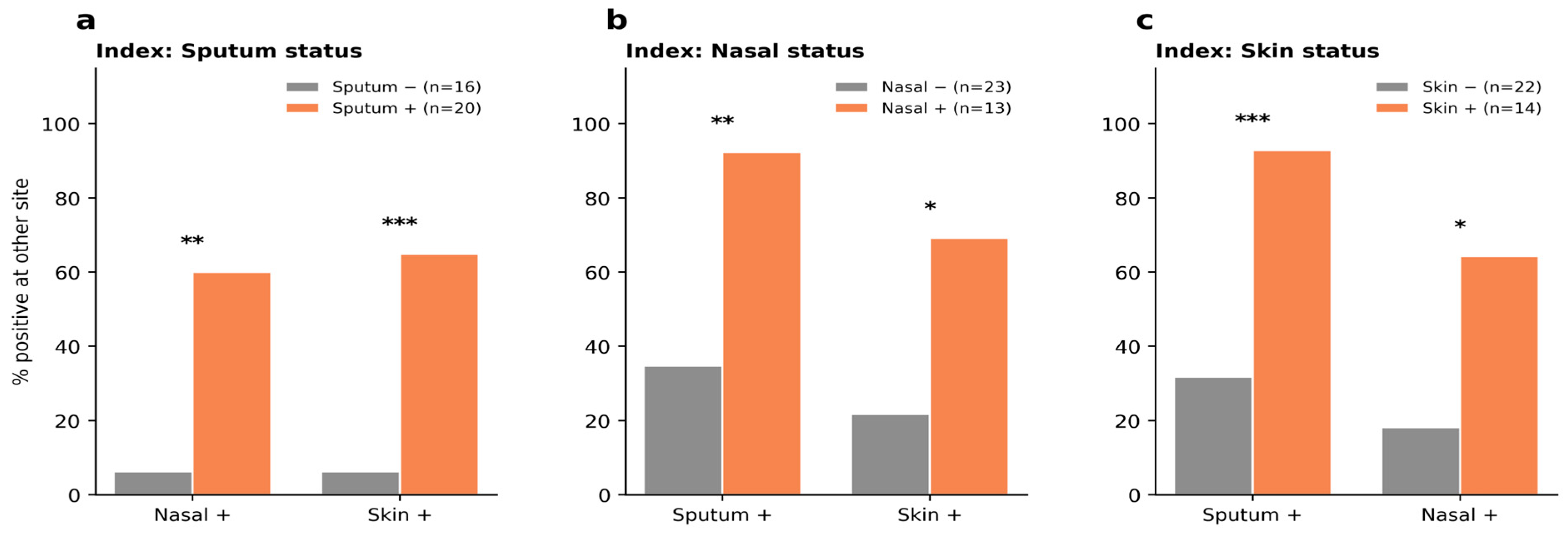
Figure 1a,d). Colonization showed significant concordance across anatomical sites, with moderate agreement between sputum, nasal and skin specimens (Cohen’s κ = 0.47–0.57; 95% CIs in
Table S4). Pairwise site associations, assessed using Fisher’s exact test, were statistically significant for sputum and nasal specimens (
p = 0.001), sputum and skin specimens (
p < 0.001), and nasal and skin specimens (
p = 0.011) (
Table S4;
Figure 3).
2.4. Polymicrobial Colonization
Polymicrobial colonization was identified in a substantial proportion of colonized patients. Among the 22 colonized individuals, 14 (63.6%) harboured two or more distinct bacterial species across the sampled anatomical sites, yielding a total of 42 species-level colonization events and an average of 1.9 bacterial species per colonized patient (
Figure 1c; 1.6 pathogenic species among the 19 patients carrying validated MDR pathogens). Co-colonization most frequently involved combinations of validated MDR pathogens, although resistant commensal bacteria were also detected in mixed colonization patterns. The distribution of co-colonizing organism pairs is presented in
Figure 2a.
2.5. Commensal and Low-Virulence Organisms on Selective Media
Commensal and low-virulence organisms that grew on the antimicrobial-containing screening media were recovered from a substantial proportion of colonized patients. These included
Neisseria flavescens (five patients),
Neisseria mucosa (three patients),
Moraxella catarrhalis (two patients), and
Lacticaseibacillus casei (one patient). Commensal
Neisseria species were detected exclusively in sputum specimens, whereas
Moraxella catarrhalis was recovered only from nasal specimens. Phenotypic testing demonstrated reduced susceptibility to multiple antimicrobial classes including β-lactams, fluoroquinolones, tetracyclines, and glycopeptides, depending on the organism (
Figure 2b and
Figure S3,
Table S5). Because CLSI interpretive breakpoints are unavailable for several of these organisms, their reduced susceptibility is reported descriptively (agar screen and disc diffusion testing) and is not directly comparable to CLSI-categorized resistance; throughout this article, the term “resistant” is reserved for isolates categorized against validated CLSI breakpoints.
3. Discussion
To our knowledge, this study is among the first to examine bacterial colonization in ambulatory cardiovascular outpatients from an ecological perspective. In this cohort, organisms with reduced antimicrobial susceptibility were recovered from 61.1% of participants, and validated multidrug-resistant (MDR) pathogens from 52.8%. Colonization was polymicrobial and extended across multiple anatomical sites; 72.7% of colonized patients were positive at two or more sites and 40.9% at all three and included commensal and low-virulence organisms that grew on the antimicrobial-containing screening media. Carriage at one anatomical site was strongly associated with carriage at the others, and prior hospitalization and concurrent antibiotic use were the factors most consistently associated with colonization.
Taken together, these observations indicate that resistant colonization in these patients behaves as a co-occurring, cross-site pattern rather than a series of isolated events, consistent with a whole-body colonization state. That more than half of these non-critically ill outpatients carried MDR bacteria, a burden approaching that reported in inpatient surveillance [
5,
13] and higher than would be expected outside hospital [
14], suggests that the ambulatory cardiovascular clinic is itself part of the community reservoir of antimicrobial resistance. This whole-body colonization state is in keeping with the ability of
Staphylococcus aureus to establish stable multi-site colonization [
15] and with gastrointestinal carriage acting as a reservoir for
Klebsiella pneumoniae dissemination [
16]. This pattern is biologically consistent with the widespread, unsupervised community use of antibiotics reported in Egypt and comparable settings, which suppresses susceptible flora and selects for resistant organisms [
8,
9]. The parallel recovery of recognized pathogens and commensal or low-virulence organisms on selective media is in line within this framework: naturally transformable commensal
Neisseria and gut-derived
Enterobacterales are recognized in the wider literature as potential reservoirs of resistance determinants [
6,
7], although here, their role is inferred from recovery under antibiotic selection rather than demonstrated genetically.
Several limitations bound these interpretations. As a single-centre, cross-sectional study of 36 patients with sparse cell counts, the analyses are exploratory and hypothesis-generating: the sample size was calculated to estimate the prevalence of MDR colonization, not to detect associations between individual clinical factors and colonization, so because the study was designed primarily for prevalence estimation rather than hypothesis testing, the observed clinical correlates should be regarded as exploratory, and non-significant findings should not be interpreted as evidence of absence of association. The cross-sectional design captures a single time point and cannot establish temporality or causation; in particular, because the exact timing of the last antibiotic dose was not recorded, the association between community antibiotic use and colonization cannot be interpreted causally. Growth on antimicrobial-containing media was treated as a phenotypic screening signal rather than a validated susceptibility category, and for commensal and low-virulence organisms lacking CLSI breakpoints, susceptibility is reported descriptively only. Sputum quality was not scored by Gram stain, so oropharyngeal carry-over cannot be excluded, and no resistance-gene detection or sequencing was performed on the commensal isolates.
Within these limits, the findings carry two practical messages. First, because colonization was common and distributed across multiple sites even among patients without a documented hospital history, single-site sampling is likely to underestimate carriage; this provides a rationale for future studies to evaluate whether pre-procedural screening for resistant bacterial carriage identifies patients at increased risk of subsequent infection, and whether targeted preventive strategies improve clinical outcomes—outcomes which the present cross-sectional study did not measure and cannot demonstrate. Second, the frequent recovery of resistant Gram-negative bacilli from hand (skin) swabs reinforces the importance of routine hand hygiene and appropriate use of personal protective equipment when examining and sampling this population [
17,
18,
19], measures that should be regarded as basic safeguards even in the outpatient setting, where they are often relaxed. In parallel, community-level antimicrobial stewardship is directly relevant to reducing this reservoir at its source, and novel anti-colonization strategies such as antimicrobial peptides derived from previously untapped microbial niches [
20] warrant exploration. Larger, longitudinal, multicentre studies incorporating whole-genome sequencing are needed to confirm the genetic relatedness and reservoir role suggested here and to test whether pre-procedural screening and targeted decolonization improve clinical outcomes.
4. Materials and Methods
4.1. Study Design, Setting and Reporting
This observational, cross-sectional study was conducted at the outpatient cardiology clinics of the Ain Shams Cardio-Thoracic Academy, a tertiary cardiovascular referral centre affiliated with El-Demerdash Hospital, Ain Shams University, Cairo, Egypt, between May and November 2025, with microbiological processing performed at the hospital’s Microbiology Laboratory. Reporting follows the STROBE statement for observational studies [
21]. This manuscript is derived from the Master’s thesis of the first author (Y.A.A.), Faculty of Science, Ain Shams University. The analysis focused on the overall colonization burden, the commensal and low-virulence flora, and polymicrobial multisite carriage, using the complete organism set and paired statistical methods suited to the multisite design.
4.2. Participants and Sampling
We consecutively enrolled all eligible, consenting adults with established cardiovascular disease who attended the outpatient cardiology clinics during the study window; no random or purposive selection was applied. Eligibility was assessed against the following predefined criteria.
Inclusion criteria:
Age ≥ 18 years;
Established cardiovascular disease;
Attendance at the outpatient cardiology clinic during the study period (May–November 2025);
Provision of written informed consent.
Exclusion criteria:
Because prior hospitalization, recent antibiotic exposure, active infection and cardiovascular comorbidity were exposures of interest in this study, patients were deliberately not excluded on these grounds. A structured questionnaire captured age, sex, comorbidities, previous hospitalization, prior percutaneous coronary intervention and any antibiotic taken during the month of specimen collection, ascertained through patient and physician reports.
The sample size was calculated to estimate the prevalence of multidrug-resistant bacterial pathogens among cardiovascular patients (the study’s primary outcome) rather than to detect associations between individual clinical variables and colonization; the analyses of clinical correlates are therefore exploratory. The calculation was based on the prevalence of multidrug-resistant bacterial pathogens among cardiovascular patients reported by Ren et al. [
11] (0.6–12%), with a 5% alpha error and 80% study power, computed in STATA 10 (StataCorp, College Station, TX, USA), yielding a minimum required sample size of 35 patients; 36 patients were enrolled. It was performed a priori by the Faculty of Medicine statistical unit as a mandatory, documented component of the approved study protocol. From every participant, three specimens were obtained under aseptic conditions, namely an anterior-nares swab, a saline-moistened hand (skin) swab, and an expectorated deep-cough sputum, giving 108 specimens overall, each transported and processed within 30 min. Sputum specimens were collected after oral rinsing by deep coughing into sterile containers; specimen adequacy was assessed to distinguish sputum from saliva, and specimens considered unsuitable for analysis were not accepted. Specimens were processed on the day of collection. No minimum sputum volume was prespecified, and formal Gram-stain-based specimen-quality scoring was not performed.
4.3. Isolation, Identification and Susceptibility Testing
Recovery of resistant organisms used antimicrobial-supplemented selective media prepared in the laboratory from commercial dehydrated bases (mannitol salt agar, MacConkey agar and blood agar base; MG, supplied by Science and Technology Center, Giza, Egypt), each used within its labelled shelf-life: cefoxitin mannitol salt agar (cefoxitin 10 mg/L) for methicillin-resistant staphylococci; vancomycin blood agar (vancomycin 8 mg/L, with 5% sheep blood) for vancomycin-non-susceptible Gram-positives; and a MacConkey plate divided into three sectors for resistant Gram-negative bacilli (sector 1, ceftazidime (2 mg/L) + amoxicillin-clavulanate (32 mg/L, amoxicillin component); sector 2, ciprofloxacin (1 mg/L) + doxycycline (4 mg/L); and sector 3, meropenem (1 mg/L)). Bases were reconstituted in distilled water as per the manufacturer’s instructions, and then autoclaved (121 °C, 15 min) and cooled to 45–50 °C before aseptic addition of antibiotic stocks (full lot-preparation details in
Table S1). Growth within an antibiotic-containing medium or sector was recorded as a positive phenotypic screen (a signal of non-susceptibility to the corresponding agent), not a definitive susceptibility category.
Selective media were prepared in the laboratory and stored at 4 °C until the following day. Before each sample collection session, six plates from each prepared batch were tested: two uninoculated plates to confirm sterility, and four plates inoculated with previously characterized control isolates routinely used in the laboratory at El-Demerdash Teaching Hospital to confirm performance. The control isolates comprised methicillin-resistant Staphylococcus aureus, a susceptible S. aureus isolate, multidrug-resistant Escherichia coli, and a susceptible E. coli isolate; these had been previously identified using BioFire FilmArray system (BioFire Diagnostics, Salt Lake City, UT, USA). All plates were incubated aerobically at 37 °C for 24 h. A batch was accepted only when the uninoculated plates showed no growth and the resistant control isolates grew on the corresponding antimicrobial-containing medium while the susceptible control isolates were inhibited, confirming both the sterility and the discriminatory capacity of each formulation. Formulations showing suboptimal discriminatory performance were excluded, and the selected formulations were verified before use for each collection session. On the day of specimen collection, plates were removed from refrigeration approximately one hour before sampling.
Plates were incubated aerobically at 37 °C for 24–48 h; recovered isolates were sub-cultured on nutrient agar (37 °C, 24 h) to obtain pure colonies. Species-level identification was performed using the VITEK MS system (bioMérieux, Marcy-l’Étoile, France; software version 2025) [
22], calibrated for each acquisition group with
Escherichia coli ATCC 8739; a single-choice confidence value ≥ 60.0% (values > 99% denoting high confidence) was accepted as species-level identification, low-discrimination results (2–4 candidates) were reported at genus level, and absence of a database match was recorded as non-identification. We did not use 16S rRNA gene sequencing for routine identification, as MALDI-TOF provides accurate, reproducible species-level identification for the organisms studied. Susceptibility was determined on the VITEK 2 Compact system (bioMérieux, Marcy-l’Étoile, France) system using the AST-GN (Gram-negative bacilli) and AST-GP (Gram-positive cocci) cards from a 0.50–0.63 McFarland suspension, interpreted per CLSI M100 (35th ed.) [
23]. For all isolates with available CLSI interpretive criteria, minimum inhibitory concentrations (MICs) were determined quantitatively on the VITEK 2 system and confirmed the reduced-susceptibility phenotypes first detected on the screening media (representative values in
Table S2). Multidrug resistance followed Magiorakos et al.’s definition (acquired non-susceptibility to ≥1 agent in ≥3 categories, excluding intrinsic resistance) [
1]. For organisms outside the VITEK 2 repertoire or lacking CLSI breakpoints
Burkholderia contaminans, commensal
Neisseria species,
Moraxella catarrhalis and
Lacticaseibacillus casei, susceptibility was assessed descriptively using Kirby–Bauer disc diffusion and by growth on the screening media, and is reported without categorical interpretation.
4.4. Classification of Pathogens and Commensal Flora
Recovered organisms were classified a priori as validated bacterial pathogens (S. aureus, Klebsiella pneumoniae, Escherichia coli, Proteus mirabilis, Providencia rettgeri, Acinetobacter johnsonii and Burkholderia contaminans; the last is a recognized pathogen for which validated CLSI breakpoints are unavailable and whose susceptibility is therefore reported descriptively) or as commensal/low-virulence flora of the respiratory tract and skin (Neisseria flavescens, Neisseria mucosa, Moraxella catarrhalis and Lacticaseibacillus casei). This assignment was a clinical ecological classification made from the established literature on each organism’s pathogenic potential and natural habitat and did not itself rely on genotypic testing. Commensal organisms recovered on selective media were characterized descriptively and were not subjected to genotypic resistance-gene testing; their appearance on antimicrobial-supplemented media was interpreted as a phenotypic screening signal of reduced susceptibility rather than confirmed carriage of specific resistance genes.
4.5. Statistical Analysis
Data were analyzed in IBM SPSS Statistics v27 (IBM Corp., Armonk, NY, USA) and Python 3 (SciPy, statsmodels). Categorical variables are presented as frequencies and percentages; prevalences are accompanied by Wilson 95% confidence intervals (CIs). Because the three anatomical sites represent repeated observations within the same 36 patients, the overall difference in positivity across sites was tested with Cochran’s
Q. Pairwise concordance between site pairs was assessed with Fisher’s exact test to determine whether carriage at one site was associated with carriage at another, and the strength of agreement was quantified with Cohen’s κ; the same Fisher’s exact framework underlies the concordance shown in
Figure 3. Cohen’s κ is reported with a nonparametric bootstrap 95% CI (5000 resamples). Associations between clinical variables and colonization, and pairwise organism co-occurrence, were assessed with Fisher’s exact test (two-tailed) among factors or organisms present in at least two patients, with odds ratios and 95% CIs estimated by the log-odds-ratio standard-error method (Haldane–Anscombe correction where a zero cell was present) and
p values adjusted for multiple comparisons with the Benjamini–Hochberg false-discovery-rate procedure (q values); underlying 2 × 2 cell counts, odds ratios, 95% CIs and q values are provided in
Tables S3 and S4. Given the sample size and resulting sparse cells, chi-square approximations were avoided, and associations are interpreted as exploratory and hypothesis-generating. A two-sided
p < 0.05 was considered significant.
5. Conclusions
Ambulatory cardiovascular outpatients in Cairo harboured a substantial burden of bacterial colonization characterized by polymicrobial communities, multisite carriage, and a co-existing commensal flora with reduced antimicrobial susceptibility. Together, these findings suggest that colonization in this population reflects a co-occurring, cross-site pattern that may be shaped, at least in part, by widespread unsupervised community antibiotic use. Here, “ambulatory” denotes outpatients not admitted to hospital at the time of sampling; it does not imply the absence of prior hospitalization, which 55.6% reported.
These findings have two important clinical implications. First, because resistant carriage was common even among patients without previous hospitalization, these findings provide a rationale for future studies to evaluate whether targeted screening of the anatomical site planned for invasive cardiac or vascular procedures reduces device- or surgical-site infection; the present cross-sectional design did not follow patients to procedures or infection outcomes and cannot demonstrate that screening prevents infection or is cost-effective. In addition, the frequent recovery of resistant organisms from hand swabs reinforces the importance of routine hand hygiene and appropriate use of personal protective equipment by healthcare workers when managing this patient population. The recovery of reduced-susceptibility commensal flora alongside validated MDR pathogens highlights the potential contribution of the commensal microbiota to the ecology of antimicrobial resistance. These observations further support the need for community antimicrobial stewardship and public awareness regarding the risks associated with unsupervised antibiotic use [
24,
25]. Larger, longitudinal, multicentre studies incorporating genotypic characterization of commensal organisms are warranted to confirm these findings, clarify their role as potential reservoirs of antimicrobial resistance determinants, and evaluate targeted screening and stewardship strategies in cardiovascular outpatients.
Supplementary Materials
The following supporting information can be downloaded at
https://www.mdpi.com/article/10.3390/antibiotics15090823/s1, Table S1: Preparation of antibiotic stock solutions and final concentrations used for the selective screening media; Table S2: Representative VITEK 2 minimum inhibitory concentrations (MICs) for the validated multidrug-resistant pathogens with available CLSI breakpoints; Table S3: Pairwise bacterial co-occurrence across all participants (Fisher’s exact test), with 2 × 2 cell counts and 95% confidence intervals; Table S4: Concordance of multisite colonization across anatomical sampling sites; Table S5: Phenotypic screening profiles of commensal and low-virulence organisms recovered on antimicrobial-containing selective media; Figure S1: Prevalence of bacterial colonization across anatomical sites with Wilson 95% confidence intervals (n = 36). Point estimates (red) and interval limits (blue) are shown for overall and site-specific positivity and for the principal organisms; Figure S2: Conceptual framework of colonization ecology in cardiovascular outpatients. The schematic summarizes the proposed pathway underlying the study: widespread community antibiotic exposure exerts selection pressure that favours commensal and low-virulence organisms with reduced susceptibility, which coexist with recognized pathogens as polymicrobial, multisite colonization; this colonized state provides a reservoir for potential transmission and, in patients undergoing cardiovascular procedures, contributes to pre-procedural infection risk. The figure illustrates the study hypothesis and does not present new data; Figure S3: Macroscopic colony morphology, microscopic Gram stains, and antimicrobial susceptibility of selected bacterial isolates. (a, b)
N. flavescens: colony morphology on agar (a) and corresponding microscopic appearance (b). (c, d)
N. mucosa: colony morphology (c) and microscopic appearance (d). (e, f)
Lacticaseibacillus casei: colony morphology (e) and microscopic appearance (f). (g, h)
Moraxella catarrhalis: colony morphology (g) and microscopic appearance (h). (i) Kirby-Bauer disk diffusion for
Moraxella catarrhalis on blood agar; disks: 1 erythromycin (E, 15 µg), 2 tetracycline (TE, 30 µg), 3 penicillin (P, 10 µg), 4 azithromycin (AZM, 15 µg), 5 levofloxacin (LV, 5 µg). The profile confirms resistance to penicillins and tetracyclines (no zones of inhibition) with susceptibility to macrolides and fluoroquinolones.
Author Contributions
Conceptualization, Y.A. and H.M.G.; Methodology, Y.A., D.H.A., H.M.G. and H.A.E.S.; Investigation, Y.A.; Formal Analysis, Y.A.; Data Curation, Y.A. and A.M.H.K.; Resources, H.M.G. and H.A.E.S.; Writing—Original Draft, Y.A.; Writing—Review and Editing, D.H.A., H.M.G., H.A.E.S. and A.M.H.K.; Visualization, Y.A. and A.M.H.K.; Supervision, H.M.G. and H.A.E.S.; Clinical Investigation, A.M.H.K.; Project Administration, Y.A. All authors have read and agreed to the published version of the manuscript.
Funding
This research received no external funding.
Institutional Review Board Statement
The study was conducted in accordance with the Declaration of Helsinki and approved by the Research Ethics Committee, Faculty of Medicine, Ain Shams University (protocol code FMASU MS/812/2024; FWA00017585) and the Ethics Committee of the Faculty of Science, Ain Shams University (ASU-SCI/MICR/2025/2/3).
Informed Consent Statement
Informed consent was obtained from all subjects involved in the study.
Data Availability Statement
The original contributions presented in this study are included in the article and
Supplementary Materials. Further inquiries can be directed to the corresponding author.
Acknowledgments
The authors thank the cardiovascular patients who participated in this study. This work is derived from the Master’s thesis of Y.A., Faculty of Science, Ain Shams University.
Conflicts of Interest
The authors declare no conflicts of interest.
References
- Magiorakos, A.P.; Srinivasan, A.; Carey, R.B.; Carmeli, Y.; Falagas, M.E.; Giske, C.G.; Harbarth, S.; Hindler, J.F.; Kahlmeter, G.; Olsson-Liljequist, B.J.C.M.; et al. Multidrug-resistant, extensively drug-resistant and pandrug-resistant bacteria: An international expert proposal for interim standard definitions for acquired resistance. Clin. Microbiol. Infect. 2012, 18, 268–281. [Google Scholar] [CrossRef] [Scilit] [PubMed]
- Murray, C.J.; Ikuta, K.S.; Sharara, F.; Swetschinski, L.; Robles Aguilar, G.; Gray, A.; Han, C.; Bisignano, C.; Rao, P.; Wool, E.; et al. Global burden of bacterial antimicrobial resistance in 2019: A systematic analysis. Lancet 2022, 399, 629–655. [Google Scholar] [CrossRef] [Scilit] [PubMed]
- GBD 2021 Antimicrobial Resistance Collaborators. Global burden of bacterial antimicrobial resistance 1990–2021: A systematic analysis with forecasts to 2050. Lancet 2024, 404, 1199–1226. [Google Scholar] [CrossRef] [Scilit] [PubMed]
- El-Kholy, A.; El-Mahallawy, H.A.; Elsharnouby, N.; Abdel Aziz, M.; Helmy, A.M.; Kotb, R. Landscape of multidrug-resistant gram-negative infections in Egypt: Survey and literature review. Infect. Drug Resist. 2021, 14, 1905–1920. [Google Scholar] [CrossRef] [Scilit]
- Fahim, N.A.E. Prevalence and antimicrobial susceptibility profile of multidrug-resistant bacteria among intensive care unit patients at Ain Shams University Hospitals in Egypt: A retrospective study. J. Egypt. Public Health Assoc. 2021, 96, 7. [Google Scholar] [CrossRef] [Scilit]
- Donskey, C.J. Antibiotic regimens and intestinal colonization with antibiotic-resistant gram-negative bacilli. Clin. Infect. Dis. 2006, 43, S62–S69. [Google Scholar] [CrossRef] [Scilit]
- Manoharan-Basil, S.S.; González, N.; Laumen, J.G.E.; Kenyon, C. Horizontal gene transfer of fluoroquinolone resistance-conferring genes from commensal Neisseria to Neisseria gonorrhoeae: A global phylogenetic analysis of 20,047 isolates. Front. Microbiol. 2022, 13, 793612. [Google Scholar] [CrossRef] [Scilit]
- Li, J.; Zhou, P.; Wang, J.; Li, H.; Xu, H.; Meng, Y.; Ye, F.; Tan, Y.; Gong, Y.; Yin, X. Worldwide dispensing of non-prescription antibiotics in community pharmacies and associated factors: A mixed-methods systematic review. Lancet Infect. Dis. 2023, 23, e361–e370. [Google Scholar] [CrossRef] [Scilit]
- Sabry, N.A.; Farid, S.F.; Dawoud, D.M. Antibiotic dispensing in Egyptian community pharmacies: An observational study. Res. Soc. Adm. Pharm. 2014, 10, 168–184. [Google Scholar] [CrossRef] [Scilit]
- Vîrtosu, D.M.; Munteanu Dragomir, A.; Crișan, S.; Luca, S.; Pătru, O.; Băghină, R.M.; Lazăr, M.A.; Cozlac, A.R.; Iurciuc, S.; Luca, C.T. Prevalence of healthcare-associated infections in patients with cardiovascular diseases: A literature review. J. Clin. Med. 2025, 14, 4941. [Google Scholar] [CrossRef] [Scilit]
- Ren, J.; Duan, S.; Wu, Y.; Wen, M.; Zhang, J.; Liu, Y.; Zhu, G. Multidrug-resistant bacterial infection in adult patients following cardiac surgery: Clinical characteristics and risk factors. BMC Cardiovasc. Disord. 2023, 23, 472. [Google Scholar] [CrossRef] [Scilit]
- Budianu, M.A.; Ciurea, C.N.; Moraru, L.; Voidazan, S. Burden of healthcare-associated infections and antimicrobial resistance in a Romanian cardiovascular and transplant center: Factors associated with mortality. Antibiotics 2025, 14, 926. [Google Scholar] [CrossRef] [Scilit]
- Rattanaumpawan, P.; Choorat, C.; Takonkitsakul, K.; Tangkoskul, T.; Seenama, C.; Thamlikitkul, V. A prospective surveillance study for multidrug-resistant bacteria colonization in hospitalized patients at a Thai University Hospital. Antimicrob. Resist. Infect. Control 2018, 7, 102. [Google Scholar] [CrossRef] [Scilit]
- Sulis, G.; Sayood, S.; Gandra, S. Antimicrobial resistance in low- and middle-income countries: Current status and future directions. Expert Rev. Anti-Infect. Ther. 2022, 20, 147–160. [Google Scholar] [CrossRef] [Scilit]
- Hasanpour, A.H.; Sepidarkish, M.; Mollalo, A.; Ardekani, A.; Almukhtar, M.; Mechaal, A.; Hosseini, S.R.; Bayani, M.; Javanian, M.; Rostami, A. The global prevalence of methicillin-resistant Staphylococcus aureus colonization in residents of elderly care centers: A systematic review and meta-analysis. Antimicrob. Resist. Infect. Control 2023, 12, 4. [Google Scholar] [CrossRef] [Scilit]
- Gorrie, C.L.; Mirčeta, M.; Wick, R.R.; Edwards, D.J.; Thomson, N.R.; Strugnell, R.A.; Pratt, N.F.; Garlick, J.S.; Watson, K.M.; Pilcher, D.V.; et al. Gastrointestinal carriage is a major reservoir of Klebsiella pneumoniae infection in intensive care patients. Clin. Infect. Dis. 2017, 65, 208–215. [Google Scholar] [CrossRef] [Scilit]
- Grice, E.A.; Segre, J.A. The skin microbiome. Nat. Rev. Microbiol. 2011, 9, 244–253. [Google Scholar] [CrossRef] [Scilit]
- Pongbangli, N.; Oniem, N.; Chaiwarith, R.; Nantsupawat, T.; Phrommintikul, A.; Wongcharoen, W. Prevalence of Staphylococcus aureus nasal carriage and surgical site infection rate among patients undergoing elective cardiac surgery. Int. J. Infect. Dis. 2021, 106, 409–414. [Google Scholar] [CrossRef] [Scilit]
- Ji, B.; Ye, W. Prevention and control of hospital-acquired infections with multidrug-resistant organism: A review. Medicine 2024, 103, e37018. [Google Scholar] [CrossRef] [Scilit]
- Fan, S.; Qin, P.; Lu, J.; Wang, S.; Zhang, J.; Wang, Y.; Cheng, A.; Cao, Y.; Ding, W.; Zhang, W. Bioprospecting of culturable marine biofilm bacteria for novel antimicrobial peptides. iMeta 2024, 3, e244. [Google Scholar] [CrossRef] [Scilit]
- von Elm, E.; Altman, D.G.; Egger, M.; Pocock, S.J.; Gøtzsche, P.C.; Vandenbroucke, J.P. The Strengthening the Reporting of Observational Studies in Epidemiology (STROBE) statement: Guidelines for reporting observational studies. Lancet 2007, 370, 1453–1457. [Google Scholar] [CrossRef] [Scilit] [PubMed]
- Clark, A.E.; Kaleta, E.J.; Arora, A.; Wolk, D.M. Matrix-assisted laser desorption ionization-time of flight mass spectrometry: A fundamental shift in the routine practice of clinical microbiology. Clin. Microbiol. Rev. 2013, 26, 547–603. [Google Scholar] [CrossRef] [Scilit]
- Clinical and Laboratory Standards Institute. Performance Standards for Antimicrobial Susceptibility Testing, 35th ed.; CLSI Supplement M100; CLSI: Wayne, PA, USA, 2025. [Google Scholar]
- Tamma, P.D.; Heil, E.L.; Justo, J.A.; Mathers, A.J.; Satlin, M.J.; Bonomo, R.A. Infectious Diseases Society of America 2024 guidance on the treatment of antimicrobial-resistant gram-negative infections. Clin. Infect. Dis. 2024, ciae403. [Google Scholar] [CrossRef] [Scilit] [PubMed]
- Armbruster, C.E.; Mobley, H.L.T.; Pearson, M.M. Pathogenesis of Proteus mirabilis infection. EcoSal Plus 2018, 8, 1–73. [Google Scholar] [CrossRef] [Scilit]
| Disclaimer/Publisher’s Note: The statements, opinions and data contained in all publications are solely those of the individual author(s) and contributor(s) and not of MDPI and/or the editor(s). MDPI and/or the editor(s) disclaim responsibility for any injury to people or property resulting from any ideas, methods, instructions or products referred to in the content. |




 ,
,