Emerging Global Patterns of Terbinafine Resistance in Trichophyton Species: A Systematic Review and Meta-Analysis

 ,
,  and
and 
Abstract
1. Introduction
2. Results
2.1. Study Characteristics
2.2. Reported SQLE Mutation Patterns
2.3. Subgroup Analyses
3. Discussion
4. Materials and Methods
4.1. Study Design
4.2. Literature Search Strategy
4.3. Inclusion and Exclusion Criteria
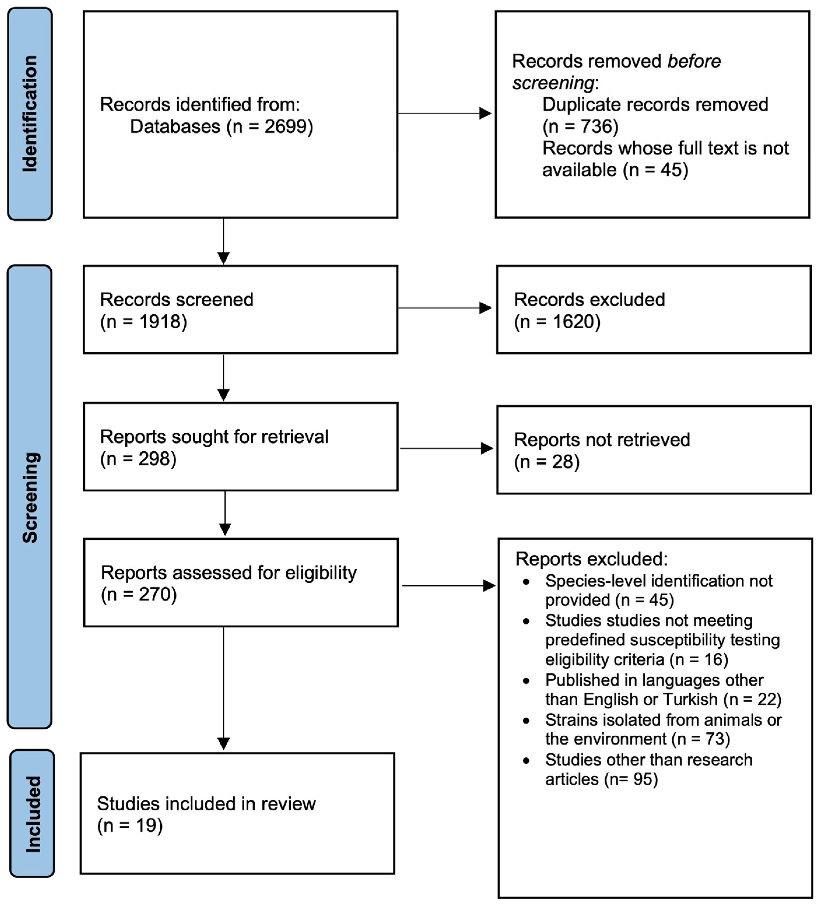
4.4. Study Selection and Data Extraction
4.5. Risk of Bias Assessment
4.6. Statistical Analysis
5. Conclusions
Author Contributions
Funding
Institutional Review Board Statement
Informed Consent Statement
Data Availability Statement
Conflicts of Interest
Appendix A

| Section and Topic | Item # | Checklist Item | Location Where Item Is Reported |
|---|---|---|---|
| TITLE | |||
| Title | 1 | Identify the report as a systematic review. | 1 |
| ABSTRACT | |||
| Abstract | 2 | See the PRISMA 2020 for Abstracts checklist. | 1 |
| INTRODUCTION | |||
| Rationale | 3 | Describe the rationale for the review in the context of existing knowledge. | 2–3 |
| Objectives | 4 | Provide an explicit statement of the objective(s) or question(s) the review addresses. | 3 |
| METHODS | |||
| Eligibility criteria | 5 | Specify the inclusion and exclusion criteria for the review and how studies were grouped for the syntheses. | 4–6 |
| Information sources | 6 | Specify all databases, registers, websites, organizations, reference lists and other sources searched or consulted to identify studies. Specify the date when each source was last searched or consulted. | 4–6 |
| Search strategy | 7 | Present the full search strategies for all databases, registers and websites, including any filters and limits used. | 4–6 |
| Selection process | 8 | Specify the methods used to decide whether a study met the inclusion criteria of the review, including how many reviewers screened each record and each report retrieved, whether they worked independently, and if applicable, details of automation tools used in the process. | 4–6 |
| Data collection process | 9 | Specify the methods used to collect data from reports, including how many reviewers collected data from each report, whether they worked independently, any processes for obtaining or confirming data from study investigators, and if applicable, details of automation tools used in the process. | 4–6 |
| Data items | 10a | List and define all outcomes for which data were sought. Specify whether all results that were compatible with each outcome domain in each study were sought (e.g., for all measures, time points, analyses), and if not, the methods used to decide which results to collect. | 4–6 |
| 10b | List and define all other variables for which data were sought (e.g., participant and intervention characteristics, funding sources). Describe any assumptions made about any missing or unclear information. | 4–6 | |
| Study risk of bias assessment | 11 | Specify the methods used to assess risk of bias in the included studies, including details of the tool(s) used, how many reviewers assessed each study and whether they worked independently, and if applicable, details of automation tools used in the process. | 4–6 |
| Effect measures | 12 | Specify for each outcome the effect measure(s) (e.g., risk ratio, mean difference) used in the synthesis or presentation of results. | 4–6 |
| Synthesis methods | 13a | Describe the processes used to decide which studies were eligible for each synthesis (e.g., tabulating the study intervention characteristics and comparing against the planned groups for each synthesis (item #5)). | 4 |
| 13b | Describe any methods required to prepare the data for presentation or synthesis, such as handling of missing summary statistics, or data conversions. | 4 | |
| 13c | Describe any methods used to tabulate or visually display results of individual studies and syntheses. | 4 | |
| 13d | Describe any methods used to synthesize results and provide a rationale for the choice(s). If meta-analysis was performed, describe the model(s), method(s) to identify the presence and extent of statistical heterogeneity, and software package(s) used. | 3, 4 | |
| 13e | Describe any methods used to explore possible causes of heterogeneity among study results (e.g., subgroup analysis, meta-regression). | 4 | |
| 13f | Describe any sensitivity analyses conducted to assess robustness of the synthesized results. | 4 | |
| Reporting bias assessment | 14 | Describe any methods used to assess risk of bias due to missing results in a synthesis (arising from reporting biases). | 4–6 |
| Certainty assessment | 15 | Describe any methods used to assess certainty (or confidence) in the body of evidence for an outcome. | 4–6 |
| RESULTS | |||
| Study selection | 16a | Describe the results of the search and selection process, from the number of records identified in the search to the number of studies included in the review, ideally using a flow diagram. | 7–15 |
| 16b | Cite studies that might appear to meet the inclusion criteria, but which were excluded, and explain why they were excluded. | 7–15 | |
| Study characteristics | 17 | Cite each included study and present its characteristics. | 7–15 |
| Risk of bias in studies | 18 | Present assessments of risk of bias for each included study. | 7–15 |
| Results of individual studies | 19 | For all outcomes, present, for each study: (a) summary statistics for each group (where appropriate) and (b) an effect estimate and its precision (e.g., confidence/credible interval), ideally using structured tables or plots. | 7–15 |
| Results of syntheses | 20a | For each synthesis, briefly summarize the characteristics and risk of bias among contributing studies. | 7–15 |
| 20b | Present results of all statistical syntheses conducted. If meta-analysis was done, present for each the summary estimate and its precision (e.g., confidence/credible interval) and measures of statistical heterogeneity. If comparing groups, describe the direction of the effect. | 7–15 | |
| 20c | Present results of all investigations of possible causes of heterogeneity among study results. | 7–15 | |
| 20d | Present results of all sensitivity analyses conducted to assess the robustness of the synthesized results. | 7–15 | |
| Reporting biases | 21 | Present assessments of risk of bias due to missing results (arising from reporting biases) for each synthesis assessed. | 16–18 |
| Certainty of evidence | 22 | Present assessments of certainty (or confidence) in the body of evidence for each outcome assessed. | 16–18 |
| DISCUSSION | |||
| Discussion | 23a | Provide a general interpretation of the results in the context of other evidence. | 16–18 |
| 23b | Discuss any limitations of the evidence included in the review. | 16–18 | |
| 23c | Discuss any limitations of the review processes used. | 16–18 | |
| 23d | Discuss implications of the results for practice, policy, and future research. | 16–18 | |
| OTHER INFORMATION | |||
| Registration and protocol | 24a | Provide registration information for the review, including register name and registration number, or state that the review was not registered. | 1, 4 |
| 24b | Indicate where the review protocol can be accessed, or state that a protocol was not prepared. | 1, 4 | |
| 24c | Describe and explain any amendments to information provided at registration or in the protocol. | 1, 4 | |
| Support | 25 | Describe sources of financial or non-financial support for the review, and the role of the funders or sponsors in the review. | - |
| Competing interests | 26 | Declare any competing interests of review authors. | 13 |
| Availability of data, code and other materials | 27 | Report which of the following are publicly available and where they can be found: template data collection forms; data extracted from included studies; data used for all analyses; analytic code; any other materials used in the review. | 13 |
| Database | Search Terms |
|---|---|
| PubMed/MEDLINE | (“Dermatophytes”[Mesh] OR dermatophyte* OR “Trichophyton”[Mesh] OR “Trichophyton rubrum” OR “Trichophyton interdigitale” OR “Trichophyton mentagrophytes” OR “Trichophyton indotineae”) AND (“Terbinafine”[Mesh] OR terbinafine) AND (“Drug Resistance, Fungal”[Mesh] OR resistance OR “antifungal resistance” OR susceptibility OR MIC OR “minimum inhibitory concentration”) AND (prevalence OR epidemiology) |
| Scopus | TITLE-ABS-KEY (dermatophyte* OR “Trichophyton rubrum” OR “Trichophyton interdigitale” OR “Trichophyton mentagrophytes” OR “Trichophyton indotineae”) AND TITLE-ABS-KEY (terbinafine) AND TITLE-ABS-KEY (“antifungal resistance” OR resistance OR susceptibility OR MIC OR prevalence) |
| Web of Science | TS = (dermatophyte* OR “Trichophyton rubrum” OR “Trichophyton interdigitale” OR “Trichophyton mentagrophytes” OR “Trichophyton indotineae”) AND TS = (terbinafine) AND TS = (“antifungal resistance” OR resistance OR susceptibility OR MIC OR prevalence) |
| Embase | (‘dermatophyte’/exp OR dermatophyte*:ti,ab OR ‘Trichophyton’/exp OR “Trichophyton rubrum”:ti,ab OR “Trichophyton interdigitale”:ti,ab OR “Trichophyton mentagrophytes”:ti,ab OR “Trichophyton indotineae”:ti,ab) AND (‘terbinafine’/exp OR terbinafine:ti,ab) AND (‘antifungal resistance’/exp OR resistance:ti,ab OR susceptibility:ti,ab OR MIC:ti,ab OR prevalence:ti,ab) |
| Group | Study | Species | Total Sample | Number of Terbinafine Resistant Strains | Terbinafine Resistance Rate (%) |
|---|---|---|---|---|---|
| 1 | Cañete-Gibas et al., 2023 [17] | T. rubrum | 117 | 21 | 17.95 |
| 1 | Cañete-Gibas et al., 2023 [17] | T. indotineae | 21 | 21 | 100.00 |
| 1 | Nojo et al., 2025 [18] | T. rubrum | 188 | 9 | 4.79 |
| 1 | Nojo et al., 2025 [18] | T. indotineae | 1 | 0 | 0.00 |
| 1 | Nojo et al., 2025 [18] | T. interdigitale | 122 | 1 | 0.82 |
| 1 | Ohara et al., 2025 [19] | T. rubrum | 353 | 16 | 4.53 |
| 1 | Ohara et al., 2025 [19] | T. interdigitale | 82 | 1 | 1.22 |
| 1 | Pashootan et al., 2022 [20] | T. rubrum | 15 | 0 | 0.00 |
| 1 | Pashootan et al., 2022 [20] | T. indotineae | 10 | 6 | 60.00 |
| 1 | Pashootan et al., 2022 [20] | T. interdigitale | 28 | 0 | 0.00 |
| 1 | Taghipour et al., 2020 [21] | T. mentagrophytes | 45 | 5 | 11.11 |
| 1 | Taghipour et al., 2020 [21] | T. interdigitale | 96 | 0 | 0.00 |
| 1 | Mori et al., 2025 [8] | T. mentagrophytes | 199 | 1 | 0.50 |
| 1 | Mori et al., 2025 [8] | T. rubrum | 16 | 16 | 100.00 |
| 1 | Mori et al., 2025 [8] | T. indotineae | 2 | 1 | 50.00 |
| 1 | Mori et al., 2025 [8] | T. interdigitale | 119 | 2 | 1.68 |
| 2 | Moreno-Sabater et al., 2022 [22] | T. rubrum | 436 | 1 | 0.23 |
| 2 | Moreno-Sabater et al., 2022 [22] | T. indotineae | 136 | 1 | 0.74 |
| 2 | Moreno-Sabater et al., 2022 [22] | T. interdigitale | 8 | 1 | 12.50 |
| 2 | Siopi et al., 2021 [23] | T. mentagrophytes | 24 | 9 | 37.50 |
| 2 | Siopi et al., 2021 [23] | T. rubrum | 70 | 0 | 0.00 |
| 2 | Siopi et al., 2021 [23] | T. interdigitale | 12 | 0 | 0.00 |
| 2 | Jiang et al., 2021 [24] | T. rubrum | 62 | 0 | 0.00 |
| 2 | Kolarczyková et al., 2023 [25] | T. mentagrophytes | 240 | 6 | 2.50 |
| 2 | Kolarczyková et al., 2023 [25] | T. rubrum | 514 | 0 | 0.00 |
| Studies | 1. Was the Sample Frame Appropriate to Address the Target Population? | 2. Were Study Participants Sampled in an Appropriate Way? | 3. Was the Sample Size Adequate? | 4. Were the Study Subjects and the Setting Described in Detail? | 5. Was the Data Analysis Conducted with Sufficient Coverage of the Identified Sample? | 6. Were Valid Methods Used for the Identification of the Condition? | 7. Was the Condition Measured in a Standard, Reliable Way for All Participants? | 8. Was There Appropriate Statistical Analysis? | 9. Was the Response Rate Adequate, and If Not, Was the Low Response Rate Managed Appropriately? | General Risk |
|---|---|---|---|---|---|---|---|---|---|---|
| Cañete-Gibas et al., 2023 [17] | Y | Y | Y | Y | U | Y | Y | Y | NA | Low |
| Nojo et al., 2025 [18] | Y | Y | Y | Y | Y | Y | Y | N | NA | Moderate |
| Ohara et al., 2025 [19] | Y | U | Y | Y | Y | Y | Y | N | NA | Moderate |
| Pashootan et al., 2022 [20] | Y | U | Y | Y | Y | Y | Y | Y | NA | Low |
| Taghipour et al., 2020 [21] | Y | U | Y | Y | Y | Y | Y | N | NA | Moderate |
| Mori et al., 2025 [8] | Y | U | Y | Y | Y | Y | Y | Y | NA | Low |
| Moreno-Sabater et al., 2022 [22] | Y | Y | Y | Y | Y | Y | Y | N | NA | Low |
| Siopi et al., 2021 [23] | Y | U | Y | Y | Y | Y | Y | Y | NA | Low |
| Jiang et al., 2021 [24] | Y | U | Y | Y | Y | Y | Y | Y | NA | Low |
| Kolarczyková et al., 2023 [25] | Y | Y | Y | Y | Y | Y | Y | N | NA | Low |
| Astvad et al., 2022 [14] | N | N | U | Y | U | Y | Y | Y | NA | Moderate |
| Amin et al., 2024 [9] | N | Y | Y | Y | Y | Y | Y | N | NA | Moderate |
| Sardana et al., 2018 [26] | N | Y | U | Y | Y | Y | Y | N | NA | Moderate |
| Bhuiyan et al., 2024 [27] | N | N | Y | Y | Y | Y | Y | N | NA | Moderate |
| McTaggart et al., 2025 [28] | N | N | Y | Y | Y | Y | Y | N | NA | Moderate |
| De Paepe et al., 2024 [29] | N | N | N | Y | Y | Y | Y | N | NA | Moderate |
| Bidaud et al., 2023 [30] | N | N | Y | Y | Y | Y | Y | N | NA | Moderate |
| Shankarnarayan et al., 2023 [31] | N | U | Y | Y | Y | Y | Y | N | NA | Moderate |
| Oguz et al., 2024 [32] | N | N | U | Y | Y | U | Y | Y | NA | Moderate |
References
- Qu, J.; Lv, X. Cryptococcal meningitis in apparently immunocompetent patients. Crit. Rev. Microbiol. 2024, 50, 76–86. [Google Scholar]
- Haghani, I.; Hedayati, M.T.; Shokohi, T.; Kermani, F.; Ghazanfari, M.; Javidnia, J.; Khojasteh, S.; Roohi, B.; Badali, H.; Fathi, M.; et al. Onychomycosis due to Fusarium species in different continents: Literature review on diagnosis and treatment. Mycoses 2024, 67, e13652. [Google Scholar] [PubMed]
- Vitiello, A.; Ferrara, F.; Boccellino, M.; Ponzo, A.; Cimmino, C.; Comberiati, E.; Zovi, A.; Clemente, S.; Sabbatucci, M. Antifungal drug resistance: An emergent health threat. Biomedicines 2023, 11, 1063. [Google Scholar] [CrossRef] [Scilit] [PubMed]
- Crotti, S.; Cruciani, D.; Sabbatucci, M.; Spina, S.; Piscioneri, V.; Torricelli, M.; Calcaterra, R.; Farina, C.; Pisano, L.; Papini, M. Terbinafine resistance in Trichophyton strains isolated from humans and animals: A retrospective cohort study in Italy, 2016 to May 2024. J. Clin. Med. 2024, 13, 5493. [Google Scholar] [CrossRef] [Scilit] [PubMed]
- Hill, R.C.; Caplan, A.S.; Elewski, B.; Gold, J.A.W.; Lockhart, S.R.; Smith, D.J.; Lipner, S.R. Expert panel review of skin and hair dermatophytoses in an era of antifungal resistance. Am. J. Clin. Dermatol. 2024, 25, 359–389. [Google Scholar] [CrossRef] [Scilit] [PubMed]
- Saunte, D.M.L.; Pereiro-Ferreirós, M.; Rodríguez-Cerdeira, C.; Sergeev, A.Y.; Arabatzis, M.; Prohić, A.; Piraccini, B.M.; Lecerf, P.; Nenoff, P.; Kotrekhova, L.P.; et al. Emerging antifungal treatment failure of dermatophytosis in Europe: Take care or it may become endemic. J. Eur. Acad. Dermatol. Venereol. 2021, 35, 1582–1586. [Google Scholar] [CrossRef] [Scilit] [PubMed]
- Kidd, S.E.; Abdolrasouli, A.; Hagen, F. Fungal Nomenclature: Managing Change is the Name of the Game. Open Forum Infect. Dis. 2023, 10, ofac559. [Google Scholar] [CrossRef] [Scilit] [PubMed]
- Mori, Y.; Yamada, T.; Ban, S.; Yoshioka, I.; Yaguchi, T. Survey on the prevalence of terbinafine-resistant Trichophyton spp. with squalene epoxidase mutations. Med. Mycol. J. 2025, 66, 125–130. [Google Scholar] [CrossRef] [Scilit] [PubMed]
- Amin, N.; Shenoy, M.M.; Pai, V. Antifungal susceptibility of dermatophyte isolates from patients with chronic and recurrent dermatophytosis. Indian Dermatol. Online J. 2024, 16, 110–115. [Google Scholar] [CrossRef] [Scilit] [PubMed]
- Patel, N.H.; Sardana, K.; Shenoy, M.M.; Rengasamy, M.; Khurana, A.; Ghate, S.; Venkata, C.K.; Marfatiya, Y.; Bhunia, D.; Jayaraman, J.; et al. Recalcitrant dermatophytosis position statement on super bioavailable itraconazole. Indian Dermatol. Online J. 2023, 15, 1–7. [Google Scholar] [CrossRef] [Scilit] [PubMed]
- Mukherjee, P.; Leidich, S.D.; Isham, N.; Leitner, I.; Ryder, N.S.; Ghannoum, M.A. Clinical Trichophyton rubrum strain exhibiting primary resistance to terbinafine. Antimicrob. Agents Chemother. 2003, 47, 82–86. [Google Scholar] [CrossRef] [Scilit] [PubMed]
- Osborne, C.S.; Leitner, I.; Favre, B.; Ryder, N.S. Amino acid substitution in Trichophyton rubrum squalene epoxidase associated with resistance to terbinafine. Antimicrob. Agents Chemother. 2005, 49, 2840–2844. [Google Scholar] [CrossRef] [Scilit] [PubMed]
- Yamada, T.; Maeda, M.; Alshahni, M.M.; Tanaka, R.; Yaguchi, T.; Bontems, O.; Salamin, K.; Fratti, M.; Monod, M. Terbinafine resistance of Trichophyton clinical isolates caused by specific point mutations in the squalene epoxidase gene. Antimicrob. Agents Chemother. 2017, 61, e00115-17. [Google Scholar] [CrossRef] [Scilit] [PubMed]
- Astvad, K.M.T.; Hare, R.K.; Jørgensen, K.M.; Saunte, D.M.L.; Thomsen, P.K.; Arendrup, M.C. Increasing terbinafine resistance in Danish Trichophyton isolates 2019–2020. J. Fungi 2022, 8, 150. [Google Scholar] [CrossRef] [Scilit]
- Gupta, A.K.; Cooper, E.A.; Wang, T.; Ravi, S.P.; Lincoln, S.A.; Piguet, V.; McCarthy, L.R.; Bakotic, W.L. Detection of squalene epoxidase mutations in United States patients with onychomycosis: Implications for management. J. Investig. Dermatol. 2023, 143, 2476–2483. [Google Scholar] [CrossRef] [Scilit] [PubMed]
- Mohammadi, L.Z.; Shams-Ghahfarokhi, M.; Salehi, Z.; Razzaghi-Abyaneh, M. Increased terbinafine resistance among clinical genotypes of Trichophyton mentagrophytes/Trichophyton interdigitale species complex harboring squalene epoxidase gene mutations. J. Mycol. Méd. 2024, 34, 101495. [Google Scholar] [CrossRef] [Scilit] [PubMed]
- Cañete-Gibas, C.F.; Mele, J.; Patterson, H.P.; Sanders, C.J.; Ferrer, D.; Garcia, V.; Fan, H.; David, M.; Wiederhold, N.P. Terbinafine-resistant dermatophytes and the presence of Trichophyton indotineae in North America. J. Clin. Microbiol. 2023, 61, e00562-23. [Google Scholar] [CrossRef] [Scilit] [PubMed]
- Nojo, H.; Hiruma, J.; Noguchi, H.; Shimizu, T.; Hiruma, M.; Harada, K.; Makimura, K.; Kano, R. Terbinafine-resistant dermatophytes isolated in Japan. Med. Mycol. J. 2025, 66, 11–15. [Google Scholar] [CrossRef] [Scilit] [PubMed]
- Ohara, S.; Noguchi, H.; Matsumoto, T.; Kubo, M.; Hayashi, D.; Kashiwada-Nakamura, K.; Yaguchi, T.; Kano, R. Emerging antifungal-resistant onychomycosis in a dermatology clinic in Kumamoto, Japan. Med. Mycol. J. 2025, 66, 61–67. [Google Scholar] [CrossRef] [Scilit] [PubMed]
- Pashootan, N.; Shams-Ghahfarokhi, M.; Chaichi Nusrati, A.; Salehi, Z.; Asmar, M.; Razzaghi-Abyaneh, M. Phylogeny, antifungal susceptibility, and point mutations of SQLE gene in major pathogenic dermatophytes isolated from clinical dermatophytosis. Front. Cell. Infect. Microbiol. 2022, 12, 851769. [Google Scholar] [CrossRef] [Scilit] [PubMed]
- Taghipour, S.; Shamsizadeh, F.; Pchelin, I.M.; Rezaei-Matehhkolaei, A.; Zarei Mahmoudabadi, A.; Valadan, R.; Ansari, S.; Katiraee, F.; Pakshir, K.; Zomorodian, K.; et al. Emergence of terbinafine resistant Trichophyton mentagrophytes in Iran harboring mutations in the squalene epoxidase (SQLE) gene. Infect. Drug Resist. 2020, 13, 845–850. [Google Scholar] [CrossRef] [Scilit] [PubMed]
- Moreno-Sabater, A.; Normand, A.C.; Bidaud, A.L.; Cremer, G.; Foulet, F.; Brun, S.; Bonnal, C.; Aït-Ammar, N.; Jabet, A.; Ayachi, A.; et al. Terbinafine resistance in dermatophytes: A French multicenter prospective study. J. Fungi 2022, 8, 220. [Google Scholar] [CrossRef] [Scilit]
- Siopi, M.; Efstathiou, I.; Theodoropoulos, K.; Pournaras, S.; Meletiadis, J. Molecular epidemiology and antifungal susceptibility of Trichophyton isolates in Greece: Emergence of terbinafine-resistant Trichophyton mentagrophytes type VIII locally and globally. J. Fungi 2021, 7, 419. [Google Scholar] [CrossRef] [Scilit]
- Jiang, Y.; Luo, W.; Verweij, P.E.; Song, Y.; Zhang, B.; Shang, Z.; Al-Hatmi, A.M.S.; Ahmed, S.A.; Wan, Z.; Li, R.; et al. Regional differences in antifungal susceptibility of the prevalent dermatophyte Trichophyton rubrum. Mycopathologia 2021, 186, 53–70. [Google Scholar] [PubMed]
- Kolarczyková, D.; Lysková, P.; Švarcová, M.; Kuklová, I.; Dobiáš, R.; Mallátová, N.; Kolařík, M.; Hubka, V. Terbinafine resistance in Trichophyton mentagrophytes and Trichophyton rubrum in the Czech Republic: A prospective multicentric study. Mycoses 2024, 67, e13708. [Google Scholar] [CrossRef] [Scilit] [PubMed]
- Sardana, K.; Kaur, R.; Arora, P.; Goyal, R.; Ghunawat, S. Is antifungal resistance a cause for treatment failure in dermatophytosis: A study focused on tinea corporis and cruris from a tertiary centre? Indian Dermatol. Online J. 2018, 9, 90–95. [Google Scholar] [CrossRef] [Scilit] [PubMed]
- Bhuiyan, M.S.I.; Verma, S.B.; Illigner, G.M.; Uhrlaß, S.; Klonowski, E.; Burmester, A.; Noor, T.; Nenoff, P. Trichophyton mentagrophytes ITS genotype VIII/Trichophyton indotineae infection and antifungal resistance in Bangladesh. J. Fungi 2024, 10, 768. [Google Scholar] [CrossRef] [Scilit]
- McTaggart, L.R.; Cronin, K.; Ruscica, S.; Patel, S.N.; Kus, J.V. Emergence of terbinafine-resistant Trichophyton indotineae in Ontario, Canada, 2014–2023. J. Clin. Microbiol. 2025, 63, e01535-24. [Google Scholar] [CrossRef] [Scilit] [PubMed]
- De Paepe, R.; Normand, A.C.; Uhrlaß, S.; Nenoff, P.; Piarroux, R.; Packeu, A. Resistance profile, terbinafine resistance screening and MALDI-TOF MS identification of the emerging pathogen Trichophyton indotineae. Mycopathologia 2024, 189, 29. [Google Scholar] [CrossRef] [Scilit] [PubMed]
- Bidaud, A.L.; Moreno-Sabater, A.; Normand, A.C.; Cremer, G.; Foulet, F.; Brun, S.; Ayachi, A.; Imbert, S.; Chowdhary, A.; Dannaoui, E. Evaluation of gradient concentration strips for detection of terbinafine resistance in Trichophyton spp. Antimicrob. Agents Chemother. 2023, 67, e01716-22. [Google Scholar] [CrossRef] [Scilit]
- Shankarnarayan, S.A.; Shaw, D.; Sharma, A.; Chakrabarti, A.; Dogra, S.; Kumaran, M.S.; Kaur, H.; Ghosh, A.; Rudramurthy, S.M. Rapid detection of terbinafine resistance in Trichophyton species by amplified refractory mutation system-polymerase chain reaction. Sci. Rep. 2020, 10, 1297. [Google Scholar] [CrossRef] [Scilit] [PubMed]
- Oğuz, I.D.; Direkel, Ş.; Akşan, B.; Kulakli, S.; Aksoy, S. In vitro antifungal susceptibility of the dermatophytes: A single center study from eastern Black Sea region of Turkey. Eur. Rev. Med. Pharmacol. Sci. 2024, 28, 2144–2154. [Google Scholar] [CrossRef] [Scilit] [PubMed]
- Balakumar, S.; Rajan, S.; Thirunalasundari, T.; Jeeva, S. Epidemiology of dermatophytosis in and around Tiruchirapalli, Tamil Nadu, India. Asian Pac. J. Trop. Dis. 2012, 2, 286–289. [Google Scholar] [CrossRef] [Scilit]
- Singh, A.; Masih, A.; Monroy-Nieto, J.; Singh, P.K.; Bowers, J.; Travis, J.; Khurana, A.; Engelthaler, D.M.; Meis, J.F.; Chowdhary, A. A unique multidrug-resistant clonal Trichophyton population distinct from Trichophyton mentagrophytes/Trichophyton interdigitale complex causing an ongoing alarming dermatophytosis outbreak in India: Genomic insights and resistance profile. Fungal Genet. Biol. 2019, 133, 103266. [Google Scholar] [CrossRef] [Scilit] [PubMed]
- Rhodes, J.; Hui, S.T.; Dellière, S.; Summerbell, R.C.; Scott, J.A.; Kaur, A.; Barton, R.C.; Leitao, R.; Hemmings, S.; Goiriz, R.; et al. Emerging terbinafine-resistant Trichophyton indotineae between 2018 and 2023: A multinational genomic epidemiology study. Lancet Microbe 2026, 7, 101273. [Google Scholar] [CrossRef] [Scilit] [PubMed]
- Shah, S.R.; Vyas, H.R.; Shah, B.J.; Jangid, N.C.; Choudhary, A.; Gehlawat, T.; Mistry, D.; Joshi, R. A clinical-mycological study of dermatophytosis in western India with focus on antifungal drug resistance as a factor in recalcitrance. Indian J. Dermatol. 2023, 68, 234. [Google Scholar] [CrossRef] [Scilit] [PubMed]
- Jegathees, T.; Holmes, Z.P.; Martin, C.; Kalai, C.; Voutier, C.; Spelman, D.; Robertson, G.; Kern, J.S. Terbinafine-resistant Trichophyton dermatophytosis in Australia. Australas. J. Dermatol. 2026; in press. [CrossRef] [Scilit] [PubMed]
- Jartarkar, S.R.; Patil, A.; Goldust, Y.; Cockerell, C.J.; Schwartz, R.A.; Grabbe, S.; Goldust, M. Pathogenesis, immunology and management of dermatophytosis. J. Fungi 2022, 8, 39. [Google Scholar]
- Hammoudi Halat, D.; Younes, S.; Mourad, N.; Rahal, M. Allylamines, benzylamines, and fungal cell permeability: A review of mechanistic effects and usefulness against fungal pathogens. Membranes 2022, 12, 1171. [Google Scholar] [CrossRef] [Scilit] [PubMed]
- Zhou, X.; de Hoog, S. A Chinese warning: Terbinafine-resistant Trichophyton indotineae, a global dermatological challenge. J. Eur. Acad. Dermatol. Venereol. 2026, 40, 148–149. [Google Scholar] [CrossRef] [Scilit] [PubMed]
- Gold, J.A.W.; Benedict, K.; Lockhart, S.R.; Lutfy, C.; Lyman, M.; Smith, D.J.; Polgreen, P.M.; Beekmann, S.E. Recognition of antifungal-resistant dermatophytosis by infectious diseases specialists, United States. Emerg. Infect. Dis. 2024, 30, 1978–1980. [Google Scholar] [CrossRef] [Scilit] [PubMed]
- Shen, J.J.; Arendrup, M.C.; Verma, S.; Saunte, D.M.L. The emerging terbinafine-resistant Trichophyton epidemic: What is the role of antifungal susceptibility testing? Dermatology 2022, 238, 60–79. [Google Scholar] [PubMed]
- Posso-De Los Rios, C.J.; Tadros, E.; Summerbell, R.C.; Scott, J.A. Terbinafine resistant Trichophyton indotineae isolated in patients with superficial dermatophyte infection in Canadian patients. J. Cutan. Med. Surg. 2022, 26, 371–376. [Google Scholar] [CrossRef] [Scilit] [PubMed]
- Edriss, M.T.; Parker, J.J.; Pritchett, E.N. Response to Gu et al.’s “Treatment-resistant dermatophytosis: A representative case highlighting an emerging public health threat”. JAAD Case Rep. 2022, 32, 88–89. [Google Scholar] [PubMed]
- Moher, D.; Shamseer, L.; Clarke, M.; Ghersi, D.; Liberati, A.; Petticrew, M.; Shekelle, P.; Stewart, L.A. Preferred reporting items for systematic review and meta-analysis protocols (PRISMA-P) 2015 statement. Syst. Rev. 2015, 4, 1. [Google Scholar] [CrossRef] [PubMed]
- Munn, Z.; Moola, S.; Lisy, K.; Riitano, D.; Tufanaru, C. Systematic reviews of prevalence and incidence. In JBI Manual for Evidence Synthesis; Aromataris, E., Munn, Z., Eds.; JBI: Adelaide, Australia, 2020; pp. 117–217. [Google Scholar]

| Group | Study | Country | Sample | Identification Method | Species | Method for Detection of Terbinafine Resistance | Guideline | Total Sample | Number of Terbinafine Resistant Strains | Terbinafine Resistance Rate (%) | Cut-Off Values |
|---|---|---|---|---|---|---|---|---|---|---|---|
| 1 | Cañete-Gibas et al., 2023 [17] | North America | Skin samples (including skin scrapings and tissue specimens) | PCR | T. rubrum, T. indotineae | BMD | CLSI M38 (2022) | 271 | 50 | 18.45 | ≥1 µg/mL |
| 1 | Nojo et al., 2025 [18] | Japan | Skin and nail samples | Microscopy and culture-based morphological identification | T. rubrum, T. indotineae, T. interdigitale | BMD | CLSI M38-A2 (2008) | 311 | 10 | 3.22 | ≥1 µg/mL |
| 1 | Ohara et al., 2025 [19] | Japan | Nail samples | PCR | T. rubrum, T. interdigitale | BMD | CLSI M38-A2 (2017) | 477 | 17 | 3.56 | ≥1 µg/mL |
| 1 | Pashootan et al., 2022 [20] | Iran | - | ITS sequencing | T. rubrum, T. indotineae, T. interdigitale | BMD | CLSI M38-A2 (2008) | 123 | 6 | 4.88 | ≥1 µg/mL |
| 1 | Taghipour et al., 2020 [21] | Iran | Skin, nail and hair samples | PCR | T. mentagrophytes, T. interdigitale | BMD | CLSI M38-A2 (2008) | 141 | 5 | 3.55 | ≥1 µg/mL |
| 1 | Mori et al., 2025 [8] | Japan | Skin and nail samples | PCR | T. mentagrophytes, T. rubrum, T. indotineae, T. interdigitale | BMD | CLSI M38-A2 (2017) | 701 | 20 | 2.85 | ≥1 µg/mL |
| 2 | Moreno-Sabater et al., 2022 [22] | France | Skin and nail samples | PCR | T. rubrum, T. indotineae, T. interdigitale | BMD | EUCAST E.DEF 11.0 (2021) | 580 | 3 | 0.52 | 0.25–0.5 µg/mL |
| 2 | Siopi et al., 2021 [23] | Greece | Skin and nail samples | PCR | T. mentagrophytes, T. rubrum, T. interdigitale | BMD | EUCAST E.DEF 11.0 (2021) | 112 | 9 | 8.04 | 0.25–0.5 µg/mL |
| 2 | Jiang et al., 2021 [24] | China | Skin and hair samples | ITS sequencing | T. rubrum | BMD | CLSI M38-A3 (2017) | 62 | 0 | 0.00 | 0.25–0.5 µg/mL |
| 2 | Kolarczyková et al., 2023 [25] | Czech Republic | Skin and nail samples | PCR | T. mentagrophytes, T. rubrum | BMD | EUCAST E.DEF 11.0 (2021) | 754 | 6 | 0.80 | >4 mg/L |
| 3 | Astvad et al., 2022 [14] | Denmark | - | ITS sequencing | T. rubrum, T. indotineae | - | EUCAST E.DEF 11.0 (2021) | 63 | 38 | 60.32 | ≥1 µg/mL |
| 3 | Amin et al., 2024 [9] | India | Skin samples (including skin scrapings) | - | T. mentagrophytes, T. rubrum | BMD | CLSI M38-A2 (2017) | 60 | 0 | 0.00 | ≥1 µg/mL |
| 3 | Sardana et al., 2018 [26] | India | Skin samples (including skin scrapings) | Microscopy and culture-based morphological identification | T. mentagrophytes, T. rubrum | BMD | CLSI M38-A2 (2008) | 40 | 0 | 0.00 | ≥1 µg/mL |
| 3 | Bhuiyan et al., 2024 [27] | Bangladesh | Skin samples | PCR | T. mentagrophytes, T. rubrum | In vitro agar-based susceptibility test | EUCAST E.DEF 11.0 (2021) | 80 | 49 | 61.25 | 0.125 µg/mL |
| 3 | McTaggart et al., 2025 [28] | Canada | - | PCR | T. indotineae | BMD | CLSI M38-A2 (2017) | 50 | 36 | 72.00 | ≥1 µg/mL |
| 3 | De Paepe et al., 2024 [29] | Germany | - | MALDI-TOF MS | T. indotineae | BMD | EUCAST E.DEF 11.0 (2022) | 20 | 9 | 45.00 | ≥0.5 µg/mL |
| 3 | Bidaud et al., 2023 [30] | France | - | ITS sequencing | T. indotineae, T. interdigitale | BMD | EUCAST E.DEF (2021) | 79 | 10 | 12.66 | 0.25–0.5 µg/mL |
| 3 | Shankarnarayan et al., 2023 [31] | India | Skin samples (including skin scrapings) | PCR | T. mentagrophytes, T. rubrum | - | CLSI M38-A2 (2008) | 136 | 15 | 11.03 | ≥1 µg/mL |
| 3 | Oguz et al., 2024 [32] | Türkiye | Skin, nail and hair samples | Microscopy and culture-based morphological identification | T. mentagrophytes, T. rubrum | BMD | CLSI | 51 | 14 | 27.45 | ≥0.5 µg/mL |
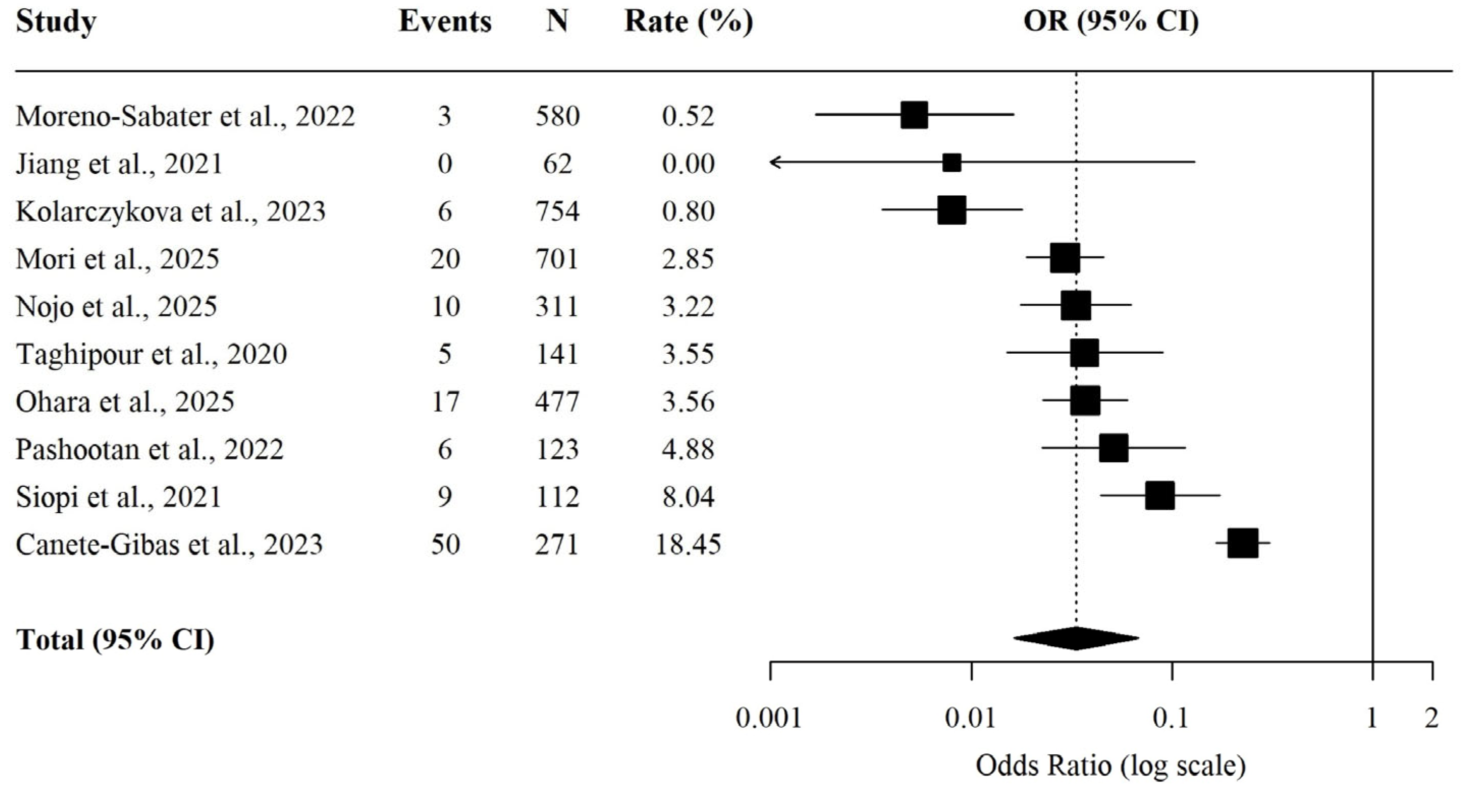
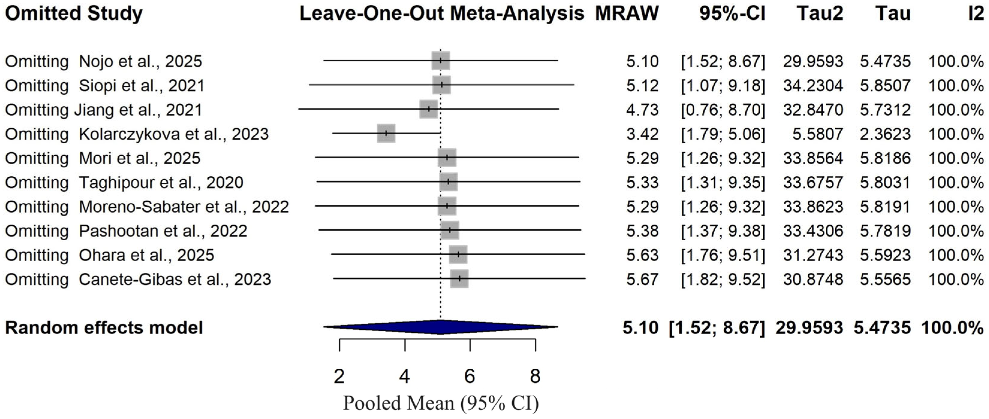
| Model | Fixed | Random | |
|---|---|---|---|
| Effect size and 95% interval | Number Studies | 10 | 10 |
| Point estimate | 0.060 | 0.032 | |
| Lower limit | 0.050 | 0.015 | |
| Upper limit | 0.071 | 0.067 | |
| Test of null (2-Tail) | Z-value | −29.532 | −8.564 |
| p-value | 0.000 | 0.000 | |
| Heterogeneity | Q-value | 135.085 | |
| df (Q) | 9 | ||
| p-value | 0.000 | ||
| i2 | 93.338 | ||
| Tau-squared | Tau2 | 1.370 | |
| Standard error | 0.890 |
Disclaimer/Publisher’s Note: The statements, opinions and data contained in all publications are solely those of the individual author(s) and contributor(s) and not of MDPI and/or the editor(s). MDPI and/or the editor(s) disclaim responsibility for any injury to people or property resulting from any ideas, methods, instructions or products referred to in the content. |
© 2026 by the authors. Licensee MDPI, Basel, Switzerland. This article is an open access article distributed under the terms and conditions of the Creative Commons Attribution (CC BY) license.
Share and Cite
Kilbas, I.; Kahraman Kilbas, E.P.; Ciftci, I.H.; Varga, N.-I.; Horhat, F.G. Emerging Global Patterns of Terbinafine Resistance in Trichophyton Species: A Systematic Review and Meta-Analysis. Antibiotics 2026, 15, 699. https://doi.org/10.3390/antibiotics15070699
Kilbas I, Kahraman Kilbas EP, Ciftci IH, Varga N-I, Horhat FG. Emerging Global Patterns of Terbinafine Resistance in Trichophyton Species: A Systematic Review and Meta-Analysis. Antibiotics. 2026; 15(7):699. https://doi.org/10.3390/antibiotics15070699
Chicago/Turabian StyleKilbas, Imdat, Elmas Pinar Kahraman Kilbas, Ihsan Hakki Ciftci, Norberth-Istvan Varga, and Florin George Horhat. 2026. "Emerging Global Patterns of Terbinafine Resistance in Trichophyton Species: A Systematic Review and Meta-Analysis" Antibiotics 15, no. 7: 699. https://doi.org/10.3390/antibiotics15070699
APA StyleKilbas, I., Kahraman Kilbas, E. P., Ciftci, I. H., Varga, N.-I., & Horhat, F. G. (2026). Emerging Global Patterns of Terbinafine Resistance in Trichophyton Species: A Systematic Review and Meta-Analysis. Antibiotics, 15(7), 699. https://doi.org/10.3390/antibiotics15070699