Article

Cefiderocol Susceptibility in Japanese Clinical Enterobacterales Isolates and the Effect of IMP-Type Carbapenemases on Resistance

1
Department of Infection Control and Prevention, Graduate School of Medicine, The University of Osaka, 2-2 Yamadaoka, Suita, Osaka 565-0871, Japan
2
Department of Transformative Protection to Infectious Disease, Graduate School of Medicine, The University of Osaka, 2-2 Yamadaoka, Suita, Osaka 565-0871, Japan
3
Osaka Institute of Public Health, 1-3-3 Nakamichi, Higashinari-ku, Osaka 537-0025, Japan
4
Department of Clinical Laboratory, The University of Osaka Hospital, 2-15 Yamadaoka, Suita, Osaka 565-0871, Japan
5
Research Institute for Microbial Diseases, The University of Osaka, 3-1 Yamadaoka, Suita, Osaka 565-0871, Japan
*
Author to whom correspondence should be addressed.
Antibiotics 2026, 15(7), 667; https://doi.org/10.3390/antibiotics15070667
Submission received: 5 June 2026 / Revised: 6 July 2026 / Accepted: 6 July 2026 / Published: 8 July 2026
(This article belongs to the Section Mechanism and Evolution of Antibiotic Resistance)

Abstract

Objectives: Carbapenem-resistant Enterobacterales (CRE) limit treatment options, and cefiderocol’s efficacy can be compromised by regional carbapenemases. In this study, we aimed to characterize cefiderocol susceptibility distribution among Enterobacterales in Japan—specifically endemic strains producing IMP-type carbapenemases (IMP)—and explore the effect of IMP in mediating resistance. Methods: The susceptibility of 556 evaluable Enterobacterales isolates (from an initial 560 screened) to cefiderocol was assessed using disk diffusion and broth microdilution methods based on Clinical and Laboratory Standards Institute (CLSI) guidelines. Minimum inhibitory concentration (MIC) distributions across groups were compared using Kruskal–Wallis and Mann–Whitney U tests. Results: A total of 556 isolates yielded evaluable results. The cefiderocol susceptibility rate was 99.5 and 98.7% based on CLSI and European Committee on Antimicrobial Susceptibility Testing breakpoints, respectively. Notably, under both criteria, IMP-producing isolates exhibited a susceptibility rate of 99.6%, whereas non-CRE and non-extended-spectrum β-lactamase (ESBL) exhibited a susceptibility rate of 99.4%. Statistical analysis revealed that the ESBL-only group had higher MICs than the non-CRE/non-ESBL and IMP-only groups (both p < 0.001), whereas no significant difference was observed between the latter two (p = 0.083). Notably, the ESBL-only group exhibited higher MICs than those of isolates harboring both IMP and ESBL. Conclusions: These findings indicate a non-additive effect, in which the coexistence of multiple resistance enzymes does not necessarily increase cefiderocol resistance. The association between IMP and cefiderocol resistance may be limited. In specific enzyme combinations, its presence was even associated with lower MICs.

1. Introduction

Antimicrobial resistance (AMR) has emerged as a global public health crisis, threatening the efficacy of modern medical treatments [1]. The global dissemination of extended-spectrum β-lactamase (ESBL) and carbapenemase-producing Enterobacterales (CPE) has compromised the efficacy of standard therapies, resulting in increased mortality and significant healthcare burden [2]. These resistance trends require effective therapeutic alternatives.
Cefiderocol is a novel siderophore cephalosporin designed to overcome common resistance mechanisms in Gram-negative bacteria, including β-lactamase production, porin mutations, and efflux pumps [3]. Structurally, it combines the features of cefepime and ceftazidime with a chlorocatechol moiety that enables siderophore-mediated iron transport into bacterial cells, thereby enhancing antibiotic uptake [4,5].
Cefiderocol demonstrates stability against carbapenemases, including metallo-β-lactamases, such as IMP-type carbapenemases (IMP), Verona integron-encoded metallo-β-lactamase (VIM), and New Delhi metallo-β-lactamase (NDM) [6]. However, resistance to cefiderocol has been associated with multiple mechanisms, including alterations in siderophore receptor genes, porin mutations, efflux pump activation, and modifications of penicillin-binding protein 3 (PBP3) [7]. Individual mechanisms alone are usually inadequate to confer clinical resistance—resistance commonly results from combinations of multiple factors [7]. Consequently, evaluating how these multi-layered genetic combinations influence real-world diagnostic performance is critical, particularly in regions where specific carbapenemase genotypes are heavily endemic.
Cefiderocol was first approved in the United States in 2019 for complicated urinary tract infections and later for hospital-acquired and ventilator-associated pneumonia [8]. In Europe, it was approved in 2020 for Gram-negative pathogen-caused infections with limited treatment options [9]. In Japan, cefiderocol was approved on 30 November 2023, for various infections caused by strains resistant to carbapenem antibiotics [10].
Globally, molecular and phenotypic surveillance of cefiderocol susceptibility has been extensively documented, particularly through large multinational programs in Europe and North America. In the SIDERO-WT surveillance studies, cefiderocol showed potent in vitro activity against a broad range of Gram-negative pathogens, including carbapenem-resistant isolates and carbapenemase producers, across more than 46,000 clinical isolates collected between 2014 and 2019 [11]. In Japan, large-scale surveillance studies provide an important epidemiologic baseline; however, there remains limited information on how specific co-existing resistance determinants affect categorical agreement between routine susceptibility methods and broth microdilution [12].
Surveillance data from the Japan Antimicrobial Resistant Bacterial Surveillance program reported cefiderocol susceptibility of 97.7 and 99.2% for CPE and IMP-type isolates, respectively [13]. However, the effect of IMP on cefiderocol susceptibility remains unclear. Additionally, susceptibility data for common clinical Enterobacterales isolates that are neither carbapenem-resistant Enterobacterales (CRE) nor ESBL producers remain limited in Japan.
To bridge this gap, the primary objective of this study was to characterize cefiderocol susceptibility and evaluate disk diffusion (DD) performance against a well-defined cohort of Enterobacterales clinical isolates collected in Osaka, Japan, across two distinct timelines (2020–2024 and 2015–2016). This regional focus provides precise, institutional diagnostic and therapeutic insights tailored for local nosocomial management.

2. Results

Among the 560 clinical isolates, four isolates were excluded because of inadequate growth during testing, leaving 556 evaluable isolates for final analysis.

2.1. Cefiderocol Susceptibility Among Enterobacterales Isolates

Using the DD method, six isolates were categorized as non-susceptible (Table 1); however, only two isolates (OU_24 and OU_186) were classified as non-susceptible by the broth microdilution (BMD) method. Additionally, one IMP-6-producing Klebsiella aerogenes isolate (OU_551) demonstrated discordant susceptibility results, being categorized as susceptible by the DD method but resistant by the BMD method.

2.2. Genomic Analysis of Cefiderocol Non-Susceptible Isolates

To assess the molecular mechanisms underlying reduced cefiderocol susceptibility, whole-genome sequencing (WGS) was performed for the three isolates identified as non-susceptible by the BMD method.
OU_24 was an ESBL-producing E. coli belonging to ST2011 and was categorized as intermediate by both DD and BMD methods. WGS analysis identified the β-lactamase gene (blaCTX-M-55), with no mutations or deletions observed in PBP3 or colicin I receptor A (cirA). However, multiple amino acid insertions were identified in outer membrane protein C (ompC) and outer membrane protein F (ompF), potentially altering the outer membrane permeability.
OU_186 was identified as Enterobacter bugandensis and classified as a non-CRE/non-ESBL AmpC β-lactamase (AmpC)-producing isolate. Multi-locus sequence typing (MLST) analysis revealed a novel sequence type (ST3387) that was newly assigned by the PubMLST database. OU_186 was categorized as resistant by both the DD and BMD methods. Genomic analysis identified the presence of blaACT-77, Leu313 deletion (ΔL313) in the R2 loop of the AmpC, cirA gene deletion, and specific amino acid substitution (Y432F) in PBP3.
OU_551 was an ESBL- and IMP-producing Klebsiella aerogenes belonging to ST44231. The isolate carried blaCTX-M-2, blaIMP-6, and blaAmpC-Kaer-1 genes. Although no deletion was observed in the R2 loop of AmpC, the isolate exhibited deletions in cirA and fiu, along with three amino acid substitutions in PBP3 (S261T, A290S, and I563V).

2.3. Cefiderocol Susceptibility Rates

Cefiderocol exhibited potent antibacterial activity across different breakpoints. For the 556 evaluable isolates, susceptibility was 99.5% (553/556) using Clinical and Laboratory Standards Institute (CLSI) and 98.7% (549/556) using European Committee on Antimicrobial Susceptibility Testing (EUCAST) standards. Among the CPE isolates, the susceptibility rate of IMP-producing isolates reached 99.6% (258/259) based on CLSI or EUCAST. The susceptibility observed in the non-CRE/non-ESBL group was 99.4% (156/157) based on CLSI or EUCAST standards.
These results indicate that while IMP exert a minimal impact, ESBL production elevates cefiderocol MICs in this clinical isolate collection. However, this phenotypic elevation rarely caused the isolates to exceed the clinical resistance breakpoints, meaning cefiderocol retains strong overall efficacy.

3. Discussion

3.1. Analysis of Resistance Mechanisms

In the Kinki region of Japan, epidemiological surveillance indicates that blaIMP variants remain the overwhelmingly predominant carbapenemase genes among Enterobacterales [14]. Our data reinforce this baseline, showing that while IMP production remains a formidable challenge for traditional β-lactams, cefiderocol maintains a high susceptibility rate against these MBL producers. The baseline cefiderocol susceptibility remained remarkably high and statistically indistinguishable between IMP-producing and non-IMP-producing isolates.
β-lactamases, AmpC enzymes, efflux pumps, or PBP3 mutations alone are generally inadequate to confer cefiderocol resistance [7]. Instead, resistance commonly emerges from multifactorial mechanisms involving concurrent alterations in iron transport systems, outer membrane permeability, and enzymatic activity [7]. Our findings align with previously reported data, indicating that reduced susceptibility to cefiderocol is likely a cumulative effect of complex, multifactorial resistance mechanisms.
To date, although the Phe313Leu mutation within the R2 loop has been reported to contribute to ceftazidime hydrolysis [15], there is no direct experimental evidence explicitly demonstrating that the Leu313 deletion (ΔL313) in AmpC independently confers resistance to cefiderocol. However, the structural integrity of the R2 loop in AmpC enzymes plays a critical role in substrate recognition. Amino acid deletions or mutations within the R2 loop of AmpC can result in reduced susceptibility to cefiderocol and other advanced cephalosporins [16,17]. Structural and functional studies showed that an R2-loop deletion increased the hydrolysis of cefiderocol and reduced susceptibility to ceftazidime-avibactam [16]. Furthermore, in a clinical setting reported that cefepime exposure selected for an R2-loop deletion that caused cefepime resistance and reduced cefiderocol susceptibility [17]. In this context, the available evidence is consistent with a potential role of the ΔL313 alteration in modulating cefiderocol activity; however, it does not by itself establish causality. The high-level resistance phenotype observed in Enterobacter bugandensis OU_186 is, therefore, more plausibly explained by the combined effects of multiple resistance mechanisms, including the ΔL313-associated PBP3 alteration and cirA deletion, rather than by any single determinant alone.
Enterobacter bugandensis is an increasingly recognized member of the Enterobacter cloacae complex, and previous reports have linked this species to severe neonatal infections and enhanced virulence in experimental models [18]. More broadly, E. cloacae complex organisms display substantial genomic diversity and a strong capacity to acquire resistance determinants, including β-lactamases and permeability-associated mechanisms [19,20]. Although E. bugandensis is not currently the most frequently recovered species in routine surveillance at our institution, its identification in the present study is microbiologically noteworthy.

3.2. Agreement Between DD and BMD Methods

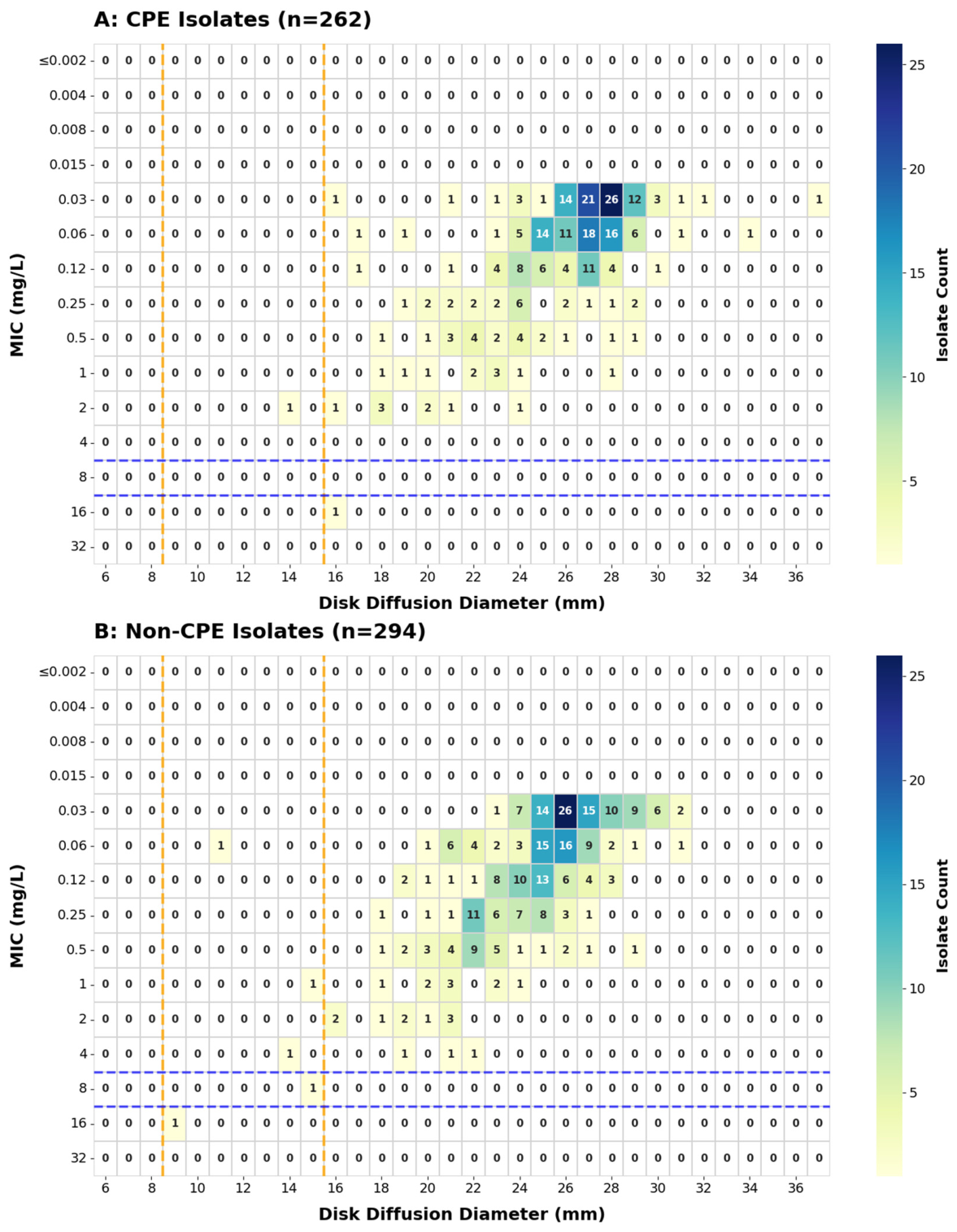
Minimum inhibitory concentration (MIC) determination using the BMD method requires more specialized techniques than those of the DD method, which remains a more practical method for routine susceptibility testing [21,22]. The agreement between the DD and BMD methods for cefiderocol susceptibility testing was assessed using heatmap correlation analysis (Figure 1). Our research indicated a moderate-to-strong inverse correlation between the DD zone diameter and MIC values (Spearman r = −0.64, p < 0.001), supporting the use of cefiderocol DD testing as a reliable screening method—specifically for the rapid management of infections.
However, minor discrepancies and critical categorical errors were observed when inhibition zone diameters were close to the susceptibility breakpoint. Some isolates exhibited resistant MICs by BMD but were categorized as susceptible by DD, raising concern for potential very major errors (VMEs). A representative example was isolate OU_551, identified as Klebsiella aerogenes, which showed a discordant cefiderocol susceptibility result: the isolate was interpreted as susceptible by DD, with an inhibition zone diameter of 16 mm, whereas BMD demonstrated high-level resistance, with a cefiderocol MIC of 16 μg/mL. Molecular characterization revealed that OU_551 co-harbored blaIMP-6 together with ESBL and AmpC determinants.
IMP-6 is a well-characterized IMP variant in Japan; it differs from IMP-1 by a single amino acid substitution, S214G, and is associated with an altered substrate profile characterized by more efficient hydrolysis of meropenem than imipenem [23]. This biochemical feature may result in a clinically important “stealth” phenotype, in which carbapenemase-producing isolates retain relatively low MICs [24,25]. In the present isolate, the coexistence of IMP-6, ESBL, and AmpC enzymes may have contributed to reduced cefiderocol activity; however, the precise contribution of each β-lactamase to the cefiderocol-resistant phenotype cannot be determined from genotype alone.
The under-calling of cefiderocol resistance by DD in this genetic background has important diagnostic and therapeutic implications [26]. Misclassification of a phenotypically resistant isolate as susceptible could lead to inappropriate cefiderocol use and potential clinical failure. A plausible explanation for this DD–BMD discordance is the particular methodological sensitivity of cefiderocol susceptibility testing. Cefiderocol activity depends on bacterial iron-transport pathways, and MIC determination by BMD requires iron-depleted cation-adjusted Mueller–Hinton broth to approximate iron-restricted in vivo conditions [27,28]. By contrast, DD is performed on routine Mueller–Hinton agar rather than iron-depleted agar, and local cefiderocol diffusion, agar composition, iron availability, and zone-edge interpretation may influence categorical assignment, especially for isolates with inhibition zones near the breakpoint [27,28,29]. Despite these rare but critical baseline discrepancies, the two methods demonstrated a high categorical agreement (CA) for most isolates. Accounting for all discordant results, the CA between the DD and BMD methods was 98.9% (550/556). This indicates that DD testing still provides a highly reliable screening method for cefiderocol susceptibility with a minimal risk of overall misclassification. However, for isolates with inhibition zones close to the breakpoint, especially CRE, confirmatory MIC testing should be performed to minimize the risk of false susceptibility assignment [30,31,32].

3.3. Activity Against IMP-Producing Isolates

For presentation purposes, the bacterial isolates were definitively stratified into groups:
Non-CRE/non-ESBL: Isolates negative for both carbapenemase and ESBL production. ESBL-only: Isolates positive for ESBL but negative for carbapenemase.
IMP-only: Isolates positive for IMP but negative for ESBL.
IMP + ESBL: Isolates co-harboring IMP and producing ESBL.
In this study, the susceptibility rate of IMP-producing isolates was highly consistent with that observed among non-CRE/non-ESBL isolates. Similar findings have been reported previously, with studies demonstrating susceptibility rates over 99% among IMP-producing Enterobacterales [13,33,34], indicating that cefiderocol retains excellent activity against IMP [35].
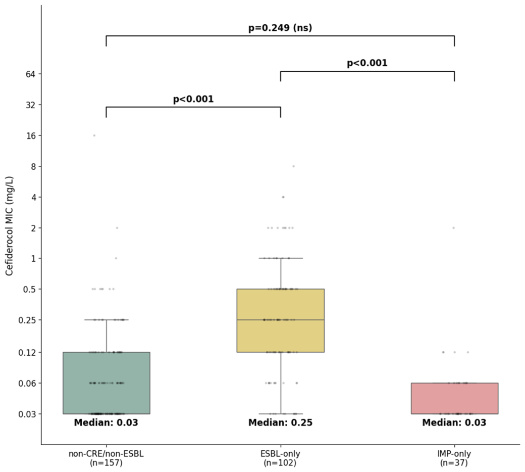
Based on the statistical analysis, cefiderocol MICs differed significantly among non-CRE/non-ESBL isolates (n = 157), ESBL-only isolates (n = 102), and IMP-only isolates (n = 37) (overall p < 0.001; Figure 2), indicating that the enzyme type affects cefiderocol MIC distributions. However, MICs were significantly higher in the ESBL-only group than in the non-CRE/non-ESBL and IMP-only groups (both p < 0.001). In contrast, the difference between the non-CRE/non-ESBL and IMP-only groups was not significant (p = 0.083), indicating that IMP enzymes provide minimal protection against cefiderocol.
These results indicate that although ESBL production is associated with reduced cefiderocol susceptibility, IMP exert a limited effect on cefiderocol resistance.

3.4. Non-Additive Effect of Cefiderocol

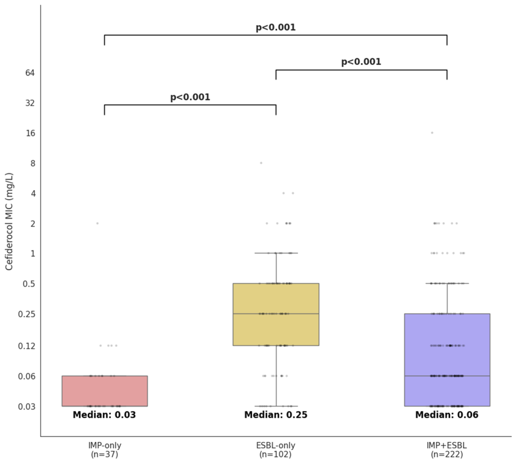
Recently, bacterial resistance to cefiderocol has involved the synergistic and additive effects of multiple mechanisms [36,37]. However, an interesting observation from this study was the non-additive effect between resistance mechanisms and cefiderocol activity. Cefiderocol MICs differed among IMP-only (n = 37), ESBL-only (n = 102), and IMP + ESBL (n = 222) isolates (overall p < 0.001; Figure 3), with pairwise comparisons exhibiting significant differences (all p < 0.001). The ESBL-only group exhibited a higher median MIC than that of the IMP + ESBL group, despite carrying fewer resistance determinants. This phenomenon indicates a non-additive interaction between the resistance mechanisms. The presence of specific enzymes, such as IMP, does not produce a cumulative increase in resistance and may, in specific combinations, be associated with reduced MICs.
One plausible explanation is that cefiderocol susceptibility in these isolates reflects a non-additive interaction between resistance burden, bacterial physiology, and drug-uptake pathways rather than a simple sum of β-lactamase activities. Plasmid-mediated resistance typically imposes significant fitness costs, driving compensatory evolution that subsequently remodels bacterial physiology [38]. Furthermore, antimicrobial resistance (AMR) epistasis dictates that the combined phenotype of multiple resistance determinants can deviate substantially from additivity. In that context, the lower cefiderocol MICs in the IMP + ESBL group may reflect a background-dependent epistatic trade-off: co-carriage of IMP and ESBL genes may alter plasmid burden, expression balance, or envelope homeostasis in a way that partially offsets β-lactamase-mediated resistance, paradoxically resulting in lower baseline MICs compared to the ESBL-only background [39].
Nonetheless, these observed variations in MIC distributions across the genomic groups may also be fundamentally influenced by other epidemiological confounding factors. As only a small number of isolates in our cohort exhibited phenotypic resistance to cefiderocol, these multi-layered molecular interaction remain just initial hypothetical frameworks that require rigorous functional validation in future genetic and biochemical studies.

3.5. Limitations and Future Perspectives

This study has some limitations that warrant consideration. First, the clinical isolates were collected from restricted geographical regions and specific facilities, which may limit the direct generalizability of our findings to the entirety of Japan. Second, a distinct temporal gap exists between the two collection cohorts (2015–2016 for the Osaka Institute of Public Health vs. 2020–2024 for The University of Osaka Hospital). While this decade-long separation could theoretically mask recent subtle shifts in regional molecular epidemiology or clonal lineage distributions, it is unlikely to compromise our core methodological conclusions, as the biochemical interactions between stable β-lactamase phenotypes and cefiderocol remain consistent regardless of the isolation year. Third, because WGS was selectively performed on only three non-susceptible strains to dissect outlier phenotypes, we could not account for the broader background resistome or other unmeasured resistance determinants across the rest of the cohort. Lastly, variations in underlying bacterial species between the ESBL-only and IMP + ESBL groups may have introduced unexpected confounding effects into the overall MIC distributions. Consequently, future prospective multi-center studies integrating larger, cohort-wide genomic datasets, plasmid copy-number determinations, and functional transcriptomic or membrane permeability analyses are strictly warranted to fully clarify the dynamic mechanisms underlying evolving cefiderocol resistance and to continuously optimize clinical susceptibility testing strategies.

4. Materials and Methods

4.1. Bacterial Isolates

Isolate Enrollment and Chronological Framework: To minimize selection bias, we initially targeted all consecutive, non-duplicate clinical isolates of Enterobacterales recovered at Osaka University Hospital during a contemporary period (March 2020 to October 2024), yielding 302 isolates. However, the baseline prevalence of CRE isolated purely within our facility was insufficient to provide adequate statistical power for robust molecular stratification. To address this sample limitation and establish a statistically viable evaluation platform, we strategically supplemented our collection with 258 distinct CRE clinical isolates consecutively referred to the Osaka Institute of Public Health during an earlier defined timeframe (November 2015 to January 2016).
In total, 560 Enterobacterales clinical isolates were initially screened across these two complementary cohorts. All isolates were definitively identified at the species level using matrix-assisted laser desorption/ionization–time-of-flight mass spectrometry (MALDI-TOF MS) via a MALDI Biotyper system (Bruker Daltonics, Bremen, Germany). Of the initial 560 isolates, four were excluded because of growth failure in subsequent susceptibility testing, leaving 556 isolates included in the final analysis.
Among Enterobacterales isolates, CRE was defined as isolates resistant to meropenem or producing carbapenemases. Carbapenemase production was characterized using the modified carbapenem inactivation method and the NG-Test CARBA 5 (NG Biotech, Guipry, France). Specifically, carbapenemase-producing Enterobacterales were categorized as CPE, whereas those without detectable carbapenemases were classified as non-CPE. A comprehensive antimicrobial susceptibility testing panel (Beckman Coulter, Brea, CA, USA) was utilized, which included meropenem (MEM), imipenem (IPM), ceftazidime (CAZ), cefotaxime (CTX), ceftriaxone (CRO), cefepime (FEP), and aztreonam (ATM).
The following procedures were used for the detection and classification of carbapenemase and ESBL production:
i.
CPE Detection: Isolates exhibiting a MEM MIC of ≥0.5 mg/L were initially screened for carbapenemase production using the modified carbapenem inactivation method (mCIM). For mCIM-positive strains, the presence of carbapenemase activity—specifically covering IMP type, VIM type, NDM type, KPC type, and OXA-48 type enzymes—was further confirmed using the NG-Test CARBA 5 (NG Biotech, Guipry, France).
ii.
ESBL Detection: ESBL production was characterized strictly according to the CLSI M100-Ed34 criteria [29]. Initial screening and characterization of the resistance phenotypes were based on the MIC criteria for a comprehensive panel of cephalosporins and monobactams, followed by phenotypic confirmatory testing using combined disks (Becton Dickinson, Franklin Lakes, NJ, USA).
iii.
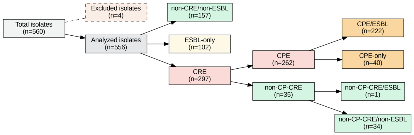
IMP Group Classification: blaIMP (n = 259) was the predominant genotype in the CPE group—222 isolates co-harbored blaIMP and ESBL genes (CPE/ESBL), whereas 37 isolates carried blaIMP alone. The other three CPE isolates were also ESBL-negative, including one carrying blaIMI-1 and two carrying blaGES-24. These 40 ESBL-negative isolates constitute the “CPE-only” group shown in Figure 4.

4.2. Antimicrobial Susceptibility Testing

Previous studies have demonstrated a high consistency between cefiderocol DD zone diameters and MIC values under CLSI criteria [40]. However, discrepancies have been reported in certain CRE isolates [30,31], highlighting the need for methodological validation in specific resistance genotypes, such as IMP. Therefore, in this study, all isolates were tested using both the DD and BMD methods.
DD testing was performed using 30 μg cefiderocol disks (Mast Group Ltd., Bootle, Merseyside, UK) on unsupplemented Mueller–Hinton agar (BD BBL™, Becton, Dickinson and Company, Franklin Lakes, NJ, USA) with a 0.5 McFarland inoculum. Readings were performed after incubation at 35 ± 2 °C for 20 ± 2 h. For BMD testing, commercial cefiderocol panels from Shionogi & Co., Ltd. (Osaka, Japan) were used following the manufacturer’s protocol. The same 0.5 McFarland inoculum used for the DD method was used for the BMD panels.
The results were interpreted based on the CLSI M100 (34th edition) [29] and EUCAST version 16.0 breakpoints [41]. For comparability, CLSI breakpoints were used for the analysis because previous nationwide surveillance studies conducted by Japanese committees have consistently reported susceptibility rates based on the CLSI criteria [13,35]. Currently, the gold standard for cefiderocol susceptibility testing is BMD-based MIC determination using cation-adjusted iron-depleted Mueller–Hinton broth [42]. In this study, the BMD results were used as the definitive standard for categorizing susceptibility.

4.3. WGS

For isolates identified as cefiderocol non-susceptible by BMD, WGS was performed to confirm species identification and characterize the molecular basis of resistance. Genomic DNA libraries were prepared using the Illumina DNA PCR-Free Prep kit and sequenced on the Illumina MiSeq platform (Illumina, San Diego, CA, USA) with 251-bp paired-end reads. Raw sequencing read quality was assessed using FastQC (v0.11.3). Subsequently, reads were assembled de novo using Unicycler (v0.4.4) [43] and annotated using the DFAST pipeline (v1.2.15) [44].
Antimicrobial resistance genes were identified using AMRFinderPlus (v4.0.23) [45] and the Comprehensive Antibiotic Resistance Database (CARD v4.0.1; RGI v6.0.5) [46]. Beyond the identification of β-lactamase genes, we assessed genetic alterations in three pathways: (i) iron uptake systems, including deletions or mutations in genes encoding catecholate siderophore receptors (cirA and fiu); (ii) target site modifications, focusing on amino acid substitutions or insertions in PBP3; and (iii) outer membrane permeability, by assessing structural variations in porin-encoding genes (ompC and ompF). Sequence alignments were performed against reference wild-type strains to identify specific mutations. MLST was determined using the PubMLST database [47], including the assignment of novel sequence types.

4.4. Statistical Analysis

The correlation between DD zone diameters and MIC values was assessed using Spearman’s rank correlation coefficient. The MIC distributions were compared among groups using the Kruskal–Wallis test, and post hoc pairwise comparisons were performed using the Mann–Whitney U test with Bonferroni correction to adjust for multiple comparisons. Adjusted p-values were calculated by multiplying the raw p-values by the number of total pairwise comparisons within each experiment (n = 3); a threshold of p < 0.05 was considered statistically significant.
All statistical analyses and data visualizations were implemented in Python (version 3.12.13). Specifically, the SciPy library (version 1.16.3) was used for statistical testing, whereas Pandas (version 2.2.2) and Matplotlib (version 3.10.0) were used for data management and visualization.

5. Conclusions

This study provides comprehensive baseline data on cefiderocol susceptibility among Enterobacterales clinical isolates in Japan, specifically focusing on those producing IMP. Our findings demonstrate that cefiderocol exhibits potent in vitro activity against IMP-producing strains and confirm a high CA (98.9%) between the routine DD and reference BMD methods. Notably, our statistical and genomic analyses highlight the highly complex, multifactorial nature of reduced cefiderocol susceptibility, as underscored by the unique non-additive effect observed in IMP + ESBL co-carriage isolates and the multi-layered molecular synergy involving AmpC mutations and receptor loss. These insights support the clinical utility of DD testing for routine cefiderocol surveillance while emphasizing that phenotypic outcomes are governed by dynamic physiological and genomic counterbalances rather than the simple accumulation of β-lactamase activities.
In conclusion, our evaluation of regional clinical isolates from Osaka demonstrates that cefiderocol retains high in vitro activity against carbapenemase-producing Enterobacterales. These findings establish a regional baseline that should be interpreted as an initial hypothesis warranting future multi-center validation across Japan.

Author Contributions

Conceptualization, G.Y. and S.H.; methodology, G.Y. and Y.T.; validation, G.Y. and M.C.; formal analysis, M.C. and D.M.; investigation, M.C.; resources, A.U. and R.K.; data curation, M.C.; writing—original draft preparation, M.C.; writing—review and editing, S.K., S.H., A.U. and R.K.; visualization, M.C.; supervision, S.K., G.Y. and S.H.; project administration, G.Y. All authors have read and agreed to the published version of the manuscript.

Funding

This research received no external funding.

Institutional Review Board Statement

The requirement for informed consent was waived by The University of Osaka Hospital Ethical Review Board because the clinical bacterial isolates used in this study were completely anonymized prior to analysis, with no access to patients’ personal or identifiable data.

Informed Consent Statement

Patient consent was waived due to the use of clinical bacterial isolates that had been completely anonymized prior to analysis, with no access by the investigators to patients’ personal or identifiable information. Written informed consent for publication was not applicable because this article contains no identifiable patient information.

Data Availability Statement

The datasets supporting the findings of this study are publicly archived in the FigShare repository. The whole-genome sequencing (WGS) data and antimicrobial susceptibility test results can be accessed via the Digital Object Identifier (DOI): 10.6084/m9.figshare.32446335.

Conflicts of Interest

The authors declare no conflicts of interest.

References

  1. Macesic, N.; Uhlemann, A.C.; Peleg, A.Y. Multidrug-resistant Gram-negative bacterial infections. Lancet 2025, 405, 257–272. [Google Scholar] [CrossRef] [PubMed]
  2. Wilson, H.; Török, M.E. Extended-spectrum β-lactamase-producing and carbapenemase-producing Enterobacteriaceae. Microb. Genom. 2018, 4, e000197. [Google Scholar] [CrossRef] [PubMed]
  3. Roustaye Gourabi, M.J.; Khanbabaei, B.; Yarahmadi Saki, Y.; Nikoo, A.; Kargar, M.; Hashemi, A.; Yasbolaghi Sharahi, J. Cefiderocol: A Comprehensive Review of Chemistry, Mechanisms of Resistance, and Clinical Applications in the Era of Multidrug Resistance. Curr. Microbiol. 2025, 83, 25. [Google Scholar] [CrossRef] [PubMed]
  4. Sato, T.; Yamawaki, K. Cefiderocol: Discovery, Chemistry, and In Vivo Profiles of a Novel Siderophore Cephalosporin. Clin. Infect. Dis. 2019, 69, S538–S543. [Google Scholar] [CrossRef] [PubMed]
  5. Aoki, T.; Yoshizawa, H.; Yamawaki, K.; Yokoo, K.; Sato, J.; Hisakawa, S.; Hasegawa, Y.; Kusano, H.; Sano, M.; Sugimoto, H.; et al. Cefiderocol (S-649266), A new siderophore cephalosporin exhibiting potent activities against Pseudomonas aeruginosa and other gram-negative pathogens including multi-drug resistant bacteria: Structure activity relationship. Eur. J. Med. Chem. 2018, 155, 847–868. [Google Scholar] [CrossRef] [PubMed]
  6. Ito, A.; Sato, T.; Ota, M.; Takemura, M.; Nishikawa, T.; Toba, S.; Kohira, N.; Miyagawa, S.; Ishibashi, N.; Matsumoto, S.; et al. In Vitro Antibacterial Properties of Cefiderocol, a Novel Siderophore Cephalosporin, against Gram-Negative Bacteria. Antimicrob. Agents Chemother. 2018, 62, e01454-17. [Google Scholar] [CrossRef] [PubMed]
  7. Karakonstantis, S.; Rousaki, M.; Kritsotakis, E.I. Cefiderocol: Systematic Review of Mechanisms of Resistance, Heteroresistance and In Vivo Emergence of Resistance. Antibiotics 2022, 11, 723. [Google Scholar] [CrossRef] [PubMed]
  8. Shionogi & Co., Ltd. Fetcroja (Cefiderocol) Prescribing Information. 2019. Available online: https://www.shionogi.com/wp-content/themes/pdfs/fetroja.pdf (accessed on 5 June 2026).
  9. Shionogi & Co., Ltd. Fetcroja. Summary of Product Characteristics. 2020. Available online: https://www.ema.europa.eu/documents/product-information/fetcroja-epar-product-information_en.pdf (accessed on 5 June 2026).
  10. Shionogi & Co., Ltd. New Siderophore Cephalosporin Antibiotic Fetroja (Cefiderocol) Intravenous Infusion 1g Vial in Japan [Press Release]. 2023. Available online: https://www.shionogi.com/global/en/news/2023/12/20231220.html (accessed on 5 June 2026).
  11. Karlowsky, J.A.; Hackel, M.A.; Takemura, M.; Yamano, Y.; Echols, R.; Sahm, D.F. In Vitro Susceptibility of Gram-Negative Pathogens to Cefiderocol in Five Consecutive Annual Multinational SIDERO-WT Surveillance Studies, 2014 to 2019. Antimicrob. Agents Chemother. 2022, 66, e0199021. [Google Scholar] [CrossRef] [PubMed]
  12. Ohkura, T.; Watarai, R.; Takekoshi, M.; Ohara, M.; Osada, Y.; Morioka, H.; Iguchi, M.; Oka, K.; Yagi, T. Susceptibility to cefiderocol and other novel antibiotics against carbapenem-non-susceptible gram-negative bacilli. Jpn. J. Infect. Dis. 2026, in press. [Google Scholar] [CrossRef] [PubMed]
  13. Kayama, S.; Kawakami, S.; Kondo, K.; Kitamura, N.; Yu, L.; Hayashi, W.; Yahara, K.; Sugawara, Y.; Sugai, M. In vitro activity of cefiderocol against carbapenemase-producing and meropenem-non-susceptible Gram-negative bacteria collected in the Japan Antimicrobial Resistant Bacterial Surveillance. J. Glob. Antimicrob. Resist. 2024, 38, 12–20. [Google Scholar] [CrossRef] [PubMed]
  14. Kuchibiro, T.; Nakamura, T.; Niki, M.; Yamasaki, K.; Nakamura, A.; Sawa, K.; Komatsu, M. In vitro activity of essential antimicrobial agents, including ceftazidime/avibactam, imipenem/relebactam, and cefiderocol, against carbapenem-resistant Gram-negative bacteria primarily harboring the IMP-type carbapenemase gene in the Kinki region of Japan. Diagn. Microbiol. Infect. Dis. 2026, 114, 117138. [Google Scholar] [CrossRef] [PubMed]
  15. Pérez-Llarena, F.J.; Kerff, F.; Zamorano, L.; Fernández, M.C.; Nuñez, M.L.; Miró, E.; Oliver, A.; Navarro, F.; Bou, G. Characterization of the new AmpC β-lactamase FOX-8 reveals a single mutation, Phe313Leu, located in the R2 loop that affects ceftazidime hydrolysis. Antimicrob. Agents Chemother. 2013, 57, 5158–5161. [Google Scholar] [CrossRef] [PubMed]
  16. Kawai, A.; McElheny, C.L.; Iovleva, A.; Kline, E.G.; Sluis-Cremer, N.; Shields, R.K.; Doi, Y. Structural Basis of Reduced Susceptibility to Ceftazidime-Avibactam and Cefiderocol in Enterobacter cloacae Due to AmpC R2 Loop Deletion. Antimicrob. Agents Chemother. 2020, 64, e00198-20. [Google Scholar] [CrossRef] [PubMed]
  17. Shields, R.K.; Iovleva, A.; Kline, E.G.; Kawai, A.; McElheny, C.L.; Doi, Y. Clinical Evolution of AmpC-Mediated Ceftazidime-Avibactam and Cefiderocol Resistance in Enterobacter cloacae Complex Following Exposure to Cefepime. Clin. Infect. Dis. 2020, 71, 2713–2716. [Google Scholar] [CrossRef] [PubMed]
  18. Girlich, D.; Ouzani, S.; Emeraud, C.; Gauthier, L.; Bonnin, R.A.; Le Sache, N.; Mokhtari, M.; Langlois, I.; Begasse, C.; Arangia, N.; et al. Uncovering the novel Enterobacter cloacae complex species responsible for septic shock deaths in newborns: A cohort study. Lancet Microbe 2021, 2, e536–e544. [Google Scholar] [CrossRef] [PubMed]
  19. Davin-Regli, A.; Lavigne, J.P.; Pagès, J.M. Enterobacter spp.: Update on Taxonomy, Clinical Aspects, and Emerging Antimicrobial Resistance. Clin. Microbiol. Rev. 2019, 32, e00002-19. [Google Scholar] [CrossRef] [PubMed]
  20. Matsuo, A.; Matsumura, Y.; Mori, K.; Noguchi, T.; Yamamoto, M.; Nagao, M. Molecular epidemiology and β-lactam resistance mechanisms of Enterobacter cloacae complex isolates obtained from bloodstream infections, Kyoto, Japan. Microbiol. Spectr. 2025, 13, e0248524. [Google Scholar] [CrossRef] [PubMed]
  21. Miller, L.A.; Rittenhouse, S.F.; Utrup, L.J.; Poupard, J.A. Comparison of three methods for determination of a single MIC of an antimicrobial agent. J. Clin. Microbiol. 1994, 32, 1373–1375. [Google Scholar] [CrossRef] [PubMed]
  22. Liu, Y.; Ding, L.; Han, R.; Zeng, L.; Li, J.; Guo, Y.; Hu, F. Assessment of cefiderocol disk diffusion versus broth microdilution results when tested against Acinetobacter baumannii complex clinical isolates. Microbiol. Spectr. 2023, 11, e0535522. [Google Scholar] [CrossRef] [PubMed]
  23. Yano, H.; Kuga, A.; Okamoto, R.; Kitasato, H.; Kobayashi, T.; Inoue, M. Plasmid-encoded metallo-beta-lactamase (IMP-6) conferring resistance to carbapenems, especially meropenem. Antimicrob. Agents Chemother. 2001, 45, 1343–1348. [Google Scholar] [CrossRef] [PubMed]
  24. Shigemoto, N.; Kuwahara, R.; Kayama, S.; Shimizu, W.; Onodera, M.; Yokozaki, M.; Hisatsune, J.; Kato, F.; Ohge, H.; Sugai, M. Emergence in Japan of an imipenem-susceptible, meropenem-resistant Klebsiella pneumoniae carrying blaIMP-6. Diagn. Microbiol. Infect. Dis. 2012, 72, 109–112. [Google Scholar] [CrossRef] [PubMed]
  25. Yonekawa, S.; Mizuno, T.; Nakano, R.; Nakano, A.; Suzuki, Y.; Asada, T.; Ishii, A.; Kakuta, N.; Tsubaki, K.; Mizuno, S.; et al. Molecular and Epidemiological Characteristics of Carbapenemase-Producing Klebsiella pneumoniae Clinical Isolates in Japan. mSphere 2020, 5, e00490-20. [Google Scholar] [CrossRef] [PubMed]
  26. Hirabayashi, A.; Yahara, K.; Kajihara, T.; Sugai, M.; Shibayama, K. Geographical distribution of Enterobacterales with a carbapenemase IMP-6 phenotype and its association with antimicrobial use: An analysis using comprehensive national surveillance data on antimicrobial resistance. PLoS ONE 2020, 15, e0243630. [Google Scholar] [CrossRef] [PubMed]
  27. Simner, P.J.; Patel, R. Cefiderocol Antimicrobial Susceptibility Testing Considerations: The Achilles’ Heel of the Trojan Horse? J. Clin. Microbiol. 2020, 59, e00951-20. [Google Scholar] [CrossRef] [PubMed]
  28. Matuschek, E.; Longshaw, C.; Takemura, M.; Yamano, Y.; Kahlmeter, G. Cefiderocol: EUCAST criteria for disc diffusion and broth microdilution for antimicrobial susceptibility testing. J. Antimicrob. Chemother. 2022, 77, 1662–1669. [Google Scholar] [CrossRef] [PubMed]
  29. Clinical and Laboratory Standards Institute (CLSI). Performance Standards for Antimicrobial Susceptibility Testing, 34th ed.; CLSI Supplement M100; CLSI: Wayne, PA, USA, 2024. [Google Scholar]
  30. Morris, C.P.; Bergman, Y.; Tekle, T.; Fissel, J.A.; Tamma, P.D.; Simner, P.J. Cefiderocol Antimicrobial Susceptibility Testing against Multidrug-Resistant Gram-Negative Bacilli: A Comparison of Disk Diffusion to Broth Microdilution. J. Clin. Microbiol. 2020, 59, e01649-20. [Google Scholar] [CrossRef] [PubMed]
  31. Duggan, C.; Cantillon, D.; Lawrie, D.; Neal, T.; Cruise, J.; Graf, F.E.; Owen, V.; Fraser, A.J.; Lewis, J.M.; Brookfield, C.; et al. Comparison of multiple cefiderocol susceptibility testing methods against genomic determinants of resistance in blaNDM carbapenemase producing Enterobacterales. bioRxiv 2026. [Google Scholar] [CrossRef]
  32. Bonnin, R.A.; Emeraud, C.; Jousset, A.B.; Naas, T.; Dortet, L. Comparison of disk diffusion, MIC test strip and broth microdilution methods for cefiderocol susceptibility testing on carbapenem-resistant Enterobacterales. Clin. Microbiol. Infect. 2022, 28, 1156.e1–1156.e5. [Google Scholar] [CrossRef] [PubMed]
  33. Yamashiro, H.; Nakai, R.; Takemura, M.; Yamano, Y. In Vitro Activity of Cefiderocol against IMP-Producing Enterobacterales Isolated in Japan. Open Forum Infect. Dis. 2022, 9, ofac492.1351. [Google Scholar] [CrossRef]
  34. Takemura, M.; Kazmierczak, K.; Hackel, M.; Sahm, D.F.; Echols, R.; Yamano, Y. In Vitro Activity of Cefiderocol Against Metallo-β-Lactamase-Producing Gram-Negative Bacteria Collected in North America and Europe Between 2014 and 2017: SIDERO-WT-2014–2016 Studies. Open Forum Infect. Dis. 2020, 7, S643–S644. [Google Scholar] [CrossRef]
  35. Matsui, K.; Sakurai, A.; Matsumura, Y.; Hosoda, T.; Suzuki, M.; Saito, S.; Hase, R.; Kato, H.; Hashimoto, T.; Matono, T.; et al. In vitro activity of cefiderocol against carbapenem-resistant Gram-negative pathogens in Japan. J. Infect. Chemother. 2026, 32, 102905. [Google Scholar] [CrossRef] [PubMed]
  36. Lasarte-Monterrubio, C.; Guijarro-Sánchez, P.; Vázquez-Ucha, J.C.; Alonso-Garcia, I.; Alvarez-Fraga, L.; Outeda, M.; Martinez-Guitian, M.; Peña-Escolano, A.; Maceiras, R.; Lence, E.; et al. Antimicrobial Activity of Cefiderocol against the Carbapenemase-Producing Enterobacter cloacae Complex and Characterization of Reduced Susceptibility Associated with Metallo-β-Lactamase VIM-1. Antimicrob. Agents Chemother. 2023, 67, e01505-22. [Google Scholar] [CrossRef] [PubMed]
  37. González-Pinto, L.; Blanco-Martín, T.; Alonso-García, I.; Rodríguez-Pallares, S.; Outeda-García, M.; Gomis-Font, M.A.; Fraile-Ribot, P.A.; Vázquez-Ucha, J.C.; González-Bello, C.; Beceiro, A.; et al. Impact of transferable β-lactamases and intrinsic AmpC amino acid substitutions on the activity of cefiderocol against wild-type and iron uptake-deficient mutants of Pseudomonas aeruginosa. J. Antimicrob. Chemother. 2024, 79, 3023–3028. [Google Scholar] [CrossRef] [PubMed]
  38. San Millan, A. Evolution of Plasmid-Mediated Antibiotic Resistance in the Clinical Context. Trends Microbiol. 2018, 26, 978–985. [Google Scholar] [CrossRef] [PubMed]
  39. Andersson, D.I.; Hughes, D. Antibiotic resistance and its cost: Is it possible to reverse resistance? Nat. Rev. Microbiol. 2010, 8, 260–271. [Google Scholar] [CrossRef] [PubMed]
  40. Lin, Y.; Lin, H.; Chen, C.; Tseng, K.; Ho, M.; Hsueh, P. Interpretive agreement of susceptibility between broth microdilution and disk diffusion methods for cefiderocol, using criteria from the Clinical and Laboratory Standards Institute, European Committee on Antimicrobial Susceptibility Testing, and the Food and Drug Administration. J. Clin. Microbiol. 2026, 64, e0125525. [Google Scholar] [CrossRef] [PubMed]
  41. The European Committee on Antimicrobial Susceptibility Testing (EUCAST). Breakpoint Tables for Interpretation of MICs and Zone Diameters; Version 16.0; EUCAST: Växjö, Sweden, 2026. [Google Scholar]
  42. Irigoyen-von-Sierakowski, A.; Ocaña, A.; Sánchez-Mayoral, R.; Cercenado, E.; GEIRAS-SEIMC Study Group. Real-world performance of susceptibility testing for cefiderocol: Insights from a prospective multicentre study on Gram-negative bacteria. JAC Antimicrob. Resist. 2024, 6, dlae169. [Google Scholar] [CrossRef] [PubMed]
  43. Wick, R.R.; Judd, L.M.; Gorrie, C.L.; Holt, K.E. Unicycler: Resolving bacterial genome assemblies from short and long sequencing reads. PLoS Comput. Biol. 2017, 13, e1005595. [Google Scholar] [CrossRef] [PubMed]
  44. Tanizawa, Y.; Fujisawa, T.; Nakamura, Y. DFAST: A flexible prokaryotic genome annotation pipeline for faster genome publication. Bioinformatics 2018, 34, 1037–1039. [Google Scholar] [CrossRef] [PubMed]
  45. Feldgarden, M.; Brover, V.; Gonzalez-Escalona, N.; Frye, J.G.; Haendiges, J.; Haft, D.H.; Hoffmann, M.; Pettengill, J.B.; Prasad, A.B.; Tillman, G.E.; et al. AMRFinderPlus and the Reference Gene Catalog facilitate examination of the genomic links among antimicrobial resistance, stress response, and virulence. Sci. Rep. 2021, 11, 12728. [Google Scholar] [CrossRef] [PubMed]
  46. Alcock, B.P.; Raphenya, A.R.; Lau, T.T.Y.; Tsang, K.K.; Bouchard, M.; Edalatmand, A.; Huynh, W.; Nguyen, A.L.V.; Cheng, J.K.; Liu, S.; et al. CARD 2023: Expanded curation, support for machine learning, and resistome prediction at the Comprehensive Antibiotic Resistance Database. Nucleic Acids Res. 2023, 51, D690–D699. [Google Scholar] [CrossRef] [PubMed]
  47. Jolley, K.A.; Bray, J.E.; Maiden, M.C.J. Open-access bacterial population genomics: BIGSdb software, the PubMLST.org website and their applications. Wellcome Open Res. 2018, 3, 124. [Google Scholar] [CrossRef] [PubMed]
Figure 1. Correlation and distribution matrices of cefiderocol MICs and disk diffusion zone diameters. Heatmap matrices illustrate the two-dimensional distribution and correlation between cefiderocol MICs (mg/L) determined by broth microdilution and disk diffusion inhibition zone diameters (mm) across clinical isolates. (A) CPE isolates (n = 262). (B) Non-CPE Enterobacterales isolates (n = 294). Color intensity gradients represent the specific isolate counts within each coordinate, with cells containing zero isolates displayed in white. The vertical dashed orange lines flanking the 9–15 mm zone and horizontal dashed blue lines flanking the value of 8 mg/L indicate the provisional or established clinical breakpoints for susceptibility categorization.
Antibiotics 15 00667 g001
Figure 2. Distribution of cefiderocol MICs among non-CRE/non-ESBL, ESBL-only, and imipenemase-only isolates. Boxplots indicate the median and interquartile range, with whiskers extending to 1.5 times the interquartile range. Individual isolates are overlaid as jittered points. MIC values across groups exhibited a significant overall difference (p < 0.001). p-values were calculated using the Mann–Whitney U test and adjusted using the Bonferroni correction for multiple pairwise comparisons (ns, not significant). Median MIC values are annotated on each box. Abbreviations: non-CRE/non-ESBL, isolates without ESBL and CRE classification; ESBL-only, isolates with ESBL and without CRE classification; IMP-only, isolates harboring an IMP-type carbapenemase without ESBL.
Antibiotics 15 00667 g002
Figure 3. Distribution of cefiderocol MICs among IMP-only, ESBL-only, and IMP + ESBL isolates. Boxplots indicate the median and interquartile range, with whiskers extending to 1.5 times the interquartile range. Individual isolates are overlaid as jittered points. MIC values compared across groups exhibited a significant overall difference (p < 0.001), Pairwise statistical significance was determined by the Mann–Whitney U test with Bonferroni correction to control for Type I error inflation. Median MIC values are indicated next to each box plot. Abbreviations: IMP-only, isolates harboring an IMP-type carbapenemase without ESBL; ESBL-only, isolates with ESBL but without an IMP-type carbapenemase; IMP + ESBL, isolates harboring both an IMP-type carbapenemase and ESBL.
Antibiotics 15 00667 g003
Figure 4. Flowchart of isolate distribution. A total of 556 Enterobacterales isolates are classified based on their CRE and ESBL phenotypes, and their CPE genotypes. Note: Four isolates from the initial collection are excluded from this flowchart because of inadequate growth during testing. Abbreviations: CRE, carbapenem-resistant Enterobacterales; CPE, carbapenemase-producing Enterobacterales; CP, carbapenemase; ESBL, extended-spectrum β-lactamase.
Antibiotics 15 00667 g004
Table 1. Isolates demonstrating non-susceptibility to cefiderocol.
Isolate IDSpeciesβ-LactamasesDD MethodBMD Method
Zone Diameter (mm)CategoryMinimum Inhibitory Concentration (μg/mL)Category
OU_24Escherichia coliESBL15I 18I
OU_38Klebsiella pneumoniaeESBL14I4S 2
OU_46Klebsiella pneumoniaeESBL15I1S
OU_91Proteus mirabilisESBL11I0.06S
OU_136Enterobacter cloacaeIMP 3 14I2S
OU_186Enterobacter bugandensisAmpC 49I16R 5
OU_551Klebsiella aerogenesIMP + ESBL + AmpC16S16R
1 Intermediate. 2 Susceptible. 3 IMP-type carbapenemases. 4 AmpC β-lactamase. 5 Resistant.
Disclaimer/Publisher’s Note: The statements, opinions and data contained in all publications are solely those of the individual author(s) and contributor(s) and not of MDPI and/or the editor(s). MDPI and/or the editor(s) disclaim responsibility for any injury to people or property resulting from any ideas, methods, instructions or products referred to in the content.