Article

Molecular Characterization of Multidrug-Resistant Escherichia coli Isolated from Beef and Chicken Meat Products in Samsun, Türkiye


Department of Food Hygiene and Technology, Faculty of Veterinary Medicine, Ondokuz Mayis University, 55139 Samsun, Türkiye
*
Author to whom correspondence should be addressed.
Antibiotics 2026, 15(7), 668; https://doi.org/10.3390/antibiotics15070668
Submission received: 22 May 2026 / Revised: 4 July 2026 / Accepted: 6 July 2026 / Published: 8 July 2026

Abstract

Background: Foodborne pathogenic Escherichia coli is a major public health concern due to its frequent association with meat and meat products and its potential to harbor virulence factors and antimicrobial resistance (AMR) determinants. Objective: This study aimed to investigate the prevalence, virulence gene profiles, and AMR patterns of E. coli isolates obtained from beef and chicken meat products. Methods: A total of 200 beef and chicken meat product samples were collected from retail markets in Samsun, Türkiye. Isolation of E. coli was performed using conventional culture-based methods, and PCR targeting the uspA gene was used for molecular confirmation. The presence of virulence genes (stx1, stx2, eae, and hlyA) was investigated by PCR. Antimicrobial susceptibility testing was conducted using the disk diffusion method, and multidrug resistance (MDR) and multiple antibiotic resistance (MAR) indices were evaluated. Results: Among the 200 samples analyzed, 80 (40%) were positive for E. coli, including 38 (38%) beef and 42 (42%) chicken meat samples. A total of 185 E. coli isolates were recovered and confirmed by PCR. Virulence gene analysis showed that stx2 was the most prevalent gene (51.4%), followed by eae (37.3%), hlyA (13.0%), and stx1 (6.5%). Antimicrobial susceptibility testing demonstrated high resistance rates to tetracycline (69.7%), ampicillin (58.4%), trimethoprim–sulfamethoxazole (48.1%), streptomycin (40.5%), nalidixic acid (40.0%), chloramphenicol (40.0%), and ciprofloxacin (34.1%). In contrast, the lowest resistance rates were observed for imipenem (2.2%), amoxicillin–clavulanate (4.9%), and amikacin (7.6%). Moreover, 126 isolates (68.1%) were identified as MDR, exhibiting resistance to at least three antimicrobial agents. The MAR index ranged from 0.06 to 1.00. Conclusions: The coexistence of virulence-associated genes and high AMR rates among E. coli isolates from meat products indicates a potential public health risk. These findings highlight the importance of continuous monitoring of pathogenic and antimicrobial-resistant E. coli throughout the food production chain.

1. Introduction

Meat is an important component of the human diet due to its high nutritional value, particularly its rich content of proteins, vitamins, and minerals [1]. However, its high water activity, nutrient availability, and near-neutral pH provide favorable conditions for microbial growth, making meat and meat products highly susceptible to spoilage and microbial contamination. During slaughtering, processing, transportation, packaging, and distribution, cross-contamination may occur at multiple stages, thereby increasing the microbial load and compromising food safety [2]. For this reason, meat and meat products are considered an important public health concern with respect to several pathogenic microorganisms, particularly Salmonella spp., Campylobacter jejuni, Escherichia coli (E. coli) and Listeria monocytogenes [3,4].
E. coli is a Gram-negative, facultative anaerobic, non-spore-forming bacterium commonly found in the intestinal microbiota of humans and warm-blooded animals [5]. Contamination of meat products may occur through fecal material during slaughter and processing or through environmental sources and handling practices. Therefore, E. coli is considered both an indicator organism and an important foodborne pathogen associated with meat and meat products [4,6]. Raw or undercooked meat products, particularly minced meat and hamburger products, play a major role in the transmission of pathogenic E. coli strains to humans [7]. In addition, certain pathogenic strains, especially Shiga toxin-producing E. coli (STEC), are capable of causing severe gastrointestinal diseases, including hemorrhagic colitis and hemolytic uremic syndrome (HUS), which may result in life-threatening complications [8].
The pathogenicity of E. coli is largely associated with specific virulence factors, particularly the Shiga toxin genes (stx1 and stx2), the adhesion-related intimin gene (eae), and the hemolysin gene (hlyA) [9,10]. The stx1 and stx2 genes encode Shiga toxins that inhibit protein synthesis in host cells and contribute to severe clinical manifestations such as hemorrhagic colitis and HUS [11,12]. The eae gene encodes intimin, an outer membrane adhesion protein responsible for intimate attachment to intestinal epithelial cells and the formation of attaching-and-effacing lesions [9]. The hlyA gene encodes α-hemolysin, an RTX (repeats-in-toxin) family toxin associated with membrane damage, cell lysis, and increased disease severity [13]. Detection of these virulence-associated genes in meat products is therefore considered an important indicator of potential public health risk.
In recent years, antimicrobial resistance (AMR) has emerged as a major global public health concern, threatening the effectiveness of antimicrobial agents used in both human and veterinary medicine. The extensive and often uncontrolled use of antimicrobials in food-producing animals has contributed substantially to the emergence and dissemination of resistant bacteria throughout the food chain [14,15]. Among foodborne pathogens, E. coli is considered a key indicator organism for AMR surveillance because of its widespread distribution and its ability to acquire and transfer resistance determinants through mobile genetic elements. In particular, MDR E. coli strains have increasingly been reported in retail meat products worldwide, posing a considerable risk to consumers [16,17,18].
The present study aimed to determine the occurrence of E. coli in beef and chicken meat products, characterize the prevalence of major virulence genes (stx1, stx2, eae, and hlyA), and evaluate the AMR profiles of the isolates. These findings may contribute to ongoing surveillance programs and support One Health strategies aimed at limiting the dissemination of AMR foodborne pathogens throughout the food production chain.

2. Results

2.1. Prevalence and Virulence Gene Profiles of E. coli Isolates

E. coli was detected in 80 (40%) of the 200 samples analyzed. Of these, 38/100 (38%) beef meat product samples and 42/100 (42%) chicken meat product samples were positive for E. coli. Among beef meat products, the highest contamination rate was observed in hamburger samples (13/20, 65%), followed by minced beef (11/20, 55%) and meatballs (8/20, 40%), whereas diced beef and sausage showed lower contamination levels (3/20, 15% each). Among chicken meat products, chicken breast (15/20, 75%) and chicken drumstick (13/20, 65%) exhibited the highest contamination rates, followed by chicken wing (11/20, 55%) and chicken sausage (3/20, 15%), while no E. coli was detected in chicken burger samples. No statistically significant difference was observed in E. coli prevalence between beef and chicken meat products (χ2 = 0.33, p = 0.56).
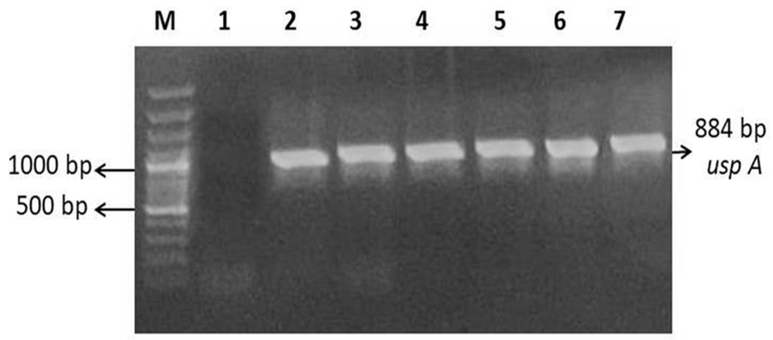
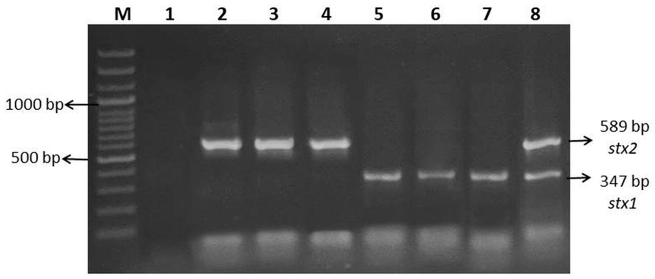
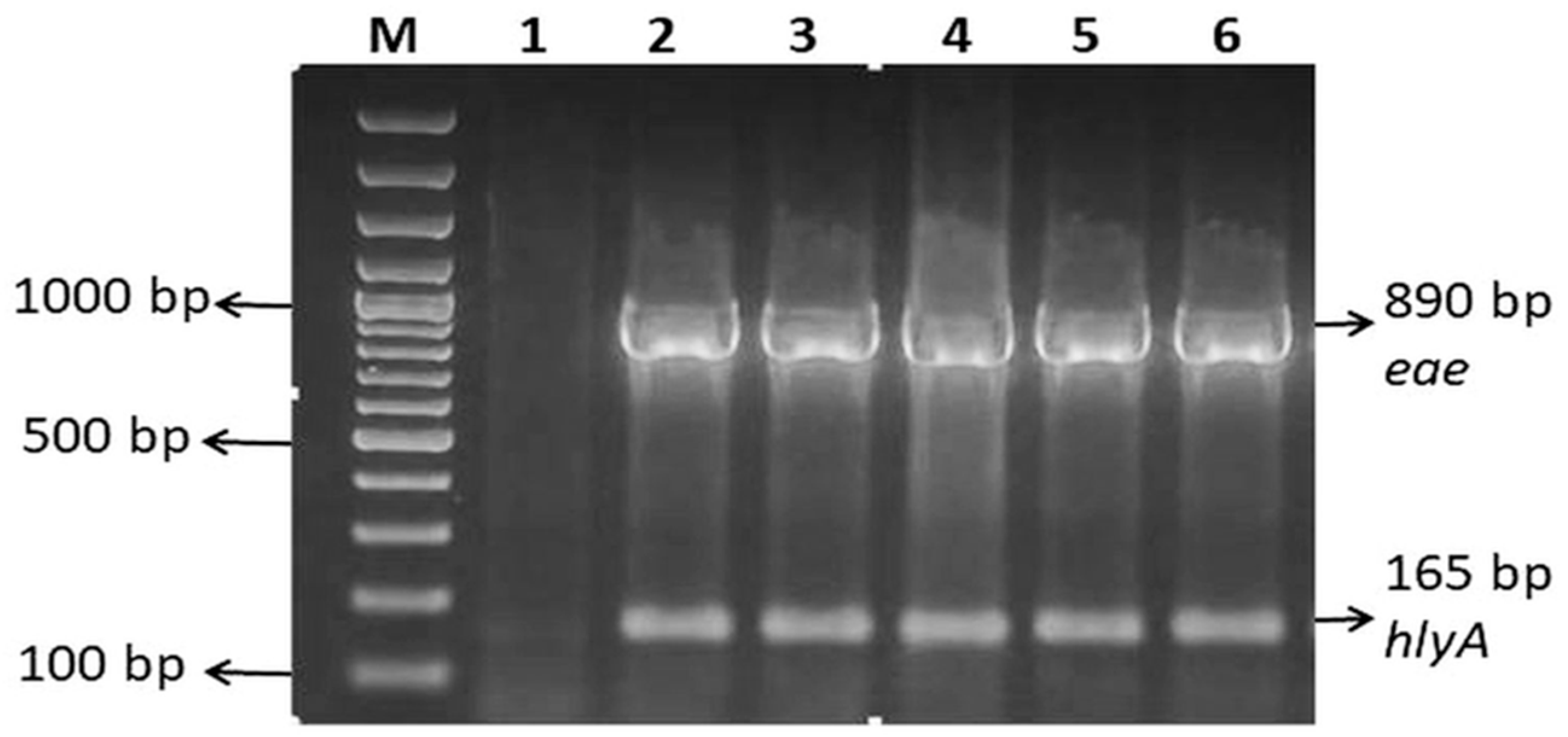
The prevalence and virulence profiles of the E. coli isolates are summarized in Table 1 and Table 2. A total of 185 E. coli isolates were recovered and confirmed by PCR targeting the uspA gene. The expected amplicon sizes were 884 bp for uspA, 347 bp for stx1, 589 bp for stx2, 890 bp for eae, and 165 bp for hlyA (Figure 1, Figure 2 and Figure 3). Virulence gene analysis revealed that 12/185 (6.5%) isolates carried stx1, 95/185 (51.4%) carried stx2, 69/185 (37.3%) carried eae, and 24/185 (13.0%) carried hlyA. Among the investigated virulence genes, stx2 was the most prevalent, followed by eae, hlyA, and stx1. No statistically significant differences were observed between beef and chicken isolates regarding stx1, stx2, and eae genes (p > 0.05), while hlyA showed a significantly higher prevalence in beef isolates (p < 0.001). Co-occurrence analysis of virulence genes revealed multiple combination patterns among the E. coli isolates (Table 3). The most prevalent combination was stx2 + eae, detected in 43 (23.2%) isolates, including 24 (25.8%) beef-derived and 19 (20.7%) chicken-derived isolates.
The distribution of virulence genes according to MDR status in beef and chicken isolates is presented in Table 4. In chicken-derived isolates, MDR prevalence was notably high among stx2-positive (93.8%) and eae-positive (92.1%) isolates. In beef isolates, MDR prevalence was more evenly distributed across virulence gene profiles. Overall, stx2-positive isolates showed a higher proportion of MDR (70.5%) compared to other virulence genes. However, variability in MDR distribution among virulence gene profiles was observed, indicating heterogeneous resistance patterns across different genotypes and meat sources.

2.2. Antimicrobial Resistance and Multidrug Resistance Profiles of E. coli Isolates

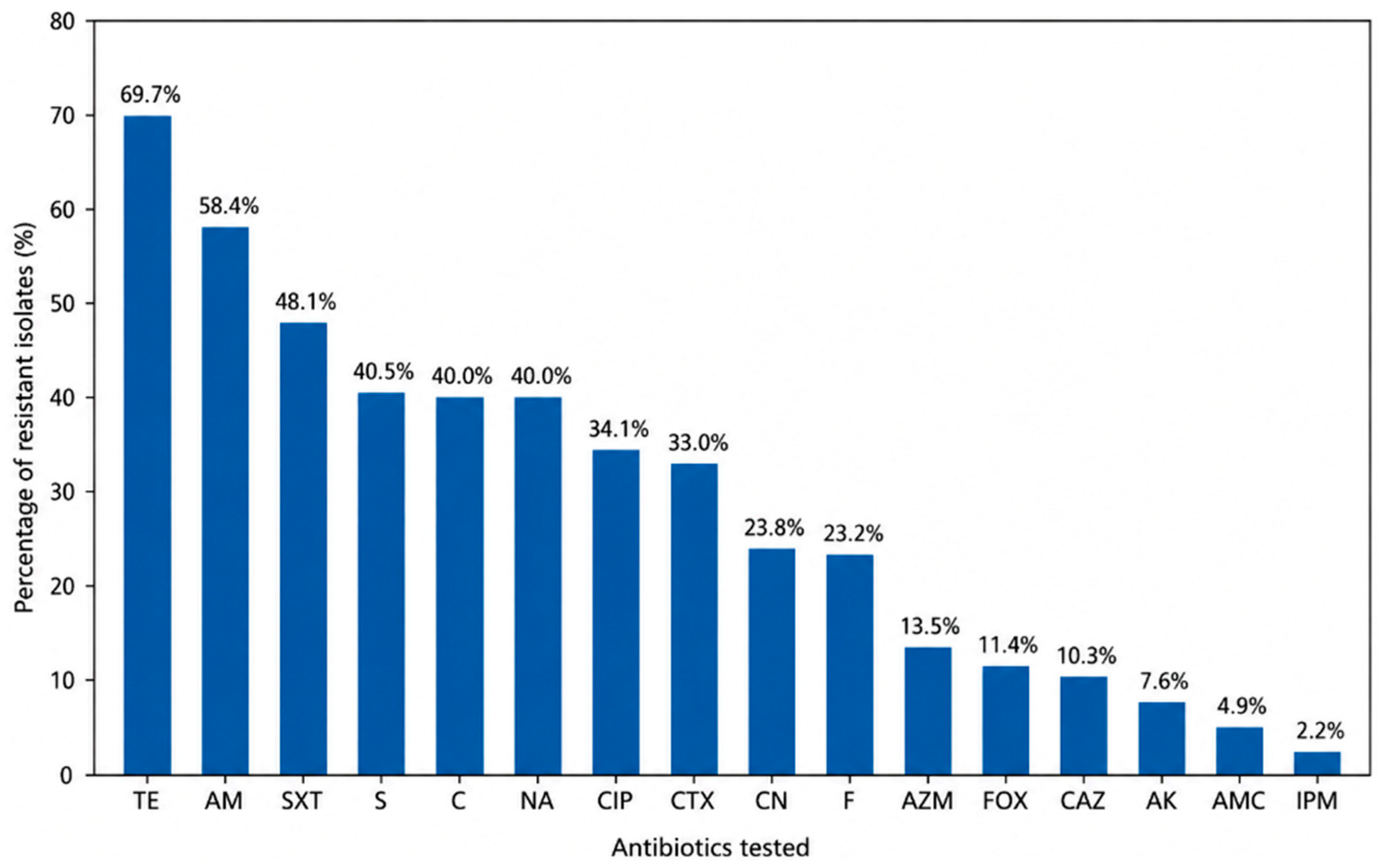
AMR profiles of E. coli isolates are summarized in Table 5 and Figure 4. High resistance rates were observed for tetracycline (69.7%), ampicillin (58.4%), trimethoprim–sulfamethoxazole (48.1%), streptomycin (40.5%), nalidixic acid (40.0%), and chloramphenicol (40.0%). Moderate resistance was observed for ciprofloxacin (34.1%), whereas low resistance rates were detected for amikacin (7.6%), amoxicillin–clavulanate (4.9%), and imipenem (2.2%).
The multiple antibiotic resistance (MAR) index values and distribution patterns are presented in Table 6. The MAR index was calculated as MAR = a/b, where a represents the number of antibiotics to which an isolate is resistant and b the total number tested. The MAR index ranged from 0.06 to 1.00, with an overall mean value of 0.285 ± 0.22. Significant differences were observed between meat types, with higher MAR index values in chicken isolates (0.406 ± 0.18) compared to beef isolates (0.165 ± 0.19) (Mann–Whitney U test, p < 0.001). Of the 185 isolates, 113 (61.1%) exhibited MAR values > 0.2, while 72 (38.9%) rall, 126 isolates (68.1%) were classified as MDR, defined as resistance to at least three antimicrobial agents. Resistance pattern analysis demonstrated a wide distribution of resistance phenotypes among the isolates. The proportions of isolates resistant to 1, 2, 3, 4, 5, 6, 7, 8, 9, 10, 11, 12, and 16 antibiotics were 9.7%, 4.8%, 7.0%, 9.1%, 15.1%, 8.1%, 5.9%, 5.4%, 7.5%, 4.8%, 3.2%, 1.1%, and 0.5%, respectively. Notably, a considerable proportion of isolates exhibited resistance to six or more antibiotics, and one isolate showed resistance to all tested antimicrobials.
Figure 1. PCR amplification of the uspA gene in E. coli isolates. M: 100 bp DNA marker; Lane 1: negative control (no template), Lane 2: positive control (E. coli ATCC 43888); Lanes 3–7: uspA-positive E. coli isolates obtained from beef and chicken meat products (isolates 2a, 9a, 14a, 28b and 43c), respectively.
Antibiotics 15 00668 g001
Representative positive isolates are shown; all 185 isolates were analyzed.
Figure 2. PCR amplification of the stx1 and stx2 genes in E. coli isolates. M: 100 bp DNA ladder; Lane 1: negative control (no template DNA); Lane 2: positive control (E. coli ATCC 43889, stx2-positive); Lanes 3–4: stx2-positive E. coli isolates obtained from minced beef (isolate 14a) and meatball samples (isolate 9a), respectively; Lane 5: positive control (E. coli ATCC 43890, stx1-positive); Lanes 6–7: stx1-positive E. coli isolates obtained from hamburger (isolate 73b) and sausage samples (isolate 76a), respectively; Lane 8: stx1/stx2-positive E. coli isolate obtained from a chicken breast sample (isolate 28b).
Antibiotics 15 00668 g002
Figure 3. PCR amplification of the eae and hlyA genes in E. coli isolates. M: 100 bp DNA marker; Lane 1: negative control (no template), Lane 2: positive control (E. coli O157:H7 ATCC 43888, hlyA+, eae+); Lanes 3–6: eae and hlyA-positive E. coli isolates obtained from chicken wing (isolate 19a), meatball (isolate 61a), hamburger (isolate 66a) and diced beef samples (isolate 43c), respectively.
Antibiotics 15 00668 g003
Figure 4. Antimicrobial resistance rate of E. coli isolates (n = 185). TE, Tetracycline; AM, Ampicillin; SXT, Trimethoprim–Sulfamethoxazole; S, Streptomycin; C, Chloramphenicol; NA, Nalidixic Acid; CIP, Ciprofloxacin; CTX, Cefotaxime; CN, Gentamicin; F, Nitrofurantoin; AZM, Azithromycin; FOX, Cefoxitin; CAZ, Ceftazidime; AK, Amikacin; AMC, Amoxicillin–Clavulanic Acid; IPM, Imipenem.
Antibiotics 15 00668 g004

3. Discussion

3.1. E. coli Prevalence in Red Meat and Poultry

E. coli is recognized as one of the most important foodborne pathogens, with certain pathogenic strains capable of causing severe and potentially life-threatening illnesses in humans. Therefore, its presence in meat and meat products represents a significant concern for both food safety and public health. In the present study, E. coli was detected in 42% of poultry meat and poultry products and 38% of red meat and meat samples, with no statistically significant difference between the two groups (χ2 = 0.33, p = 0.56). The prevalence observed in red meat indicates a considerable level of contamination at the retail level and highlights its potential role as a vehicle for foodborne transmission.
Previous studies have consistently reported the occurrence of E. coli in retail red meat products, although prevalence rates vary considerably among regions and production systems, including studies conducted in Egypt, Senegal, Vietnam, and other African countries [19,20,21,22,23]. Reported prevalence values ranging from 3% to 62% reflect substantial variability, which is strongly influenced by slaughter hygiene, carcass handling, processing infrastructure, and retail-level sanitation practices. These findings emphasize the critical role of regional food safety systems and production standards in determining contamination levels.
In poultry products, contamination levels observed in the present study were consistent with findings reported in studies conducted in Pakistan, the United States (Washington, D.C. and Southern California), Iran, Egypt, and several European countries, where the prevalence of E. coli in poultry meat has been reported to range from 8% to 68% [23,24,25,26,27]. Poultry is widely recognized as a major reservoir of E. coli due to its high intestinal bacterial load and intensive processing conditions during slaughter, evisceration, and distribution. This variability suggests that contamination is influenced by multiple factors along the production chain, including farm-level hygiene, slaughtering practices, processing environments, and retail handling.
Overall, the present study demonstrated a higher prevalence of E. coli in poultry meat compared to red meat, which is consistent with previous investigations [24,28]. Poultry generally harbors a higher intestinal bacterial load, increasing the likelihood of contamination during slaughter, particularly during evisceration. In addition, high-speed processing lines and extensive handling during poultry processing may facilitate cross-contamination between carcasses. Furthermore, the structural characteristics of poultry skin provide a favorable surface for bacterial attachment and persistence. The combination of these factors may promote the dissemination of E. coli throughout the processing chain and contribute to the higher contamination levels observed in poultry products. In contrast, contamination in red meat is often more localized to surface tissues and may occur at lower levels under relatively more controlled processing conditions.
Among red meat samples, the highest contamination rate was observed in hamburgers (65%), followed by minced beef (55%) and meatballs (40%), whereas diced beef and sausage showed considerably lower contamination levels (15%). In poultry meat samples, chicken breast (75%) and drumstick (65%) exhibited the highest contamination rates, followed by chicken wing (55%). In contrast, lower contamination levels were detected in chicken sausage (15%), while E. coli was not detected in chicken burger samples. These findings indicate that contamination levels may vary not only between meat species but also according to product type and processing characteristics. Comparable patterns have also been reported in previous studies, in which minced and processed meat products were identified as important sources of E. coli contamination [19].
These findings demonstrate notable differences not only between meat types but also among product categories. The higher contamination rates observed in comminuted meat products, such as minced beef and hamburgers, may be attributed to increased surface area, disruption of muscle structure, and a greater risk of cross-contamination during processing. Grinding disrupts the natural protective barriers of whole-muscle meat and facilitates the distribution of microorganisms throughout the product, resulting in a more homogeneous contamination pattern, even when initial bacterial loads are low. In addition, repeated handling, mixing processes, and contact with processing equipment may increase opportunities for microbial dissemination. Similar observations regarding the increased susceptibility of minced and processed meat products to E. coli contamination have also been reported previously, including the detection of E. coli O157 in minced beef and beef burger products [7]. Collectively, these factors reinforce the role of minced and processed meat products as high-risk matrices for foodborne pathogens such as E. coli.
In addition, inadequate storage conditions, including temperature abuse and interruptions in the cold chain, may further contribute to bacterial survival and proliferation in poultry meat products. Insufficient hygiene practices during processing and handling may also play a critical role in the persistence and spread of E. coli contamination. To reduce this public health risk, the implementation of strict food safety management systems is essential. This includes regular hygiene training for personnel, proper application of Hazard Analysis and Critical Control Point (HACCP) principles, effective sanitation of equipment and processing environments, and strict maintenance of the cold chain during storage and distribution. Strengthening these control measures can significantly reduce the risk of E. coli contamination and improve overall food safety.
A limitation of this study is that samples were collected from a single geographic region (Samsun, Türkiye), which may limit the generalizability of the findings to other regions of Türkiye. In addition, no molecular typing or clonal relatedness analysis was performed; therefore, isolates cannot be confirmed as genetically distinct strains and should be interpreted as phenotypically selected colonies. Furthermore, molecular characterization of AMR determinants was not conducted, and the genetic basis of the observed resistance phenotypes could not be determined. Future studies incorporating resistance gene profiling, molecular typing, and broader geographical sampling would provide a more comprehensive understanding of the epidemiology and public health significance of these isolates.

3.2. Virulence Gene Profiles of E. coli Isolates

The detection of virulence-associated genes in E. coli isolates recovered from retail meat products provides important insight into their pathogenic potential and public health significance. In the present study, stx2 was the most prevalent gene (51.4%), followed by eae (37.3%), hlyA (13.0%), and stx1 (6.5%). The predominance of stx2 is particularly important because this gene is strongly associated with severe clinical outcomes such as hemorrhagic colitis and hemolytic uremic syndrome (HUS) [10]. Similar findings have been reported by Zeinali et al. [27], who also observed stx2 as the dominant virulence gene in chicken meat isolates, suggesting a consistent epidemiological pattern across food sources.
stx2-positive STEC strains have been reported to be more frequently associated with severe human diseases than strains carrying only stx1. Therefore, the high prevalence of stx2 may indicate the circulation of E. coli populations with enhanced pathogenic potential within the meat production chain. In addition, Shiga toxin-encoding bacteriophages play a crucial role in the horizontal transfer of virulence determinants, facilitating the dissemination and persistence of highly virulent genotypes in food-producing animals and retail meat products.
The eae gene, encoding the adhesion protein intimin, was detected in 37.3% of the isolates, indicating a considerable proportion of strains with enhanced colonization potential. This prevalence was higher than those reported in several previous studies, including Zeinali et al. [27], who conducted a study in Iran, and Fatima et al. [26], who reported relatively low eae positivity rates (7–11%) in poultry-associated isolates from Pakistan. In contrast, Thierry et al. [29] reported a higher prevalence of eae-positive STEC isolates (47.6%) in animal-derived food sources from Mauritius, indicating variability in adhesion-associated virulence profiles across different studies. Since intimin plays a critical role in bacterial attachment to intestinal epithelial cells, eae-positive isolates may have an increased ability to establish infection and persist within the host. These differences may be attributed to variations in animal sources, geographic regions, and production practices.
Co-occurrence analysis revealed the presence of stx2 + eae in a substantial proportion of isolates, which is of particular concern due to its strong association with enterohemorrhagic E. coli (EHEC) pathotypes. Notably, stx2 + eae was the most frequent co-occurring virulence profile (23.2%), particularly in beef-derived isolates. Strains harboring both genes possess an enhanced ability to adhere to intestinal epithelium and produce Shiga toxins, thereby increasing their pathogenic potential. This combination of virulence factors may therefore represent a greater public health risk than isolates carrying only a single virulence determinant.
The hlyA gene, encoding enterohemolysin, was detected in 13.0% of the isolates, with a higher frequency observed in beef-derived samples. This finding may suggest a greater contribution of red meat products to the dissemination of hemolysin-producing E. coli. Enterohemolysin is considered an accessory virulence factor that may act synergistically with Shiga toxins and adhesion factors, potentially contributing to increased tissue damage and disease severity. Similar prevalence rates of hlyA-positive E. coli isolates from retail meat products have been reported by Martínez-Vázquez et al. [30].
The present study investigated the association between virulence gene carriage and MDR phenotype in E. coli isolates. A higher proportion of MDR was observed among stx2 and eae positive isolates, particularly in chicken-derived samples, where MDR rates exceeded 90%. This finding suggests that poultry-associated E. coli strains may serve as important reservoirs for both virulence and AMR determinants. However, the distribution was not uniform across all genes, indicating that virulence and resistance traits may be partially independent. These results are consistent with previous reports indicating that AMR and virulence determinants are often carried on different mobile genetic elements, leading to heterogeneous associations in foodborne E. coli populations.

3.3. Antimicrobial Resistance

The emergence and dissemination of AMR E. coli in foods of animal origin represent a major public health concern worldwide because resistant strains may be transmitted to humans through the food chain. In the present study, high resistance rates were observed particularly for tetracycline (69.7%), ampicillin (58.3%), trimethoprim–sulfamethoxazole (48.1%), streptomycin (40.5%), nalidixic acid (40.0%), and chloramphenicol (40.0%). Similar resistance patterns have been reported in previous studies investigating meat-derived E. coli isolates [31,32,33]. The high prevalence of resistance to these commonly used antimicrobials is likely driven by their extensive and long-term use in food animal production systems, which exerts selective pressure and promotes the persistence of resistant bacterial populations.
In particular, the high resistance to tetracycline and ampicillin may reflect the long-term and extensive use of these antimicrobial classes in animal husbandry. Tetracycline resistance is frequently mediated by transferable tet genes, whereas resistance to β-lactam antibiotics such as ampicillin is commonly associated with plasmid-mediated β-lactamase production. However, molecular detection of β-lactamase genes was not performed in the present study, which represents a limitation. The dissemination of mobile genetic elements may facilitate the co-selection and accumulation of multiple resistance determinants within bacterial populations. Future studies incorporating molecular characterization of resistance genes are therefore warranted to better elucidate the genetic basis of AMR.
The present study also demonstrated a high prevalence of MDR (68.1%), which was higher than the MDR rates reported in several previous studies [31,32]. In addition, the MAR index ranged from 0.06 to 1.00, indicating the circulation of isolates with diverse resistance profiles. Notably, one isolate exhibited resistance to all tested antimicrobials, highlighting the potential public health risk associated with highly resistant E. coli strains in retail meat products. The occurrence of isolates resistant to multiple antimicrobial classes may indicate sustained antimicrobial selection pressure and suggests that resistant bacteria and resistance genes may persist and disseminate throughout the food production chain. The higher MAR burden in poultry-derived isolates was statistically significant (p < 0.001), further supporting the role of chicken meat as a potential reservoir of MDR E. coli of public health concern.
Resistance to critically important antimicrobials such as ciprofloxacin was also considerable (34.1%) in the present study. Similar findings have been reported in retail meat isolates worldwide, although resistance levels vary depending on regional antimicrobial usage practices, farm management systems, and regulatory policies [23,32]. In Türkiye, Sahin et al. [34] reported the circulation of ESBL-producing and MDR E. coli isolates in poultry, while Dishan et al. [35] demonstrated high levels of AMR and virulence-associated characteristics among STEC isolates recovered from retail chicken meat. These findings further support the role of poultry-associated food products as important reservoirs for AMR determinants and potentially pathogenic E. coli strains. The widespread occurrence of MDR E. coli in meat products further supports concerns regarding the role of retail foods as reservoirs for AMR determinants.
In addition to antimicrobial selection pressure, differences in the physiological environments of poultry and cattle gastrointestinal tracts may contribute to the observed variation in AMR patterns among meat-derived E. coli isolates. In ruminants, the presence of multiple stomach compartments, particularly the rumen with an acidic environment (pH approximately 5), may impose distinct selective pressures on bacterial populations compared with the avian intestinal tract. These conditions may influence bacterial survival and stress adaptation, thereby shaping the composition of E. coli populations entering the food chain. In support of this concept, Zhang et al. [36] demonstrated that the global regulators RpoS and Crp coordinate acid stress responses in E. coli by regulating metabolic pathways, membrane-associated functions, and stress adaptation under acidic conditions.
The emergence of MDR E. coli in retail meat products may therefore involve not only the acquisition of AMR determinants but also enhanced bacterial fitness under selective environmental pressures. The interplay between stress response systems and AMR may promote the persistence and dissemination of MDR strains through potential co-selection mechanisms. However, the mechanistic relationship between acid tolerance and AMR remains incompletely understood. Further studies investigating stress response phenotypes in MDR isolates would provide valuable insights into their ecological success in food production chains.

4. Materials and Methods

4.1. Sample Collection

A total of 200 retail meat samples, including 100 beef products (diced beef, minced beef, meatballs, hamburgers, and sausages) and 100 chicken products (chicken breast, drumsticks, wings, sausages, and burgers), were collected from various retail outlets, local butcher shops, and supermarket chains in Samsun, Türkiye, between November 2024 and February 2025. Beef products were obtained from 11 local butcher shops and two national supermarket chains, while chicken products were collected from seven local poultry retailers, local butcher shops, and supermarket chains operating in the study area. All samples consisted of retail meat products; no live animals were included in this study. Samples reflect locally available products within the regional retail market and may represent meats distributed through local and regional supply chains. All samples were transported to the laboratory under cold-chain conditions and processed promptly upon arrival.
The sample size was determined based on feasibility, availability of retail meat products, and in accordance with previously published similar studies. Samsun was selected as the study area due to its status as the largest province in the Middle Black Sea Region and its role as a major regional center for meat production and food distribution. Information regarding the farm of origin, slaughterhouse, production region, or supply chain of the animals was not available for the sampled products. Therefore, the results should be interpreted as representative of retail meat products marketed in Samsun rather than specific livestock production systems or geographical regions.

4.2. Isolation and Characterization of E. coli

The presence of E. coli in meat and meat products was determined using conventional culture-based methods. For analysis, 10 g of each food sample was homogenized with 90 mL of Maximum Recovery Diluent (Merck, Darmstadt, Germany) using a stomacher for 2 min. Subsequently, 0.1 mL aliquots of the homogenate were spread onto Violet Red Bile Agar (VRBA; Merck, Germany) and incubated at 37 °C for 24 h for coliform enumeration. Red–pink colonies on VRBA were transferred to Eosin Methylene Blue (EMB) agar (Merck, Germany) and incubated at 37 °C for 24 h. Presumptive colonies exhibiting a metallic sheen on EMB agar were subcultured on Tryptic Soy Agar (TSA; Merck, Germany) and incubated at 37 °C for 18–24 h for biochemical confirmation. Colonies showing typical and/or distinct morphological characteristics were considered for further analysis. From each positive sample, 2–3 confirmed E. coli isolates were randomly selected from morphologically distinct colonies to ensure balanced representation and to avoid overrepresentation of any individual sample. Presumptive E. coli isolates were preserved in cryovials containing Tryptic Soy Broth (TSB) supplemented with 20% glycerol and stored at −20 °C [37,38].

4.3. DNA Extraction

Genomic DNA from bacterial isolates was extracted using the boiling method, a rapid and cost-effective DNA preparation approach previously used for PCR-based detection of bacterial virulence genes. The boiling method was selected because it provides DNA of sufficient quality for PCR-based molecular analyses and has been successfully applied in previous studies for the detection of bacterial pathogens [39]. Isolates stored at −20 °C were first revived in Tryptic Soy Broth (TSB; Merck, Darmstadt, Germany) and incubated at 37 °C for 18–24 h. The cultures were then streaked onto Eosin Methylene Blue (EMB) agar, and colonies showing a metallic sheen were subcultured on Tryptic Soy Agar (TSA; Merck, Darmstadt, Germany) and incubated at 37 °C for 18–24 h. The obtained colonies were suspended in 200 µL of sterile deionized water in Eppendorf tubes. Denaturation was performed at 95 °C for 10 min using a dry block heater (Biosan TDB-120, Biosan, Riga, Lithuania). The samples were then centrifuged at 10,000× g for 5 min at 4 °C (Hettich Universal 320R, Tuttlingen, Germany). The supernatant was transferred to a sterile Eppendorf tube and stored at −20 °C until molecular analysis.
The quality and purity of the extracted genomic DNA were assessed using a spectrophotometer by determining the A260/A280 absorbance ratio. Additionally, DNA integrity was checked by 1.5% agarose gel electrophoresis and visualized under UV illumination. The suitability of the extracted DNA for downstream PCR applications was further confirmed by amplification of the uspA gene, which served as a species-specific internal control marker for the identification of E. coli.

4.4. PCR Confirmation of E. coli Isolates Using the uspA Gene

Presumptive E. coli colonies were confirmed by PCR targeting the uspA gene. All primers used in this study are listed in Table 7. The PCR mixture was prepared in a final volume of 25 µL containing 1× PCR buffer (500 mM KCl, 200 mM Tris–HCl), 0.2 mM dNTPs, 2 mM MgCl2, 0.5 µM of each primer, 1 U Taq DNA polymerase, and 2 µL of template DNA. PCR reactions were performed using a thermal cycler (MJ Mini, PTC-1148; Bio-Rad Laboratories, Hercules, CA, USA). The amplification conditions consisted of an initial denaturation at 94 °C for 5 min, followed by 32 cycles of denaturation at 94 °C for 30 s, annealing at 63 °C for 30 s, and extension at 72 °C for 1.5 min, with a final extension at 72 °C for 5 min.

4.5. Detection of Virulence Genes in E. coli Isolates by PCR

PCR assays were performed for the detection of virulence genes (stx1, stx2, hlyA, and eae) in E. coli isolates. The PCR mixture was prepared in a final volume of 25 µL containing 1× PCR buffer (500 mM KCl, 200 mM Tris–HCl), 0.2 mM dNTPs, 2 mM MgCl2, 0.8 µM of each primer, 1 U Taq DNA polymerase, and 2 µL of template DNA. The amplification conditions consisted of an initial denaturation at 95 °C for 5 min, followed by 35 cycles of denaturation at 95 °C for 30 s, annealing at 57 °C for 45 s for stx1 and stx2 genes or 55 °C for 45 s for hlyA and eae genes, and extension at 72 °C for 45 s, with a final extension at 72 °C for 7 min. All PCR assays were performed in duplicate to ensure reproducibility of the results.

4.6. Agarose Gel Electrophoresis

PCR products were analyzed on 1.5% agarose gel prepared in 1× TBE buffer (Tris–borate–EDTA; 89 mM Tris, 89 mM boric acid, 2 mM EDTA, pH 8.3) and stained with 0.5 µg/mL ethidium bromide. Electrophoresis was carried out at 80 V for 1 h using a horizontal electrophoresis system (multiSUB Horizontal System, Cleaver Scientific, Rugby, UK). The uspA gene was visualized at 884 bp, stx1 at 347 bp, stx2 at 589 bp, eae at 890 bp, and hlyA at 165 bp under a UV transilluminator (Wise-UV WUV-L50, DAIHAN Scientific, Seoul, Republic of Korea).

4.7. Antimicrobial Susceptibility Testing

Antimicrobial susceptibility of the E. coli isolates against various antibiotics was determined using the disk diffusion method according to the guidelines of the Clinical and Laboratory Standards Institute (CLSI) [44,45]. The antibiotic panel was selected to represent major antimicrobial classes commonly used in human and veterinary medicine, covering different mechanisms of action and clinically relevant drugs for the treatment of E. coli infections. The selection was also consistent with widely used antimicrobial susceptibility testing panels and the WHO Critically Important Antimicrobials list. The antibiotics used in this study are listed in Table 5. For this purpose, E. coli isolates were grown in Mueller–Hinton Broth (MHB; Merck, Darmstadt, Germany) at 35 °C for 18–24 h. After incubation, fresh cultures were adjusted to a turbidity equivalent to 0.5 McFarland standard (~1 × 10⁸ CFU/mL) using a densitometer (DEN-1, Biosan, Riga, Latvia). Then, 100 µL of the bacterial suspension was uniformly spread on Mueller–Hinton Agar (MHA; Merck, Darmstadt, Germany) plates using a sterile cotton swab and allowed to dry for 15 min. Antibiotic discs (Bioanalyse, Ankara, Turkey) were then placed on the agar surface, and the plates were incubated at 35 °C for 18–24 h. Following incubation, the diameters of inhibition zones around each disc were measured. The results were interpreted as susceptible, intermediate, or resistant according to CLSI-recommended breakpoints [46]. E. coli ATCC 25922 was used as a quality control strain to ensure the reliability and accuracy of the disk diffusion method, and all results were within the CLSI-recommended quality control ranges.

4.8. Multidrug Resistance (MDR) and Multiple Antibiotic Resistance (MAR) Index

Isolates showing resistance to at least one agent in three or more antimicrobial classes were classified as MDR according to the criteria proposed by Magiorakos et al. [47]. The multiple antibiotic resistance (MAR) index for each isolate was calculated using the following formula: MAR index = a/b, where a represents the number of antibiotics to which the isolate was resistant and b represents the total number of antibiotics tested [48]. High MAR index values were considered indicative of exposure to environments with frequent antimicrobial use.

4.9. Statistical Analysis

Statistical analyses were performed using SPSS Statistics software (Version 22.0; IBM Corp., Armonk, NY, USA). Differences in the prevalence of E. coli isolates, AMR patterns, and virulence gene frequencies between beef and chicken samples were evaluated using Pearson’s Chi-square test. Comparisons of MAR index values between beef- and chicken-derived isolates were performed using the Mann–Whitney U test. A p-value of <0.05 was considered statistically significant.

5. Conclusions

In conclusion, the present study demonstrated that retail beef and chicken meat products may serve as important reservoirs of E. coli harboring virulence determinants and MDR traits. The coexistence of virulence-associated genes and AMR determinants among the isolates increases the potential public health risk associated with contaminated meat products. Therefore, continuous surveillance programs, prudent antimicrobial use in food animal production, and improved hygienic practices during slaughtering and meat processing remain essential for limiting the dissemination of AMR E. coli throughout the food chain. Furthermore, additional molecular characterization of resistance and virulence determinants, particularly through whole-genome sequencing-based studies, may provide deeper insight into the epidemiology and transmission dynamics of pathogenic E. coli strains associated with retail meat products.

Author Contributions

Conceptualization, G.T.G.; methodology, G.T.G.; supervision, G.T.G.; project administration, G.T.G.; funding acquisition, G.T.G.; investigation, G.T.G., S.K. and E.E.; data curation, S.K. and E.E.; formal analysis, G.T.G.; writing—original draft preparation, G.T.G.; writing—review and editing, G.T.G., S.K. and E.E. All authors have read and agreed to the published version of the manuscript.

Funding

This research was funded by the Scientific Research Projects Coordination Unit of Ondokuz Mayıs University (Project No. BAP01-2024-5051).

Institutional Review Board Statement

Not applicable.

Informed Consent Statement

Not applicable.

Data Availability Statement

The raw data supporting the conclusions of this article will be made available by the authors on request.

Acknowledgments

During the preparation of this manuscript, the authors used ChatGPT (GPT-5.5-mini, OpenAI, San Francisco, CA, USA) for the purposes of language editing and improving the clarity and readability of the manuscript. The authors have reviewed and edited the output and take full responsibility for the content of this publication.

Conflicts of Interest

The authors declare no conflicts of interest.

Abbreviations

The following abbreviations are used in this manuscript:
AMRAntimicrobial Resistance
ATCCAmerican Type Culture Collection
CFUColony-Forming Unit
CLSIClinical and Laboratory Standards Institute
EHECEnterohemorrhagic Escherichia coli
EMBEosin Methylene Blue Agar
HUSHemolytic Uremic Syndrome
MDRMultidrug-Resistant
MARMultiple Antibiotic Resistance
MHAMueller–Hinton Agar
MHBMueller–Hinton Broth
STECShiga Toxin-Producing Escherichia coli
TSATryptic Soy Agar
TSBTryptic Soy Broth
VRBAViolet Red Bile Agar

References

  1. McAfee, A.J.; McSorley, E.M.; Cuskelly, G.J.; Moss, B.W.; Wallace, J.M.; Bonham, M.P.; Fearon, A.M. Red Meat Consumption: An overview of the risks and benefits. Meat Sci. 2010, 84, 1–13. [Google Scholar] [CrossRef]
  2. Zelalem, A.; Sisay, M.; Vipham, J.L.; Abegaz, K.; Kebede, A.; Terefe, Y. The prevalence and antimicrobial resistance profiles of bacterial isolates from meat and meat products in Ethiopia: A systematic review and meta-analysis. Int. J. Food Contam. 2019, 6, 1. [Google Scholar] [CrossRef]
  3. Constantin, C.E.; Holban, A.M.; Iordache, F.; Curutiu, C. Antimicrobial nanomaterials in the food industry: Applications in meat packaging. Materials 2026, 19, 1160. [Google Scholar] [CrossRef] [PubMed]
  4. Sofos, J.N. Challenges to meat safety in the 21st century. Meat Sci. 2008, 78, 3–13. [Google Scholar] [CrossRef] [PubMed]
  5. Tenaillon, O.; Skurnik, D.; Picard, B.; Denamur, E. The population genetics of commensal Escherichia coli. Nat. Rev. Microbiol. 2010, 8, 207–217. [Google Scholar] [CrossRef] [PubMed]
  6. Nørrung, B.; Buncic, S. Microbial Safety of Meat in the European Union. Meat Sci. 2008, 78, 14–24. [Google Scholar] [CrossRef] [PubMed]
  7. Cagney, C.; Crowley, H.; Duffy, G.; Sheridan, J.J.; O’Brien, S.; Carney, E.; Bishop, R.H. Prevalence and numbers of Escherichia coli O157:H7 in minced beef and beef burgers from butcher shops and supermarkets in the republic of Ireland. Food Microbiol. 2004, 21, 203–212. [Google Scholar] [CrossRef]
  8. Kaper, J.B.; Nataro, J.P.; Mobley, H.L. Pathogenic Escherichia coli. Nat. Rev. Microbiol. 2004, 2, 123–140. [Google Scholar] [CrossRef] [PubMed]
  9. Nataro, J.P.; Kaper, J.B. Diarrheagenic Escherichia coli. Clin. Microbiol. Rev. 1998, 11, 142–201. [Google Scholar] [CrossRef] [PubMed]
  10. Karmali, M.A. Infection by shiga toxin-producing Escherichia coli: An overview. Mol. Biotechnol. 2004, 26, 117–122. [Google Scholar] [PubMed]
  11. Melton-Celsa, A.R. Shiga toxin (Stx) classification, structure, and function. Microbiol. Spectr. 2014, 2, 1–13. [Google Scholar] [CrossRef]
  12. Obrig, T.G. Escherichia coli Shiga toxin mechanisms of action in renal disease. Toxins 2010, 2, 2769–2794. [Google Scholar] [CrossRef] [PubMed]
  13. Schmidt, H.; Beutin, L.; Karch, H. Molecular analysis of the plasmid-encoded hemolysin of Escherichia coli O157:H7 strain EDL 933. Infect. Immun. 1995, 63, 1055–1061. [Google Scholar] [PubMed]
  14. EFSA. The European Union Summary Report on Antimicrobial Resistance in Zoonotic and Indicator Bacteria from Humans, Animals and Food in 2020/2021. EFSA J. 2023, 21, e07867. [Google Scholar] [CrossRef] [PubMed]
  15. Economou, V.; Gousia, P. Agriculture and food animals as a source of antimicrobial-resistant bacteria. Infect. Drug Resist. 2015, 8, 49–61. [Google Scholar] [CrossRef] [PubMed]
  16. Poirel, L.; Madec, J.Y.; Lupo, A.; Schink, A.K.; Kieffer, N.; Nordmann, P.; Schwarz, S. Antimicrobial resistance in Escherichia coli. Microbiol. Spectr. 2018, 6, 1–27. [Google Scholar] [CrossRef]
  17. Van Boeckel, T.P.; Brower, C.; Gilbert, M.; Grenfell, B.T.; Levin, S.A.; Robinson, T.P.; Laxminarayan, R. Global trends in antimicrobial use in food animals. Proc. Natl. Acad. Sci. USA 2015, 112, 5649–5654. [Google Scholar] [CrossRef] [PubMed]
  18. Aworh, M.K.; Thakur, S.; Gensler, C.; Harrell, E.; Harden, L.; Fedorka-Cray, P.J.; Jacob, M. Characteristics of antimicrobial resistance in Escherichia coli isolated from retail meat products in North Carolina. PLoS ONE 2024, 19, e0294099. [Google Scholar] [CrossRef] [PubMed]
  19. Elsawy, M.S.; Abdel-Monem, M.O.; Yassine, M.H.; El-Sapagh, S. Molecular characterization of antibiotic resistance patterns among gram-negative bacteria isolated from red meat. Benha J. Appl. Sci. 2024, 9, 97–114. [Google Scholar] [CrossRef]
  20. Mansour, A.M.; Shehab, S.A.; Nossair, M.A.; Ayyad, A.S.; Tawfik, R.G.; El-Lami, S.A.; Eskander, M. Molecular characterization of Shiga toxin-producing Escherichia coli isolated from some food products as well as human stool in Alexandria, Egypt. J. Adv. Vet. Res. 2023, 13, 1056–1062. [Google Scholar]
  21. Loubamba, L.; Diallo, A.A.; Musabyemariya, B.; Moyen, R.; Sylla, K.S.B.; Alambédji, R.B. Virulence genes (stx1/stx2/eae) and O serogroups of Escherichia coli strains isolated from slaughtered cattle and ground meat sold in retail markets in Dakar, Senegal. Microbe 2023, 1, 100024. [Google Scholar] [CrossRef]
  22. Duc, H.M.; Ha, C.T.T.; Hoa, T.T.K.; Hung, L.V.; Thang, N.V.; Son, H.M. Prevalence, molecular characterization, and antimicrobial resistance profiles of shiga toxin-producing Escherichia coli isolated from raw beef, pork, and chicken meat in Vietnam. Foods 2024, 13, 2059. [Google Scholar] [CrossRef] [PubMed]
  23. Yi, S.; Alexander, K.A.; Bywater, A.; Dintwe, G.; Sies, A.N.; Haidl, T.H.; Ponder, M.A. Imported retail beef and chicken meat products serve as reservoirs for emerging antibiotic-resistant pathotypes of Escherichia coli in Pristine areas free from agricultural activity. MicrobiologyOpen 2026, 15, e70273. [Google Scholar] [PubMed]
  24. Zhao, C.; Ge, B.; De Villena, J.; Sudler, R.; Yeh, E.; Zhao, S.; Meng, J. Prevalence of Campylobacter spp., Escherichia coli, and Salmonella serovars in retail chicken, turkey, pork, and beef from the Greater Washington, DC area. Appl. Environ. Microbiol. 2001, 67, 5431–5436. [Google Scholar] [PubMed]
  25. Deag, S.A.; Haggag, Y.N.; Nossair, M.A.; Mansour, A.M.; Elaadli, H. Molecular characterization of diarrheagenic Escherichia coli isolated from some poultry products. Alex. J. Vet. Sci. 2021, 71, 46. [Google Scholar] [CrossRef]
  26. Fatima, A.; Ali, S.; Raut, R. Comparative analysis of virulence genes and antimicrobial resistance in Escherichia coli from poultry meat and poultry farm environments in Pakistan. DNA 2025, 5, 42. [Google Scholar] [CrossRef]
  27. Zeinali, T.; Arefinejad, A.; Kabiri-Rad, H.; Khodadadi, M. Detection of virulence genes (stx, eae and ehxa) and serogroup in Escherichia coli isolates collected from chicken in East of Iran. Food Humanit. 2025, 5, 100833. [Google Scholar] [CrossRef]
  28. Lee, K.Y.; Lavelle, K.; Huang, A.; Atwill, E.R.; Pitesky, M.; Li, X. Assessment of prevalence and diversity of antimicrobial resistant Escherichia coli from retail meats in Southern California. Antibiotics 2023, 12, 782. [Google Scholar] [CrossRef] [PubMed]
  29. Thierry, S.I.L.; Gannon, J.E.; Jaufeerally-Fakim, Y.; Santchurn, S.J. Shiga-toxigenic Escherichia coli from animal food sources in mauritius: Prevalence, serogroup diversity and virulence profiles. Int. J. Food Microbiol. 2020, 324, 108589. [Google Scholar] [CrossRef] [PubMed]
  30. Martínez-Vázquez, A.V.; Rivera-Sánchez, G.; Lira-Méndez, K.; Reyes-López, M.Á.; Bocanegra-García, V. Prevalence, antimicrobial resistance and virulence genes of Escherichia coli isolated from retail meat in Tamaulipas, Mexico. J. Glob. Antimicrob. Resist. 2018, 14, 266–272. [Google Scholar] [CrossRef] [PubMed]
  31. Adzitey, F.; Huda, N.; Shariff, A.H.M. Phenotypic antimicrobial susceptibility of Escherichia coli from raw meats, ready-to-eat meats, and their related samples in one health context. Microorganisms 2021, 9, 326. [Google Scholar] [CrossRef] [PubMed]
  32. Boudjerda, D.; Lahouel, M. Virulence and antimicrobial resistance of Escherichia coli isolated from chicken meat, beef, and raw milk. Aust. J. Vet. Sci. 2022, 54, 115–125. [Google Scholar] [CrossRef]
  33. Bratfelan, D.O.; Tabaran, A.; Colobatiu, L.; Mihaiu, R.; Mihaiu, M. Prevalence and antimicrobial resistance of Escherichia coli isolates from chicken meat in Romania. Animals 2023, 13, 3488. [Google Scholar] [CrossRef] [PubMed]
  34. Sahin, S.; Celil Ozaslan, B.G.; Mogulkoc, M.N.; Karadag, M.; Hammerl, J.A.; Grobbel, M.; Kurekci, C. Antimicrobial resistance profiles and whole-genome sequence analysis of extended-spectrum β-lactamase (ESBL) production in commensal Escherichia coli from poultry in Türkiye. PLoS ONE 2026, 21, e0344717. [Google Scholar] [PubMed]
  35. Dishan, A.; Hizlisoy, H.; Barel, M.; Disli, H.B.; Gungor, C.; Ertas Onmaz, N.; Gonulalan, Z.; Al, S.; Yildirim, Y. Biofilm formation, antibiotic resistance and genotyping of Shiga toxin-producing Escherichia coli isolated from retail chicken meats. Br. Poult. Sci. 2023, 64, 63–73. [Google Scholar] [CrossRef] [PubMed]
  36. Zhang, W.; Zhang, L.; Xu, J.; Huang, Q.; Wang, L.; Liu, H.; Chen, X.; Sun, Z.; Zhou, Q.; Wu, Y.; et al. Quantitative Transcriptomic and Metabolic Analyses Reveal the Roles of RpoS and Crp in the Acid Resistance System 1 in Escherichia coli. Microbiol. Spectr. 2026, 14, e02063-25. [Google Scholar]
  37. Fahim, K.M.; Ismael, E.; Khalefa, H.S.; Farag, H.S.; Hamza, D.A. Isolation and characterization of E. coli strains causing intramammary infections from dairy animals and wild birds. Int. J. Vet. Sci. Med. 2019, 7, 61–70. [Google Scholar] [CrossRef] [PubMed]
  38. Vanderzant, C.; Splittstoesser, D.F. Compendium of Methods for the Microbiological Examination of Foods; American Public Health Association: Washington, DC, USA, 1992. [Google Scholar]
  39. Talukdar, P.K.; Rahman, M.; Rahman, M.; Nabi, A.; Islam, Z.; Hoque, M.M.; Endtz, H.P.; Islam, M.A. Antimicrobial resistance, virulence factors and genetic diversity of Escherichia coli isolates from household water supply in Dhaka, Bangladesh. PLoS ONE 2013, 8, e61090. [Google Scholar] [CrossRef] [PubMed]
  40. Chen, J.; Griffiths, M.W. PCR differentiation of Escherichia coli from other Gram-negative bacteria using primers derived from the nucleotide sequences flanking the gene encoding the universal stress protein. Lett. Appl. Microbiol. 1998, 27, 369–371. [Google Scholar] [CrossRef] [PubMed]
  41. Fujioka, M.; Otomo, Y.; Ahsan, C.R. A novel single-step multiplex polymerase chain reaction assay for the detection of diarrheagenic Escherichia coli. J. Microbiol. Methods 2013, 92, 289–292. [Google Scholar] [CrossRef] [PubMed]
  42. Gannon, V.P.; D’Souza, S.; Graham, T.; King, R.K.; Rahn, K.; Read, S. Use of the flagellar H7 gene as a target in multiplex PCR assays and improved specificity in identification of enterohemorrhagic Escherichia coli strains. J. Clin. Microbiol. 1997, 35, 656–662. [Google Scholar] [CrossRef] [PubMed]
  43. Fratamico, P.M.; Sackitey, S.K.; Wiedmann, M.; Deng, M.Y. Detection of Escherichia coli O157:H7 by multiplex PCR. J. Clin. Microbiol. 1995, 33, 2188–2191. [Google Scholar] [CrossRef] [PubMed]
  44. Bauer, A.W.; Kirby, W.M.M.; Sherris, J.C.; Turck, M. Antibiotic susceptibility testing by a standardized single disk method. Am. J. Clin. Pathol. 1966, 45, 493–496. [Google Scholar] [CrossRef] [PubMed]
  45. Clinical and Laboratory Standards Institute. Methods for Dilution Antimicrobial Susceptibility Tests for Bacteria That Grow Aerobically, 12th ed.; CLSI standard M07; Clinical and Laboratory Standards Institute: Wayne, PA, USA, 2024. [Google Scholar]
  46. Clinical and Laboratory Standards Institute. Performance Standards for Antimicrobial Susceptibility Testing, 35th ed.; CLSI Standard M100; Clinical and Laboratory Standards Institute: Wayne, PA, USA, 2025. [Google Scholar]
  47. Magiorakos, A.P.; Srinivasan, A.; Carey, R.B.; Carmeli, Y.; Falagas, M.E.; Giske, C.G.; Harbarth, S.; Hindler, J.F.; Kahlmeter, G.; Olsson-Liljequist, B.; et al. Multidrug-resistant, extensively drug-resistant and pandrug-resistant bacteria: An ınternational expert proposal for ınterim standard definitions for acquired resistance. Clin. Microbiol. Infect. 2012, 18, 268–281. [Google Scholar] [PubMed]
  48. Krumperman, P.H. Multiple antibiotic resistance indexing of Escherichia coli to identify high-risk sources of fecal contamination of Foods. Appl. Environ. Microbiol. 1983, 46, 165–170. [Google Scholar] [CrossRef] [PubMed]

Table 1. Prevalence of E. coli in beef and chicken meat products.
Type of SampleNo. of Samples Examined (n)Positive Sample
n (%)
No. of E. coli (+) Isolates
Beef meat products
Diced beef203 (15%)11
Minced beef2011 (55%)21
Meatball208 (40%)20
Hamburger2013 (65%)34
Sausage203 (15%)7
Total beef10038 (38%)93
Chicken meat products
Breast2015 (75%)30
Drumstick2013 (65%)33
Wing2011 (55%)26
Sausage203 (15%)3
Burger20NDND
Total chicken10042 (42%)92
Overall total20080 (40%)185
ND: not detected, (+): Positive.
Table 2. Distribution of virulence genes among E. coli isolates recovered from beef and chicken meat products.
Type of SampleNo. of E. coli
uspA (+) Isolates
stx1
n (%)
stx2
n (%)
eae
n (%)
hlyA
n (%)
Beef meat products
Diced beef110 (0.0%)3 (27.3%)3 (27.3%)1 (9.1%)
Minced beef215 (23.8%)10 (47.6%)7 (33.3%)6 (28.6%)
Meatball201 (5.0%)15 (75.0%)7 (35.0%)6 (30.0%)
Hamburger342 (5.9%)12 (35.3%)14 (41.2%)1 (2.9%)
Sausage71 (14.3%)7 (100.0%)0 (0.0%)6 (85.7%)
Total beef939 (9.7%)47 (50.5%)31 (33.3%)20 (21.5%)
Chicken meat products
Breast300 (0.0%)16 (53.3%)14 (46.7%)1 (3.3%)
Drumstick331 (3.0%)16 (48.5%)12 (36.4%)1 (3.0%)
Wing262 (7.7%)14 (53.8%)9 (34.6%)2 (7.7%)
Sausage30 (0.0%)2 (66.7%)3 (100.0%)0 (0.0%)
BurgerNDNDNDNDND
Total chicken923 (3.3%)48 (52.2%)38 (41.3%)4 (4.3%)
Overall total18512 (6.5%)95 (51.4%)69 (37.3%)24 (13.0%)
ND: not detected, (+): Positive.
Table 3. Selected virulence gene co-occurrence profiles among E. coli isolates.
Virulence Gene
Combination
Beef Meat Products n (%)
(n = 93)
Chicken Meat Products n (%)
(n = 92)
Total Sample n (%)
(n = 185)
stx2 + eae24 (25.8%)19 (20.7%)43 (23.2%)
stx2 + eae + hlyA4 (4.3%)0 (0%)4 (2.2%)
stx2 + hlyA15 (16.1%)3 (3.3%)18 (9.7%)
stx1 + stx28 (8.6%)2 (2.2%)10 (5.4%)
stx1 + stx2 + eae + hlyA2 (2.1%)0 (0%)2 (1.1%)
stx1 + stx2 + eae4 (4.3%)1 (1.1%)5 (2.7%)
eae + hlyA4 (4.3%)1 (1.1%)5 (2.7%)
Table 4. Distribution of virulence genes according to MDR status in beef and chicken E. coli isolates.
Virulence GeneBeef Meat ProductsChicken Meat ProductsTotal
MDR (+)MDR (−)MDR (+)MDR (−)MDR (+)MDR (−)
stx1 (n = 12)5 (55.6%)4 (44.4%)2 (66.7%)1 (33.3%)7 (58.3%)5 (41.7%)
stx2 (n = 95)22 (46.8%)25 (53.2%)45 (93.8%)3 (6.3%)67 (70.5%)28 (29.5%)
eae (n = 69)12 (38.7%)19 (61.3%)35 (92.1%)3 (7.9%)47 (68.1%)22 (31.9%)
hlyA (n = 24)10 (50.0%)10 (50.0%)4 (100.0%)0 (0.0%)14 (58.3%)10 (41.7%)
(+): Positive; (−): Negative.
Table 5. Antibiotic resistance profiles of E. coli isolates (n = 185).
No. (%) of E. coli Isolates
AntibioticsBeef Samples (n = 93)Chicken Samples (n = 92)Total Samples (n = 185)
(Disk Content)RISRISRISp-Value
AK (30 µg)11 (11.8%)1 (1.1%)81 (87.1%)3 (3.3%)0 (0.0%)89 (96.7%)14 (7.6%)1 (0.5%)170 (91.9%)0.051
AM (10 µg)28 (30.1%)10 (10.8%)55 (59.1%)80 (87.0%)4 (4.3%)8 (8.7%)108 (58.4%)14 (7.6%)63 (34.1%)<0.001
AMC (20 + 10 μg)3 (3.2%)10 (10.8%)80 (86.0%)6 (6.5%)18 (19.6%)68 (73.9%)9 (4.9%)28 (15.1%)148 (80.0%)0.119
AZM (15 µg)2 (2.2%)12 (12.9%)79 (84.9%)23 (25.0%)6 (6.5%)63 (68.5%)25 (13.5%)18 (9.7%)142 (76.8%)<0.001
CAZ (30 µg)10 (10.8%)8 (8.6%)75 (80.6%)9 (9.8%)24 (26.1%)59 (64.1%)19 (10.3%)32 (17.3%)134 (72.4%)0.007
FOX (30 µg)16 (17.2%)2 (2.2%)75 (80.6%)5 (5.4%)6 (6.5%)81 (88.0%)21 (11.4%)8 (4.3%)156 (84.3%)0.018
CTX (30 µg)15 (16.1%)2 (2.2%)76 (81.7%)46 (50.0%)6 (6.5%)40 (43.4%)61 (33.0%)8 (4.3%)116 (62.7%)<0.001
CIP (5 µg)14 (15.1%)23 (24.7%)56 (60.2%)49 (53.3%)34 (37.0%)9 (9.8%)63 (34.1%)57 (30.8%)65 (35.1%)<0.001
C (30 µg)22 (23.7%)3 (3.2%)68 (73.1%)52 (56.5%)16 (17.4%)24 (26.1%)74 (40.0%)19 (10.3%)92 (49.7%)<0.001
CN (10 µg)12 (12.9%)0 (0.0%)81 (87.1%)32 (34.8%)4 (4.3%)56 (60.9%)44 (23.8%)4 (2.2%)137 (74.1%)<0.001
IPM (10 µg)1 (1.1%)2 (2.2%)90 (96.8%)3 (3.3%)2 (2.2%)87 (94.6%)4 (2.2%)4 (2.2%)177 (95.7%)0.593
NA (30 µg)17 (18.3%)0 (0.0%)76 (81.7%)57 (62.0%)19 (20.7%)16 (17.4%)74 (40.0%)19 (10.3%)92 (49.7%)<0.001
F (300 µg)13 (14.0%)5 (5.4%)75 (80.6%)30 (32.6%)8 (8.7%)54 (58.7%)43 (23.2%)13 (7.0%)129 (69.7%)0.005
TE (30 µg)47 (50.5%)0 (0.0%)46 (49.5%)82 (89.1%)2 (2.2%)10 (10.9%)129 (69.7%)2 (1.1%)56 (30.3%)<0.001
SXT (1.25/23.75 µg)19 (20.4%)1 (1.1%)73 (78.5%)70 (76.1%)5 (5.4%)17 (18.5%)89 (48.1%)6 (3.2%)90 (48.6%)<0.001
S (10 µg)20 (21.5%)15 (16.1%)58 (62.4%)55 (59.8%)4 (4.3%)33 (35.9%)75 (40.5%)19 (10.3%)91 (49.2%)<0.001
AK, amikacin; AM, ampicillin; AMC, amoxicillin/clavulanic acid; AZM, azithromycin; CAZ, ceftazidime; CIP, ciprofloxacin; C, chloramphenicol; CN, gentamicin; FOX, cefoxitin; CTX, cefotaxime; IPM, imipenem; NA, nalidixic acid; S, streptomycin; SXT, trimethoprim/sulfamethoxazole; TE, tetracycline; F, nitrofurantoin. Data are presented as n (%). R, resistant; I, intermediate; S, susceptible. Differences in susceptibility distributions (R/I/S) between beef and chicken isolates were assessed using Pearson’s chi-square test. Statistical significance was set at p < 0.05.
Table 6. Multiple antibiotic resistance (MAR) index of E. coli isolates (n = 185).
Number of AntibioticsBeef
Isolates n (%)
(n = 93)
Chicken Isolates n (%)
(n = 92)
Total
Isolates n (%)
(n = 185)
MAR Index
117 (18.3%)1 (1.1%)18 (9.7%)0.06
28 (8.6%)1 (1.1%)9 (4.8%)0.12
38 (8.6%)5 (5.4%)13 (7.0%)0.18
47 (7.5%)10 (10.9%)17 (9.1%)0.25
511 (11.8%)17 (18.5%)28 (15.1%)0.31
61 (1.1%)14 (15.2%)15 (8.1%)0.37
76 (6.5%)5 (5.4%)11 (5.9%)0.43
82 (2.2%)8 (8.7%)10 (5.4%)0.50
90 (0%)14 (15.2%)14 (7.5%)0.56
103 (3.2%)6 (6.5%)9 (4.8%)0.62
110 (0%)6 (6.5%)6 (3.2%)0.68
120 (0%)2 (2.2%)2 (1.1%)0.75
≥131 (1.1%)0 (0%)1 (0.5%)1.00
Mean ± SD0.165 ± 0.190.406 ± 0.180.285 ± 0.22
Note: Thirty-two (17.2%) E. coli isolates showed no resistance to any of the tested antibiotics (MAR = 0.00). MAR indices were significantly higher in chicken isolates than in beef isolates (p < 0.001).
Table 7. Primers used in this study.
PrimerPrimer Sequences (5′–3′)Product Size (bp)Annealing (°C)Reference
uspA-F
uspA-R
5′-CCGATACGCTGCCAATCAGT-3′
5′-ACGCAGACCGTAGGCCAGAT-3′
88463Chen & Griffiths [40]
stx1-F
stx1-R
5′-AGTTAATGTGGTGGCGAAGG-3′
5′-CACCAGACAATGTAACCGC-3′
34757Fujioka et al. [41]
stx2-F
stx2-R
5′-TTCGGTATCCTATTCCCGG-3′
5′-CGTCATCGTATACACAGGAG-3′
58957Fujioka et al. [41]
eae-F
eae-R
5′-GTGGCGAATACTGGCGAGACT-3′
5′-CCCCATTCTTTTTCACCGTCG-3′
89055Gannon et al. [42]
hlyA-F
hlyA-R
5′-ACGATGTGGTTTATTCTGGA-3′
5′-CTTCACGTGACCATACATAT-3′
16555Fratamico et al. [43]
Disclaimer/Publisher’s Note: The statements, opinions and data contained in all publications are solely those of the individual author(s) and contributor(s) and not of MDPI and/or the editor(s). MDPI and/or the editor(s) disclaim responsibility for any injury to people or property resulting from any ideas, methods, instructions or products referred to in the content.