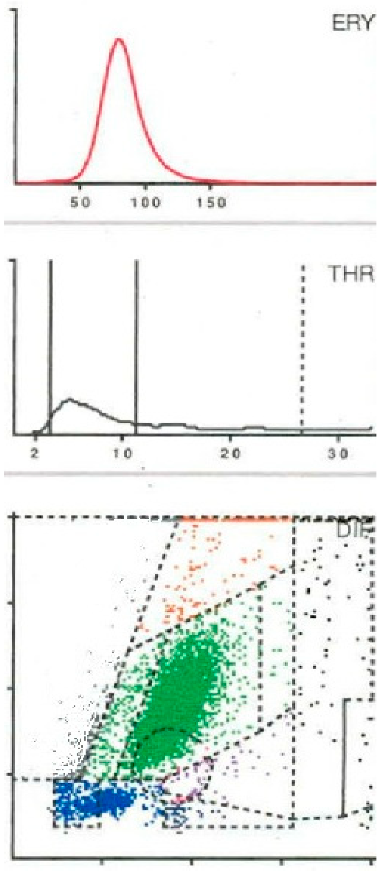
Additional Erythrocyte Field Is Helpful for Graphic Type Differentiation of Cell Count Determination Between Acute Periprosthetic Joint Infection and Hematoma
 , ,
, ,
Abstract
1. Introduction
2. Results
3. Discussion
4. Materials and Methods
5. Conclusions
Author Contributions
Funding
Institutional Review Board Statement
Informed Consent Statement
Data Availability Statement
Conflicts of Interest
References
- Bedair, H.; Ting, N.; Jacovides, C.; Saxena, A.; Moric, M.; Parvizi, J.; Della Valle, C.J. Diagnosis of early postoperative TKA infection using synovial fluid analysis. Clin. Orthop. Relat. Res. 2011, 469, 34–40. [Google Scholar] [CrossRef]
- Kim, S.G.; Kim, J.G.; Jang, K.M.; Han, S.B.; Lim, H.C.; Bae, J.H. Diagnostic value of synovial white blood cell count and serum C-reactive protein for acute periprosthetic joint infection after knee arthroplasty. J. Arthroplast. 2017, 32, 3724–3728. [Google Scholar] [CrossRef]
- Yi, P.H.; Cross, M.B.; Moric, M.; Sporer, S.M.; Berger, R.A.; Della Valle, C.J. Diagnosis of infection in the early postoperative period after total hip arthroplasty. Clin. Orthop. Relat. Res. 2014, 472, 424–429. [Google Scholar] [CrossRef]
- Tornero, E.; Soriano, A. Importance of Selection and Duration of Antibiotic Regimen in Prosthetic Joint Infections Treated with Debridement and Implant Retention—Authors’ Response. J. Antimicrob. Chemother. 2016, 71, 3627. [Google Scholar] [CrossRef]
- Yu, B.Z.; Fu, J.; Chei, W.; Hao, L.B.; Chen, J.Y. Neutrophil to lymphocyte ratio as a predictor for diagnosis of early periprosthetic joint infection. BMC Muscoloskeltal. Disord. 2020, 21, 706. [Google Scholar] [CrossRef] [PubMed]
- Parvizi, J.; Zmistowski, B.; Berbari, E.F.; Bauer, T.W.; Springer, B.D.; Della Valle, C.J.; Garvin, K.L.; Mont, M.A.; Wongworawat, M.D.; Zalavras, C.G. New definition for periprosthetc joint infection: From the Workgroup of the Musculoskeletal Infection Society. Clin. Orthop. Relat. Res. 2011, 469, 2992–2994. [Google Scholar] [CrossRef]
- Parvizi, J.; Tan, T.L.; Goswami, K.; Higuera, C.; Della Valle, C.; Chen, A.F.; Shohat, N. The 2018 Definition of Periprosthetic Hip and Knee Infection: An Evidence-Based and Validated Criteria. J. Arthroplast. 2018, 33, 1309–1314. [Google Scholar] [CrossRef] [PubMed]
- Workgroup Convened by the Musculoskeletal Infection Society. New definition for periprosthetic joint infection. J. Arthroplast. 2011, 26, 1136–1138. [CrossRef]
- Parvizi, J.; Gehrke, T. International Consensus Group on Periprosthetic Joint Infection. Definition of periprosthetic joint infection. J. Arthroplast. 2014, 29, 1331. [Google Scholar] [CrossRef]
- Sukhonthamarn, K.; Tan, T.I.; Xu, C.; Ku, F.C.; Lee, M.S.; Citak, M.; Gehrke, T.; Goswani, K.; Parvizi, J. Determining diagnostic thresholds for acute postoperative periprosthetic joint infection. J. Bone. Joint. Surg. Am. 2020, 102, 2043–2048. [Google Scholar] [CrossRef] [PubMed]
- Fink, B.; Hoyka, M.; Weissbarth, E.; Schuster, P.; Berger, I. The graphical representation of cell count representation. A new procedure for the diagnosis of periprosthetic joint infections. Antibiotics 2021, 10, 346. [Google Scholar] [CrossRef] [PubMed]
- Fink, B.; Hoyka, M.; Weissbarth, E.; Schuster, P.; Berger, I. A new graphic type differentiation for distinguishing acute periprosthetic joint infection from hemarthrosis. Antibiotics 2022, 11, 1284. [Google Scholar] [CrossRef] [PubMed]
- Fink, B.; Hoyka, M.; Blersch, B.P.; Baum, H.; Sax, F.H. Graphic type differentiation of cell count data for diagnosis of early and late periprosthetic joint infection: A new method. Technol. Health Care 2023, 32, 3669–3680. [Google Scholar] [CrossRef] [PubMed]
- Sigmund, I.K.; Puchner, S.E.; Windhager, R. Serum inflammatory biomarkers in the diagnosis of periprosthetic joint infections. Biomedicines 2021, 9, 1128. [Google Scholar] [CrossRef]
- Berbari, E.; Mabry, T.; Tsaras, G.; Spangehl, M.; Erwin, P.J.; Murad, M.H.; Steckelberg, J.; Osmon, D. Inflammatory blood laboratory levels as markers of prosthetic joint infection: A systematic review and meta-analysis. J. Bone. Joint. Surg. Am. 2010, 92, 2102–2109. [Google Scholar] [CrossRef]
- Meier, M.-P.; Bauer, I.J.; Maheshwari, A.K.; Husen, M.; Jäckle, K.; Hubert, J.; Hawellek, J.; Lehmann, W.; Saul, D. Predicting the expection—CRP and primary hip arthroplasty. J. Clin. Med. 2021, 10, 4985. [Google Scholar] [CrossRef]
- Xu, C.; Tan, L.; Kuo, F.C.; Goswami, K.; Wang, Q.; Parvizi, J. Reevaluating current cutoffs for acute periprosthetic joint infection: Current thresholds are insensitive. J. Arthroplast. 2019, 30, 2744–2748. [Google Scholar] [CrossRef]
- Schäfer, P.; Fink, B.; Sandow, D.; Margull, A.; Berger, I.; Frommelt, L. Prolonged bacterial culture to identify late periprosthetic joint infection: A promising strategy. Clin. Inf. Dis. 2008, 47, 1403–1409. [Google Scholar] [CrossRef]
- Siekmeier, R.; Bierlich, A.; Jaross, W. The white blood cell differential: Three methods compared. Clin. Chem. Lab. Med. 2001, 39, 432–445. [Google Scholar] [CrossRef]
- Arroyo, M.E.; Tabernero, M.D.; Garcia-Marcos, M.A.; Orfao, A. Analytic performance of the PENTRA 80 automated blood cell analyser for the evaluation of normal and pathologic WBCs. Am. J. Clin. Pathol. 2005, 123, 206–214. [Google Scholar] [CrossRef]
- Danise, P.; Maconi, M.; Rovetti, A.; Avino, D.; Di Palma, A.; Gerardo Pirofalo, M.; Esposito, C. Cell counting of body fluids: Comparison between three automated haematology analysers and the manual microscope method. Int. J. Lab. Hematol. 2013, 35, 608–613. [Google Scholar] [CrossRef]
- Krenn, V.; Otto, M.; Morawietz, L.; Hopf, T.; Jakobs, M.; Klauser, W.; Schwantes, B.; Gehrke, T. Histopathologic diagnostics in endoprosthetics: Periprosthetic neosynovialitis, hypersensitivity reaction, and arthrofibrosis. Orthopäde 2009, 38, 520–530. [Google Scholar] [CrossRef] [PubMed]
- Krenn, V.; Morawietz, L.; Perino, G.; Kienapfel, H.; Ascherl, R.; Hassenpflug, G.J.; Thomsen, M.; Thomas, P.; Huber, M.; Kendoff, D.; et al. Revised histopathological consensus classification of joint implant related pathology. Pathol. Res. Pract. 2014, 210, 779–786. [Google Scholar] [CrossRef] [PubMed]
- Müller, M.; Morawietz, L.; Hasart, O.; Strube, P.; Perka, C.; Tohtz, S. Histopathological diagnosis of periprosthetic joint infection following total hip arthroplasty: Use of a standardized classification system of the periprosthetic interface membrane. Orthopäde 2009, 38, 1087–1096. [Google Scholar] [CrossRef] [PubMed]



| Likelihood Ratio Negative | Likelihood Ratio Positive | |||||||
|---|---|---|---|---|---|---|---|---|
| LHR neg. | LHR pos. | 98.1% | Accuracy | Infection | ||||
| 0.03 | 73.62 | 96.9% | Sensitivity | no | yes | |||
| 98.7% | Specificity | 32 | 1 | 31 | pos. | LMNE type 2/6 | ||
| 96.9% | PPW | 76 | 75 | 1 | neg. | |||
| 98.7% | NPW | 108 | 76 | 32 | ||||
| LHR neg. | LHR pos. | 75.0% | Accuracy | Infection | ||||
| 0.66 | 3.86 | 40.6% | Sensitivity | no | yes | |||
| 89.5% | Specificity | 21 | 8 | 13 | pos. | CRP > 100 mg/dL | ||
| 61.9% | PPW | 87 | 68 | 19 | neg. | |||
| 78.2% | NPW | 108 | 76 | 32 | ||||
| LHR neg. | LHR pos. | 75.9% | Accuracy | Infection | ||||
| 0.63 | 4.16 | 43.8% | Sensitivity | no | yes | |||
| 89.5% | Specificity | 22 | 8 | 14 | pos. | CRP > 90 mg/dL | ||
| 63.6% | PPW | 86 | 68 | 18 | neg. | |||
| 79.1% | NPW | 108 | 76 | 32 | ||||
| LHR neg. | LHR pos. | 75.0% | Accuracy | Infection | ||||
| 0.58 | 3.45 | 50.0% | Sensitivity | no | yes | |||
| 85.5% | Specificity | 27 | 11 | 16 | pos. | CRP > 75 mg/dL | ||
| 59.3% | PPW | 81 | 65 | 16 | neg. | |||
| 80.2% | NPW | 108 | 76 | 32 | ||||
| LHR neg. | LHR pos. | 83.3% | Accuracy | Infection | ||||
| 0.43 | 9.03 | 59.4% | Sensitivity | no | yes | |||
| 93.4% | Specificity | 24 | 5 | 19 | pos. | Cell Count ≥ 10,000 | ||
| 79.2% | PPW | 84 | 71 | 13 | neg. | |||
| 84.5% | NPW | 108 | 76 | 32 | ||||
| LHR neg. | LHR pos. | 88.0% | Accuracy | Infection | ||||
| 0.41 | #DIV/0! | 59.4% | Sensitivity | no | yes | |||
| 100.0% | Specificity | 19 | 0 | 19 | pos. | Cell Count ≥ 10,000 | ||
| 100.0% | PPW | 89 | 76 | 13 | neg. | +LMNE pos. | ||
| 85.4% | NPW | 108 | 76 | 32 | ||||
| LHR neg. | LHR pos. | 83.3% | Accuracy | Infection | ||||
| 0.46 | 13.13 | 56.3% | Sensitivity | no | yes | |||
| 95.7% | Specificity | 21 | 3 | 18 | pos. | PMN ≥ 79.5% | ||
| 85.7% | PPW | 81 | 67 | 14 | neg. | |||
| 82.7% | NPW | 102 | 70 | 32 | ||||
| LHR neg. | LHR pos. | 72.5% | Accuracy | Infection | ||||
| 0.86 | 10.94 | 15.6% | Sensitivity | no | yes | |||
| 98.6% | Specificity | 6 | 1 | 5 | pos. | PMN ≥ 90% | ||
| 83.3% | PPW | 96 | 69 | 27 | neg. | |||
| 71.9% | NPW | 102 | 70 | 32 | ||||
| LHR neg. | LHR pos. | 86.3% | Accuracy | Infection | ||||
| 0.44 | #DIV/0! | 56.3% | Sensitivity | no | yes | |||
| 100.0% | Specificity | 18 | 0 | 18 | pos. | PMN ≥ 79.5% | ||
| 100.0% | PPW | 84 | 70 | 14 | neg. | +Cell Count ≥ 10,000 | ||
| 83.3% | NPW | 102 | 70 | 32 | ||||
| LHR neg. | LHR pos. | 73.5% | Accuracy | Infection | ||||
| 0.84 | #DIV/0! | 15.6% | Sensitivity | no | yes | |||
| 100.0% | Specificity | 5 | 0 | 5 | pos. | PMN ≥ 90% | ||
| 100.0% | PPW | 97 | 70 | 27 | neg. | +Cell Count ≥ 10,000 | ||
| 72.2% | NPW | 102 | 70 | 32 | ||||
| LHR neg. | LHR pos. | 90.7% | Accuracy | Infection | ||||
| 0.31 | #DIV/0! | 68.8% | Sensitivity | no | yes | |||
| 100.0% | Specificity | 22 | 0 | 22 | pos. | Culture | ||
| 100.0% | PPW | 86 | 76 | 10 | neg. | |||
| 88.4% | NPW | 108 | 76 | 32 | ||||
| LHR neg. | LHR pos. | 93.0% | Accuracy | Infection | ||||
| 0.09 | 18.26 | 91.3% | Sensitivity | no | yes | |||
| 95.0% | Specificity | 22 | 1 | 21 | pos. | Histo > five cells | ||
| 95.5% | PPW | 21 | 19 | 2 | neg. | |||
| 90.5% | NPW | 43 | 20 | 23 | ||||
| LHR neg. | LHR pos. | 95.3% | Accuracy | Infection | ||||
| 0.05 | 19.13 | 95.7% | Sensitivity | no | yes | |||
| 95.0% | Specificity | 23 | 1 | 22 | pos. | Histo type 2 | ||
| 95.7% | PPW | 20 | 19 | 1 | neg. | |||
| 95.0% | NPW | 43 | 20 | 23 | ||||
Disclaimer/Publisher’s Note: The statements, opinions and data contained in all publications are solely those of the individual author(s) and contributor(s) and not of MDPI and/or the editor(s). MDPI and/or the editor(s) disclaim responsibility for any injury to people or property resulting from any ideas, methods, instructions or products referred to in the content. |
© 2026 by the authors. Licensee MDPI, Basel, Switzerland. This article is an open access article distributed under the terms and conditions of the Creative Commons Attribution (CC BY) license.
Share and Cite
Sax, F.H.; Hoyka, M.; Blersch, B.P.; Weissbarth, E.; Schuster, P.; Berger, I.; Baum, H.; Fink, B. Additional Erythrocyte Field Is Helpful for Graphic Type Differentiation of Cell Count Determination Between Acute Periprosthetic Joint Infection and Hematoma. Antibiotics 2026, 15, 122. https://doi.org/10.3390/antibiotics15020122
Sax FH, Hoyka M, Blersch BP, Weissbarth E, Schuster P, Berger I, Baum H, Fink B. Additional Erythrocyte Field Is Helpful for Graphic Type Differentiation of Cell Count Determination Between Acute Periprosthetic Joint Infection and Hematoma. Antibiotics. 2026; 15(2):122. https://doi.org/10.3390/antibiotics15020122
Chicago/Turabian StyleSax, Florian Hubert, Marius Hoyka, Benedikt Paul Blersch, Elke Weissbarth, Philipp Schuster, Irina Berger, Hansjörg Baum, and Bernd Fink. 2026. "Additional Erythrocyte Field Is Helpful for Graphic Type Differentiation of Cell Count Determination Between Acute Periprosthetic Joint Infection and Hematoma" Antibiotics 15, no. 2: 122. https://doi.org/10.3390/antibiotics15020122
APA StyleSax, F. H., Hoyka, M., Blersch, B. P., Weissbarth, E., Schuster, P., Berger, I., Baum, H., & Fink, B. (2026). Additional Erythrocyte Field Is Helpful for Graphic Type Differentiation of Cell Count Determination Between Acute Periprosthetic Joint Infection and Hematoma. Antibiotics, 15(2), 122. https://doi.org/10.3390/antibiotics15020122