Article

Serratia marcescens Isolates from Bovine Mastitic Milk: Antimicrobial Resistance and Virulence Features

1
School of Medicine and Biomedical Sciences, ICBAS-UP, University of Porto, Rua de Jorge Viterbo Ferreira, nr. 228, 4050-313 Porto, Portugal
2
Associate Laboratory for Animal and Veterinary Science (AL4AnimalS), 1300-477 Lisboa, Portugal
3
Centro de Estudos de Ciência Animal (CECA), Instituto de Ciências, Tecnologias e Agroambiente (ICETA), University of Porto, Rua D. Manuel II, Apartado 55142, 4051-401 Porto, Portugal
4
Research Center in Biodiversity and Genetic Resources (CIBIO/InBIO), University of Porto, Rua Padre Amando Quintas, nr. 7, 4485-661 Vairão, Portugal
5
BIOPOLIS Program in Genomics, Biodiversity and Land Planning, CIBIO, Campus de Vairão, 4485-661 Vairão, Portugal
*
Author to whom correspondence should be addressed.
Antibiotics 2025, 14(9), 892; https://doi.org/10.3390/antibiotics14090892
Submission received: 30 June 2025 / Revised: 4 August 2025 / Accepted: 8 August 2025 / Published: 3 September 2025

Abstract

Background: Bovine mastitis (BM) is a major disease affecting dairy herds (DHs), with Serratia marcescens (S. marcescens) being increasingly implicated as a causative agent. The growing concern over antimicrobial resistance (AMR) extends to BM-associated S. marcescens isolates, where resistance patterns are emerging. Methods: Here, four BM Gram-negative isolates were investigated: 1-DH1, 2-DH1, 3-DH2, and 4-DH3. Phenotypic characterization was performed using the Neg-Urine-Combo98 panel on a MicroScan WalkAway Plus system. Whole-genome sequencing (WGS) was performed to characterize and identify AMR and virulence factors (VF) genes and plasmids in isolates 1-DH1, 3-DH2, and 4-DH3, and phylogenomic analyses were conducted for a visual comparison of the genomes. Results: Phenotypically, isolates 1-DH1, 2-DH1, and 4-DH3 were identified as S. marcescens, and 3-DH2 as Serratia odorifera (confirmed as S. marcescens by WGS). A 28.00% (n = 25) prevalence of phenotypic AMR for isolates 1-DH1, 2-DH1, and 4-DH3 against Aug-E, AM, To, Cfx, Crm, Cl, and Fd was shown, and 24.00% (n = 25) for isolate 3-DH2 against Aug-E, AM, To, Crm, Cl, and Fd. The AMR genes AAC(6′)-Ic, aac(6′)-Ic_1, aac(6′)-Ial, H-NS, SRT-2, oqxB, oqxB_1, oqxB25, mexI, CRP, and blaSST-1, and flgH, fliP, fliM, and fliG VF genes were identified in the whole genome of the S. marcescens sequenced isolates 1-DH1, 2-DH1, and 4-DH3. In addition, a phylogenomic analysis of these three isolates revealed that WGS genomes are more closely related to S. marcescens prevenient from environmental sources. Conclusions: This study reports, for the first time, AMR resistance to tobramycin, cefuroxime, colistin, and nitrofurantoin in BM S. marcescens isolates. Genomic analysis revealed the presence of multiple AMR and VF genes, further highlighting the pathogenic potential of these isolates. Phylogenomic analysis revealed that the genome of the three BM S. marcescens isolates is more closely related to environmental S. marcescens strains.

1. Introduction

Bovine milk consumption is estimated at billions of liters worldwide, with the majority consumed as pasteurized milk [1]. However, the consumption of raw milk (RM) and unpasteurized dairy products poses a substantial public health risk, as they may be contaminated by pathogenic microorganisms originating from environmental sources or infected animals. Exposure to zoonotic pathogens through ingestion or direct contact can lead to zoonotic diseases (ZDs), with the consumption of RM widely recognized as one of the primary risk factors [1,2]. Nevertheless, non-zoonotic pathogens also pose a threat to animal health and welfare, and indirectly to public health, as they can serve as reservoirs of transferable antimicrobial resistance genes [3]. Overall, deliberately adopting the “One Health” concept within dairy herds (DHs) [4] could be a strategic framework for controlling ZDs, and good farming practices and effective post milk processing in a herd [1] could contribute to putting into practice both concepts.
Bovine mastitis (BM), defined as the inflammation of the mammary gland, is globally considered one of the most important diseases within DH due to its economic impact. It leads to considerable losses not only through decreased milk production and quality but also due to the reduced reproductive capacity of the affected animals, discarded milk, increased treatment costs, and higher culling rates [5,6,7]. Bovine mastitis can be classified as clinical (CM), subclinical (SCM), and chronic according to the degree of the mammary gland inflammation [8], and there are reported studies suggesting that SCM could account for more financial losses than CM in a herd [9,10]. Moreover, recent studies have described an increase in the incidence of environmental mastitis and a decrease in the incidence of contagious mastitis [11]. Regarding infections, various factors including pathogens, environmental factors, and opportunistic microorganisms are the most prevalent causes of BM in DH. The bacteria most commonly associated with BM include Gram-positive cocci such as Staphylococcus aureus (S. aureus), Streptococcus agalactiae, and Streptococcus uberis, as well as Gram-negative (GN) bacilli like Escherichia coli and Klebsiella pneumoniae [2]. Besides the mentioned pathogens, Serratia marcescens (S. marcescens) and Proteus mirabilis have been described as environmental causative agents of mastitis, and S. marcescens has been described as a causative agent of CM and SCM outbreaks [12,13]. Some cases of the SCM form may resolve spontaneously. However, there are reported studies where cows become chronic carriers of S. marcescens [14,15]. Additionally, Serratia marcescens is recognized as an emerging healthcare-associated pathogen, frequently implicated in nosocomial infections in humans. It primarily affects the respiratory and urinary tracts, as well as surgical wounds and soft tissues [16].
Regarding treatment, bovine mastitis (BM) remains a widespread infectious disease with a substantial economic impact, accounting for approximately 80.00% of antimicrobial usage in dairy animals. However, the global effectiveness of antimicrobial therapy has progressively declined, largely due to the emergence of novel bacterial resistance mechanisms. These mechanisms include the reduced permeability or antimicrobial uptake resulting from porin protein modifications; increased antimicrobial efflux mediated by efflux pumps; enzymatic inactivation of antimicrobials; alterations in antimicrobial targets via spontaneous point mutations; loss of enzymes responsible for antimicrobial activation; use of alternative metabolic pathways to bypass the need for inhibited targets; and acquisition of resistance determinants through mobile genetic elements [17,18,19], resulting in an increase in antimicrobial resistance (AMR) globally [3,16,20]. According to the literature, Serratia marcescens isolates have demonstrated resistance to multiple antimicrobial agents in BM cases, such as cefazolin, chloramphenicol, ampicillin, ceftriaxone, cefotaxime, and tetracyclin, and in human clinical infections, including resistance to penicillins, cephalosporins, tetracyclines, macrolides, nitrofurantoin, and colistin [21,22,23]. Moreover, several AMR genes detected in S. marcescens isolates from BM for extended-spectrum beta-lactamases (e.g., blaTEM, blaCTX-M, blaCTX-M-15, blaCTX-M-14, blaCTX-M-65, and blaSHV-28), chloramphenicol (e.g., cmlA and floR), carbapenem (e.g., SIM-1), and efflux pumps (e.g., sdeB, sdeY, sdeR, and sdeD) were previously described [21,23]. Furthermore, mutations in the sdeS efflux pump gene have been reported to upregulate the expression of sdeAB efflux pump genes, contributing to the development of multidrug resistance (MDR) in S. marcescens isolates, including those derived from aquatic environments [24]. In addition, the pathogenicity of S. marcescens is also mediated by an arsenal of virulence factors (VFs) including hemolysin, lipase, protease, prodigiosin, nuclease, chitinase, motility, and biofilm [25,26,27], and VF genes (e.g., flhD, entB, kpn, mrkD, ycfM, bsmB, pigP, kfu, and shlB) in S. marcescens isolates from BM have also been previously reported [21].
Antimicrobial resistance (AMR) is recognized as a major public health concern in Portugal, with several studies reporting prevalence rates ranging from 50.00% to 83.00%. These studies also describe the detection of AMR genes and multidrug-resistant (MDR) profiles, varying according to the study design and context. The reported resistant isolates include methicillin-resistant Staphylococcus aureus (MRSA), S. aureus, Escherichia coli, and Enterococcus spp., recovered from human, animal (including bovine mastitis), food-producing animal, and environmental sources [28,29,30,31,32]. In this context, our investigation focused on Serratia marcescens isolates obtained from cases of bovine mastitis.

2. Results

2.1. Phenotypic Identification and Antimicrobial Susceptibility Testing

Isolates 1-DH1, 2-DH1, and 4-DH3 were phenotypically identified as Serratia marcescens with a probability of 99.99%, while isolate 3-DH2 was identified as Serratia odorifera (S. odorifera) with a probability of 94.66%, using the Neg-Urine-Combo98 panel on the MicroScan WalkAway Plus system (Table 1). Regarding antimicrobial susceptibility testing (AST), S. marcescens isolates (1-DH1, 2-DH1, and 4-DH3) exhibited a phenotypic AMR prevalence of 28.00%, with resistance detected against Aug-E and AM (β-lactams), To (aminoglycoside), Cfx and Crm (cephalosporins), Cl (polymyxin), and Fd (nitrofuran). Isolate 3-DH2 showed a 24.00% phenotypic AMR prevalence, with resistance to Aug-E and AM (β-lactams), To (aminoglycoside), Crm (cephalosporin), Cl (polymyxin), and Fd (nitrofuran) (Table 1).
With respect to antimicrobial susceptibility, S. marcescens isolates 1-DH1, 2-DH1, and 4-DH3 demonstrated 72.00% phenotypic susceptibility to Cpe, Cft, Caz/CA, Etp, Imp, and Mer (β-lactams); NA, Cp, Lvx, and Nxn (quinolones); AK and Gm (aminoglycosides); T/S (sulphonamide); AZT (monobactam); Caz (cephalosporin); Fos (phosphonic acid derivative); Cft/CA (cephalosporin/β-lactamase inhibitor); and P/T (penicillin/β-lactamase inhibitor) (Table 1).
Similarly, isolate 3-DH2 exhibited a phenotypic AMR prevalence of 24.00% against the same antimicrobials, except for Cfx, to which it was not resistant (Table 1). Notably, 3-DH2 showed 76.00% susceptibility to the remaining tested antimicrobials (Table 1).
The minimum inhibitory concentration (MIC) values, interpreted according to EUCAST guidelines (as detailed in the Section 4), are presented in Table 1.

2.2. Genome Characteristics and Bacteria Classification

For the three whole-genome-sequenced bacteria isolates (isolates 1-DH1, 3-DH2, and 4-DH3), the median genome size was 5 Mb–5.1 Mb, with a G + C content of 59.50% and an average of 4727 protein-coding sequences. Mash [33] identified all the isolates as S. marcescens, with a 99.30% identity for all the three isolates (Table 2). The genome map and the clusters of orthologous groups (COGs) of the proteins obtained for the three isolates, 1-DH1, 3-DH2, and 4-DH, by WGS are shown in Figure 1.

2.3. Genomic Analysis of Antimicrobial Resistance Genes

The AMR genes AAC(6′)-Ic, H-NS, tet(41), SRT-2, oqxB, mexI, and CRP were identified in whole-genome sequences of the S. marcescens isolates 1-DH1, 3-DH2, and 4-DH3, with identity percentages ranging from 97.10% to 100.00%, 80.85% to 100.00%, and 80.85% to 96.22%, respectively, by ABRicate using the CARD database (Table 3 and Table S2). Additionally, the AMR genes aac(6′)-Ial, tet(41), blaSST-1, and oqxB25 were identified in the genomes of all three isolates, with identity percentages ranging from 82.33% to 99.09%, as determined by ABRicate using the NCBI AMRFinderPlus database (Table 3 and Table S2). Furthermore, the genes aac(6′)-Ic_1, tet(41)_1, blaSST-1_1, and oqxB_1 were identified in the three isolates with identity percentages ranging from 82.10% to 99.03% by ABRicate using the Resfinder database (Table 3 and Table S2). The antimicrobial resistance (AMR) genes identified were associated with specific resistance mechanisms and antimicrobial classes. The genes AAC(6′)-Ic, aac(6′)-Ic_1, and aac(6′)-Ial, which encode aminoglycoside-modifying enzymes, conferred resistance to aminoglycosides, specifically amikacin (AK) and tobramycin (To), through antimicrobial inactivation. The H-NS gene, involved in global gene regulation and functioning via an efflux-based resistance mechanism, was associated with resistance to cephalosporins, fluoroquinolones, macrolides, penicillins, tetracyclines, and cephamycins. The tet(41) gene, which also operates through antimicrobial efflux, conferred resistance to tetracyclines including acycline, doxycycline, and tetracycline. The SRT-2 gene, encoding a β-lactamase, mediated resistance to cephalosporins, including ceftazidime (Cft), via antimicrobial inactivation.
Furthermore, the oqxB gene, associated with an efflux mechanism, was linked to resistance against diaminopyrimidines, fluoroquinolones, glycylcyclines, nitrofurans, and tetracyclines. Its variant, oqxB_1, conferred resistance specifically to nalidixic acid (NA) and ciprofloxacin (Cp). The mexI gene, part of the resistance–nodulation–division (RND) efflux system, was related to resistance to acridine dyes, fluoroquinolones, and tetracyclines. The CRP gene, a transcriptional regulator that modulates efflux pump expression, conferred resistance to fluoroquinolones, macrolides, and penicillins. Lastly, the blaSST-1 gene, encoding a β-lactamase enzyme, conferred resistance to cephalosporins via antimicrobial inactivation. These findings are detailed in Table 3 and Table S2.

2.4. Genomic Analysis of Virulence Factors Genes

The virulence factor (VF) genes flgH, fliP, fliM, and fliG, which encode flagellar proteins, were identified in the whole-genome sequences of Serratia marcescens isolates 1-DH1, 3-DH2, and 4-DH3. These genes exhibited identity percentages ranging from 80.12% to 82.58% across all isolates, as determined using ABRicate (BLAST) against the VFDB database (Table 3 and Table S2).

2.5. Genomic Analysis of Plasmids

The S. marcescens whole-genome sequences of the three isolates (isolates 1-DH1, 3-DH2, and 4-DH3) were evaluated for the presence of plasmids using the PlasmidFinder tool and MOBScan server, and no plasmids were identified in any of the three isolates.

2.6. Phylogenomic Analysis

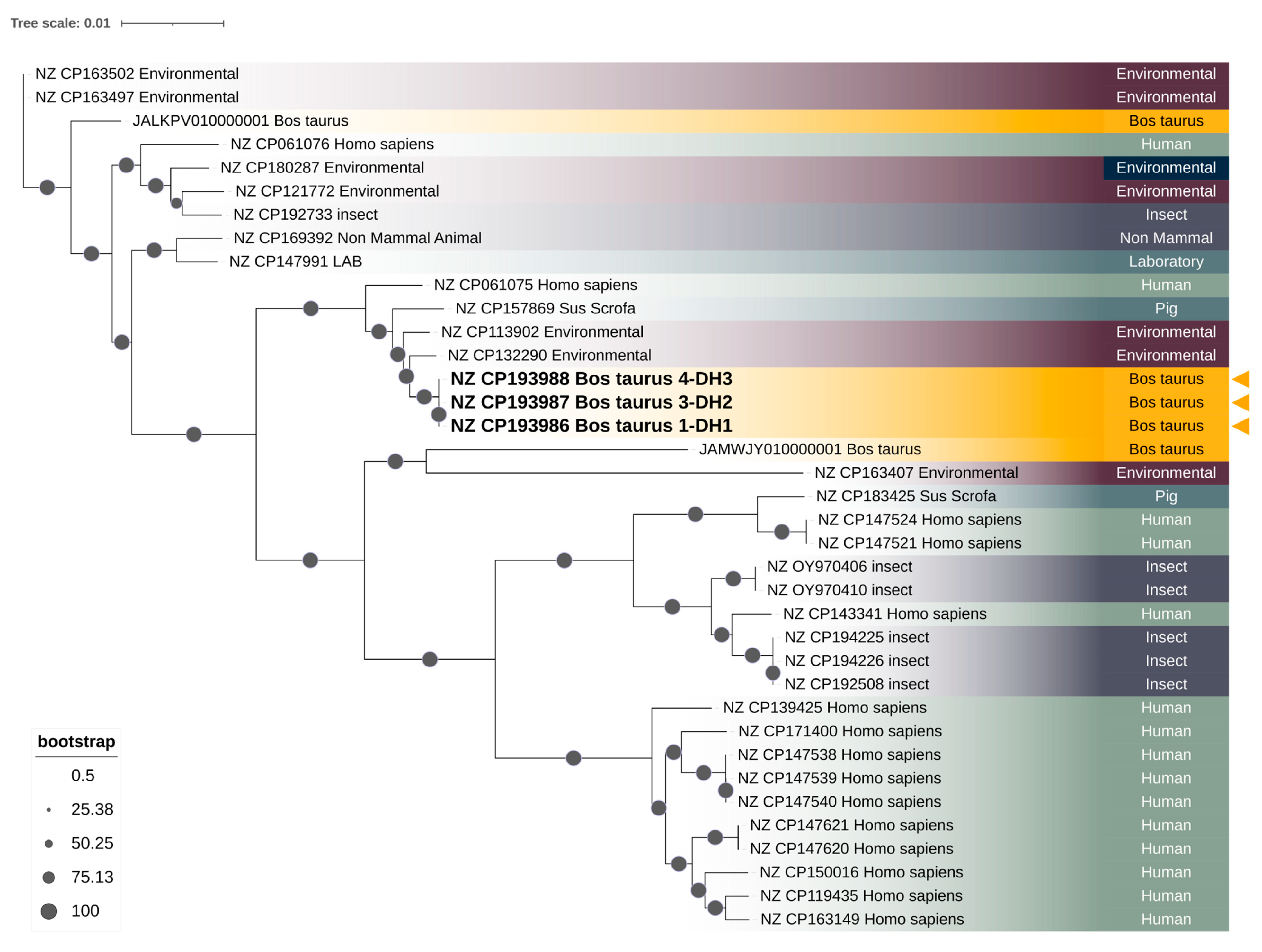
A core genome phylogenetic analysis revealed two major and strongly supported S. marcescens clusters, each comprising isolates from multiple sources (Figure 2). The first cluster includes S. marcescens isolates of environmental, human, pig, and insect origin, as well as a single Bos taurus isolate (JALKPV010000001), and is supported by confident bootstrap values (100%). This cluster displays greater phylogenetic diversity overall, with longer internal branch lengths, reflecting a broader genomic divergence among its constituent isolates. Despite this diversity, isolates within the cluster exhibit relatively similar phylogenetic distances to each other when compared internally, suggesting a degree of lineage cohesion. However, this entire cluster is clearly separated from the rest of the tree by a longer basal branch, indicating a greater evolutionary distance from the second major cluster. This suggests that the isolates in the first cluster, while heterogeneous, share a more distant common ancestor relative to the isolates grouped in the second cluster.
In contrast, the second major cluster includes S. marcescens isolates from Bos taurus (including those sequenced in this study), as well as human, pig, and environmental origins. The three Bos taurus isolates (NZ_CP193986 Bos taurus 1-DH1, NZ_CP193987 Bos taurus 3-DH2, and NZ_CP193988 Bos taurus 4-DH3) generated in this study form a tightly grouped, strongly supported monophyletic clade (bootstrap: 99%), with near-zero branch lengths (0.00000003). This clade is embedded within a subcluster that also includes two environmental S. marcescens isolates (NZ_CP132290 and NZ_CP113902), forming a broader lineage with high bootstrap support (100%). The second cluster exhibits shorter branch lengths overall, particularly within the subcluster containing the study isolates, suggesting a lower genetic divergence compared to the first cluster. Additional subclusters within this second group include human S. marcescens isolates forming distinct branches with strong support, interspersed with pig and environmental genomes.
The phylogenetic tree, thus, demonstrates the presence of genetically distinct lineages of S. marcescens with varying degrees of host and environmental association, and highlights the close genetic relationship among the Bos taurus isolates characterized in this study (Figure 2).

3. Discussion

Bovine mastitis is considered one of the most important diseases within DH worldwide due to its significant economic impact. This impact could be reflected in the decreased milk production and quality, reduced reproductive capacity, loss of discarded milk, and increased treatment costs and culling rates [5,6,7]. Recently, changes in the incidence of environmental and contagious mastitis have been reported, with increased cases of environmental mastitis and decreased cases of contagious mastitis [11]. The environmental pathogen S. marcescens has been described as a causative agent of CM and SCM outbreaks [12,13,14]. Furthermore, S. marcescens has been isolated from various farm environments, including bedding, milking parlors, feces of dairy cows, water, soil, different types of plants, and insects [34,35,36].
Bovine mastitis, as a widespread infectious disease, accounts for 80.00% of antimicrobial usage in dairy animals worldwide [37]. Moreover, the economic impact of mastitis and the risk of treatment failure highlights the significant financial burden on dairy farmers and the potential for the treatment to be ineffective [20,38].
Furthermore, AMR is also increasing due to new and emerging mechanisms of bacterial resistance that are spreading globally [3,20]. In this context, several studies have reported S. marcescens isolates from BM exhibiting AMR to antimicrobials such as cefazolin, chloramphenicol, ampicillin, ceftriaxone, cefotaxime, and tetracycline [21,23].
In this study, four GN bacterial isolates obtained from RM samples of BM cases from three DH in the northwest region of Portugal were investigated: isolate 1-DH1, isolate 2-DH1, isolate 3-DH2, and isolate 4-DH3. Using the Neg-Urine-Combo98 panel on the MicroScan WalkAway Plus system, isolates 1-DH1, 2-DH1, and 4-DH3 were phenotypically identified as S. marcescens, while isolate 3-DH2 was identified as S. odorifera. However, WGS confirmed isolate 3-DH2 as S. marcescens, indicating a misidentification by the panel-based method. The misidentification of isolate 3-DH2 highlights the inherent limitations of phenotypic methods in achieving precise species-level identification. Whole-genome sequencing (WGS), by providing comprehensive genetic information, enabled accurate taxonomic classification and unequivocally confirmed the isolate as Serratia marcescens. This genomic approach overcomes the ambiguities associated with phenotypic assays, thereby improving the reliability of bacterial identification in this study. To our knowledge, there are no published reports on the use of these panels for bacterial identification in RM from BM cases. Nonetheless, S. marcescens has been identified in human clinical samples using this technology with other panel types [39,40,41], suggesting that the panel used in this study or the others could be an alternative for S. marcescens identification and/or AST in RM samples from BM. Moreover, a study aimed at developing a direct method for rapid antimicrobial susceptibility testing and the identification of bacteria in positive blood cultures, including clinical and spiked isolates of Serratia marcescens, compared the performance of the matrix-assisted laser desorption ionization-time of flight mass spectrometry (MALDI-TOF MS), Vitek 2, and MicroScan WalkAway 96 Plus systems. The systems demonstrated categorical agreement rates of 94.90%, 97.40%, and 100.00% for MIC panels 53, 38, and MICroSTREP Plus 2, respectively. Comparable results were achieved across the tested platforms, as previously reported [41]. Regarding antimicrobial resistance (AMR) as determined by the Neg-Urine-Combo98 panel in the MicroScan WalkAway Plus system, Serratia marcescens isolates 1-DH1, 2-DH1, and 4-DH3 exhibited 28.00% phenotypic AMR prevalence to Aug-E and AM, To, Cfx, Crm, Cl, and Fd. These antimicrobials represent the β-lactam, aminoglycoside, cephalosporin, polymyxin, and nitrofuran classes, respectively. Based on these findings and in accordance with the literature, the isolates tested in this study can be classified as multidrug-resistant (MDR) bacteria [42]. Isolate 3-DH2, identified as S. odorifera by phenotypic testing but confirmed as S. marcescens via WGS, showed 24.00% resistance to the same set of antimicrobials, except for Cfx, showing additional susceptibility. Overall, the present findings suggest that antimicrobials such as Aug-E, AM, To, Cfx, Crm, Cl, and Fd may not be the most suitable options for treating BM caused by S. marcescens. Previously, the resistance of S. marcescens to Aug-E, AM, and Cfx had been described in BM, and the resistance to To, Crm, Cl, and Fd had been reported in human clinical isolates [21,22,43,44,45,46]. Resistance to To, Crm, Cl, and Fd were identified in this study. In terms of susceptibility, S. marcescens isolates 1-DH1, 2-DH1, and 4-DH3 (all 72.00%) and 3-DH2 (76.00%) were susceptible to the remaining tested antimicrobials. Overall, the antimicrobials Cpe, Cft, Caz/CA, Etp, Imp, Mer, NA, Cp, Lvx, Nxn, AK, Gm, T/S, AZT, Caz, Fos, Cft/CA, and P/T may represent better alternative treatment options for cows of all tested isolates (1-DH1, 2-DH1, 3-DH2, and 4-DH3). However, some of these agents are primarily intended for human use and are classified as critically important by the WHO or categorized as Category A by the EMA, which prohibits their use as veterinary medicines within the European Union. Nevertheless, antimicrobial susceptibility of S. marcescens to Cpe, Etp, Imp, NA, Cp, Lvx, Gm, T/S, Caz, Cft, Mer, AK, AZT, Fos, P/T, Cfx, and Cft/CA has previously been described in BM, as well as in human clinical outbreaks [21,22,44,45,47]. However, resistance to Cft/CA, Caz/CA, and Nxn has only been reported in human clinical cases [48,49,50]. On the other hand, emerging therapeutic alternatives for the control of BM, particularly, a wide range of natural products derived from plants, animals, and bacteria, have been reported to possess potential activity against BM and environmental causative agents of mastitis, including Serratia marcescens. These alternatives may provide added value in the management of BM and offer a strategy to circumvent the restrictions on the use of antimicrobials primarily intended for human medicine, supported by scientific evidence indicating their potential efficacy in treating BM [8,51,52,53,54]
Whole-genome sequencing (WGS) was performed for isolates 1-DH1, 3-DH2, and 4-DH3. Isolate 2-DH1 was not subjected to WGS as it originated from the same DH as isolate 1-DH1 and yielded comparable results in both bacterial identification and AST using the Neg-Urine-Combo98 panel on the MicroScan WalkAway Plus system. Based on the WGS analysis, all three sequenced isolates (1-DH1, 3-DH2, and 4-DH3) were identified as S. marcescens, with an average nucleotide identity of 99.30% compared to reference S. marcescens strains’ genomes. Moreover, the whole-genome analysis of the three S. marcescens isolates reveled hits for the AMR genes AAC(6′)-Ic, aac(6′)-Ic_1, and aac(6′)-Ial (all aminoglycoside acetyltransferase genes, conferring resistance to AK and To), H-NS (histone-like protein encoding gene H-NS, conferring resistance to cephalosporins, fluoroquinolones, macrolides, penicillins, tetracyclines, and cephamycins), tet(41) (tetracycline efflux pump gene, conferring resistance to tetracyclines), SRT-2 (beta-lactamase gene, conferring resistance to Cft), oqx, oqxB_1, and oqxB25 (RND efflux pump, conferring resistance to diaminopyrimidine, fluoroquinolone, glycylcycline, nitrofuran, tetracycline, phenicol, quinolone, NA, and CP), mexI (membrane transporter gene, conferring resistance to acridine_dye, fluoroquinolone, and tetracycline), CRP (multidrug efflux pump repressor gene, conferring resistance to fluoroquinolone, macrolide, and penicillin), and blaSST-1 and blaSST-1_1 (cephalosporin-hydrolyzing class C beta-lactamase gene, conferring resistance to cephalosporins) with identity percentages ranging from 80.85% to 100.00%. The AMR genes AAC(6′)-Ic, aac(6′)-Ic_1, aac(6′)-Ial, H-NS, SRT-2, oqxB, oqxB_1, oqxB25, mexI, CRP, and blaSST-1 have been previously reported in S. marcescens isolates from clinical human and environmental sources [50,55,56,57,58,59,60,61]. However, to the best of our knowledge, this is the first report of these genes in S. marcescens isolates associated with BM outbreaks, suggesting a novel finding in the context of animal health and dairy production. The resistances potentially conferred by these genes correspond only to the phenotypic resistance observed for To and Fd in the four tested isolates 1-DH1, 2-DH1, 3-DH2, and 4-DH3. However, such discrepancies are unfortunately common, and a similar phenomenon has been previously reported in Serratia sarumanii [62]. Moreover, S. marcescens is also described as intrinsically resistant to Fd [63] and the presence of oqxA/B genes linked to Fd AMR were previously reported in clinical Enterobacteriaceae [64]. Relative to the tet(41) AMR gene, it was previously described in S. marcescens from clinical human and BM isolates, and also in environmental isolates [21,65]. The AST for this antimicrobial was not performed, as it is not included in the used panel (Neg-Urine-Combo98 panel) for GN bacteria identification and AST, although the presence of the gene suggests tetracycline resistance. Overall, the observed AMR determinants are chromosome-borne, as they were identified within the chromosomal sequences of the whole-genome assemblies of the three Serratia marcescens isolates studied. Furthermore, the discrepancy observed between phenotypic and genotypic resistance profiles may be attributed to factors such as gene expression regulation, variability in efflux pump activity, or gene silencing, consistent with previous reports [56,61,66,67].
Regarding the VF genes flgH, fliP, fliM, and fliG (also chromosome-borne) homologous to flagella proteins of Yersinia enterocolitica subsp. Enterocolitica, they were identified in the genome sequences of the S. marcescens isolates 1-DH1, 3-DH2, and 4-DH3, with identity percentage ranging from 80.12% to 82.58%. These VF genes were previously described in S. marcescens or Serratia species from clinical human isolates [50,68]. These are not reported in S. marcescens from clinical BM isolates in the literature, suggesting that they are reported for the first time in the present study. Moreover, these VF genes were previously reported in S. marcescens isolates linked to biofilm production [27], and, as flagella are involved in bacterial adhesion and invasion both indirectly by providing motility towards target cells and receptors, and directly by adhering to these targets, the possibility of mammary gland tissue invasion and adhesion for biofilm formation is high, contributing to ineffective antimicrobial treatment or resulting in persistent infections [69,70,71,72,73,74]. Additionally, plasmid screening in the three sequenced S. marcescens genomes revealed no plasmids, as no extrachromosomal DNA was detected. This suggests that our isolates may lack mobile genetic elements conferring antimicrobial resistance (AMR), in agreement with the previous findings reported in the literature [75].
Concerning the phylogenetic analysis, the core genome phylogeny revealed that the three S. marcescens isolates obtained from Bos taurus in this study (NZ_CP193986 Bos taurus 1-DH1, NZ_CP193987 Bos taurus 3-DH2, and NZ_CP193988 Bos taurus 4-DH3) form a tightly clustered, strongly supported monophyletic group (bootstrap: 99%), with near-zero branch lengths (0.00000003). This pattern indicates a clonal lineage with very recent divergence. The bovine-associated clade is nested within a broader lineage that includes environmental S. marcescens isolates (NZ_CP132290 and NZ_CP113902), separated by short genetic distances (≤0.003), which suggests a recent common ancestry and supports the hypothesis of an environmental origin for the isolates described in this study. The close phylogenomic relationship between these bovine and environmental S. marcescens isolates may reflect the capacity of certain environmental strains to colonize or infect mammalian hosts under suitable conditions [14,76,77]. Furthermore, the three S. marcescens isolates from this study are phylogenetically distinct from isolates originating from human, insect, non-mammalian, laboratory, and pig outbreaks, as well as from other environmental and Bos taurus isolates. Notably, they also diverge considerably from the previously reported Bos taurus S. marcescens isolate JALKPV010000001 (BM-2019), which clusters within a separate, distantly related lineage comprising primarily insect and human S. marcescens isolates. The phylogenetic separation of JALKPV010000001 suggests it may represent an independent acquisition from an unrelated lineage, highlighting the genomic diversity of S. marcescens strains associated with cattle. These results point to the presence of at least two genetically distinct S. marcescens lineages capable of colonizing Bos taurus: one more closely related to environmental and mammal isolates, as observed in the genomes sequenced in this study, and another more distantly related lineage with distinct ecological associations. Additionally, it is important to note that the core genome phylogeny focuses on genes conserved across all isolates, providing a stable and reliable framework for reconstructing evolutionary relationships, especially for identifying clonal lineages. However, this approach also has limitations. By excluding the accessory genome, the analysis may overlook critical genetic differences relevant to pathogenicity and ecological fitness. Accessory genes are frequently carried on mobile genetic elements, and their presence or absence can greatly influence the phenotypic diversity among isolates that appear closely related based solely on the core genome. Furthermore, horizontal gene transfer events, a major driver of bacterial evolution, are often missed in core genome analyses, potentially underestimating recent adaptive changes [78]. Supporting this hypothesis, several studies have demonstrated that environmental reservoirs on farms are frequent sources of S. marcescens associated with BM. Moreover, S. marcescens has been isolated from bedding pack materials, such as sawdust, which can serve as a reservoir for infection, and genetic analysis has shown that strains from bedding and infected milk are often identical, confirming bedding material as a direct source of BM outbreaks [36,79]. Contaminated post-milking teat dip solutions and temporary containers have also been identified as significant sources of S. marcescens. Contamination typically arises at the farm level—unopened commercial products usually test negative, while containers handled on-farm often test positive [79]. The milking machine itself can facilitate the transmission of S. marcescens from environmental sources like sawdust to the udder, especially when contaminated material is aspirated during milking [79]. In contrast, S. marcescens isolates derived from humans, which are predominantly associated with nosocomial infections, form multiple distinct and genetically diverse lineages, with longer branch lengths and no immediate clustering with the bovine strains. Insect-associated S. marcescens strains were phylogenetically variable, with some forming discrete clades and others interspersed among environmental or clinical isolates, implying a possible role as environmental vectors or reservoirs [80]. Overall, the phylogeny supports a scenario in which the mastitis-causing S. marcescens isolates may have evolved from environmental ancestors, distinct from human-associated hospital strains, with a possible host adaptation to cattle [76,81].
On the other hand, exposure to zoonotic pathogens through ingestion or direct contact can lead to zoonotic diseases (ZDs), with the consumption of RM being widely recognized as one of the primary risk factors [1,2]. Within the framework of the One Health concept, several studies have investigated the potential transmission of S. marcescens zoonosis in various contexts, including the environment, food, animals (including bovine milk, and BM), materials (including DH), and humans contaminated or infected with this pathogen. This highlights the need for continued research on S. marcescens in these areas to prevent its dissemination and support the control of BM, contributing to animal and public health [14,82,83,84].

4. Materials and Methods

4.1. Samples

Four GN bacterial isolates derived from RM samples of four cows with BM from three DH in the Entre-Douro e Minho region of northwest Portugal in February 2025 were used in this study. These isolates are part of the microorganism collection of SVA Expleite, Ldª, Fradelos, Portugal. Isolates 1 and 2 belong to DH1 (1-DH1, 2-DH1), isolate 3 to DH2 (3-DH2), and isolate 4 to DH3 (4-DH3). Isolate 1-DH1 is from a cow with SCM (with 1874 somatic cell count), isolates 3-DH2 and 4-DH3 are from cows with CM (both with >5000 somatic cell count), and it was not possible to further characterize the BM associated with the isolate 2-DH1.

4.2. Isolate Identification and Antimicrobial Susceptibility Testing

The four GN bacterial isolates were streaked onto Agar MacConkey (Merck, Darmstadt, Germany) and incubated at 37 °C for 24 h. Following incubation, bacterial identification and antimicrobial minimum inhibitory concentration (MIC) testing of the grown colonies were performed using the Neg-Urine-Combo98 panel (both tests in 1 run) (Beckman Coulter, Tokyo, Japan) using a MicroScan WalkAway Plus following the MicroScan Gram-negative procedural manual instructions (Beckman Coulter, Tokyo, Japan), as previously described [47], including European Committee on Antimicrobial Susceptibility Testing (EUCAST) and Clinical and Laboratory Standards Institute (CLSI) guidelines for AST interpretation [85,86,87]
The antimicrobials included in the panels and the MIC dilutions for antimicrobial interpretation, respectively, are as follows: amikacin (AK, 8-16), amoxicillin-clavulanate acid (Aug-E, 8/2, 32/2), ampicillin (AM, 4-8), aztreonam (AZT, 1-4, 16), cefepime (Cpe, 1, 4-8), cefotaxime (Cft, 1-16), cefotaxime-clavulanate acid (Cft/CA, 0.25/4-0.5/4, 4/4), cefoxitin (Cfx, 8), ceftazidime (Caz, 1-16), ceftazidime-clavulanate acid (Caz/CA, 0.25/4-0.5/4, 2/4), cefuroxime (Crm, 8), ciprofloxacin (Cp, 0.06, 0.25-1), colistin (Cl, 2-4), ertapenem (Etp, 0.12-0.5), fosfomycin (Fos, 8, 32), gentamicin (Gm, 2-4), imipenem (Imp, 2-4), levofloxacin (Lvx, 0.5-1), meropenem (Mer, 0.12, 2-8), nalidixic acid (NA, 16), nitrofurantoin (Fd, 64), norfloxacin (Nxn, 0.5-1), piperacillin-tazobactam (P/T, 8/4-16/4), tobramycin (To, 2-4), and trimethoprim-sulfamethoxazole (T/S, 2/38-4/76). Isolates were categorized as susceptible, intermediate, or resistant to each tested antimicrobial compound following the EUCAST guidelines. Isolates 1-DH1, 3-DH2, and 4-DH3 were selected for WGS.

4.3. Isolates DNA Extraction

For bacterial genomic DNA extraction, isolates 1-DH1, 3-DH2, and 4-DH3 were inoculated into brain heart infusion (BHI) (Liofilchem, Roseto degli Abruzzi, Italy), followed by bead mill homogenization using a TissueLyser (Qiagen, Hilden, Germany) system as previously described [88,89]. Isolate 2-DH1 was not further explored since this presented the same biochemical and AMR profiles as isolate 1-DH1. Briefly, each isolate was inoculated into tubes containing 5 mL of BHI and incubated at 37 °C for 48 h. The tubes were then centrifuged at 4000 rpm for 10 min and 4 mL of the supernatant was discarded. The pellet, along with the remaining BHI, were transferred to new sterile 2 mL tubes, to which 0.1 mm precellys beads (Bertin Technologies SAS, Montigny-le-Bretonneux, France) washed with HCL (1M) were added. Tubes were subsequently placed in a TissueLyser (Qiagen, Hilden, Germany) at 15 Hz for 3 min. Genomic DNA was then extracted using the Mag-Bind® Universal Pathogen Core Kit-Tissue protocol (OMEGA BIO-TEK, Norcross, GA, USA) and quantified using the 1x ds DNA HS Assay Kit (TransGen Biothec, Beijing, China) in a Qubit™ fluorometer (Invitrogen, Carlsbad, CA, USA), following the manufactures instructions.

4.4. Whole-Genome Sequencing

Genomic DNA from isolates 1-DH1 and 3-DH2 (both with DNA concentration > 60.00 ng/μL) and isolate 4-DH3 (with DNA concentration equal to 48.30 ng/μL) were sequenced by Plasmidsaurus (Bacterial Genome sequencing, London, UK) using Oxford Nanopore Technology (ONT, Oxford, UK) via a PromethION 24 instrument, equipped with an R10.4.1 flow cell. The library preparation utilized the Native Barcoding Kit 96 V14 (SQK-NBD114.96, Oxford Nanopore Technologies, Oxford, UK), following the manufacturer’s instructions [90], followed by custom analysis and annotation. The raw FASTQ reads were basecalled in super-accurate mode, using ont-doradod-for-promethion v7.4.12, applying a Q-score of minimum 10, with adapters and barcodes trimmed via MinKNOW v6.0.4. Initial quality control of the sequencing reads was performed using NanoPlot 1.43.0 [91]. Sequencing adapters and barcodes were removed using Porechop 0.2.4 [92]. To filter the reads, NanoFilt v.2.8.0 [91] was employed, applying a minimum average quality score threshold of 10.

4.5. Whole-Genome Analysis

Long-read sequencing data were first quality-filtered by removing the bottom 5.00% of reads using Filtlong v0.2.1 [93] with default parameters. The reads were then downsampled to 250 Mb with Filtlong [29] to generate a rough assembly using Miniasm v0.3 [94]. Based on the resulting Miniasm assembly, reads were re-downsampled to approximately 100× genome coverage (if sufficient coverage was available), prioritizing the removal of low-quality reads using Filtlong with a high mean quality weight (—mean_q_weight 10). The filtered reads were assembled using Flye v2.9.1 [95], with parameters optimized for high-quality ONT reads. Assembly polishing was performed with Medaka v1.8.0 [96] using the high-quality reads from the previous step.
The resulting assembly underwent functional and structural evaluation, including genome annotation with Bakta v1.6.1 [97], and assessment of genome completeness and contamination with CheckM v1.2.2 [98]. Taxonomic and plasmid identification were performed using Mash v2.3 [99] against the RefSeq genome and plasmid database [100]. Additionally, plasmid-related sequences were analyzed using PlasmidFinder 2.1 [43], and the MOBscan [101] server. Antimicrobial resistance and VF related genes were screened using ABRicate v1.0.0 with the ResFinder [102], National Center for Biotechnology Information (NCBI) [100], and Comprehensive Antibiotic Resistance Database (CARD) [103] databases for AMR genes, and the Virulence Factor Database (VFDB) [104] for VF-associated genes. The three draft genome sequences of Serratia marcescens isolates were deposited in GenBank under the BioProject accession PRJNA1258987. For isolate 1-DH1, the accessions are SUB15321111, SAMN48480600, contig_1, and CP193986; for isolate 3-DH2, the accessions are SUB15321111, SAMN48480601, contig_1_1, and CP193987; and, for isolate 4-DH3, the accessions are SUB15321111, SAMN48480602, contig_2, and CP193988. Species from the NCBI RefSeq database were restricted to entries published within the last two years (as of 24 June 2025). In addition, two whole-genome sequence references from S. marcescens isolates recovered from BM cases were included to assess the phylogenomic positioning of host-specific strains. Genome annotation was performed using Bakta (v1.11.1) [97]. All annotated genomes were processed with Panaroo (v1.5.2) [105] using strict filtering parameters to construct a high-confidence pangenome. The built-in core genome alignment was extracted directly from Panaroo and used as the basis for phylogenomic inference. Phylogenomic reconstruction was conducted using IQ-TREE2 (v2.4.0) [106]. The best-fit nucleotide substitution model was determined using the Bayesian Information Criterion (BIC), which selected GTR + F + I + R8. To assess the robustness of the tree topology, 1000 ultrafast bootstrap replicates were performed. The resulting tree was visualized using the Interactive Tree of Life (iTOL) (v7) [107]. Isolation source was mapped onto the tree to aid in the interpretation of clustering patterns.

4.6. Statistical Analysis

Descriptive statistics were performed using Microsoft® Excel® 2016. Categorical variables were summarized as percentages.

5. Conclusions

In conclusion, the Neg-Urine-Combo98 panel used with the MicroScan WalkAway Plus system may serve as a useful tool for the phenotypic identification and antimicrobial susceptibility testing (AST) of Gram-negative isolates from clinical mastitis (CM) and subclinical mastitis (SCM) bovine mastitis (BM) cases. In this study, while the AST results were reliable, the phenotypic identification of Serratia species was less consistent. Notably, isolate 3-DH2 was misidentified phenotypically as Serratia odorifera. Whole-genome sequencing (WGS) provided higher specificity, correctly identifying all tested isolates (including 3-DH2) as S. marcescens, thus addressing the limitations of phenotypic methods. Phenotypic AST revealed resistance to tobramycin (To), clindamycin (Crm), chloramphenicol (Cl), and fidaxomicin (Fd) in S. marcescens isolates from BM, representing a novel resistance profile not previously reported. Conversely, susceptibility to antimicrobials such as cefepime (Cpe), cefotaxime (Cft), ceftazidime/clavulanic acid (Caz/CA), ertapenem (Etp), imipenem (Imp), meropenem (Mer), nalidixic acid (NA), ciprofloxacin (Cp), levofloxacin (Lvx), norfloxacin (Nxn), amikacin (AK), gentamicin (Gm), trimethoprim/sulfamethoxazole (T/S), azithromycin (AZ), cefoxitin (Cfx), ceftazidime (Caz), fosfomycin (Fos), cefotaxime/clavulanic acid (Cft/CA), and piperacillin/tazobactam (P/T) suggests potential therapeutic options.
A whole-genome sequencing analysis of S. marcescens isolates 1-DH1, 3-DH2, and 4-DH3 revealed the presence of AMR genes including AAC(6′)-Ic, aac(6′)-Ic_1, aac(6′)-Ial, H-NS, SRT-2, oqxB, oqxB_1, oqxB25, mexI, CRP, and blaSST-1, as well as virulence factor (VF) genes flgH, fliP, fliM, and fliG. To the best of our knowledge, this is the first report of these AMR and VF genes in S. marcescens isolates from bovine mastitis cases. Furthermore, a phylogenetic analysis indicated that the genomes of these isolates are more closely related to S. marcescens strains originating from environmental sources than to those from human or insect cases. Overall, these findings raise concerns relevant to public health, One Health, and zoonotic disease fields.
In future studies, investigations involving a larger number of S. marcescens isolates from each studied DH are needed to better understand local antimicrobial resistance patterns. Additionally, environmental sampling, including bedding, water, milking equipment, and potential human and animal reservoirs, should be conducted in order to identify possible sources of contamination and transmission within the frameworks of public health, One Health, and zoonotic disease risk. Furthermore, future research could explore the efficacy of natural disinfectants with antibiofilm properties as potential control strategies.

Supplementary Materials

The following supporting information can be downloaded at: https://www.mdpi.com/article/10.3390/antibiotics14090892/s1. Table S1. Clusters of orthologous groups (COG) obtained in the genome analysis of the three isolates by WGS. Table S2. Results obtained for sequences placed in the CARD, NCBI and ResFinder, and VFDB databases for AMR genes and VF identification, respectively, from WGS of isolates 1-DH1, 3-DH2 and 4-DH3.

Author Contributions

Conceptualization, G.M. and E.S.; methodology, G.M., J.R.M. and E.S.; software, G.M.; investigation, G.M., J.R.M., L.P. and E.S.; data curation, G.M., J.R.M., L.P. and E.S.; writing—original draft preparation, E.S.; writing—review and editing, G.M., J.R.M., L.P. and E.S. All authors have read and agreed to the published version of the manuscript.

Funding

This research was funded by RumiRes: Epidemiological Surveillance and Awareness of Antimicrobial Resistance and Drug Residues in Small Ruminants in the Central Region, grant number PRR-C05-i03-I-000190.

Institutional Review Board Statement

This study only involved milk collections that are part of the routine noninvasive clinical and microbiological assessment of raw milk cows with bovine mastitis. No milk samples were taken for the purpose of this study; hence, no ethical approval was required.

Informed Consent Statement

Not applicable.

Data Availability Statement

The original contributions presented in this study are included in the article. Further inquiries can be directed to the corresponding author.

Acknowledgments

The authors would like to thank sara.ferreira@biopolis.pt for her support in the management of the APC. The authors would also like to thank FCT; Work supported by National Funds through FCT-Fundação para a Ciência e a Tecnologia in the scope of the project UID/50027-Rede de Investigação em Biodiversidade e Biologia Evolutiva.

Conflicts of Interest

The authors declare no conflicts of interest.

References

  1. Sugrue, I.; Tobin, C.; Ross, R.P.; Stanton, C.; Hill, C. Chapter 12-Foodborne Pathogens and Zoonotic Diseases. In Raw Milk; Academic Press: San Diego, CA, USA, 2018; pp. 259–272. [Google Scholar] [CrossRef]
  2. Barlow, J. Letter to the Editor: Comments on “Mammary Microbial Dysbiosis Leads to the Zoonosis of Bovine Mastitis: A One-Health Perspective” by Maity and Ambatipudi. FEMS Microbiol. Ecol. 2021, 97, fiab078. [Google Scholar] [CrossRef]
  3. Naranjo-Lucena, A.; Slowey, R. Invited Review: Antimicrobial Resistance in Bovine Mastitis Pathogens: A Review of Genetic Determinants and Prevalence of Resistance in European Countries. J. Dairy Sci. 2023, 106, 1–23. [Google Scholar] [CrossRef] [PubMed]
  4. Zoonotic Disease: Emerging Public Health Threats in The Region. Available online: http://www.emro.who.int/fr/about-who/rc61/zoonotic-diseases.html (accessed on 30 June 2025).
  5. Sharun, K.; Dhama, K.; Tiwari, R.; Gugjoo, M.B.; Iqbal Yatoo, M.; Patel, S.K.; Pathak, M.; Karthik, K.; Khurana, S.K.; Singh, R.; et al. Advances in Therapeutic and Managemental Approaches of Bovine Mastitis: A Comprehensive Review. Vet. Q. 2021, 41, 107–136. [Google Scholar] [CrossRef] [PubMed]
  6. Azooz, M.F.; El-Wakeel, S.A.; Yousef, H.M. Financial and Economic Analyses of the Impact of Cattle Mastitis on the Profitability of Egyptian Dairy Farms. Vet. World 2020, 13, 1750–1759. [Google Scholar] [CrossRef]
  7. Tong, X.; Barkema, H.W.; Nobrega, D.B.; Xu, C.; Han, B.; Zhang, C.; Yang, J.; Li, X.; Gao, J. Virulence of Bacteria Causing Mastitis in Dairy Cows: A Literature Review. Microorganisms 2025, 13, 167. [Google Scholar] [CrossRef]
  8. Cheng, W.N.; Han, S.G. Bovine Mastitis: Risk Factors, Therapeutic Strategies, and Alternative Treatments—A Review. Asian-Australas. J. Anim. Sci. 2020, 33, 1699–1713. [Google Scholar] [CrossRef]
  9. Zhao, X.; Lacasse, P. Mammary Tissue Damage during Bovine Mastitis: Causes and Control. J. Anim. Sci. 2008, 86, 57–65. [Google Scholar] [CrossRef]
  10. Romero, J.; Benavides, E.; Meza, C. Assessing Financial Impacts of Subclinical Mastitis on Colombian Dairy Farms. Front. Vet. Sci. 2018, 5, 273. [Google Scholar] [CrossRef]
  11. Cervinkova, D.; Vlkova, H.; Borodacova, I.; Makovcova, J.; Babak, V.; Lorencova, A.; Vrtkova, I.; Marosevic, D.; Jaglic, Z. Prevalence of Mastitis Pathogens in Milk from Clinically Healthy Cows. Veterinární Medicína 2013, 58, 567–575. [Google Scholar] [CrossRef]
  12. Bogni, C.; Odierno, L.; Raspanti, C.; Giraudo, J.; Larriestra, A.; Reinoso, E.; Lasagno, M.; Ferrari, M.; Ducrós, E.; Frigerio, C. War against mastitis: Current concepts on controlling bovine mastitis pathogens. In Science Against Microbial Pathogens: Communicafing Current Research and Technological Advances; Mendez-Vilas, A., Ed.; World Scientific: Singapore, 2011; pp. 483–494. [Google Scholar]
  13. Tarazona-Manrique, L.E.; Villate-Hernández, J.R.; Andrade-Becerra, R.J.; Tarazona-Manrique, L.E.; Villate-Hernández, J.R.; Andrade-Becerra, R.J. Bacterial and Fungal Infectious Etiology Causing Mastitis in Dairy Cows in the Highlands of Boyacá (Colombia). Rev. Fac. Med. Vet. Zootec. 2019, 66, 208–218. [Google Scholar] [CrossRef]
  14. Friman, M.J.; Eklund, M.H.; Pitkälä, A.H.; Rajala-Schultz, P.J.; Rantala, M.H.J. Description of Two Serratia Marcescens Associated Mastitis Outbreaks in Finnish Dairy Farms and a Review of Literature. Acta Vet. Scand. 2019, 61, 54. [Google Scholar] [CrossRef]
  15. Pinzón-Sánchez, C.; Ruegg, P.L. Risk Factors Associated with Short-Term Post-Treatment Outcomes of Clinical Mastitis. J. Dairy Sci. 2011, 94, 3397–3410. [Google Scholar] [CrossRef] [PubMed]
  16. Khanna, A.; Khanna, M.; Aggarwal, A. Serratia Marcescens—A Rare Opportunistic Nosocomial Pathogen and Measures to Limit Its Spread in Hospitalized Patients. J. Clin. Diagn. Res. JCDR 2013, 7, 243–246. [Google Scholar] [CrossRef] [PubMed]
  17. Blair, J.M.A.; Webber, M.A.; Baylay, A.J.; Ogbolu, D.O.; Piddock, L.J.V. Molecular Mechanisms of Antibiotic Resistance. Nat. Rev. Microbiol. 2015, 13, 42–51. [Google Scholar] [CrossRef] [PubMed]
  18. Lamichhane, B.; Mawad, A.M.M.; Saleh, M.; Kelley, W.G.; Harrington, P.J.; Lovestad, C.W.; Amezcua, J.; Sarhan, M.M.; El Zowalaty, M.E.; Ramadan, H.; et al. Salmonellosis: An Overview of Epidemiology, Pathogenesis, and Innovative Approaches to Mitigate the Antimicrobial Resistant Infections. Antibiotics 2024, 13, 76. [Google Scholar] [CrossRef]
  19. Deekshit, V.K.; Srikumar, S. ‘To Be, or Not to Be’—The Dilemma of ‘Silent’ Antimicrobial Resistance Genes in Bacteria. J. Appl. Microbiol. 2022, 133, 2902–2914. [Google Scholar] [CrossRef]
  20. Morales-Ubaldo, A.L.; Rivero-Perez, N.; Valladares-Carranza, B.; Velázquez-Ordoñez, V.; Delgadillo-Ruiz, L.; Zaragoza-Bastida, A. Bovine Mastitis, a Worldwide Impact Disease: Prevalence, Antimicrobial Resistance, and Viable Alternative Approaches. Vet. Anim. Sci. 2023, 21, 100306. [Google Scholar] [CrossRef]
  21. Liang, Z.; Shen, J.; Liu, J.; Sun, X.; Yang, Y.; Lv, Y.; Zheng, J.; Mou, X.; Li, H.; Ding, X.; et al. Prevalence and Characterization of Serratia Marcescens Isolated from Clinical Bovine Mastitis Cases in Ningxia Hui Autonomous Region of China. Infect. Drug Resist. 2023, 16, 2727–2735. [Google Scholar] [CrossRef]
  22. Zivkovic Zaric, R.; Zaric, M.; Sekulic, M.; Zornic, N.; Nesic, J.; Rosic, V.; Vulovic, T.; Spasic, M.; Vuleta, M.; Jovanovic, J.; et al. Antimicrobial Treatment of Serratia Marcescens Invasive Infections: Systematic Review. Antibiotics 2023, 12, 367. [Google Scholar] [CrossRef]
  23. Ahmed, A.M.; Shimamoto, T. Molecular Characterization of Antimicrobial Resistance in Gram-Negative Bacteria Isolated from Bovine Mastitis in Egypt. Microbiol. Immunol. 2011, 55, 318–327. [Google Scholar] [CrossRef]
  24. Maseda, H.; Hashida, Y.; Shirai, A.; Omasa, T.; Nakae, T. Mutation in the sdeS Gene Promotes Expression of the sdeAB Efflux Pump Genes and Multidrug Resistance in Serratia Marcescens. Antimicrob. Agents Chemother. 2011, 55, 2922–2926. [Google Scholar] [CrossRef]
  25. Khayyat, A.N.; Abbas, H.A.; Khayat, M.T.; Shaldam, M.A.; Askoura, M.; Asfour, H.Z.; Khafagy, E.-S.; Abu Lila, A.S.; Allam, A.N.; Hegazy, W.A.H. Secnidazole Is a Promising Imidazole Mitigator of Serratia Marcescens Virulence. Microorganisms 2021, 9, 2333. [Google Scholar] [CrossRef] [PubMed]
  26. Hegazy, W.A.H.; Khayat, M.T.; Ibrahim, T.S.; Youns, M.; Mosbah, R.; Soliman, W.E. Repurposing of Antidiabetics as Serratia Marcescens Virulence Inhibitors. Braz. J. Microbiol. 2021, 52, 627–638. [Google Scholar] [CrossRef] [PubMed]
  27. Van Houdt, R.; Givskov, M.; Michiels, C.W. Quorum Sensing in Serratia. FEMS Microbiol. Rev. 2007, 31, 407–424. [Google Scholar] [CrossRef]
  28. Hnini, R.; Silva, E.; Pinho, L.; Najimi, M.; Thompson, G. Phenotypic Characteriza-Tion and Resistance Genes Detection of Staphylococcus Aureus Isolated from Bovine Mastitis in the Northwest of Portugal. Acta Vet. Eurasia 2023, 49, 127–136. [Google Scholar] [CrossRef]
  29. Marinho, C.M.; Santos, T.; Gonçalves, A.; Poeta, P.; Igrejas, G. A Decade-Long Commitment to Antimicrobial Resistance Surveillance in Portugal. Front. Microbiol. 2016, 7, 01650. [Google Scholar] [CrossRef]
  30. SciELO Brazil-Beta-Lactam Antimicrobials Activity and the Diversity of blaZ Gene in Staphylococcus Aureus Isolates from Bovine Mastitis in the Northwest of Portugal Beta-Lactam Antimicrobials Activity and the Diversity of blaZ Gene in Staphylococcus aureus Isolates from Bovine Mastitis in the Northwest of Portugal. Available online: https://www.scielo.br/j/rbz/a/kM7mH9RRF93SdBvVVydspNc/ (accessed on 25 July 2025).
  31. Quinteira, S.; Dantas, R.; Pinho, L.; Campos, C.; Freitas, A.R.; Brito, N.V.; Miranda, C. Dairy Cattle and the Iconic Autochthonous Cattle in Northern Portugal Are Reservoirs of Multidrug-Resistant Escherichia coli. Antibiotics 2024, 13, 1208. [Google Scholar] [CrossRef]
  32. Bento, J.T.; Gomes-Gonçalves, S.; Cruz, R.; Esteves, F.; Baptista, A.L.; Aires Pereira, M.; Caseiro, P.; Carreira, P.; Figueira, L.; Mesquita, J.R.; et al. The Prevalence of Antimicrobial Resistance Genes in the Environments of Small Ruminant Farms from Central Portugal. Antibiotics 2025, 14, 576. [Google Scholar] [CrossRef]
  33. Mash: Fast Genome and Metagenome Distance Estimation Using MinHash|Genome Biology|Full Text. Available online: https://genomebiology.biomedcentral.com/articles/10.1186/s13059-016-0997-x (accessed on 25 July 2025).
  34. Chen, S.; Blom, J.; Walker, E.D. Genomic, Physiologic, and Symbiotic Characterization of Serratia Marcescens Strains Isolated from the Mosquito Anopheles Stephensi. Front. Microbiol. 2017, 8, 1483. [Google Scholar] [CrossRef]
  35. Ruegg, P.L.; Guterbock, W.M.; Holmberg, C.A.; Gay, J.M.; Weaver, L.D.; Walton, R.W. Microbiologic Investigation of an Epizootic of Mastitis Caused by Serratia Marcescens in a Dairy Herd. J. Am. Vet. Med. Assoc. 1992, 200, 184–189. [Google Scholar] [CrossRef]
  36. Kamarudin, M.I.; Fox, L.K.; Gaskins, C.T.; Gay, J.M. Environmental Reservoirs for Serratia Marcescens Intramammary Infections in Dairy Cows. J. Am. Vet. Med. Assoc. 1996, 208, 555–558. [Google Scholar] [CrossRef]
  37. International Conference on Mastitis Control from Science to Practice. Mastitis Control: From Science to Practice, Proceedings of International Conference on Mastitis Control: From Science to Practice, The Hague, The Netherlands, 30 September–2 October 2008; Academic Publishers: Wageningen, The Netherlands, 2008. [Google Scholar]
  38. Stanek, P.; Żółkiewski, P.; Januś, E. A Review on Mastitis in Dairy Cows Research: Current Status and Future Perspectives. Agriculture 2024, 14, 1292. [Google Scholar] [CrossRef]
  39. Nagano, D.S.; Taniguchi, I.; Ono, T.; Nakamura, K.; Gotoh, Y.; Hayashi, T. Systematic Analysis of Plasmids of the Serratia Marcescens Complex Using 142 Closed Genomes. Microb. Genom. 2023, 9, 001135. [Google Scholar] [CrossRef]
  40. Saralegui, C.; Ponce-Alonso, M.; Pérez-Viso, B.; Moles Alegre, L.; Escribano, E.; Lázaro-Perona, F.; Lanza, V.F.; de Pipaón, M.S.; Rodríguez, J.M.; Baquero, F.; et al. Genomics of Serratia Marcescens Isolates Causing Outbreaks in the Same Pediatric Unit 47 Years Apart: Position in an Updated Phylogeny of the Species. Front. Microbiol. 2020, 11, 451. [Google Scholar] [CrossRef] [PubMed]
  41. Brandt, M.; McCullor, K.; Harris, D.; Ratzlaff, Z.; Thompson, E.; Pfeifer, C.M. Direct Inoculation Method for Identification and Antimicrobial Susceptibility Testing Using Matrix-Assisted Laser Desorption Ionization-Time of Flight Mass Spectrometry and Both the Vitek 2 and MicroScan Walkaway 96 Plus Systems. Proc. Bayl. Univ. Med. Cent. 2023, 36, 354–359. [Google Scholar] [CrossRef] [PubMed]
  42. Magiorakos, A.-P.; Srinivasan, A.; Carey, R.B.; Carmeli, Y.; Falagas, M.E.; Giske, C.G.; Harbarth, S.; Hindler, J.F.; Kahlmeter, G.; Olsson-Liljequist, B.; et al. Multidrug-Resistant, Extensively Drug-Resistant and Pandrug-Resistant Bacteria: An International Expert Proposal for Interim Standard Definitions for Acquired Resistance. Clin. Microbiol. Infect. 2012, 18, 268–281. [Google Scholar] [CrossRef] [PubMed]
  43. Carattoli, A.; Zankari, E.; García-Fernández, A.; Voldby Larsen, M.; Lund, O.; Villa, L.; Møller Aarestrup, F.; Hasman, H. In Silico Detection and Typing of Plasmids Using PlasmidFinder and Plasmid Multilocus Sequence Typing. Antimicrob. Agents Chemother. 2014, 58, 3895–3903. [Google Scholar] [CrossRef]
  44. Lopes, T.S.; Fussieger, C.; Rizzo, F.A.; Silveira, S.; Lunge, V.R.; Streck, A.F. Species Identification and Antimicrobial Susceptibility Profile of Bacteria Associated with Cow Mastitis in Southern Brazil. Pesqui. Veterinária Bras. 2022, 42, e06958. [Google Scholar] [CrossRef]
  45. Abdullah, A.; Aljoburi, A. Isolation and Identification of Serratia Marcescens from Bovine Mastitis Infections in Iraq and Their Susceptibility to Antibiotics. J. Entomol. Zool. Stud. 2017, 5, 489–492. [Google Scholar] [CrossRef]
  46. Awoderu, B.; Omololu-Aso, J. Susceptibility Profile among Isolates of S. Marcescens Obtained from Clinical and Non-Clinical Sources, Ile-Ife, Osun State, Nigeria. Int. Arch. Public Health Community Med. 2021, 5, 071. [Google Scholar] [CrossRef]
  47. Hayashi, W.; Yoshida, S.; Izumi, K.; Koide, S.; Soga, E.; Takizawa, S.; Arakawa, Y.; Nagano, Y.; Nagano, N. Genomic Characterisation and Epidemiology of Nosocomial Serratia Marcescens Isolates Resistant to Ceftazidime and Their Plasmids Mediating Rare blaTEM-61. J. Glob. Antimicrob. Resist. 2021, 25, 124–131. [Google Scholar] [CrossRef]
  48. Traub, W.H. Antibiotic Susceptibility of Serratia Marcescens and Serratia Liquefaciens. Chemotherapy 2000, 46, 315–321. [Google Scholar] [CrossRef] [PubMed]
  49. Chopra, D.A.; Oberoi, D.L.; Singh, D.K.; Sidhu, D.S. Serratia Marcescens Diabetic Foot: A Case Report and Review of Literature. Int. J. Health Sci. Res. 2019, 9, 316–319. [Google Scholar]
  50. Ballaben, A.; de Almeida, O.G.; Ferreira, J.; Garcia, D.; Doi, Y.; Ernst, R.; Kress, M.; Darini, A. Phenotypic and in Silico Characterization of Carbapenem-Resistant Serratia Marcescens Clinical Strains. J. Glob. Antimicrob. Resist. 2025, 42, 105–112. [Google Scholar] [CrossRef]
  51. Caneschi, A.; Bardhi, A.; Barbarossa, A.; Zaghini, A. Plant Essential Oils as a Tool in the Control of Bovine Mastitis: An Update. Molecules 2023, 28, 3425. [Google Scholar] [CrossRef]
  52. Tomanić, D.; Božin, B.; Kladar, N.; Stanojević, J.; Čabarkapa, I.; Stilinović, N.; Apić, J.; Božić, D.D.; Kovačević, Z. Environmental Bovine Mastitis Pathogens: Prevalence, Antimicrobial Susceptibility, and Sensitivity to Thymus Vulgaris L., Thymus Serpyllum L., and Origanum Vulgare L. Essential Oils. Antibiotics 2022, 11, 1077. [Google Scholar] [CrossRef]
  53. Hyyawi, S.; Abdulrasool, M.; Nadhom, B.; Al-Rubaye, S. The Growth Susceptibility Test of Serratia Marcescens in The Presence of Crude Capsicum Annum. Plant Arch. 2021, 20, 8690–8694. [Google Scholar]
  54. Galgano, M.; Pellegrini, F.; Mrenoshki, D.; Addante, L.; Sposato, A.; Del Sambro, L.; Capozzi, L.; Catalano, E.; Solito, M.; D’Amico, F.; et al. Inhibition of Biofilm Production and Determination of In Vitro Time-Kill Thymus Vulgaris L. Essential Oil (TEO) for the Control of Mastitis in Small Ruminants. Pathogens 2025, 14, 412. [Google Scholar] [CrossRef]
  55. Gómez-Lus, R.; Rivera, M.J.; Bobey, D.; Martín, C.; Navarro, M. Chromosomal Origin of Acetyltransferase AAC(6′) Specifying Amikacin Resistance in Serratia Marcescens. Microbiol. Madr. Spain 1987, 3, 185–194. [Google Scholar]
  56. Shaw, K.J.; Rather, P.N.; Sabatelli, F.J.; Mann, P.; Munayyer, H.; Mierzwa, R.; Petrikkos, G.L.; Hare, R.S.; Miller, G.H.; Bennett, P.; et al. Characterization of the Chromosomal Aac(6′)-Ic Gene from Serratia Marcescens. Antimicrob. Agents Chemother. 1992, 36, 1447–1455. [Google Scholar] [CrossRef]
  57. Tada, T.; Miyoshi-Akiyama, T.; Shimada, K.; Dahal, R.K.; Mishra, S.K.; Ohara, H.; Kirikae, T.; Pokhrel, B.M. A Novel 6′-N-Aminoglycoside Acetyltransferase, AAC(6′)-Ial, from a Clinical Isolate of Serratia Marcescens. Microb. Drug Resist. 2016, 22, 103–108. [Google Scholar] [CrossRef]
  58. Hou, J.; Mao, D.; Zhang, Y.; Huang, R.; Li, L.; Wang, X.; Luo, Y. Long-Term Spatiotemporal Variation of Antimicrobial Resistance Genes within the Serratia Marcescens Population and Transmission of S. Marcescens Revealed by Public Whole-Genome Datasets. J. Hazard. Mater. 2022, 423, 127220. [Google Scholar] [CrossRef]
  59. Franzon, J.H.; Santos, D.S. A Role for Histone-like Protein H1 (H-NS) in the Regulation of Hemolysin Expression by Serratia Marcescens. Braz. J. Med. Biol. Res. Rev. Bras. Pesqui. Medicas E Biol. 2004, 37, 1763–1769. [Google Scholar] [CrossRef]
  60. Wu, L.-T.; Tsou, M.-F.; Wu, H.-J.; Chen, H.-E.; Chuang, Y.-C.; Yu, W.-L. Survey of CTX-M-3 Extended-Spectrum Beta-Lactamase (ESBL) among Cefotaxime-Resistant Serratia Marcescens at a Medical Center in Middle Taiwan. Diagn. Microbiol. Infect. Dis. 2004, 49, 125–129. [Google Scholar] [CrossRef] [PubMed]
  61. Piccirilli, A.; Cherubini, S.; Brisdelli, F.; Fazii, P.; Stanziale, A.; Di Valerio, S.; Chiavaroli, V.; Principe, L.; Perilli, M. Molecular Characterization by Whole-Genome Sequencing of Clinical and Environmental Serratia Marcescens Strains Isolated during an Outbreak in a Neonatal Intensive Care Unit (NICU). Diagnostics 2022, 12, 2180. [Google Scholar] [CrossRef] [PubMed]
  62. Klages, L.J.; Kaup, O.; Busche, T.; Kalinowski, J.; Rückert-Reed, C. Classification of a Novel Serratia Species, Isolated from a Wound Swab in North Rhine-Westphalia: Proposal of Serratia sarumanii sp. Nov. Syst. Appl. Microbiol. 2024, 47, 126527. [Google Scholar] [CrossRef] [PubMed]
  63. Cunha, B.A.; Schoch, P.E.; Hage, J.R. Nitrofurantoin: Preferred Empiric Therapy for Community-Acquired Lower Urinary Tract Infections. Mayo Clin. Proc. 2011, 86, 1243–1244. [Google Scholar] [CrossRef]
  64. Osei Sekyere, J. Genomic Insights Into Nitrofurantoin Resistance Mechanisms and Epidemiology in Clinical Enterobacteriaceae. Future Microbiol. 2018, 4, FSO293. [Google Scholar] [CrossRef]
  65. Thompson, S.A.; Maani, E.V.; Lindell, A.H.; King, C.J.; McArthur, J.V. Novel Tetracycline Resistance Determinant Isolated from an Environmental Strain of Serratia Marcescens. Appl. Environ. Microbiol. 2007, 73, 2199–2206. [Google Scholar] [CrossRef]
  66. Bolourchi, N.; Noori Goodarzi, N.; Giske, C.G.; Nematzadeh, S.; Haririzadeh Jouriani, F.; Solgi, H.; Badmasti, F. Comprehensive Pan-Genomic, Resistome and Virulome Analysis of Clinical OXA-48 Producing Carbapenem-Resistant Serratia Marcescens Strains. Gene 2022, 822, 146355. [Google Scholar] [CrossRef]
  67. Tavares-Carreon, F.; De Anda-Mora, K.; Rojas-Barrera, I.C.; Andrade, A. Serratia Marcescens Antibiotic Resistance Mechanisms of an Opportunistic Pathogen: A Literature Review. PeerJ 2023, 11, e14399. [Google Scholar] [CrossRef]
  68. Pérez-Viso, B.; Hernández-García, M.; Rodríguez, C.M.; Fernández-de-Bobadilla, M.D.; Serrano-Tomás, M.I.; Sánchez-Díaz, A.M.; Avendaño-Ortiz, J.; Coque, T.M.; Ruiz-Garbajosa, P.; Del Campo, R.; et al. A Long-Term Survey of Serratia spp. Bloodstream Infections Revealed an Increase of Antimicrobial Resistance Involving Adult Population. Microbiol. Spectr. 2024, 12, e0276223. [Google Scholar] [CrossRef]
  69. Katsikogianni, M.; Missirlis, Y.F. Concise Review of Mechanisms of Bacterial Adhesion to Biomaterials and of Techniques Used in Estimating Bacteria-Material Interactions. Eur. Cells Mater. 2004, 8, 37–57. [Google Scholar] [CrossRef]
  70. Zahller, J.; Stewart, P.S. Transmission Electron Microscopic Study of Antibiotic Action on Klebsiella Pneumoniae Biofilm. Antimicrob. Agents Chemother. 2002, 46, 2679–2683. [Google Scholar] [CrossRef] [PubMed]
  71. Haiko, J.; Westerlund-Wikström, B. The Role of the Bacterial Flagellum in Adhesion and Virulence. Biology 2013, 2, 1242–1267. [Google Scholar] [CrossRef] [PubMed]
  72. Anderl, J.N.; Franklin, M.J.; Stewart, P.S. Role of Antibiotic Penetration Limitation in Klebsiella Pneumoniae Biofilm Resistance to Ampicillin and Ciprofloxacin. Antimicrob. Agents Chemother. 2000, 44, 1818–1824. [Google Scholar] [CrossRef] [PubMed]
  73. Melchior, M.B.; Vaarkamp, H.; Fink-Gremmels, J. Biofilms: A Role in Recurrent Mastitis Infections? Vet. J. 2006, 171, 398–407. [Google Scholar] [CrossRef]
  74. Pedersen, R.R.; Krömker, V.; Bjarnsholt, T.; Dahl-Pedersen, K.; Buhl, R.; Jørgensen, E. Biofilm Research in Bovine Mastitis. Front. Vet. Sci. 2021, 8, 656810. [Google Scholar] [CrossRef]
  75. Tang, B.; Zhao, H.; Li, J.; Liu, N.; Huang, Y.; Wang, J.; Yue, M. Detection of Clinical Serratia Marcescens Isolates Carrying BlaKPC-2 in a Hospital in China. Heliyon 2024, 10, e29702. [Google Scholar] [CrossRef]
  76. Abreo, E.; Altier, N. Pangenome of Serratia Marcescens Strains from Nosocomial and Environmental Origins Reveals Different Populations and the Links between Them. Sci. Rep. 2019, 9, 46. [Google Scholar] [CrossRef]
  77. Sandner-Miranda, L.; Vinuesa, P.; Cravioto, A.; Morales-Espinosa, R. The Genomic Basis of Intrinsic and Acquired Antibiotic Resistance in the Genus Serratia. Front. Microbiol. 2018, 9, 00828. [Google Scholar] [CrossRef] [PubMed]
  78. McNally, A.; Oren, Y.; Kelly, D.; Pascoe, B.; Dunn, S.; Sreecharan, T.; Vehkala, M.; Välimäki, N.; Prentice, M.; Ashour, A.; et al. Combined Analysis of Variation in Core, Accessory and Regulatory Genome Regions Provides a Super-Resolution View into the Evolution of Bacterial Populations. PLoS Genet. 2016, 12, e1006280. [Google Scholar] [CrossRef] [PubMed]
  79. Isaksson, A.; Holmberg, O. Serratia-mastitis in cows as a herd problem. Nord. Vet. Med. 1984, 36, 354–360. [Google Scholar] [PubMed]
  80. Ono, T.; Taniguchi, I.; Nakamura, K.; Nagano, D.S.; Nishida, R.; Gotoh, Y.; Ogura, Y.; Sato, M.P.; Iguchi, A.; Murase, K.; et al. Global Population Structure of the Serratia Marcescens Complex and Identification of Hospital-Adapted Lineages in the Complex. Microb. Genom. 2022, 8, 000793. [Google Scholar] [CrossRef]
  81. Shikov, A.E.; Merkushova, A.V.; Savina, I.A.; Nizhnikov, A.A.; Antonets, K.S. The Man, the Plant, and the Insect: Shooting Host Specificity Determinants in Serratia Marcescens Pangenome. Front. Microbiol. 2023, 14, 1211999. [Google Scholar] [CrossRef]
  82. Hejazi, A.; Falkiner, F.R. Serratia Marcescens. J. Med. Microbiol. 1997, 46, 903–912. [Google Scholar] [CrossRef]
  83. Fusco, V.; Abriouel, H.; Benomar, N.; Kabisch, J.; Chieffi, D.; Cho, G.-S.; Franz, C.M.A.P. Chapter 10-Opportunistic Food-Borne Pathogens. In Food Safety and Preservation; Grumezescu, A.M., Holban, A.M., Eds.; Academic Press: Cambridge, MA, USA, 2018; pp. 269–306. ISBN 978-0-12-814956-0. [Google Scholar]
  84. Gonzalez, T.D.J.B.; van Gelderen, B.; Harders, F.; Vloet, R.; Voorbergen-Laarman, M.; de Ruiter, B.; Haenen, O.L.M. Molecular Characterization of Serratia Marcescens Strain Isolated from Yellow Mealworms, Tenebrio Molitor, in The Netherlands. Insects 2023, 14, 770. [Google Scholar] [CrossRef]
  85. Eucast: Clinical Breakpoints and Dosing of Antibiotics. Available online: https://www.eucast.org/clinical_breakpoints (accessed on 30 June 2025).
  86. M07|Methods for Dilution Antimicrobial Susceptibility Tests for Bacteria That Grow Aerobically. Available online: https://clsi.org/shop/standards/m07/ (accessed on 30 June 2025).
  87. M100|Performance Standards for Antimicrobial Susceptibility Testing. Available online: https://clsi.org/shop/standards/m100/ (accessed on 30 June 2025).
  88. van Tongeren, S.P.; Degener, J.E.; Harmsen, H.J.M. Comparison of Three Rapid and Easy Bacterial DNA Extraction Methods for Use with Quantitative Real-Time PCR. Eur. J. Clin. Microbiol. Infect. Dis. 2011, 30, 1053–1061. [Google Scholar] [CrossRef]
  89. Smith, B.; Li, N.; Andersen, A.S.; Slotved, H.C.; Krogfelt, K.A. Optimising Bacterial DNA Extraction from Faecal Samples: Comparison of Three Methods. Open Microbiol. J. 2011, 5, 14–17. [Google Scholar] [CrossRef]
  90. Ligation Sequencing Amplicons-Native Barcoding Kit 96 V14 (SQK-NBD114.96). Available online: https://nanoporetech.com/document/ligation-sequencing-amplicons-native-barcoding-v14-sqk-nbd114-96 (accessed on 25 July 2025).
  91. De Coster, W.; Rademakers, R. NanoPack2: Population-Scale Evaluation of Long-Read Sequencing Data. Bioinformatics 2023, 39, btad311. [Google Scholar] [CrossRef]
  92. Wick, R. rrwick/Porechop 2024. Available online: https://github.com/rrwick/Porechop (accessed on 23 June 2025).
  93. Wick, R. rrwick/Filtlong 2025. Available online: https://github.com/rrwick/Filtlong (accessed on 23 June 2025).
  94. Li, H. Minimap and Miniasm: Fast Mapping and de Novo Assembly for Noisy Long Sequences. Bioinformatics 2016, 32, 2103–2110. [Google Scholar] [CrossRef] [PubMed]
  95. Kolmogorov, M.; Yuan, J.; Lin, Y.; Pevzner, P.A. Assembly of Long, Error-Prone Reads Using Repeat Graphs. Nat. Biotechnol. 2019, 37, 540–546. [Google Scholar] [CrossRef] [PubMed]
  96. Nanoporetech/Medaka 2025. Available online: https://github.com/nanoporetech/medaka (accessed on 23 June 2025).
  97. Beyvers, S.; Jelonek, L.; Goesmann, A.; Schwengers, O. Bakta Web–Rapid and Standardized Genome Annotation on Scalable Infrastructures. Nucleic Acids Res. 2025, 53, gkaf335. [Google Scholar] [CrossRef] [PubMed]
  98. Parks, D.H.; Imelfort, M.; Skennerton, C.T.; Hugenholtz, P.; Tyson, G.W. CheckM: Assessing the Quality of Microbial Genomes Recovered from Isolates, Single Cells, and Metagenomes. Genome Res. 2015, 25, 1043–1055. [Google Scholar] [CrossRef]
  99. Ondov, B.D.; Starrett, G.J.; Sappington, A.; Kostic, A.; Koren, S.; Buck, C.B.; Phillippy, A.M. Mash Screen: High-Throughput Sequence Containment Estimation for Genome Discovery. Genome Biol. 2019, 20, 232. [Google Scholar] [CrossRef]
  100. O’Leary, N.A.; Wright, M.W.; Brister, J.R.; Ciufo, S.; Haddad, D.; McVeigh, R.; Rajput, B.; Robbertse, B.; Smith-White, B.; Ako-Adjei, D.; et al. Reference Sequence (RefSeq) Database at NCBI: Current Status, Taxonomic Expansion, and Functional Annotation. Nucleic Acids Res. 2016, 44, D733–D745. [Google Scholar] [CrossRef]
  101. Garcillán-Barcia, M.P.; Redondo-Salvo, S.; Vielva, L.; de la Cruz, F. MOBscan: Automated Annotation of MOB Relaxases. In Horizontal Gene Transfer. Methods in Molecular Biology; Humana: New York, NY, USA, 2020; Volume 2075, pp. 295–308. [Google Scholar] [CrossRef]
  102. Seemann, T. Tseemann/Abricate 2025. Available online: https://github.com/tseemann/abricate (accessed on 23 June 2025).
  103. Jia, B.; Raphenya, A.R.; Alcock, B.; Waglechner, N.; Guo, P.; Tsang, K.K.; Lago, B.A.; Dave, B.M.; Pereira, S.; Sharma, A.N.; et al. CARD 2017: Expansion and Model-Centric Curation of the Comprehensive Antibiotic Resistance Database. Nucleic Acids Res. 2017, 45, D566–D573. [Google Scholar] [CrossRef]
  104. Chen, L.; Yang, J.; Yu, J.; Yao, Z.; Sun, L.; Shen, Y.; Jin, Q. VFDB: A Reference Database for Bacterial Virulence Factors. Nucleic Acids Res. 2005, 33, D325–D328. [Google Scholar] [CrossRef]
  105. Tonkin-Hill, G.; MacAlasdair, N.; Ruis, C.; Weimann, A.; Horesh, G.; Lees, J.A.; Gladstone, R.A.; Lo, S.; Beaudoin, C.; Floto, R.A.; et al. Producing Polished Prokaryotic Pangenomes with the Panaroo Pipeline. Genome Biol. 2020, 21, 180. [Google Scholar] [CrossRef]
  106. Minh, B.Q.; Schmidt, H.A.; Chernomor, O.; Schrempf, D.; Woodhams, M.D.; von Haeseler, A.; Lanfear, R. IQ-TREE 2: New Models and Efficient Methods for Phylogenetic Inference in the Genomic Era. Mol. Biol. Evol. 2020, 37, 1530–1534. [Google Scholar] [CrossRef]
  107. Letunic, I.; Bork, P. Interactive Tree of Life (iTOL) v4: Recent Updates and New Developments. Nucleic Acids Res. 2019, 47, W256–W259. [Google Scholar] [CrossRef]
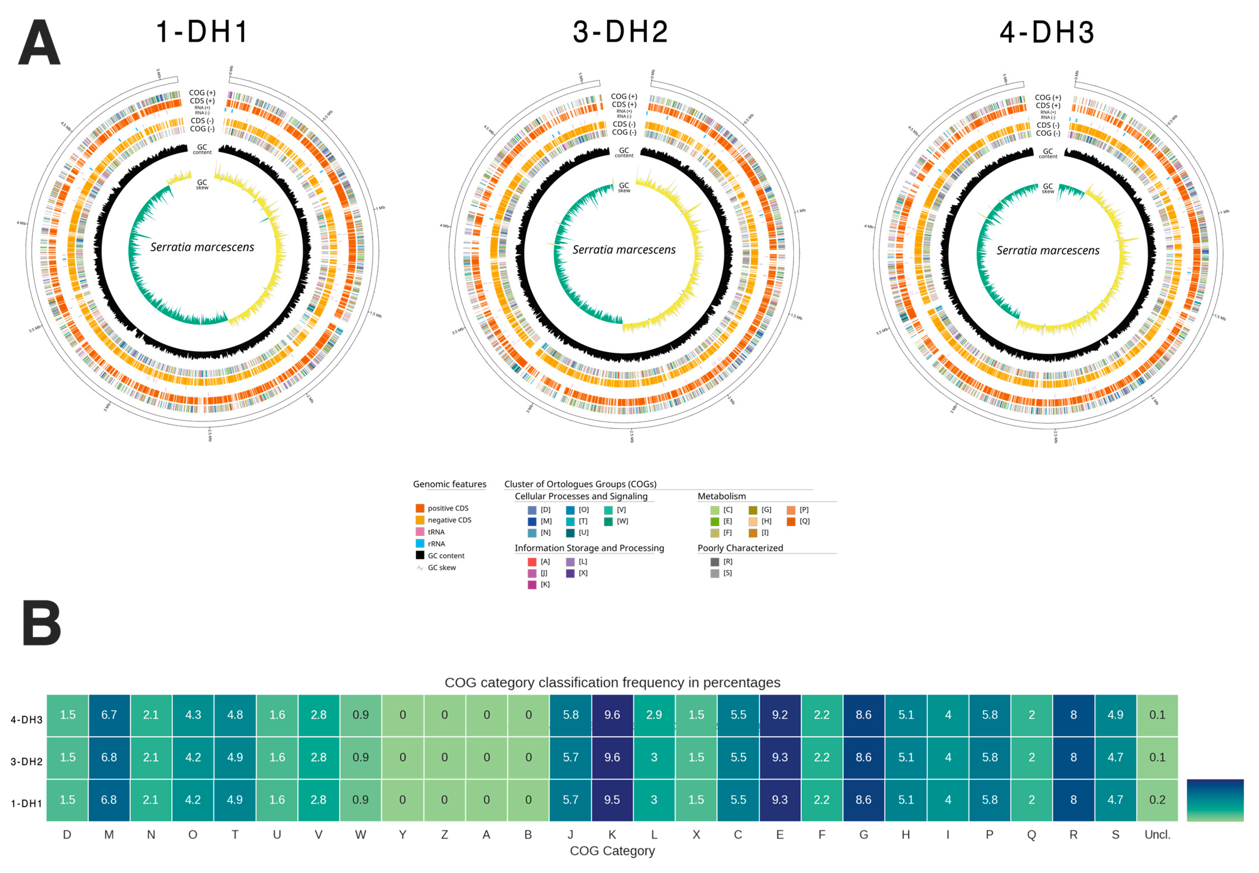
Figure 1. Serratia marcescens whole genome obtained for the three isolates, 1-DH1, 3-DH2, and 4-DH3, by WGS. (A) Whole-genome map of the three S. marcescens isolates, 1-DH1, 3-DH2, and 4-DH3. The median bacterial chromosome genome size was 5–5.1 Mb. The outer concentric circles (orange) indicate the coding sequence (CDS, +), the inner, dark yellow circle represents the CDS (-), the black circle indicates the % of GC content, and the inner circle indicates the GC skew [(G–C)/(G+C)], positive (light yellow) and negative (green). (B) Clusters of orthologous groups (COGs) of proteins category classification frequency in percentages obtained for the three isolates. The COGs obtained in the genome analysis of the three isolates by WGS could be seen in Table S1.
Antibiotics 14 00892 g001
Figure 2. Phylogenomic analysis of the three S. marcescens whole-genome generated in this study, belonging to the S. marcescens isolates 1-DH1, 3-DH2, and 4-DH3 prevenient from BM. The phylogenetic tree was constructed based on pangenome core genes, as assigned by Panaroo, using the maximum-likelihood method based on the GTR + F + I + R8 model. Serratia marcescens sequences generated in this study are highlighted in bold.
Antibiotics 14 00892 g002
Table 1. Phenotypic identification and antimicrobial susceptibility testing obtained from the four isolates from raw milk cows with BM by MicroScan WalkAway Plus.
Neg-Urine-Combo98 PanelIsolate
1-DH1
Isolate
2-DH1
Isolate
3-DH2
Isolate
4-DH3
Bacteria Identification
% of probability
S. marcescens
99.99
S. marcescens
99.99
S. odorifera
94.66
S. marcescens
99.99
Antimicrobials
β-lactam
Amoxicillin-clavulanate acid-E (Aug-E)R * (>32.00)R * (>32.00)R * (>32.00)R * (32.00)
Ampicillin (AM)R * (>8.00)R * (>8.00)R * (>8.00)R * (>8.00)
Cefepime (Cpe)S * (≤1.00)S * (≤1.00)S * (≤1.00)S * (≤1.00)
Cefotaxime (Cft) S * (<1.00)S * (<1.00)S * (<1.00)S * (<1.00)
Ceftazidime-clavulanate acid (Caz/CA)S * (≤0.25)S * (≤0.25)S * (≤0.25)S * (≤0.25)
Ertapenem (Etp)S * (≤0.12)S * (≤0.12)S * (≤0.12)S * (≤0.12)
Imipenem (Imp)S * (≤2.00)S * (≤2.00)S * (≤2.00)S * (≤2.00)
Meropenem (Mer)S * (≤0.12)S * (≤0.12)S * (≤0.12)S * (≤0.12)
Quinolone
Nalidixic Acid (NA)S * (≤0.16)S * (≤0.16)S * (≤0.16)S * (≤0.16)
Ciprofloxacin (Cp)S * (0.25)S * (0.25)S * (0.25)S * (≤0.06)
Levofloxacin (Lvx)S * (≤0.50)S * (≤0.50)S * (≤0.50)S * (≤0.50)
Norfloxacine (Nxn)S * (≤0.50)S * (≤0.50)S * (≤0.50)S * (≤0.50)
Aminoglycoside
Amikacin (AK)S * (≤8.00)S * (≤8.00)S * (≤8.00)S * (≤8.00)
Gentamicin (Gm)S * (≤0.20)S * (≤0.20)S * (≤0.20)S * (≤0.20)
Tobramycin (To)R * (4.00)R * (4.00)R * (4.00)R * (≤0.20)
Sulphonamide
Trimethoprim-Sulfamethoxazole (T/S)S * (≤2/38)S * (≤2/38)S * (≤2/38)S * (≤2/38)
Monobactam
Aztreonam (AZT)S * (≤1.00)S * (≤1.00)S * (≤1.00)S * (≤1.00)
Cephalosporin
Cefoxitin (Cfx)R * (≤8.00)R * (≤8.00)S * (≤8.00)R * (≤8.00)
Ceftazidime (Caz)S * (≤1.00)S * (≤1.00)S * (≤1.00)S * (≤1.00)
Cefuroxime (Crm)R * (>8.00)R * (>8.00)R * (>8.00)R * (>8.00)
Polymyxin
Colistin (Cl)R * (>4.00)R * (>4.00)R * (>4.00)R * (>4.00)
Phosphonic
Fosfomycin (Fos)S * (≤8.00)S * (≤8.00)S * (≤8.00)S * (≤8.00)
Nitrofuran
Nitrofurantoin (Fd)R * (>64.00)R * (>64.00)R * (>64.00)R * (>64.00)
Cephalosporin/β-lactam
Cefotaxime-clavulanate acid (Cft/CA)S * (≤0.25)S * (≤0.25)S * (≤0.25)S * (≤0.25)
Penicillin/β-lactam
Piperacillin-Tazobactam (P/T)S * (≤8.00)S * (≤8.00)S * (≤8.00)S * (≤8.00)
Total25252525
% Susceptible72.0072.0076.0072.00
% Intermediate0.000.000.000.00
% Resistant28.0028.0024.0028.00
DH (Dairy herd), S (Susceptible), R (Resistant), * (Minimum inhibitory concentrations (MICs) achieved in this study).
Table 2. Genome characterization obtained for the three isolates, 1-DH1, 3-DH2, and 4-DH3, by WGS.
IsolateLength (bp)Bacterial Species% Identity
1-DH15,083,761NZ_JGVB01000001.1 Serratia marcescens strain ATCC 14041 contig00199.30
3-DH25,083,758NZ_JGVB01000001.1 Serratia marcescens strain ATCC 14041 contig00199.30
4-DH35,082,903NZ_JGVB01000001.1 Serratia marcescens strain ATCC 14041 contig00199.30
DH (Dairy herd), bp (Base pair).
Table 3. Antimicrobial resistance and VF genes identified in the S. marcescens whole-genome sequences of the three isolates, 1-DH1, 3-DH2, and 4-DH3, by ABRicate (BLAST) using CARD, ResFinder and NCBI, and VFDB databases, respectively.
Database CARD NCBI ResFinder* AMR in CARD, NCBI or ResFinder
AMR Gene/Isolate 1 3 4 1 3 4 1 3 4
AAC(6′)-Icxxx Aminoglycoside
aac(6′)-Ic_1 xxxAmikacin; tobramycin
aac(6′)-Ial xxx Aminoglycoside
H-NSxxx Cephalosporin, fluoroquinolone, macrolide, penicillin, tetracycline, cephamycin
tet(41)xxxxxxxxxAcycline, doxycycline, tetracycline
SRT-2xxx Cephalosporin, cefotaxime
oqxBxxx Diaminopyrimidine, fluoroquinolone, glycylcycline, nitrofuran, tetracycline
oqxB_1 xxxNalidixic acid; ciprofloxacin
oqxB25 xxx Phenicol, quinolone
mexIxxx Acridine_dye, fluoroquinolone, tetracycline
CRPxxx fluoroquinolone, macrolide, penicillin
blaSST-1 xxx Cephalosporin
blaSST-1_1 xxxN/A
Database VFDB ** VF in VFDB
VF gene/Isolate 1 3 4
fliG x x x Flagellar motor switch protein G
fliM x x x Flagellar motor switch protein FliM
fliP x x x Flagellar biosynthetic protein FliP
flgH x x x Flagellar L-ring protein precursor FlgH
AMR (Antimicrobial resistance), VF (Virulence factor), x (Gene presented), N/A (Not available), CARD (Comprehensive Antibiotic Resistance Database), NCBI (National Center for Biotechnology Information), * AMR genes results obtained in CARD, NCBI, or ResFinder could be seen in Table S2, ** VF genes results obtained in VFDB (Virulence Factor Database) could be seen in Table S2.
Disclaimer/Publisher’s Note: The statements, opinions and data contained in all publications are solely those of the individual author(s) and contributor(s) and not of MDPI and/or the editor(s). MDPI and/or the editor(s) disclaim responsibility for any injury to people or property resulting from any ideas, methods, instructions or products referred to in the content.