Article

Streamlining Bacillus Strain Selection Against Listeria monocytogenes Using a Fluorescence-Based Infection Assay Integrated into a Multi-Tiered Validation Pipeline

by
Blanca Lorente-Torres
1,
Pablo Castañera
1,
Helena Á. Ferrero
1,
Sergio Fernández-Martínez
1,
Suleiman Adejoh Ocholi
2,
Jesús Llano-Verdeja
1,
Farzaneh Javadimarand
1,
Yaiza Carnicero-Mayo
1,
Amanda Herrero-González
3,
Alba Puente-Sanz
3,
Irene Sainz Machín
1,
Isabel Karola Voigt
1,
Silvia Guerrero Villanueva
1,
Álvaro López García
1,
Eva Martín Gómez
1,
James C. Ogbonna
2,
José M. Gonzalo-Orden
1,
Jesús F. Aparicio
1,
Luis M. Mateos
1,4,
Álvaro Mourenza
1,* and
Michal Letek
1,5,*
add Show full author list remove Hide full author list
1
Departamento de Biología Molecular, Área de Microbiología, Universidad de León, 24071 León, Spain
2
Department of Microbiology, University of Nigeria, University Road 1, Nsukka 410105, Nigeria
3
Neural Therapies SL. Edif. Institutos de Investigación, Planta baja, Local B43. Campus de Vegazana s/n, 24071 León, Spain
4
Instituto de Biología Molecular, Genómica y Proteómica (INBIOMIC), Universidad de León, 24071 León, Spain
5
Instituto de Desarrollo Ganadero y Sanidad Animal (INDEGSAL), Universidad de León, 24071 León, Spain
*
Authors to whom correspondence should be addressed.
Antibiotics 2025, 14(8), 765; https://doi.org/10.3390/antibiotics14080765
Submission received: 22 June 2025 / Revised: 25 July 2025 / Accepted: 26 July 2025 / Published: 29 July 2025

Abstract

Background/Objectives: Listeria monocytogenes is a foodborne pathogen of major public health concern due to its ability to invade host cells and cause severe illness. This study aimed to develop and validate a multi-tiered screening pipeline to identify Bacillus strains with probiotic potential against L. monocytogenes. Methods: A total of 26 Bacillus isolates were screened for antimicrobial activity, gastrointestinal resilience, and host cell adhesion. A fluorescence-based infection assay using mCherry-expressing HCT 116 cells was used to assess cytoprotection against L. monocytogenes NCTC 7973. Eight strains significantly improved host cell viability and were validated by quantification of intracellular CFU. Two top candidates were tested in a murine model of listeriosis. The genome of the lead strain was sequenced to evaluate safety and biosynthetic potential. Results: B. subtilis CECT 8266 completely inhibited intracellular replication of L. monocytogenes in HCT 116 cells, reducing bacterial recovery to undetectable levels. In vivo, it decreased splenic bacterial burden by approximately 6-fold. Genomic analysis revealed eight bacteriocin biosynthetic clusters and silent antibiotic resistance genes within predicted genomic islands, as determined by CARD and Alien Hunter analysis. The strain also demonstrated bile and acid tolerance, as well as strong adhesion to epithelial cells. Conclusions: The proposed pipeline enables efficient identification of probiotic Bacillus strains with intracellular protective activity. B. subtilis CECT 8266 is a promising candidate for translational applications in food safety or health due to its efficacy, resilience, and safety profile.

1. Introduction

Listeria monocytogenes is a Gram-positive, facultative intracellular pathogen responsible for listeriosis, a severe foodborne disease with high mortality, particularly among immunocompromised individuals, pregnant women, and the elderly [1,2,3,4]. The ability of L. monocytogenes to replicate at refrigeration temperatures, tolerate high salt concentrations, and form biofilms underlies its persistence in food production environments and contributes to contamination of ready-to-eat products [5,6,7,8,9,10,11]. As a result, there is a growing interest in identifying bacterial strains or derived compounds that can actively prevent L. monocytogenes invasion and replication within host cells [12]. However, the lack of predictive, high-throughput functional assays has limited the efficiency with which such candidates can be identified and prioritised for translational applications.
Among candidate genera, Bacillus spp. have emerged as promising antagonists of L. monocytogenes due to their ability to produce diverse antimicrobial compounds, tolerate gastrointestinal conditions, and colonise mucosal surfaces [13,14,15,16,17,18]. Strains belonging to Bacillus spp. are particularly attractive for food and health-related applications, in part because of their Generally Recognised as Safe (GRAS) status and robustness under industrial and physiological stressors [19,20,21]. Numerous studies have reported bacteriocins and lipopeptides from Bacillus strains with potent anti-Listeria activity [10,22,23,24,25]. However, most of these findings are based on agar diffusion assays or bacteriocin purification protocols, which provide limited insight into functional efficacy within host-associated contexts. Crucially, the capacity of Bacillus strains to prevent host cell invasion by L. monocytogenes has rarely been assessed using standardised, predictive infection models. Recent studies have explored host cell-based screening methods to evaluate the efficacy of probiotic and biocontrol candidates against foodborne pathogens in vitro [13,26].
In this study, we developed and validated an integrated pipeline to identify Bacillus strains with protective activity against L. monocytogenes. Starting from a diverse panel of isolates, we applied sequential selection criteria including in vitro antimicrobial activity, survival under simulated gastrointestinal conditions, and host cell adhesion. A fluorescence-based infection model was then used as a rapid screening tool to identify strains that confer intracellular protection, with results corroborated by traditional CFU enumeration in a subset of candidates. The most promising strains were further tested in a murine challenge model, and genomic analysis was performed to assess bacteriocin potential and safety attributes. This stepwise approach streamlines the selection of probiotic Bacillus strains by ensuring functional efficacy, safety, and regulatory compliance of the candidates.

2. Results

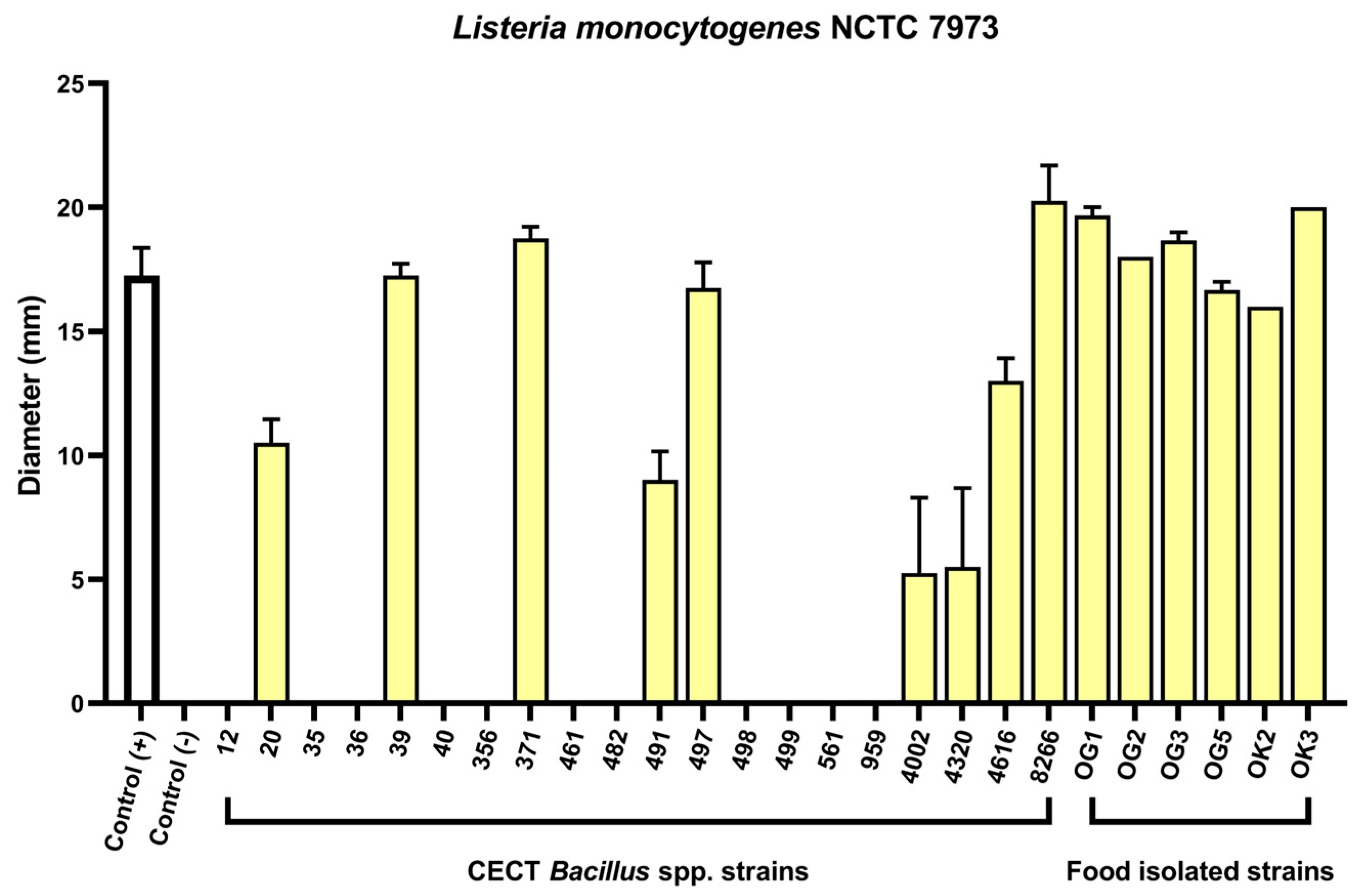
2.1. Agar Well Diffusion Screening of Bacillus spp. Reveals Anti-Listeria Activity

The antimicrobial activity of 20 Bacillus spp. strains from the CECT collection (Table S1) was evaluated using agar well diffusion assays with cell-free culture supernatants, thereby focusing on secreted bioactive compounds. Supernatants were tested against a panel of Gram-positive and Gram-negative pathogens, including Staphylococcus aureus, Salmonella enterica serovar Typhimurium, and Listeria monocytogenes (Figure 1 and Figure S1). Twelve strains showed inhibitory activity against at least one target organism, with nine exhibiting clear zones of inhibition against L. monocytogenes NCTC 7973 (Figure 1). To expand the strain repertoire, we screened eight African fermented foods, recognised as rich microbial reservoirs [27]. Four products—Ogiri, Iru, Okpeye, and Ukpaka—yielded 22 Gram-positive, spore-forming rod-shaped isolates. Among them, six strains, all isolated from Ogiri (OG) or Okpeye (OK), produced antimicrobial compounds effective against L. monocytogenes NCTC 7973 (Figure 1). These results allowed the initial selection of strains exhibiting strong anti-listerial potential for further testing in gastrointestinal-like conditions.

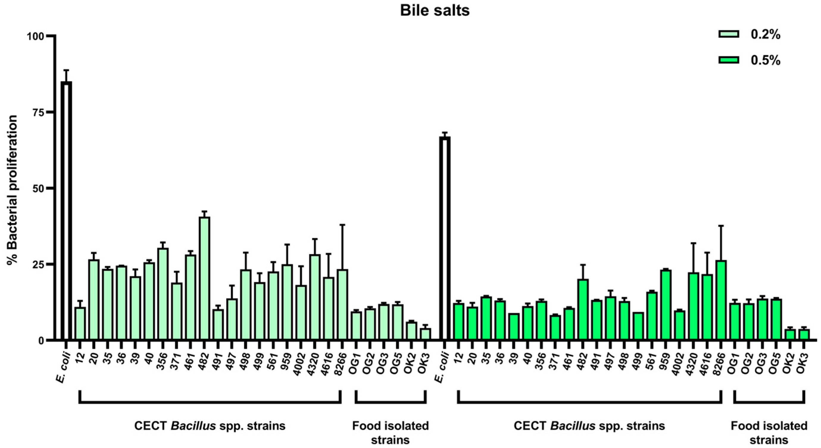
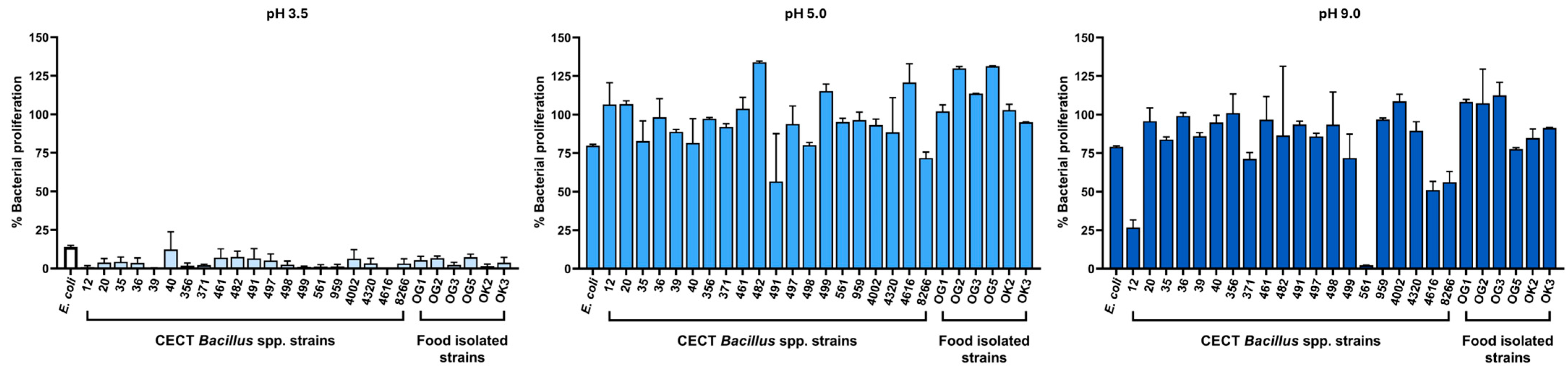
2.2. Tolerance to Gastrointestinal Stressors: Bile Salts and pH

Although Bacillus endospores are known for their resilience [28], the ability of vegetative cells to tolerate gastrointestinal stressors is a more relevant indicator of in situ activity. We therefore assessed the capacity of all Bacillus spp. strains to grow in the presence of bile salts (up to 0.5%) and at acidic pH values (as low as pH 3.5) to simulate small intestinal conditions. In adults, bile salt concentrations in the duodenum can reach ~10 mM (≈0.46% w/v) postprandially, while pH increases rapidly from ~3 in the stomach to ~7 in the upper small intestine, reflecting a major physicochemical transition relevant for probiotic survival [29].
As expected, bile salts inhibited vegetative growth to varying degrees. Notably, B. subtilis CECT 8266 displayed the highest tolerance among CECT-derived strains, maintaining measurable growth even at 0.5% bile salts (Figure 2). In contrast, several food-derived strains, including OK2 and OK3, were more susceptible to bile stress.
Acid tolerance was evaluated by monitoring bacterial growth at pH values ranging from 3.5 to 9.0. Most strains grew optimally between pH 5.0 and 7.0, with reduced proliferation observed at pH 9.0 and a marked decrease at pH 3.5 (Figure 3). Among the tested strains, B. subtilis CECT 40 showed the broadest pH tolerance, although several other isolates—including CECT 8266—also sustained growth under acidic conditions.
Thirteen strains that exhibited both anti-Listeria activity and resilience to pH and bile salt stress were selected for downstream functional evaluation using host cell-based infection assays. All food-derived isolates chosen for further analysis were taxonomically identified at this stage as B. subtilis based on 16S rDNA and tuf gene sequencing (Figures S2 and S3).

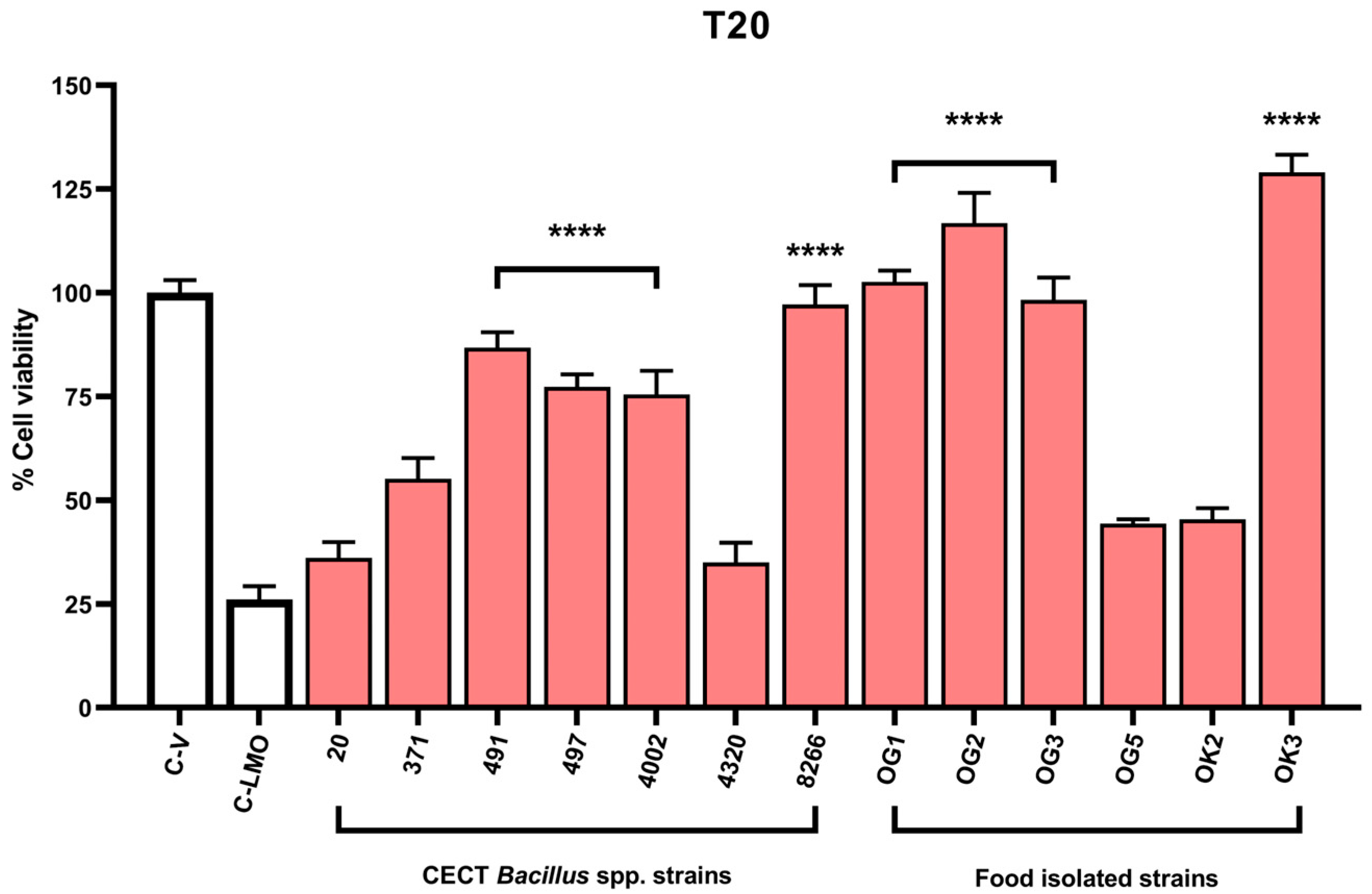
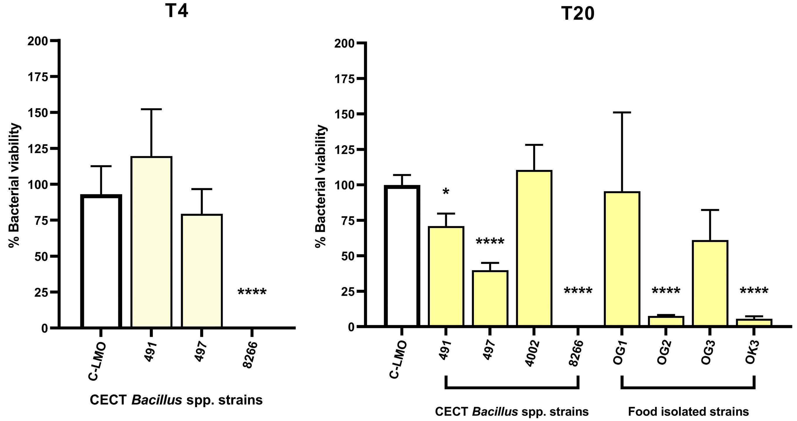
2.3. A Fluorescence-Based Infection Assay Determines Host Cell Protection Against L. monocytogenes

To streamline the identification of protective strains, a fluorescence-based infection model was used to screen preselected candidates for their ability to protect host cells from L. monocytogenes–induced cytotoxicity. We applied a high-throughput, fluorescence-based assay using HCT 116 human intestinal epithelial cells transduced for constitutive expression of the red fluorescent protein mCherry. This system enables the direct quantification of host cell viability following bacterial challenge, independent of host metabolism, which can be compromised during infection and lead to unreliable results in conventional assays based on the reduction of MTT or lactate dehydrogenase release [30,31].
We first validated the assay by correlating mCherry fluorescence intensity with MTT-derived viability measurements across varying cell densities. A strong linear correlation was observed (Figure 4; correlation coefficient = 0.92), confirming that mCherry fluorescence is a reliable proxy for cell viability under our experimental conditions.
Next, we applied the assay to assess whether pre-exposure to Bacillus strains conferred protection to mCherry-HCT 116 cells infected with L. monocytogenes. Thirteen strains were tested, and mCherry fluorescence was measured at 20 h post-infection as an indicator of host cell survival. Eight strains significantly improved cell viability compared to infected, untreated controls (C-LMO), indicating effective protection against pathogen-induced cytotoxicity (Figure 5). Notably, five of these strains—despite showing strong antimicrobial activity in agar diffusion assays—failed to protect host cells, suggesting that in vitro antimicrobial activity alone is not a sufficient predictor of functional efficacy in infection models.
To validate the performance of the fluorometric assay, we quantified intracellular bacteria via CFU enumeration for the top eight candidates. B. subtilis CECT 8266 completely inhibited intracellular bacterial replication at both 4- and 20-h post-infection (Figure 6), indicating potent blockade of pathogen internalisation or early intracellular survival. B. subtilis OK3 also reduced intracellular burden, but the effect was less pronounced.
Together, these results demonstrate that the fluorescence-based infection assay provides a reliable and high-throughput platform to assess the ability of Bacillus strains to protect host epithelial cells from L. monocytogenes-induced cytotoxicity. The observed differences between strains with comparable in vitro antimicrobial activity highlight the importance of functional screening in host-relevant models.

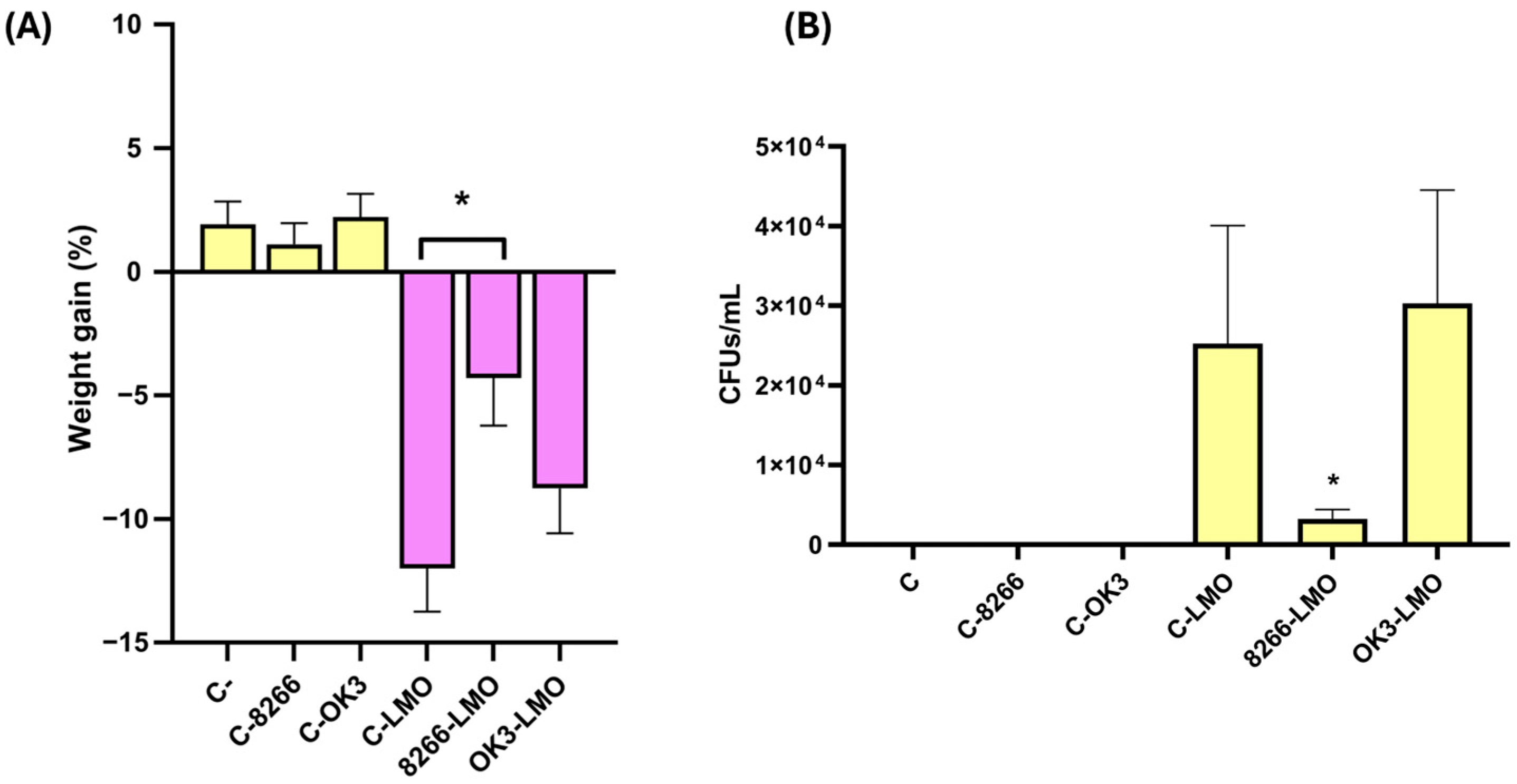
2.4. In Vivo Validation of Protective Strains in a Murine Model of Listeriosis

To evaluate whether the protective effects observed in vitro translated into host protection in vivo, we selected two top-performing strains—B. subtilis CECT 8266 and OK3—for testing in a murine model of L. monocytogenes infection. These strains were prioritised based on their consistent performance across multiple in vitro assays, as they exhibited strong inhibition zones against L. monocytogenes, demonstrated tolerance to gastrointestinal stressors, and provided substantial protection to HCT 116 enterocytes in the fluorescence-based infection assay. In addition, both B. subtilis CECT 8266 and OK3 inhibited multiple L. monocytogenes isolates in agar diffusion assays (Figure S4), suggesting a broad inhibitory spectrum that may further enhance their translational relevance.
Therefore, BALB/c mice were orally administered either B. subtilis CECT 8266, OK3, or PBS for ten consecutive days before challenge with L. monocytogenes NCTC 7973. Mice were monitored for weight loss and signs of disease, and were sacrificed 48 h post-infection for quantification of the bacterial burden in the spleen. Survival was also monitored throughout the experiment. One mouse in the infected, untreated control group died before the experimental endpoint was reached. All other animals, including those treated with B. subtilis strains, survived until necropsy. Both Bacillus-treated groups tolerated the strains well, with no weight loss or signs of discomfort observed in uninfected animals (Figure 7A), confirming the safety of these strains under the tested conditions.
In infected animals, weight loss was significantly reduced in the group treated with B. subtilis CECT 8266 compared to the infected, untreated controls (Figure 7A). Moreover, spleen homogenates from this group revealed a ~6-fold reduction in L. monocytogenes CFU counts relative to both the untreated group and the OK3-treated group (Figure 7B). In contrast, mice treated with B. subtilis OK3 exhibited no significant reduction in splenic bacterial burden, despite the moderate protection observed in vitro. These findings suggest that B. subtilis CECT 8266 confers a biologically meaningful protection against L. monocytogenes infection in vivo, as evidenced by improved survival, reduced weight loss, and lower bacterial burden.

2.5. Host Cell Adhesion and Growth Dynamics

To further explore traits that may contribute to host protection, we assessed bacterial adhesion to HCT 116 cells and compared growth kinetics across selected Bacillus strains. Adhesion was quantified by co-incubating bacteria with HCT 116 monolayers, followed by extensive washing and CFU enumeration. Both B. subtilis CECT 8266 and OK3 exhibited significantly higher adhesion to host cells compared to the type strain B. subtilis CECT 39T (Figure 8A), suggesting enhanced capacity for mucosal colonisation.
We also monitored bacterial growth in rich media by measuring absorbance at 600 nm (Abs600) over time. Growth kinetics were analysed by plotting ln(Abs600) versus time during the exponential phase and applying linear regression to estimate the specific growth rate (μ, h−1). The resulting slopes indicated similar exponential growth rates across the three strains: B. subtilis CECT 39T (μ = 1.295 ± 0.051), B. subtilis CECT 8266 (μ = 1.266 ± 0.047), and B. subtilis OK3 (μ = 1.292 ± 0.030), with no statistically significant differences observed between them.
Despite these comparable growth rates, final biomass accumulation differed. B. subtilis OK3 reached the highest absorbance during stationary phase (1.4 ± 0.1), followed by B. subtilis CECT 8266 (1.2 ± 0.0), and B. subtilis CECT 39T (0.9 ± 0.2). However, these in vitro parameters did not correlate with in vivo performance. Only B. subtilis CECT 8266 conferred protection in the murine infection model, despite similar growth rates across all strains and higher biomass in OK3. These results indicate that growth kinetics and biomass accumulation alone do not explain the protective effect of B. subtilis CECT 8266, highlighting the importance of strain-specific factors in its probiotic potential.
Previous studies have suggested that strong epithelial adhesion may support the competitive exclusion of L. monocytogenes in the gut [26]. However, in our study, adhesion capacity and growth rate were not reliable predictors of protective efficacy. For example, B. subtilis OK3 exhibited robust epithelial adhesion and rapid growth but failed to confer full protection in both in vitro and in vivo infection models.

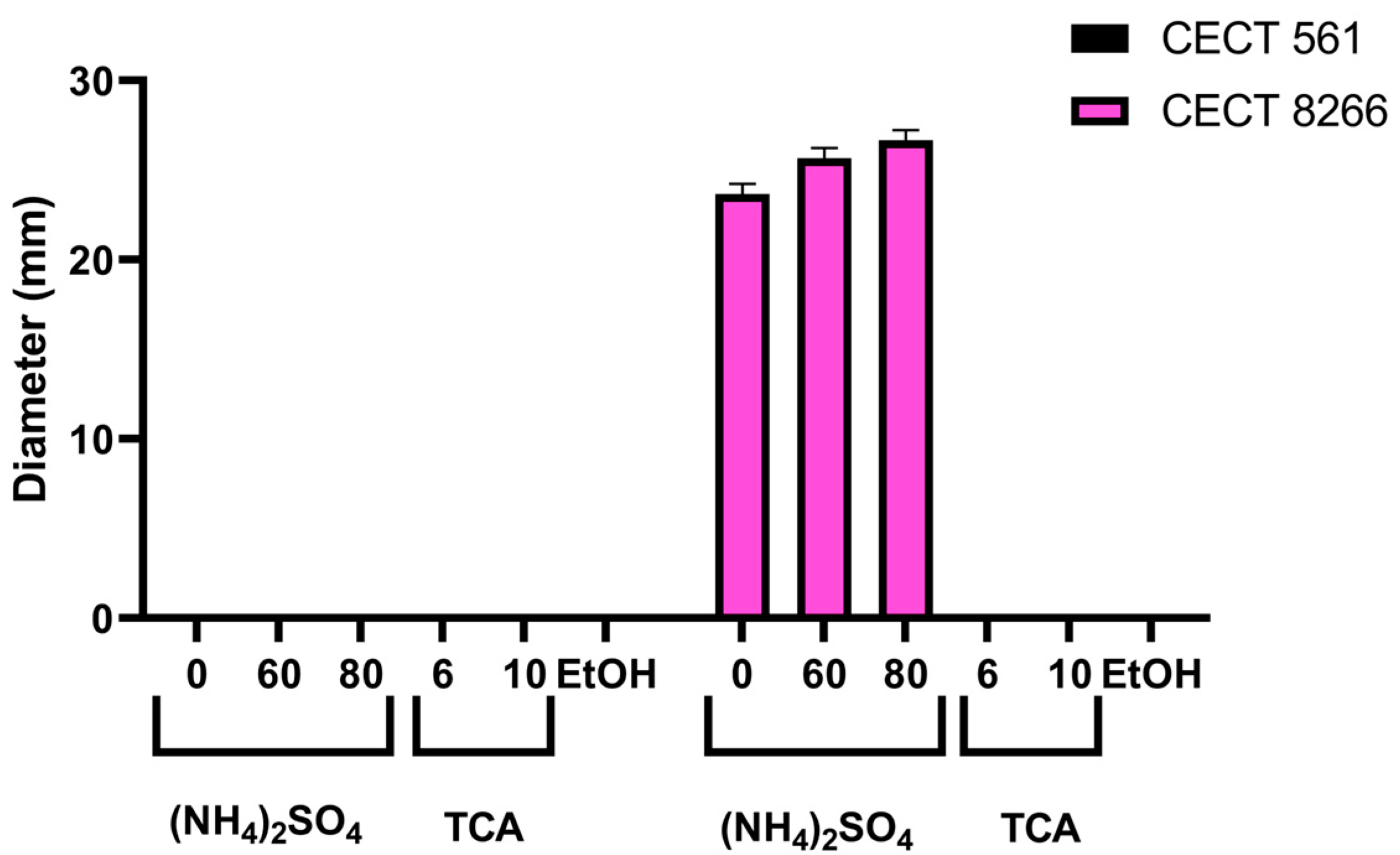
2.6. Evidence of Bacteriocin-Mediated Antimicrobial Activity in B. subtilis CECT 8266

Numerous studies have suggested that the anti-listerial activity of Bacillus strains is mediated by the production of bacteriocins [25,32,33,34,35,36,37,38,39,40]. To assess whether extracellular bacteriocins contribute to the antimicrobial activity of B. subtilis CECT 8266, we compared its secreted protein fractions with those of B. subtilis CECT 561, a strain lacking detectable activity against L. monocytogenes in agar diffusion assays and thus used as a negative control (Figure 1).
Both strains were processed in parallel following established protocols for bacteriocin purification [41,42,43]. Ammonium sulphate precipitation was selected as the initial step for recovering bacteriocin activity due to its mild, tunable conditions, which allowed for the successful recovery of bioactive peptides from B. subtilis CECT 8266. In contrast, other methods, such as trichloroacetic acid or ethanol precipitation, did not yield active fractions under our experimental conditions [38,44,45]. It is important to note that this approach enriches the proteinaceous fraction of the culture supernatant, selectively concentrating bacteriocins while excluding low-molecular-weight antimicrobial compounds that remain soluble [38,44].
Ammonium sulphate precipitation at 60% and 80% saturation successfully concentrated active compounds from B. subtilis CECT 8266 supernatants, yielding inhibition halos of 26 mm and 27 mm, respectively (Figure 9). In contrast, no antimicrobial activity was detected in equivalent fractions from B. subtilis CECT 561.
Attempts to recover active fractions using trichloroacetic acid or ethanol precipitation were unsuccessful for both strains, likely due to protein denaturation or poor solubility. These results indicate that the observed anti-listerial activity of B. subtilis CECT 8266 is due to secreted proteinaceous compounds—consistent with bacteriocins—that are stable under mild ammonium sulphate precipitation conditions but are inactivated by harsher extraction methods.

2.7. Genomic Analysis of B. subtilis CECT 8266 Reveals Bacteriocin-Encoding Genes

To better understand the molecular basis of the pronounced anti-listerial activity exhibited by B. subtilis CECT 8266 both in vitro and in vivo, we sequenced its genome (Figure S5). The resulting assembly spans 4,163,299 base pairs and includes 4145 predicted coding sequences.
AntiSMASH and BAGEL5 analyses revealed a diverse array of biosynthetic gene clusters (BGCs) associated with secondary metabolite production (Table S2). These include non-ribosomal peptide synthetase (NRPS) clusters for surfactin, fengycin, and bacillibactin biosynthesis—compounds with documented antimicrobial, antifungal, and iron-scavenging functions [46,47,48]. Additional BGCs encode bacillaene, subtilosin A, pulcherriminic acid, bacilysin, and terpenes, many of which have demonstrated antibacterial activity [17,49,50,51]. Several terpene BGCs were also detected, suggesting the potential for volatile bioactive compound production [52]. Furthermore, a Type III polyketide synthase (T3PKS) gene cluster of unknown function was present.
All of these genomic features are part of the Bacillus pangenome, which suggests that the potent anti-listerial activity of B. subtilis CECT 8266 is not due to the presence of novel or unique bacteriocin genes. Instead, it likely results from the combined action of multiple well-characterised and conserved anti-listerial compounds—such as subtilosin A, surfactin, and fengycin [35,36,38]—whose synergistic interactions may underlie the robust phenotype observed.

2.8. Antibiotic Resistance

Antibiotic resistance must be absent for Bacillus strains to be considered safe candidates for microbial-based therapies or supplements, in line with the safety criteria established by the European Food Safety Authority (EFSA) and recent evaluations of probiotic suitability [19,53]. Therefore, we evaluated the antibiotic susceptibility of B. subtilis CECT 8266 by determining its Minimum Inhibitory Concentrations (MIC) values against a panel of EFSA-recommended antibiotics (Table S3) [53]. No resistance was observed, including to aminoglycosides and glycopeptides, two critically important classes of antibiotics. In contrast, the type strain B. subtilis CECT 39T exhibited resistance to streptomycin and clindamycin. Notably, B. subtilis OK3 exhibited resistance to all tested antibiotics except chloramphenicol, likely reflecting the historical lack of antibiotic regulation in Nigeria, which has only recently begun to be addressed [54].
To complement this phenotypic assessment, we screened the genome of B. subtilis CECT 8266 using the Comprehensive Antibiotic Resistance Database (CARD). Several genes commonly associated with antimicrobial resistance were identified (Table S4). However, these genes represent either intrinsic features widely conserved in Bacillus spp. (e.g., efflux pumps or regulatory proteins), or distant homologs of acquired resistance determinants not known to be functional in this genus. Importantly, although a subset of these genes (vanT, vanW, BcI, and aadK) were located within regions predicted to be horizontally acquired, no corresponding phenotypic resistance to vancomycin or streptomycin was detected in MIC testing. These findings indicate that these genomic features do not confer clinically relevant resistance in this context.
Alien Hunter analysis detected several horizontally acquired genomic islands, characterized by altered GC content and absent from the reference genomes (Figure S5). One of these regions, spanning positions 4,004,525 to 4,009,686, encodes several hypothetical proteins and a bacitracin transport ATP-binding protein (BcrA3). This is one of three bcrA homologs in the genome, all of which are implicated in bacitracin resistance.
Together, the combined genomic and phenotypic data confirm that B. subtilis CECT 8266 does not harbour acquired antimicrobial resistance traits of safety concern. These results meet EFSA criteria, which prioritise the absence of transferable resistance genes and phenotypic resistance to clinically relevant antibiotics.

3. Discussion

Our results demonstrate that Bacillus strains exhibit pronounced specificity against L. monocytogenes (Table S1), likely reflecting an evolutionary adaptation driven by ecological niche competition [55]. B. subtilis and L. monocytogenes frequently co-occur in soil, silage, fermented foods, and food-processing environments, where persistent cohabitation may have selected for B. subtilis strains capable of producing targeted antimicrobials such as bacteriocins and lipopeptides [56,57,58,59]. This selective pressure may explain why strains like B. subtilis CECT 8266 display strong and specific anti-listerial activity. Notably, B. subtilis can allocate up to 5% of its genome to the synthesis of secondary metabolites with antimicrobial function [60].
Interestingly, all food-derived isolates that progressed through our screening were identified as B. subtilis based on 16S rRNA and tuf gene sequencing. This aligns with its known dominance in traditional fermentations and its ability to produce antimicrobials in situ [61]. Such prevalence suggests fermented foods are a valuable reservoir of anti-Listeria Bacillus strains. While this specificity is advantageous for food safety, the limited activity against Gram-negative bacteria implies minimal disruption of the gut microbiota, which is desirable in the context of targeted probiotic applications [62].
Our stepwise screening pipeline revealed that anti-listerial activity alone was insufficient to qualify a strain as a probiotic candidate. Many isolates failed to tolerate low pH or bile salts—key barriers to gastrointestinal survival—leading to a high attrition rate, in line with previous probiotic discovery efforts [63]. Only about half of the strains met all basic functional criteria, underscoring the importance of combining antimicrobial testing with resilience assays early in the selection process.
Antibiotic susceptibility is another critical safety parameter. According to EFSA guidelines, probiotic strains must not harbour acquired resistance genes [53]. While B. subtilis OK3 showed promising antimicrobial effects, it exhibited multidrug resistance, disqualifying it as a safe candidate. In contrast, B. subtilis CECT 8266 was sensitive to all tested antibiotics. This clean profile, together with acid and bile tolerance and epithelial adhesion, made B. subtilis CECT 8266 the only isolate (~4% of our initial pool) to meet all probiotic selection criteria—consistent with previous reports indicating that true candidates emerge at low frequency [63].
Remarkably, in vitro inhibition of L. monocytogenes did not always translate to protection in host cell assays. Approximately one-third of the strains with strong agar diffusion activity failed to prevent infection in the mCherry-based fluorescence assay. This limitation is well-documented: pathogen inhibition in bacteriological assays does not necessarily reflect protection of host cells or modulation of infection outcomes [64,65]. Functional screening in physiologically relevant models is therefore essential. This approach not only refines selection but also reduces the number of strains requiring validation in animal models, aligning with the 3Rs principle (Replacement, Reduction, and Refinement) [66].
In the murine model of listeriosis, only B. subtilis CECT 8266 reduced splenic bacterial burden and mitigated weight loss. Full eradication is rare in oral Listeria challenge models [67]. Therefore, we considered a ~6-fold reduction in spleen CFUs, coupled with improved clinical signs and 100% survival, a biologically meaningful effect. Variability in organ colonisation and technical limitations in tissue homogenization may have contributed to noise in pathogen recovery, but the spleen data and weight monitoring support the protective capacity of B. subtilis CECT 8266. Notably, our protocol employed a prophylactic approach, with Bacillus administered before infection. Future studies should investigate the continuous administration of this approach to optimise efficacy.
Whole-genome sequencing of B. subtilis CECT 8266 revealed eight biosynthetic gene clusters linked to anti-listerial activity, including subtilosin A, surfactin, and fengycin [35,36,38]. These compounds likely act synergistically to produce the observed inhibition. While our extraction methods favoured proteinaceous antimicrobials such as bacteriocins, we may have missed hydrophobic metabolites like polyketides or organic acids. Further transcriptomic or metabolomic profiling will be necessary to fully characterise the active antimicrobial components.
Crucially, genome mining using CARD and Alien Hunter, combined with MIC determination, confirmed the absence of functional resistance determinants. Although genes such as vanT, vanW or aadK, were detected, none conferred phenotypic resistance to vancomycin, streptomycin or kanamycin—suggesting they are either non-functional or not expressed, a finding consistent with reports in other bacteria [68]. This supports the regulatory safety profile of B. subtilis CECT 8266.
As a spore-former, B. subtilis CECT 8266 is suitable for oral administration and formulation as a stable supplement or food additive [69,70,71]. Given its genetic stability, the strain could be further improved through synthetic biology—for instance, by overexpressing antimicrobial biosynthetic genes or deleting latent resistance loci—to enhance both efficacy and safety.

4. Materials and Methods

4.1. Bacterial Strains and Culture Conditions

We screened twenty Bacillus spp. strains from the Colección Española de Cultivos Tipo (CECT) for attributes linked to gut colonisation and antimicrobial activity; genome data are publicly available for four of them (Table S1). The L. monocytogenes strains used in this study included NCTC 7973 (reference strain used for infection models), ATCC 7644, CECT 911 (ATCC 19112; serotype 1/2c, cerebrospinal fluid isolate), CECT 940 (ATCC 19117; sheep isolate), CECT 4031 (ATCC 15313; rabbit isolate), and ST4C (serotype 4c; poultry isolate). Strain information and origin are detailed in Table S1.
All these strains were cultured on Trypticase Soy Agar or Broth (TSA or TSB; Biokar Diagnostics, Solabia Group, Paris La Défense, France) and incubated at 37 °C, with shaking at 200 rpm for liquid cultures. All remaining bacterial strains used here were cultured on Brain Heart Infusion (BHI) Agar or Broth (Condalab, Laboratorios Conda S.A., Torrejón de Ardoz, Madrid, Spain), also at 37 °C and shaking at 200 rpm for liquid cultures, as previously reported [72].
We also expanded our collection by including additional strains sourced from eight traditional Nigerian fermented foods and condiments: Ogiri, Iru, Okpeye, Ukpaka, Garri, Fufu, Owo, and Fura (Table S1). For this, one gram of each solid sample was mixed with 9 mL of distilled water and thoroughly shaken. The mixtures were then heated in a water bath at 80 °C for 15 min to eliminate vegetative (non-spore-forming) bacteria. After heat treatment, 100 μL of each suspension was spread onto TSA plates and incubated at 37 °C to promote bacterial growth. Colonies with distinct morphologies were carefully selected and subcultured for further analysis. The identity of Bacillus species among the isolates was confirmed by Gram staining, followed by sequencing of the 16S rDNA and tuf genes (see Section 4.10).
Growth curves of B. subtilis strains were obtained by culturing the bacteria in BHI medium using 96-well U-bottom plates. Each well was inoculated with a 1:1000 dilution of an overnight culture adjusted to an absorbance measured at a wavelength of 600 nm (Abs600) of 1. Plates were incubated at 37 °C with continuous shaking in a Victor Nivo plate reader (Revvity, Waltham, MA, USA). Abs600 was recorded hourly over 24 h to monitor bacterial growth.
Bacterial growth was monitored by measuring absorbance at 600 nm (Abs600) at regular time intervals using a spectrophotometer. To determine the specific growth rate (μ, in h−1), a semi-logarithmic approach was applied. Absorbance values were transformed to their natural logarithm (ln Abs600), and the exponential growth phase for each strain was visually identified. A simple linear regression was applied to the ln-transformed Abs600 values as a function of time (t, in hours), using the equation: ln(Abs600) = μ × t + b, where μ is the slope of the line (specific growth rate) and b is the intercept. The exponential growth phase was defined as follows: for B. subtilis CECT 39T and B. subtilis CECT 8266, it was between 5 and 8 h; for B. subtilis OK3, it was between 2 and 9 h.

4.2. Agar-Well Diffusion Tests and Determination of the Minimum Inhibitory Concentrations

The antimicrobial activity of the selected Bacillus spp. strains was evaluated against various bacteria using the agar-well diffusion method. This assay was conducted according to the guidelines provided by the Clinical and Laboratory Standards Institute (CLSI) for antimicrobial susceptibility testing [73], with some modifications. The day before the assay, Bacillus spp. strains were cultured in TSB. On the day of the assay, 2 mL of each culture were collected, centrifuged in Eppendorf tubes at maximum speed for 10 min, and the supernatants were filtered through 0.45 µm pore filters using 5 mL syringes, except for B. subtilis CECT 39T and CECT 371, which had to be filtered with 0.22 µm pore filters to prevent bacterial growth in the borders of the wells. The filtrates were transferred to sterile Eppendorf tubes for immediate use. Although samples could be stored at 4 °C for up to 24 h without significant loss of activity, prolonged storage resulted in a marked reduction of antimicrobial efficacy. Notably, freezing the cell-free supernatants completely abolished their antimicrobial activity.
A range of bacterial strains was prepared for the assay by embedding them as test strains in agar plates. Test strains were cultured in TSB the day before and harvested at an Abs600 in the range of 0.8 to 1.2. The final volume added to the bioassay medium was adjusted to an Abs600 of 0.03. Then, TSA was melted and cooled to around 45 °C and 2 mL of the TSB medium containing the test strain was added to 100 mL of TSA agar, mixed thoroughly, and poured into four 100 mm Petri dishes. Once solidified, equidistant wells were made in the agar using the mouth of a sterile Pasteur pipette, and the plugs were removed with sterile toothpicks. Each well was filled with 35 µL of the Bacillus spp. cell-free supernatants, and the plates were incubated at 37 °C for 24 h. The plates were then examined for the presence of inhibition halos, and their diameters were measured with a calliper.
The MIC of a selected panel of EFSA-recommended antibiotics was determined following the CLSI guidelines [73]. Briefly, Bacillus spp. strains were cultured in Mueller-Hinton broth to the exponential growth phase (Abs600 = 1), then diluted to a final concentration of 2 × 10⁵ cells per 100 µL in each well of 96-well microtiter plates. Serial dilutions of the antibiotics were prepared in triplicate, and the plates were incubated at 37 °C for 16 h. Each experiment included a negative control containing only Mueller-Hinton broth.

4.3. Biliary Salts and pH Tests

Vegetative growth of Bacillus spp. strains was assessed under varying pH conditions (3.5, 5.0, 7.0, and 9.0) and bile salt concentrations (0–0.5%). For each condition, 5 µL of overnight cultures were inoculated into 96-well plates containing 95 µL of TSB adjusted accordingly. Cultures were incubated at 37 °C with shaking for 24 h, and growth was quantified by measuring absorbance at 600 nm using a Victor Nivo plate reader. Values were normalised against blank wells containing only media and compared to control conditions (pH 7.0 or TSB without bile salts).

4.4. Cellular Lines and Culture Conditions

Human epithelial colon HCT 116 cells (CCL-247, American Type Culture Collection, Manassas, VA, USA) were cultured in 100 mm cell culture plates with Dulbecco’s Modified Eagle’s Medium (DMEM; Gibco, Thermo Fisher Scientific, Waltham, MA, USA) supplemented with pyruvate, glucose, glutamine, 10% heat-inactivated foetal bovine serum (FBS; Gibco), and 5% penicillin-streptomycin solution (Corning, NY, USA). HCT 116 cells expressing constitutively mCherry were generated by transduction with the p12-MMP-mCherry vector as described previously [74]. To ensure selective growth, hygromycin (Sigma-Aldrich, MilliporeSigma, St. Louis, MO, USA) was added to the medium to eliminate non-transduced cells. The cells were maintained at 37 °C in a 5% CO2 atmosphere. The expression of mCherry was confirmed by measuring fluorescence in a Victor Nivo plate reader with an excitation filter of 580 nm and an emission filter of 625 nm [75].

4.5. Cell Viability Assay

HCT 116 cells expressing mCherry were seeded at varying densities in black-walled, clear-bottom 96-well plates containing complete DMEM. The plate was incubated at 37 °C with 5% CO2 for 24 h and then washed twice with phosphate-buffered saline (PBS; Gibco, Thermo Fisher Scientific, Waltham, MA, USA). The mCherry fluorescence was measured as described above. Then, PBS was replaced with 100 µL of DMEM containing antibiotics, and 10 µL of tetrazolium dye 3-(4,5-dimethylthiazol-2-yl)-2,5-diphenyltetrazolium bromide (MTT) solution (Alfa Aesar, Thermo Fisher Scientific, Ward Hill, MA, USA) was added to each well. The plate was incubated for 4 h at 37 °C with 5% CO2. After incubation, 100 µL of isopropanol (Fisher BioReagents, Thermo Fisher Scientific, Pittsburgh, PA, USA) with 0.04 N HCl (Fisher Chemical, Thermo Fisher Scientific, Waltham, MA, USA) was added to each well, and the plate was incubated for an additional hour. Absorbance was then measured at 570 nm with a reference wavelength of 630 nm using a Victor Nivo plate reader.

4.6. Infection Assay

HCT 116 cells were seeded in 96-well plates (8 × 10⁴ cells per well) with complete DMEM medium (without antibiotics) and incubated O/N at 37 °C with 5% CO2. On the day of the assay, the medium was replaced with 50 µL of antibiotic-free media containing Bacillus spp. strains, adjusted to a final concentration of 8 × 10⁵ CFUs per well. The plate was centrifuged at 800× g for 5 min, then incubated at 37 °C with 5% CO2 for 2 h. Following this incubation, the cells were washed with PBS and subsequently infected with L. monocytogenes at a multiplicity of infection (MOI) of 0.1. For this, 150 µL of antibiotic-free media containing the adjusted concentration of L. monocytogenes was added to each well. The plate was centrifuged again and incubated under the same conditions. After the incubation period, the medium was replaced with complete DMEM containing gentamicin sulphate (100 µg/mL; MP Biomedicals, Santa Ana, CA, USA) to eliminate extracellular bacteria. The fluorescence of viable mCherry-expressing cells was measured as described above at 20 h post-infection.
To determine the number of intracellular CFUs of L. monocytogenes, HCT 116 cells were seeded in a 24-well plate at a density of 2.4 × 10⁵ cells per well and incubated overnight at 37 °C with 5% CO2. On the day of the assay, the medium was replaced with 500 µL of media containing Bacillus spp. strains, adjusted to a concentration of 2.4 × 10⁶ CFUs per well. The plate was centrifuged at 800× g for 5 min and then incubated at 37 °C with 5% CO2 for 2 h. Following incubation, the cells were washed with PBS, and 500 µL of antibiotic-free DMEM containing L. monocytogenes, adjusted to an MOI of 0.1, was added to each well. The plate was centrifuged and incubated as previously described. After incubation with Bacillus strains and L. monocytogenes, the medium was replaced with complete DMEM containing gentamicin (100 µg/mL) to kill any extracellular bacteria. At 4 and 20 h post-infection, the cells were washed with PBS and lysed with 100 µL of 0.1% Triton X-100 (Sigma-Aldrich, MilliporeSigma, St. Louis, MO, USA) for 3 min at room temperature. After lysis, 900 µL of PBS was added to each well. The lysates were then serially diluted and plated on BHI agar for CFU counting.

4.7. Adhesion Assay

HTC 116 cells were seeded in 24-well plates at a density of 1.5 × 10⁵ cells per well in complete DMEM. On the day of the experiment, Bacillus spp. strains, previously cultured under standard conditions, were washed and resuspended in antibiotic-free DMEM. Bacterial suspensions were adjusted to an OD600 of 1.0. Cell monolayers were gently washed with PBS before adding 1 mL of a 1:10 dilution of the OD-normalised bacterial suspension (~1 × 10⁷ CFU/well; MOI ~10). Untreated wells served as negative controls. After one hour of incubation at 37 °C, the medium was aspirated, and wells were washed three times with PBS. Cells were lysed with 0.1% Triton X-100, and lysates were resuspended in 1 mL of PBS. Serial dilutions were plated on TSA to determine the number of adherent Bacillus spp. by CFU enumeration.

4.8. Evaluation of Protective Effects of Bacillus Strains in a Mouse Model of L. monocytogenes Infection

Forty-eight female BALB/c mice, 6–8 weeks old, were obtained from Charles River (France). Mice were housed in Type III cages in a dedicated biosecurity level 2 room. Cages were environmentally enriched with paper and cardboard, and food and water were provided ad libitum. The room was maintained under a 12-h light/dark cycle, with a relative humidity of 50–60% and a temperature of 20–25 °C. Mice were allowed to acclimatize for five days before the experiment and were randomly assigned to six groups, with eight mice per group. The experimental groups consisted of a PBS control, two groups administered B. subtilis CECT 8266 or B. subtilis OK3, and three matching groups that were subsequently challenged with L. monocytogenes NCTC 7973.
B. subtilis strains CECT 8266 and OK3 were cultured in TSB for 24 h. Cells were harvested by centrifugation, washed three times with PBS (pH 7.4), and resuspended to a final concentration of 5 × 10¹¹ CFU/mL. A 100 µL suspension containing 5 × 10¹⁰ CFUs was administered orally by gavage once daily for 10 days to the designated groups, while the control groups received 100 µL of PBS.
L. monocytogenes NCTC 7973 was cultured in BHI for 12 h and prepared to a concentration of 5 × 10¹⁰ CFU/mL for BALB/c mice. On day 16, after a 5-h fasting period, 100 µL of a L. monocytogenes suspension containing 5 × 10⁹ CFUs was administered orally via gavage to the infected groups (namely control, CECT 8266, and OK3). Uninfected control groups received 100 µL of PBS.
Mice were monitored daily for weight loss, physical appearance, and gastrointestinal disturbances. Humane endpoints were established, including a weight loss exceeding 20%, signs of severe dehydration, or persistent diarrhoea lasting more than 48 h. All mice were sacrificed 48 h post-infection (day 18) using cervical dislocation. Following euthanasia, aseptic necropsies were performed. Spleens were collected and homogenised in 9 mL of PBS supplemented with 0.1% Triton X-100 (Sigma-Aldrich, MilliporeSigma, St. Louis, MO, USA). The number of CFUs in each organ was determined using Modified Oxford Agar plates, a selective medium for L. monocytogenes (Condalab, Laboratorios Conda S.A., Torrejón de Ardoz, Madrid, Spain) that was supplemented with an Oxford Listeria Selective Supplement (Condalab, Laboratorios Conda S.A., Torrejón de Ardoz, Madrid, Spain).

4.9. Purification of Bacteriocins

Purification of bacteriocins was performed following a previously described protocol using ammonium sulphate [41,42,43], with some modifications. Bacillus strains were cultured in 100 mL of TSB at 37 °C for 16 h with shaking. Cells were removed from the medium by centrifugation at 8210× g for 5 min. The cell-free supernatant was filtered through a 0.22 µm filter and subjected to protein precipitation using different agents: trichloroacetic acid (TCA), ethanol (EtOH), and ammonium sulphate. For TCA precipitation, either 10% or 6% (w/v) TCA was added to the supernatant and incubated at 4 °C for 2 h. For ethanol precipitation, two volumes of EtOH were added to one volume of supernatant, and the mixture was incubated at 4 °C overnight. For ammonium sulphate precipitation, 60% or 80% saturation was used, followed by incubation at 4 °C overnight. In all cases, the resulting pellets were washed twice with acetone and air-dried. The pellets were then resuspended in distilled water and dialysed against 4 L of deionised water overnight. The dialysed proteins were subsequently tested for antimicrobial activity, as described in Section 4.2.

4.10. DNA Sequencing, Annotation and Analysis

Pure cultures of B. subtilis CECT 8266 were grown in TSB, and harvested cells were resuspended in DNA/RNA Shield (Zymo Research, Irvine, CA, USA) and processed at MicrobesNG (Birmingham, UK). Cell suspensions (5 μL) were lysed with 120 μL of TE buffer containing lysozyme (MP Biomedicals, Irvine, CA, USA), metapolyzyme (Sigma-Aldrich, MilliporeSigma, St. Louis, MO, USA), and RNase A (ITW Reagents, Castellar del Vallès, Barcelona, Spain), followed by incubation at 37 °C for 25 min. Subsequently, Proteinase K (VWR Chemicals, Avantor, Radnor, PA, USA) was added to a final concentration of 0.1 mg/mL, and SDS (Sigma-Aldrich, MilliporeSigma, St. Louis, MO, USA) was added to a final concentration of 0.5% (v/v). This mixture was incubated at 65 °C for 5 min. Genomic DNA was purified using an equal volume of SPRI beads and resuspended in EB buffer (10 mM Tris-HCl, pH 8.0). Extracted DNA was quantified using the Quant-iT dsDNA HS assay (Thermo Fisher Scientific, Waltham, MA, USA) in an Eppendorf AF2200 plate reader (Eppendorf, Hamburg, Germany).
We then employed a hybrid sequencing approach, combining Illumina and Oxford Nanopore Technologies (ONT, Oxford, UK) sequencing methods. Illumina sequencing was performed on an Illumina NovaSeq 6000 platform (Illumina, San Diego, CA, USA) using a 250 bp paired-end protocol. ONT sequencing was conducted on a GridION platform. DNA libraries for Illumina sequencing were prepared using the Nextera XT Library Prep Kit (Illumina, San Diego, CA, USA), following the manufacturer’s protocol with modifications: the input DNA amount was doubled, and the PCR elongation time was extended to 45 s. DNA quantification and library preparation were automated using a Hamilton Microlab STAR system (Hamilton Bonaduz AG, Bonaduz, Switzerland). For ONT sequencing, high-molecular-weight DNA (200–400 ng) was used to prepare DNA libraries with the SQK-RBK114.96 kit (ONT, Oxford, UK). Barcoded samples were pooled into a single sequencing library and loaded onto a FLO-MIN114 (R.10.4.1) flow cell for sequencing on a GridION platform (ONT, Oxford, UK). Raw sequencing reads were adapter-trimmed using Trimmomatic version 0.30 with a sliding window quality cutoff of Q15 [76]. Hybrid de novo assembly of Illumina and ONT reads was performed using Unicycler version 0.4.0 [77].
The resulting contigs were annotated using Prokka version 1.11 [78]. Functional annotation was refined using InterProScan (v5.53) [79]. Functional pathway analysis was carried out using the KEGG Automatic Annotation Server (KAAS) to identify metabolic and signalling pathways [80]. Signal peptides were identified using SignalP (v5.0) [81]. Transmembrane domains were predicted using TMHMM (v2.0) [82]. Secondary metabolite biosynthetic gene clusters were predicted using antiSMASH (v6.0) [83]. Antimicrobial resistance genes were identified using the Comprehensive Antibiotic Resistance Database (CARD) (v3.2.0) [84]. Horizontal gene transfer regions were identified using Alien Hunter [85], and potential bacteriocins were predicted with Bagel 5 [86]. The complete genome sequence has been deposited in the NCBI database under BioSample accession number SAMN44006356.
All Bacillus isolates selected from fermented foods were taxonomically identified by sequencing both the 16S rDNA and tuf genes. For 16S rDNA, the universal primer pair 8FPL (5′-GCGGATCCGCGGCCGCTGCAGAGTTTGATCCTGGCTCAG-3′) and 806R (5′-GCGGATCCGCGGCCGCGGACTACCAGGGTATCTAAT-3′) was used [87]. For tuf gene amplification, the primer pair tuf2-F (5′-AVGGHTCTGCHYTDAAAGC-3′) and tuf2-R (5′-GTDAYRTCHGWWGTACGGA-3′) was employed, as described previously [88]. For Sanger sequencing, total DNA was extracted using a boiling method [87]. In summary, a loopful of a colony (10 µL) grown on TSB was suspended in 100 µL of water, boiled for 10 min, and then centrifuged for 2 min. The supernatant obtained was utilised for further analysis. Then, 16S rDNA genes were amplified into a 0.8 kb fragment using an Applied Biosystems Veriti Pro Thermal Cycler (Thermo Fisher Scientific, Foster City, CA, USA) and standard primers 8FPL/806R [87]. In addition, we also sequenced a 500 bp amplicon from tuf gene using primer pairs tuf2-F/tuf2-R [88]. The resulting gene fragments were directly used as templates for DNA sequencing, which was carried out using a 3500xL Genetic Analyser (Applied Biosystems, Thermo Fisher Scientific, Foster City, CA, USA). Similarity searches were conducted on public servers, provided by the National Centre for Biotechnology Information (Bethesda, MD, USA). Sequences were aligned using MAFFT (v7) with default settings for nucleotides [89]. Phylogenetic analysis was performed using the Neighbour-Joining method based on the Jukes-Cantor model for nucleotide substitution. Bootstrap resampling was carried out with 1000 replicates to assess the robustness of the tree topology. The resulting trees were visualised using Interactive Tree Of Life (iTOL) [90], and annotated manually for clarity.

4.11. Statistics

Statistical analysis and graph plotting were performed using GraphPad Prism software (version 8.0.1; GraphPad Software, San Diego, CA, USA). To identify significant differences between the different conditions, normality and homoscedasticity were confirmed, followed by One-way or Two-way ANOVA and a post hoc multiple comparison Dunnett’s test. Statistically significant differences were considered when the p-value was ≤0.05.

5. Conclusions

This study developed and validated a stepwise screening pipeline for selecting probiotic Bacillus strains that can protect host cells from L. monocytogenes. A total of 26 isolates were evaluated for antimicrobial activity, gastrointestinal resilience, and epithelial adhesion. A fluorescence-based infection assay using mCherry-HCT 116 cells was implemented as a predictive and high-throughput functional screen, identifying eight strains with intracellular protective activity. Among these, B. subtilis CECT 8266 showed the most potent effects, fully blocking L. monocytogenes replication in host cells, significantly reducing splenic bacterial burden in a murine model, and exhibiting strong host cell adhesion and bile/pH tolerance. Genomic analysis confirmed the absence of functional laterally acquired antibiotic resistance genes and revealed biosynthetic potential for multiple known anti-Listeria bacteriocins. These results validate the use of host cell-based fluorescence assays as efficient predictors of in vivo efficacy, supporting the translational potential of B. subtilis CECT 8266 as a safe and promising candidate for food biocontrol or therapeutic applications.

Supplementary Materials

The following supporting information can be downloaded at: https://www.mdpi.com/article/10.3390/antibiotics14080765/s1, Figure S1: Bioassay results of cell-free supernatants from twenty Bacillus spp. strains obtained from the CECT against twelve test strains; Figure S2: Phylogenetic classification of six food-derived Bacillus spp. isolates based on 16S rDNA sequences, constructed using the neighbour-joining method; Figure S3: Phylogenetic classification of six food-derived Bacillus spp. isolates based on tuf gene sequences in comparison to over 200 Bacillus spp., using the neighbour-joining method; Figure S4: Antimicrobial activity of cell-free supernatants from B. subtilis CECT 8266 and OK3 against five L. monocytogenes strains, measured by agar diffusion; Figure S5: Circular genome map of Bacillus subtilis CECT 8266; Table S1: Bacterial strains used in this work; Table S2: Results from antiSMASH and BAGEL5 analyses identifying secondary metabolites produced by B. subtilis CECT 8266; Table S3: MICs of a selection of EFSA-recommended antibacterial compounds against different Bacillus spp. The data result from three independent experiments with two technical replicates per assay; Table S4: CARD resistance genes in B. subtilis CECT 8266.

Author Contributions

M.L. conceptualisation. B.L.-T., P.C., H.Á.F., S.F.-M., S.A.O., J.L.-V., F.J., Y.C.-M., A.H.-G., A.P.-S., I.S.M., I.K.V., S.G.V., E.M.G. and Á.L.G. Performed the experimental work. J.C.O., J.M.G.-O., J.F.A., L.M.M., Á.M. and M.L. obtained funding. B.L.-T., P.C. and M.L. designed and drew the figures. B.L.-T. wrote the first draft. B.L.-T., P.C., J.L.-V., H.Á.F., S.F.-M., F.J., Á.M., L.M.M. and M.L. reviewed and edited the manuscript. All authors have read and agreed to the published version of the manuscript.

Funding

We would like to extend our heartfelt thanks for the grant CNS2022-135378 funded by MCIN/AEI/10.13039/501100011033 and by European Union NextGenerationEU/PRTR; to the Junta de Castilla y León (JCyL), Spain, for funding our research through the grant LE044P20, and to the Fundación General de la Universidad de León y de la Empresa (FGULEM) through the TCUE plans 2018–2020 and 2021–2023. We also acknowledge the fellowships that have supported our team: B.L.-T. and P.C. received a predoctoral fellowship from the JCyL; J.L.-V. and H.Á.F. earned the Beca de Colaboración from the Spanish Ministry of Education, with J.L.-V. also securing an FPU fellowship (FPU2022-00432); S.F.-M. was awarded a summer scholarship from the Asociación Española Contra el Cáncer (AECC); H.Á.F. received a PhD fellowship from the AECC; S.A. received an Erasmus+ KA107 scholarship; Á.M. received a Margarita Salas postdoctoral fellowship and M.L. was recipient of a Beatriz Galindo grant (ref. BEAGAL18/00068—BGP18/00033).

Institutional Review Board Statement

Ethical approval for the study was granted by the Universidad de León (ETICA-ULE-060-2021 and ETICA-ULE-003-2023) to work with biosafety level 2 pathogens and genetically modified human cell lines. Approval for animal experimentation was provided by the Junta de Castilla y León (OEBA-ULE-020-2023).

Informed Consent Statement

Not applicable.

Data Availability Statement

The complete genome sequence of Bacillus subtilis CECT 8266 has been deposited in the NCBI database under BioProject accession number PRJNA1167475, BioSample accession number SAMN44006356, and GenBank accession number CP170582.

Acknowledgments

During the preparation of this work, the authors used Grammarly to enhance language and readability. After using this tool, the authors reviewed and edited the content as needed and take full responsibility for the content of the publication.

Conflicts of Interest

The authors declare that they have no conflicts of interest.

References

  1. McLauchlin, J.; Mitchell, R.T.; Smerdon, W.J.; Jewell, K. Listeria monocytogenes and Listeriosis: A Review of Hazard Characterisation for Use in Microbiological Risk Assessment of Foods. Int. J. Food Microbiol. 2004, 92, 15–33. [Google Scholar] [CrossRef]
  2. Rajabi, S.; Darban, D.; Tabatabaei, R.R.; Hosseini, F. Antimicrobial Effect of Spore-Forming Probiotics Bacillus laterosporus and Bacillus megaterium against Listeria monocytogenes. Arch. Microbiol. 2020, 202, 2791–2797. [Google Scholar] [CrossRef]
  3. Buchanan, R.L.; Gorris, L.G.M.; Hayman, M.M.; Jackson, T.C.; Whiting, R.C. A Review of Listeria monocytogenes: An Update on Outbreaks, Virulence, Dose-Response, Ecology, and Risk Assessments. Food Control 2017, 75, 1–13. [Google Scholar] [CrossRef]
  4. Lungu, B.; O’Bryan, C.A.; Muthaiyan, A.; Milillo, S.R.; Johnson, M.G.; Crandall, P.G.; Ricke, S.C. Listeria monocytogenes: Antibiotic Resistance in Food Production. Foodborne Pathog. Dis. 2011, 8, 569–578. [Google Scholar] [CrossRef]
  5. Alonso-Hernando, A.; Prieto, M.; García-Fernández, C.; Alonso-Calleja, C.; Capita, R. Increase over Time in the Prevalence of Multiple Antibiotic Resistance among Isolates of Listeria monocytogenes from Poultry in Spain. Food Control 2012, 23, 37–41. [Google Scholar] [CrossRef]
  6. Halimi, B.; Dortu, C.; Arguelles-Arias, A.; Thonart, P.; Joris, B.; Fickers, P. Antilisterial Activity on Poultry Meat of Amylolysin, a Bacteriocin from Bacillus amyloliquefaciens GA1. Probiotics Antimicrob. Proteins 2010, 2, 120–125. [Google Scholar] [CrossRef] [PubMed]
  7. Swaminathan, B.; Gerner-Smidt, P. The Epidemiology of Human Listeriosis. Microbes Infect. 2007, 9, 1236–1243. [Google Scholar] [CrossRef] [PubMed]
  8. Wilson, A.; Gray, J.; Scott Chandry, P.; Fox, E.M. Phenotypic and Genotypic Analysis of Antimicrobial Resistance among Listeria monocytogenes Isolated from Australian Food Production Chains. Genes 2018, 9, 80. [Google Scholar] [CrossRef]
  9. Amrouche, T.; Noll, K.S.; Wang, Y.; Huang, Q.; Chikindas, M.L. Antibacterial Activity of Subtilosin Alone and Combined with Curcumin, Poly-Lysine and Zinc Lactate against Listeria monocytogenes Strains. Probiotics Antimicrob. Proteins 2010, 2, 250–257. [Google Scholar] [CrossRef]
  10. Jung, S.; Woo, C.; Fugaban, J.I.I.; Vazquez Bucheli, J.E.; Holzapfel, W.H.; Todorov, S.D. Bacteriocinogenic Potential of Bacillus amyloliquefaciens Isolated from Kimchi, a Traditional Korean Fermented Cabbage. Probiotics Antimicrob. Proteins 2021, 13, 1195–1212. [Google Scholar] [CrossRef]
  11. Thomas, J.; Govender, N.; McCarthy, K.M.; Erasmus, L.K.; Doyle, T.J.; Allam, M.; Ismail, A.; Ramalwa, N.; Sekwadi, P.; Ntshoe, G.; et al. Outbreak of Listeriosis in South Africa Associated with Processed Meat. N. Engl. J. Med. 2020, 382, 632–643. [Google Scholar] [CrossRef]
  12. Anupama, A.; Pattapulavar, V.; Christopher, J.G. The Past, Present, Future of Listeria monocytogenes: Understanding the Molecular Pathways, Antibiotic Resistance and Public Health Implications. Med. Microecol. 2025, 25, 100127. [Google Scholar] [CrossRef]
  13. Drolia, R.; Amalaradjou, M.A.R.; Ryan, V.; Tenguria, S.; Liu, D.; Bai, X.; Xu, L.; Singh, A.K.; Cox, A.D.; Bernal-Crespo, V.; et al. Receptor-Targeted Engineered Probiotics Mitigate Lethal Listeria Infection. Nat. Commun. 2020, 11, 6344. [Google Scholar] [CrossRef] [PubMed]
  14. Abriouel, H.; Franz, C.M.A.P.; Omar, N.B.; Galvez, A. Diversity and Applications of Bacillus Bacteriocins. FEMS Microbiol. Rev. 2011, 35, 201–232. [Google Scholar] [CrossRef] [PubMed]
  15. Khochamit, N.; Siripornadulsil, S.; Sukon, P.; Siripornadulsil, W. Antibacterial Activity and Genotypic-Phenotypic Characteristics of Bacteriocin-Producing Bacillus subtilis KKU213: Potential as a Probiotic Strain. Microbiol. Res. 2015, 170, 36–50. [Google Scholar] [CrossRef] [PubMed]
  16. Settanni, L.; Corsetti, A. Application of Bacteriocins in Vegetable Food Biopreservation. Int. J. Food Microbiol. 2008, 121, 123–138. [Google Scholar] [CrossRef]
  17. Shelburne, C.E.; An, F.Y.; Dholpe, V.; Ramamoorthy, A.; Lopatin, D.E.; Lantz, M.S. The Spectrum of Antimicrobial Activity of the Bacteriocin Subtilosin A. J. Antimicrob. Chemother. 2007, 59, 297–300. [Google Scholar] [CrossRef]
  18. Willey, J.M.; Van Der Donk, W.A. Lantibiotics: Peptides of Diverse Structure and Function. Annu. Rev. Microbiol. 2007, 61, 477–501. [Google Scholar] [CrossRef]
  19. Elshaghabee, F.M.F.; Rokana, N.; Gulhane, R.D.; Sharma, C.; Panwar, H. Bacillus as Potential Probiotics: Status, Concerns, and Future Perspectives. Front. Microbiol. 2017, 8, 1490. [Google Scholar] [CrossRef]
  20. Choyam, S.; Jain, P.M.; Kammara, R. Characterization of a Potent New-Generation Antimicrobial Peptide of Bacillus. Front. Microbiol. 2021, 12, 710741. [Google Scholar] [CrossRef]
  21. Deegan, L.H.; Cotter, P.D.; Hill, C.; Ross, P. Bacteriocins: Biological Tools for Bio-Preservation and Shelf-Life Extension. Int. Dairy J. 2006, 16, 1058–1071. [Google Scholar] [CrossRef]
  22. Alvarez-Ordóñez, A.; Begley, M.; Clifford, T.; Deasy, T.; Considine, K.; O’Connor, P.; Paul Ross, R.; Hill, C. Investigation of the Antimicrobial Activity of Bacillus licheniformis Strains Isolated from Retail Powdered Infant Milk Formulae. Probiotics Antimicrob. Proteins 2014, 6, 32–40. [Google Scholar] [CrossRef] [PubMed]
  23. Begley, M.; Cotter, P.D.; Hill, C.; Ross, R.P. Identification of a Novel Two-Peptide Lantibiotic, Lichenicidin, Following Rational Genome Mining for LanM Proteins. Appl. Environ. Microbiol. 2009, 75, 5451–5460. [Google Scholar] [CrossRef] [PubMed]
  24. Dischinger, J.; Josten, M.; Szekat, C.; Sahl, H.G.; Bierbaum, G. Production of the Novel Two-Peptide Lantibiotic Lichenicidin by Bacillus licheniformis DSM 13. PLoS ONE 2009, 4, e6788. [Google Scholar] [CrossRef] [PubMed]
  25. Saggese, A.; De Luca, Y.; Baccigalupi, L.; Ricca, E. An Antimicrobial Peptide Specifically Active against Listeria monocytogenes Is Secreted by Bacillus pumilus SF214. BMC Microbiol. 2022, 22, 3. [Google Scholar] [CrossRef]
  26. Rani, R.P.; Anandharaj, M.; Hema, S.; Deepika, R.; Ravindran, A.D. Purification of Antilisterial Peptide (Subtilosin A) from Novel Bacillus tequilensis FR9 and Demonstrate Their Pathogen Invasion Protection Ability Using Human Carcinoma Cell Line. Front. Microbiol. 2016, 7, 1910. [Google Scholar] [CrossRef]
  27. Obafemi, Y.D.; Oranusi, S.U.; Ajanaku, K.O.; Akinduti, P.A.; Leech, J.; Cotter, P.D. African Fermented Foods: Overview, Emerging Benefits, and Novel Approaches to Microbiome Profiling. NPJ Sci. Food 2022, 6, 15. [Google Scholar] [CrossRef]
  28. Bernardeau, M.; Lehtinen, M.J.; Forssten, S.D.; Nurminen, P. Importance of the Gastrointestinal Life Cycle of Bacillus for Probiotic Functionality. J. Food Sci. Technol. 2017, 54, 2570–2584. [Google Scholar] [CrossRef]
  29. Mackie, A.; Mulet-Cabero, A.I.; Torcello-Gomez, A. Simulating Human Digestion: Developing Our Knowledge to Create Healthier and More Sustainable Foods. Food Funct. 2020, 11, 9397–9431. [Google Scholar] [CrossRef]
  30. Ghasemi, M.; Turnbull, T.; Sebastian, S.; Kempson, I. The MTT Assay: Utility, Limitations, Pitfalls, and Interpretation in Bulk and Single-Cell Analysis. Int. J. Mol. Sci. 2021, 22, 12827. [Google Scholar] [CrossRef]
  31. Van den Bossche, S.; Vandeplassche, E.; Ostyn, L.; Coenye, T.; Crabbé, A. Bacterial Interference with Lactate Dehydrogenase Assay Leads to an Underestimation of Cytotoxicity. Front. Cell Infect. Microbiol. 2020, 10, 494. [Google Scholar] [CrossRef] [PubMed]
  32. Wei, Z.; Shan, C.; Zhang, L.; Ge, D.; Wang, Y.; Xia, X.; Liu, X.; Zhou, J. A Novel Subtilin-like Lantibiotics Subtilin JS-4 Produced by Bacillus subtilis JS-4, and Its Antibacterial Mechanism against Listeria monocytogenes. LWT 2021, 142, 110993. [Google Scholar] [CrossRef]
  33. Xin, B.; Xu, H.; Liu, H.; Liu, S.; Wang, J.; Xue, J.; Zhang, F.; Deng, S.; Zeng, H.; Zeng, X.; et al. Identification and Characterization of a Novel Circular Bacteriocin, Bacicyclicin XIN-1, from Bacillus Sp. Xin1. Food Control 2021, 121, 107696. [Google Scholar] [CrossRef]
  34. Wang, J.; Xu, H.; Liu, S.; Song, B.; Liu, H.; Li, F.; Deng, S.; Wang, G.; Zeng, H.; Zeng, X.; et al. Toyoncin, a Novel Leaderless Bacteriocin That Is Produced by Bacillus toyonensis XIN-YC13 and Specifically Targets B. cereus and Listeria monocytogenes. Appl. Environ. Microbiol. 2021, 87, e00185-21. [Google Scholar] [CrossRef]
  35. Lin, L.Z.; Zheng, Q.W.; Wei, T.; Zhang, Z.Q.; Zhao, C.F.; Zhong, H.; Xu, Q.Y.; Lin, J.F.; Guo, L.Q. Isolation and Characterization of Fengycins Produced by Bacillus amyloliquefaciens JFL21 and Its Broad-Spectrum Antimicrobial Potential Against Multidrug-Resistant Foodborne Pathogens. Front. Microbiol. 2020, 11, 579621. [Google Scholar] [CrossRef]
  36. Sabaté, D.C.; Audisio, M.C. Inhibitory Activity of Surfactin, Produced by Different Bacillus subtilis subsp. subtilis Strains, against Listeria monocytogenes Sensitive and Bacteriocin-Resistant Strains. Microbiol. Res. 2013, 168, 125–129. [Google Scholar] [CrossRef]
  37. Deng, S.; Liu, S.; Li, X.; Liu, H.; Li, F.; Liu, K.; Zeng, H.; Zeng, X.; Xin, B. Thuricins: Novel Leaderless Bacteriocins with Potent Antimicrobial Activity Against Gram-Positive Foodborne Pathogens. J. Agric. Food Chem. 2022, 70, 9990–9999. [Google Scholar] [CrossRef]
  38. Hong, S.W.; Kim, J.H.; Cha, H.A.; Chung, K.S.; Bae, H.J.; Park, W.S.; Ham, J.S.; Park, B.Y.; Oh, M.H. Identification and Characterization of a Bacteriocin from the Newly Isolated Bacillus subtilis HD15 with Inhibitory Effects against Bacillus cereus. J. Microbiol. Biotechnol. 2022, 32, 1462–1470. [Google Scholar] [CrossRef]
  39. Wang, Y.; Gu, Z.; Zhang, S.; Li, P. Complete Genome Sequencing Revealed the Potential Application of a Novel Weizmannia coagulans PL-W Production with Promising Bacteriocins in Food Preservative. Foods 2023, 12, 216. [Google Scholar] [CrossRef]
  40. Zhang, X.; Xin, N.; Zhu, Z.; Li, X.; Dai, D.; Pan, C.; Peng, D.; Sun, M. Three Novel Leaderless Bacteriocins Have Antimicrobial Activity against Gram-Positive Bacteria to Serve as Promising Food Biopreservative. Microb. Cell Fact. 2022, 21, 194. [Google Scholar] [CrossRef]
  41. Goyal, C.; Malik, R.K.; Pradhan, D. Purification and Characterization of a Broad Spectrum Bacteriocin Produced by a Selected Lactococcus lactis Strain 63 Isolated from Indian Dairy Products. J. Food Sci. Technol. 2018, 55, 3683–3692. [Google Scholar] [CrossRef]
  42. Zhang, J.; Yang, Y.; Yang, H.; Bu, Y.; Yi, H.; Zhang, L.; Han, X.; Ai, L. Purification and Partial Characterization of Bacteriocin Lac-B23, a Novel Bacteriocin Production by Lactobacillus plantarum J23, Isolated from Chinese Traditional Fermented Milk. Front. Microbiol. 2018, 9, 2165. [Google Scholar] [CrossRef] [PubMed]
  43. Sánchez, B.; Urdaci, M.C. Extracellular Proteins from Lactobacillus plantarum BMCM12 Prevent Adhesion of Enteropathogens to Mucin. Curr. Microbiol. 2012, 64, 592–596. [Google Scholar] [CrossRef] [PubMed]
  44. Parente, E.; Ricciardi, A. Production, Recovery and Purification of Bacteriocins from Lactic Acid Bacteria. Appl. Microbiol. Biotechnol. 1999, 52, 628–638. [Google Scholar] [CrossRef] [PubMed]
  45. Sharma, V.; Aseri, G.; Bhagwat, P.; Jain, N.; Ranveer, R.C. Production, Purification And Characterization Of A Novel Bacteriocin Produced By Bacillus subtilis Vs Isolated From Mango (Mangifera Indica L.). Braz. Arch. Biol. Technol. 2021, 64, e21190749. [Google Scholar] [CrossRef]
  46. de Araujo, L.V.; Abreu, F.; Lins, U.; de Melo Santa Anna, L.M.; Nitschke, M.; Freire, D.M.G. Rhamnolipid and Surfactin Inhibit Listeria monocytogenes Adhesion. Food Res. Int. 2011, 44, 481–488. [Google Scholar] [CrossRef]
  47. Ongena, M.; Jacques, P. Bacillus Lipopeptides: Versatile Weapons for Plant Disease Biocontrol. Trends Microbiol. 2008, 16, 115–125. [Google Scholar] [CrossRef]
  48. Nalli, Y.; Singh, S.; Gajjar, A.; Mahizhaveni, B.; Dusthackeer, V.N.A.; Shinde, P.B. Bacillibactin Class Siderophores Produced by the Endophyte Bacillus subtilis NPROOT3 as Antimycobacterial Agents. Lett. Appl. Microbiol. 2023, 76, ovac026. [Google Scholar] [CrossRef]
  49. Miao, S.; Liang, J.; Xu, Y.; Yu, G.; Shao, M. Bacillaene, Sharp Objects Consist in the Arsenal of Antibiotics Produced by Bacillus. J. Cell Physiol. 2023, 239, e30974. [Google Scholar] [CrossRef]
  50. Yuan, S.; Yong, X.; Zhao, T.; Li, Y.; Liu, J. Research Progress of the Biosynthesis of Natural Bio-Antibacterial Agent Pulcherriminic Acid in Bacillus. Molecules 2020, 25, 5611. [Google Scholar] [CrossRef]
  51. Islam, T.; Rabbee, M.F.; Choi, J.; Baek, K.H. Biosynthesis, Molecular Regulation, and Application of Bacilysin Produced by Bacillus Species. Metabolites 2022, 12, 397. [Google Scholar] [CrossRef]
  52. Grahovac, J.; Pajčin, I.; Vlajkov, V. Bacillus VOCs in the Context of Biological Control. Antibiotics 2023, 12, 581. [Google Scholar] [CrossRef] [PubMed]
  53. European Food Safety Authority. Guidance on the Assessment of Bacterial Susceptibility to Antimicrobials of Human and Veterinary Importance. EFSA J. 2012, 10, 2740. [Google Scholar] [CrossRef]
  54. Popoola, O.O.; Adepitan, D.S.; Adeyemi, A.S.; Oladeru, O.F.; Yusuff, S.I. A National Survey of the Antibiotic Use, Self-Medication Practices, and Knowledge of Antibiotic Resistance among Graduates of Tertiary Institutions in Nigeria. Sci. Afr. 2024, 23, e01978. [Google Scholar] [CrossRef]
  55. Hibbing, M.E.; Fuqua, C.; Parsek, M.R.; Peterson, S.B. Bacterial Competition: Surviving and Thriving in the Microbial Jungle. Nat. Rev. Microbiol. 2010, 8, 15–25. [Google Scholar] [CrossRef]
  56. Gray, J.; Chandry, P.S.; Kaur, M.; Kocharunchitt, C.; Fanning, S.; Bowman, J.P.; Fox, E.M. Colonisation Dynamics of Listeria monocytogenes Strains Isolated from Food Production Environments. Sci. Rep. 2021, 11, 12195. [Google Scholar] [CrossRef]
  57. Wang, H.; Huang, J.; Jiang, X. Perspectives on Using a Competitive Exclusion Approach to Control Listeria monocytogenes in Biological Soil Amendments of Animal Origin (BSAAO): A Review. Appl. Microbiol. 2023, 3, 786–804. [Google Scholar] [CrossRef]
  58. Akinsemolu, A.A.; Onyeaka, H.; Odion, S.; Adebanjo, I. Exploring Bacillus subtilis: Ecology, Biotechnological Applications, and Future Prospects. J. Basic Microbiol. 2024, 64, e2300614. [Google Scholar] [CrossRef]
  59. Vivant, A.L.; Garmyn, D.; Piveteau, P. Listeria monocytogenes, a down-to-Earth Pathogen. Front. Cell Infect. Microbiol. 2013, 4, 87. [Google Scholar] [CrossRef]
  60. Caulier, S.; Nannan, C.; Gillis, A.; Licciardi, F.; Bragard, C.; Mahillon, J. Overview of the Antimicrobial Compounds Produced by Members of the Bacillus subtilis Group. Front. Microbiol. 2019, 10, 302. [Google Scholar] [CrossRef]
  61. Li, Z.; Zheng, M.; Zheng, J.; Gänzle, M.G. Bacillus species in Food Fermentations: An Underappreciated Group of Organisms for Safe Use in Food Fermentations. Curr. Opin. Food Sci. 2023, 50, 101007. [Google Scholar] [CrossRef]
  62. Meade, E.; Slattery, M.A.; Garvey, M. Bacteriocins, Potent Antimicrobial Peptides and the Fight against Multi Drug Resistant Species: Resistance Is Futile? Antibiotics 2020, 9, 32. [Google Scholar] [CrossRef] [PubMed]
  63. Vinderola, G.; Gueimonde, M.; Gomez-Gallego, C.; Delfederico, L.; Salminen, S. Correlation between in Vitro and in Vivo Assays in Selection of Probiotics from Traditional Species of Bacteria. Trends Food Sci. Technol. 2017, 68, 83–90. [Google Scholar] [CrossRef]
  64. Cleveland, J.; Montville, T.J.; Nes, I.F.; Chikindas, M.L. Bacteriocins: Safe, Natural Antimicrobials for Food Preservation. Int. J. Food Microbiol. 2001, 71, 1–20. [Google Scholar] [CrossRef]
  65. Sánchez, B.; Bressollier, P.; Urdaci, M.C. Exported Proteins in Probiotic Bacteria: Adhesion to Intestinal Surfaces, Host Immunomodulation and Molecular Cross-Talking with the Host. FEMS Immunol. Med. Microbiol. 2008, 54, 1–17. [Google Scholar] [CrossRef]
  66. Lewis, D.I. Animal Experimentation: Implementation and Application of the 3Rs. Emerg. Top. Life Sci. 2019, 3, 675–679. [Google Scholar] [CrossRef]
  67. Hoelzer, K.; Pouillot, R.; Dennis, S. Animal Models of Listeriosis: A Comparative Review of the Current State of the Art and Lessons Learned. Vet. Res. 2012, 43, 18. [Google Scholar] [CrossRef]
  68. Deekshit, V.K.; Srikumar, S. ‘To Be, or Not to Be’—The Dilemma of ‘Silent’ Antimicrobial Resistance Genes in Bacteria. J. Appl. Microbiol. 2022, 133, 2902–2914. [Google Scholar] [CrossRef]
  69. Jeżewska-Frąckowiak, J.; Seroczyńska, K.; Banaszczyk, J.; Jedrzejczak, G.; Żylicz-Stachula, A.; Skowron, P.M. The Promises and Risks of Probiotic Bacillus Species. Acta Biochim. Pol. 2018, 65, 509–519. [Google Scholar] [CrossRef]
  70. Marseglia, G.L.; Tosca, M.; Cirillo, I.; Licari, A.; Leone, M.; Marseglia, A.; Castellazzi, A.M.; Ciprandi, G. Efficacy of Bacillus clausii Spores in the Prevention of Recurrent Respiratory Infections in Children: A Pilot Study. Ther. Clin. Risk Manag. 2007, 3, 13–17. [Google Scholar] [CrossRef]
  71. Lefevre, M.; Racedo, S.M.; Ripert, G.; Housez, B.; Cazaubiel, M.; Maudet, C.; Jüsten, P.; Marteau, P.; Urdaci, M.C. Probiotic Strain Bacillus subtilis CU1 Stimulates Immune System of Elderly during Common Infectious Disease Period: A Randomized, Double-Blind Placebo-Controlled Study. Immun. Ageing 2015, 12, 24. [Google Scholar] [CrossRef]
  72. Bravo-Santano, N.; Boll, E.J.; Capern, L.C.; Cieplak, T.M.; Keleszade, E.; Letek, M.; Costabile, A. Comparative Evaluation of the Antimicrobial and Mucus Induction Properties of Selected Bacillus Strains against Enterotoxigenic Escherichia coli. Antibiotics 2020, 9, 849. [Google Scholar] [CrossRef] [PubMed]
  73. Humphries, R.M.; Ambler, J.; Mitchell, S.L.; Castanheira, M.; Dingle, T.; Hindler, J.A.; Koeth, L.; Sei, K. CLSI Methods Development and Standardization Working Group Best Practices for Evaluation of Antimicrobial Susceptibility Tests. J. Clin. Microbiol. 2018, 56, e01934-17. [Google Scholar] [CrossRef] [PubMed]
  74. Bravo-Santano, N.; Ellis, J.K.; Mateos, L.M.; Calle, Y.; Keun, H.C.; Behrends, V.; Letek, M. Intracellular Staphylococcus aureus Modulates Host Central Carbon Metabolism to Activate Autophagy. mSphere 2018, 3, e00374-18. [Google Scholar] [CrossRef] [PubMed]
  75. Bravo-Santano, N.; Stölting, H.; Cooper, F.; Bileckaja, N.; Majstorovic, A.; Ihle, N.; Mateos, L.M.; Calle, Y.; Behrends, V.; Letek, M. Host-Directed Kinase Inhibitors Act as Novel Therapies against Intracellular Staphylococcus aureus. Sci. Rep. 2019, 9, 4876. [Google Scholar] [CrossRef]
  76. Bolger, A.M.; Lohse, M.; Usadel, B. Trimmomatic: A Flexible Trimmer for Illumina Sequence Data. Bioinformatics 2014, 30, 2114–2120. [Google Scholar] [CrossRef]
  77. Wick, R.R.; Judd, L.M.; Gorrie, C.L.; Holt, K.E. Unicycler: Resolving Bacterial Genome Assemblies from Short and Long Sequencing Reads. PLoS Comput. Biol. 2017, 13, e1005595. [Google Scholar] [CrossRef]
  78. Seemann, T. Prokka: Rapid Prokaryotic Genome Annotation. Bioinformatics 2014, 30, 2068–2069. [Google Scholar] [CrossRef]
  79. Jones, P.; Binns, D.; Chang, H.Y.; Fraser, M.; Li, W.; McAnulla, C.; McWilliam, H.; Maslen, J.; Mitchell, A.; Nuka, G.; et al. InterProScan 5: Genome-Scale Protein Function Classification. Bioinformatics 2014, 30, 1236–1240. [Google Scholar] [CrossRef]
  80. Kanehisa, M.; Furumichi, M.; Sato, Y.; Kawashima, M.; Ishiguro-Watanabe, M. KEGG for Taxonomy-Based Analysis of Pathways and Genomes. Nucleic Acids Res. 2023, 51, D587–D592. [Google Scholar] [CrossRef]
  81. Almagro Armenteros, J.J.; Tsirigos, K.D.; Sønderby, C.K.; Petersen, T.N.; Winther, O.; Brunak, S.; von Heijne, G.; Nielsen, H. SignalP 5.0 Improves Signal Peptide Predictions Using Deep Neural Networks. Nat. Biotechnol. 2019, 37, 420–423. [Google Scholar] [CrossRef] [PubMed]
  82. Kahsay, R.Y.; Gao, G.; Liao, L. An Improved Hidden Markov Model for Transmembrane Protein Detection and Topology Prediction and Its Applications to Complete Genomes. Bioinformatics 2005, 21, 1853–1858. [Google Scholar] [CrossRef] [PubMed]
  83. Blin, K.; Shaw, S.; Kloosterman, A.M.; Charlop-Powers, Z.; Van Wezel, G.P.; Medema, M.H.; Weber, T. AntiSMASH 6.0: Improving Cluster Detection and Comparison Capabilities. Nucleic Acids Res. 2021, 49, W29–W35. [Google Scholar] [CrossRef] [PubMed]
  84. Alcock, B.P.; Raphenya, A.R.; Lau, T.T.Y.; Tsang, K.K.; Bouchard, M.; Edalatmand, A.; Huynh, W.; Nguyen, A.L.V.; Cheng, A.A.; Liu, S.; et al. CARD 2020: Antibiotic Resistome Surveillance with the Comprehensive Antibiotic Resistance Database. Nucleic Acids Res. 2020, 48, D517–D525. [Google Scholar] [CrossRef]
  85. da Silva Filho, A.C.; Raittz, R.T.; Guizelini, D.; De Pierri, C.R.; Augusto, D.W.; dos Santos-Weiss, I.C.R.; Marchaukoski, J.N. Comparative Analysis of Genomic Island Prediction Tools. Front. Genet. 2018, 9, 619. [Google Scholar] [CrossRef]
  86. Hart, T.; Moffat, J. BAGEL: A Computational Framework for Identifying Essential Genes from Pooled Library Screens. BMC Bioinform. 2016, 17, 164. [Google Scholar] [CrossRef]
  87. Letek, M.; Ordóñez, E.; Fernández-Natal, I.; Gil, J.A.; Mateos, L.M. Identification of the Emerging Skin Pathogen Corynebacterium amycolatum Using PCR-Amplification of the Essential divIVA Gene as a Target. FEMS Microbiol. Lett. 2006, 265, 256–263. [Google Scholar] [CrossRef]
  88. Xu, X.; Nielsen, L.J.D.; Song, L.; Maróti, G.; Strube, M.L.; Kovács, Á.T. Enhanced Specificity of Bacillus Metataxonomics Using a tuf-Targeted Amplicon Sequencing Approach. ISME Commun. 2023, 3, 126. [Google Scholar] [CrossRef]
  89. Rozewicki, J.; Li, S.; Amada, K.M.; Standley, D.M.; Katoh, K. MAFFT-DASH: Integrated Protein Sequence and Structural Alignment. Nucleic Acids Res. 2019, 47, W5–W10. [Google Scholar] [CrossRef]
  90. Letunic, I.; Bork, P. Interactive Tree of Life (ITOL) v5: An Online Tool for Phylogenetic Tree Display and Annotation. Nucleic Acids Res. 2021, 49, W293–W296. [Google Scholar] [CrossRef]
Figure 1. Bioassay results of cell-free supernatants from all Bacillus spp. strains tested against L. monocytogenes NCTC 7973. Positive and negative controls were TSB medium with 50 µg/mL kanamycin (+) or without kanamycin (−).
Antibiotics 14 00765 g001
Figure 2. Proliferation levels of all Bacillus spp. strains cultured in TSB supplemented with varying concentrations of bile salts. Growth is depicted at different bile salt concentrations to evaluate the tolerance of each strain compared to a control grown in TSB without bile salts. E. coli ATCC 25922 was used as a reference strain.
Antibiotics 14 00765 g002
Figure 3. Proliferation levels of all Bacillus spp. strains cultured in TSB adjusted to different pH levels. Growth was measured under various pH conditions to assess the tolerance of each strain relative to growth at pH 7.0. E. coli ATCC 25922 was used as a reference strain.
Antibiotics 14 00765 g003
Figure 4. Comparison of the fluorescence signal produced by mCherry-expressing HCT 116 cells with MTT assay results, in relation to increasing cell numbers. The graphs illustrate the correlation between fluorescence intensity and MTT levels as cell density increases.
Antibiotics 14 00765 g004
Figure 5. Host cell survival of HCT 116 cells treated with different Bacillus spp. strains following infection with L. monocytogenes NCTC 7973. The figure illustrates the protective effect of Bacillus spp. on host cell viability at 20-h post-infection (T20). Data were normalized to the negative control (C-V), consisting of uninfected and untreated cells. All comparisons were made relative to the positive control (C-LMO), consisting of infected but untreated cells. Data represent the result of at least three independent experiments ± SEM. Statistical significance between groups was analysed by t-test; p-value < 0.0001 ****.
Antibiotics 14 00765 g005
Figure 6. L. monocytogenes NCTC 7973 survival during HCT 116 infection in cells treated with different Bacillus spp. strains at 4- or 20-h post-infection (T4/T20). The figure highlights the impact of B. subtilis CECT 8266 and OK3 treatment on the survival of L. monocytogenes in the infected host cells. All comparisons were performed relative to the positive control, which consisted of infected and untreated cells (C-LMO). Data represent the result of at least three independent experiments ± SEM. Statistical significance between groups was analysed by t-test; p-value < 0.05 *; p-value < 0.0001 ****.
Antibiotics 14 00765 g006
Figure 7. In vivo challenge results in BALB/c mice fed with B. subtilis CECT 8266 or OK3 strains, followed by L. monocytogenes NCTC 7973 infection. (A) Weight gain was assessed from day 11 to day 13 post-infection. (B) Bacterial load of L. monocytogenes in the spleens of infected mice on day 13 post-infection. No significant impact on body weight was observed in non-infected mice treated with either B. subtilis strain, indicating their safety. Infected mice experienced significant weight loss compared to non-infected controls ((A); purple bars). Only B. subtilis CECT 8266 significantly mitigated weight loss ((A); 8266-LMO) and reduced pathogen load in the spleens of infected mice ((B); 8266-LMO). All comparisons were performed relative to the positive control of infection, which consisted of infected and untreated mice (C-LMO). Statistical significance between groups was analysed by t-test; p-value < 0.05 *.
Antibiotics 14 00765 g007
Figure 8. (A) Adhesion of three Bacillus spp. strains to HCT 116 enterocytes. B. subtilis OK3 and CECT 8266 exhibited higher adhesion compared to the type strain B. subtilis 39T. (B) Growth kinetics of three Bacillus spp. strains monitored by measuring absorbance at 600 nm every hour over 24 h. All comparisons were performed relative to the B. subtilis 39T. Data represent the result of three independent experiments ± SEM. Statistical significance between groups was analysed by t-test; p-value < 0.0001 ****.
Antibiotics 14 00765 g008
Figure 9. Bacteriocin activity of protein fractions precipitated from culture supernatants of two Bacillus spp. strains (CECT 561 and 8266) using ammonium sulphate, ethanol, or trichloroacetic acid (TCA). Inhibition zones indicate antimicrobial activity against L. monocytogenes NCTC 7973. Left and right panels correspond to different Bacillus isolates (CECT 561 and 8266, respectively).
Antibiotics 14 00765 g009
Disclaimer/Publisher’s Note: The statements, opinions and data contained in all publications are solely those of the individual author(s) and contributor(s) and not of MDPI and/or the editor(s). MDPI and/or the editor(s) disclaim responsibility for any injury to people or property resulting from any ideas, methods, instructions or products referred to in the content.

