
Prevalence and Risk Factors for Superinfection with a Difficult-to-Treat Pathogen in Periprosthetic Joint Infections
 , ,
, ,
Abstract
1. Introduction
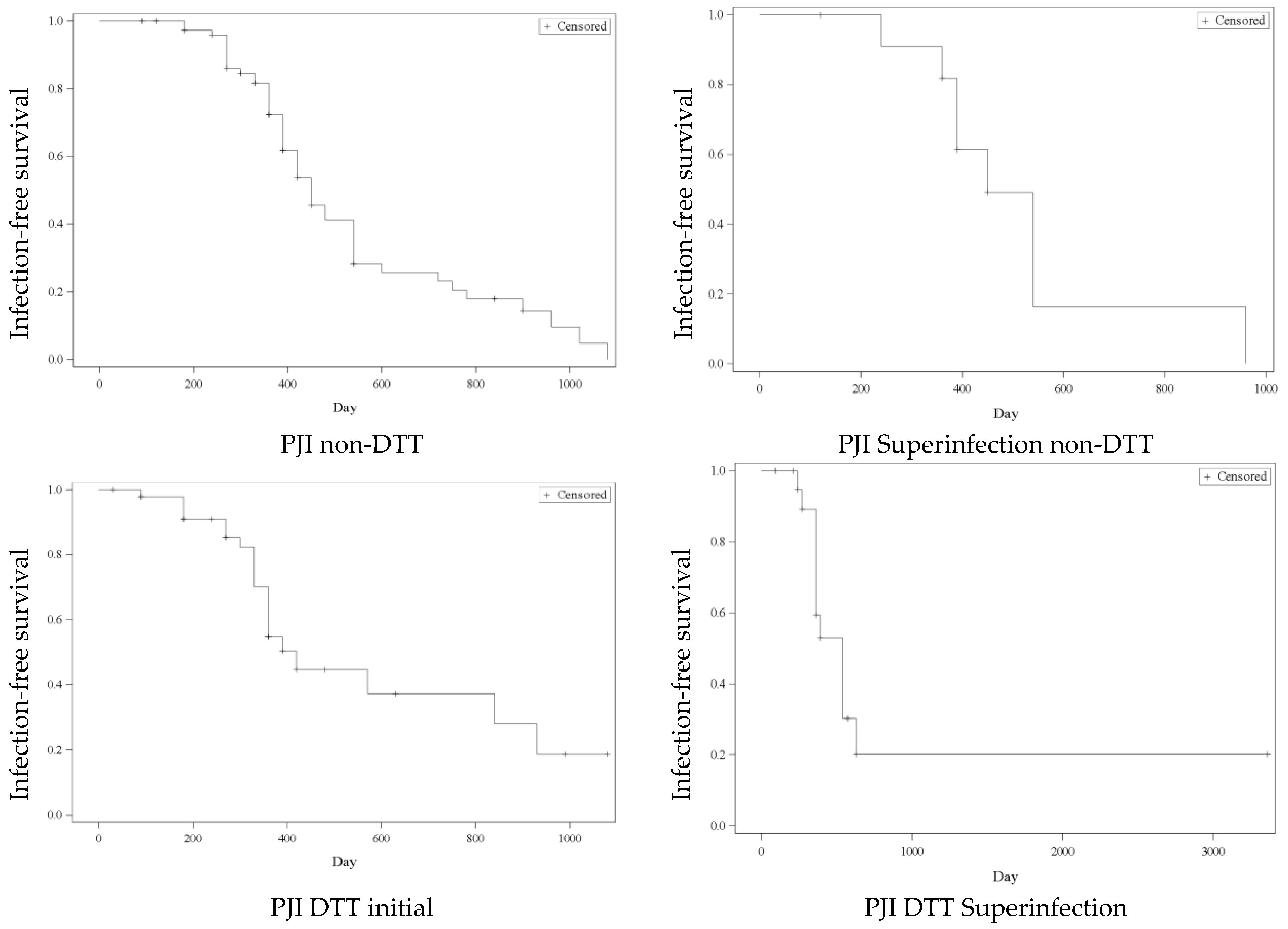
2. Results
3. Discussion
4. Materials and Methods
4.1. Study Population
4.2. Patient Parameters
4.3. Definition of Periprosthetic Joint Infection and DTT Pathogens
- -
- PJI caused by a non-DTT pathogen without superinfection.
- -
- PJI caused by a DTT pathogen without superinfection.
- -
- PJI initially caused by a non-DTT pathogen but with subsequent superinfection or pathogen switch to a DTT pathogen later in the course of the infection.
- -
- PJI with superinfection of any kind (DTT and non-DTT pathogens).
4.4. Treatment Regimen
4.5. Statistical Analysis
4.6. Ethics Approval
5. Conclusions
Author Contributions
Funding
Institutional Review Board Statement
Informed Consent Statement
Data Availability Statement
Conflicts of Interest
References
- Neuprez, A.; Neuprez, A.H.; Kaux, J.F.; Kurth, W.; Daniel, C.; Thirion, T.; Huskin, J.P.; Gillet, P.; Bruyère, O.; Reginster, J.Y. Total joint replacement improves pain, functional quality of life, and health utilities in patients with late-stage knee and hip osteoarthritis for up to 5 years. Clin. Rheumatol. 2020, 39, 861–871. [Google Scholar] [CrossRef] [PubMed]
- Egerci, O.F.; Yapar, A.; Dogruoz, F.; Selcuk, H.; Kose, O. Preventive strategies to reduce the rate of periprosthetic infections in total joint arthroplasty; a comprehensive review. Arch. Orthop. Trauma Surg. 2024, 144, 5131–5146. [Google Scholar] [CrossRef] [PubMed]
- Jämsen, E.; Stogiannidis, I.; Malmivaara, A.; Pajamäki, J.; Puolakka, T.; Konttinen, Y.T. Outcome of prosthesis exchange for infected knee arthroplasty: The effect of treatment approach. Acta Orthop. 2009, 80, 67–77. [Google Scholar] [CrossRef] [PubMed]
- Yaghmour, K.M.; Chisari, E.; Khan, W.S. Single-Stage Revision Surgery in Infected Total Knee Arthroplasty: A PRISMA Systematic Review. J. Clin. Med. 2019, 8, 174. [Google Scholar] [CrossRef] [PubMed]
- Kunutsor, S.K.; Whitehouse, M.R.; Lenguerrand, E.; Blom, A.W.; Beswick, A.D. Re-Infection Outcomes Following One- And Two-Stage Surgical Revision of Infected Knee Prosthesis: A Systematic Review and Meta-Analysis. PLoS ONE 2016, 11, e0151537. [Google Scholar] [CrossRef] [PubMed]
- Premkumar, A.; Kolin, D.A.; Farley, K.X.; Wilson, J.M.; McLawhorn, A.S.; Cross, M.B.; Sculco, P.K. Projected Economic Burden of Periprosthetic Joint Infection of the Hip and Knee in the United States. J. Arthroplast. 2021, 36, 1484–1489.e3. [Google Scholar] [CrossRef] [PubMed]
- Kurd, M.F.; Ghanem, E.; Steinbrecher, J.; Parvizi, J. Two-stage Exchange Knee Arthroplasty: Does Resistance of the Infecting Organism Influence the Outcome? Clin. Orthop. Relat. Res. 2010, 468, 2060–2066. [Google Scholar] [CrossRef] [PubMed]
- Zmistowski, B.; Fedorka, C.J.; Sheehan, E.; Deirmengian, G.; Austin, M.S.; Parvizi, J. Prosthetic Joint Infection Caused by Gram-Negative Organisms. J. Arthroplast. 2011, 26, 104–108. [Google Scholar] [CrossRef] [PubMed]
- Tornero, E.; Morata, L.; Martínez-Pastor, J.C.; Bori, G.; Mensa, J.; Soriano, A. Prosthetic joint infections due to methicillin-resistant and methicillin-susceptible staphylococci treated with open debridement and retention of the prosthesis. Rev. Esp. Quim. Quimioter. 2013, 26, 353–359. [Google Scholar]
- Kheir, M.M.; Tan, T.L.; Higuera, C.; George, J.; Della Valle, C.J.; Shen, M.; Parvizi, J. Periprosthetic Joint Infections Caused by Enterococci Have Poor Outcomes. J. Arthroplast. 2017, 32, 933–947. [Google Scholar] [CrossRef] [PubMed]
- Hsieh, P.H.; Lee, M.S.; Hsu, K.Y.; Chang, Y.H.; Shih, H.N.; Ueng, S.W. Gram-negative prosthetic joint infections: Risk factors and outcome of treatment. Clin. Infect. Dis. 2009, 49, 1036–1043. [Google Scholar] [CrossRef] [PubMed]
- Kuo, F.C.; Goswami, K.; Shohat, N.; Blevins, K.; Rondon, A.J.; Parvizi, J. Two-Stage Exchange Arthroplasty Is a Favorable Treatment Option Upon Diagnosis of a Fungal Periprosthetic Joint Infection. J. Arthroplast. 2018, 33, 3555–3560. [Google Scholar] [CrossRef] [PubMed]
- Izakovicova, P.; Borens, O.; Trampuz, A. Periprosthetic joint infection: Current concepts and outlook. EFORT Open Rev. 2019, 4, 482–494. [Google Scholar] [CrossRef] [PubMed]
- Mirza, Y.H.; Tansey, R.; Sukeik, M.; Shaath, M.; Haddad, F.S. Biofilm and the Role of Antibiotics in the Treatment of Periprosthetic Hip and Knee Joint Infections. Open Orthop. J. 2016, 10, 636–645. [Google Scholar] [CrossRef] [PubMed]
- Wimmer, M.D.; Hischebeth, G.T.R.; Randau, T.M.; Gathen, M.; Schildberg, F.A.; Fröschen, F.S.; Kohlhof, H.; Gravius, S. Difficult-to-treat pathogens significantly reduce infection resolution in periprosthetic joint infections. Diagn. Microbiol. Infect. Dis. 2020, 98, 115114. [Google Scholar] [CrossRef] [PubMed]
- Tuchscherr, L.; Heitmann, V.; Hussain, M.; Viemann, D.; Roth, J.; von Eiff, C.; Peters, G.; Becker, K.; Löffler, B. Staphylococcus aureus small-colony variants are adapted phenotypes for intracellular persistence. J. Infect. Dis. 2010, 202, 1031–1040. [Google Scholar] [CrossRef] [PubMed]
- Vuong, C.; Kidder, J.B.; Jacobson, E.R.; Otto, M.; Proctor, R.A.; Somerville, G.A. Staphylococcus epidermidis polysaccharide intercellular adhesin production significantly increases during tricarboxylic acid cycle stress. J. Bacteriol. 2005, 187, 2967–2973. [Google Scholar] [CrossRef] [PubMed]
- Akgün, D.; Perka, C.; Trampuz, A.; Renz, N. Outcome of hip and knee periprosthetic joint infections caused by pathogens resistant to biofilm-active antibiotics: Results from a prospective cohort study. Arch. Orthop. Trauma Surg. 2018, 138, 635–642. [Google Scholar] [CrossRef] [PubMed]
- Faschingbauer, M.; Bieger, R.; Kappe, T.; Weiner, C.; Freitag, T.; Reichel, H. Difficult to treat: Are there organism-dependent differences and overall risk factors in success rates for two-stage knee revision? Arch. Orthop. Trauma Surg. 2020, 140, 1595–1602. [Google Scholar] [CrossRef] [PubMed]
- Hipfl, C.; Winkler, T.; Janz, V.; Perka, C.; Müller, M. Management of Chronically Infected Total Knee Arthroplasty With Severe Bone Loss Using Static Spacers With Intramedullary Rods. J. Arthroplast. 2019, 34, 1462–1469. [Google Scholar] [CrossRef] [PubMed]
- Darwich, A.; Dally, F.J.; Abu Olba, K.; Mohs, E.; Gravius, S.; Hetjens, S.; Assaf, E.; Bdeir, M. Superinfection with Difficult-to-Treat Pathogens Significantly Reduces the Outcome of Periprosthetic Joint Infections. Antibiotics 2021, 10, 1145. [Google Scholar] [CrossRef] [PubMed]
- Chang, C.H.; Lee, S.H.; Lin, Y.C.; Wang, Y.C.; Chang, C.J.; Hsieh, P.H. Increased periprosthetic hip and knee infection projected from 2014 to 2035 in Taiwan. J. Infect. Public Health 2020, 13, 1768–1773. [Google Scholar] [CrossRef] [PubMed]
- O’Toole, P.; Maltenfort, M.G.; Chen, A.F.; Parvizi, J. Projected Increase in Periprosthetic Joint Infections Secondary to Rise in Diabetes and Obesity. J. Arthroplast. 2016, 31, 7–10. [Google Scholar] [CrossRef] [PubMed]
- Bozic, K.J.; Lau, E.; Kurtz, S.; Ong, K.; Rubash, H.; Vail, T.P.; Berry, D.J. Patient-related risk factors for periprosthetic joint infection and postoperative mortality following total hip arthroplasty in Medicare patients. J. Bone Jt. Surg. Am. 2012, 94, 794–800. [Google Scholar] [CrossRef] [PubMed]
- Poultsides, L.A.; Ma, Y.; Della Valle, A.G.; Chiu, Y.L.; Sculco, T.P.; Memtsoudis, S.G. In-hospital surgical site infections after primary hip and knee arthroplasty--incidence and risk factors. J. Arthroplast. 2013, 28, 385–389. [Google Scholar] [CrossRef] [PubMed]
- Parvizi, J.; Tan, T.L.; Goswami, K.; Higuera, C.; Della Valle, C.; Chen, A.F.; Shohat, N. The 2018 Definition of Periprosthetic Hip and Knee Infection: An Evidence-Based and Validated Criteria. J. Arthroplast. 2018, 33, 1309–1314.e2. [Google Scholar] [CrossRef] [PubMed]
- Tetreault, M.W.; Wetters, N.G.; Moric, M.; Gross, C.E.; Della Valle, C.J. Is synovial C-reactive protein a useful marker for periprosthetic joint infection? Clin. Orthop. Relat. Res. 2014, 472, 3997–4003. [Google Scholar] [CrossRef] [PubMed]
- Vanderstappen, C.; Verhoeven, N.; Stuyck, J.; Bellemans, J. Intra-articular versus serum C-reactive protein analysis in suspected periprosthetic knee joint infection. Acta Orthop. Belg. 2013, 79, 326–330. [Google Scholar] [PubMed]
- Grzelecki, D.; Kocon, M.; Mazur, R.; Grajek, A.; Kowalczewski, J. The diagnostic accuracy of blood C-reactive protein and erythrocyte sedimentation rate in periprosthetic joint infections—A 10-year analysis of 1510 revision hip and knee arthroplasties from a single orthopaedic center. J. Orthop. Surg. Res. 2025, 20, 276. [Google Scholar] [CrossRef] [PubMed]
- Unter Ecker, N.; Suero, E.M.; Gehrke, T.; Haasper, C.; Zahar, A.; Lausmann, C.; Hawi, N.; Citak, M. Serum C-reactive protein relationship in high- versus low-virulence pathogens in the diagnosis of periprosthetic joint infection. J. Med. Microbiol. 2019, 68, 910–917. [Google Scholar] [CrossRef] [PubMed]
- Ghanem, E.; Antoci, V., Jr.; Pulido, L.; Joshi, A.; Hozack, W.; Parvizi, J. The use of receiver operating characteristics analysis in determining erythrocyte sedimentation rate and C-reactive protein levels in diagnosing periprosthetic infection prior to revision total hip arthroplasty. Int. J. Infect. Dis. 2009, 13, e444–e449. [Google Scholar] [CrossRef] [PubMed]
- Grossi, O.; Asseray, N.; Bourigault, C.; Corvec, S.; Valette, M.; Navas, D.; Happi-Djeukou, L.; Touchais, S.; Bémer, P.; Boutoille, D.; et al. Gram-negative prosthetic joint infections managed according to a multidisciplinary standardized approach: Risk factors for failure and outcome with and without fluoroquinolones. J. Antimicrob. Chemother. 2016, 71, 2593–2597. [Google Scholar] [CrossRef] [PubMed]
- Baertl, S.; Lovasz, D.; Kees, M.G.; Walter, N.; Schindler, M.; Li, J.; Reinhard, J.; Alt, V.; Rupp, M. Periprosthetic Joint Infection and Concomitant Sepsis: Unveiling Clinical Manifestations, Risk Factors, and Patient Outcomes. J. Arthroplast. 2025, 40, 1827–1835. [Google Scholar] [CrossRef] [PubMed]
- Bae, K.J.; Chae, Y.J.; Jung, S.J.; Gong, H.S. Incidence and risk factors for periprosthetic joint infection: A common data model analysis. Jt. Dis. Relat. Surg. 2022, 33, 303–313. [Google Scholar] [CrossRef] [PubMed]
- Tsai, Y.; Chang, C.H.; Lin, Y.C.; Lee, S.H.; Hsieh, P.H.; Chang, Y. Different microbiological profiles between hip and knee prosthetic joint infections. J. Orthop. Surg. (Hong Kong) 2019, 27, 2309499019847768. [Google Scholar] [CrossRef] [PubMed]
- Enz, A.; Mueller, S.C.; Warnke, P.; Ellenrieder, M.; Mittelmeier, W.; Klinder, A. Periprosthetic Fungal Infections in Severe Endoprosthetic Infections of the Hip and Knee Joint-A Retrospective Analysis of a Certified Arthroplasty Centre of Excellence. J. Fungi 2021, 7, 404. [Google Scholar] [CrossRef] [PubMed]
- Tornero, E.; Senneville, E.; Euba, G.; Petersdorf, S.; Rodriguez-Pardo, D.; Lakatos, B.; Ferrari, M.C.; Pilares, M.; Bahamonde, A.; Trebse, R.; et al. Characteristics of prosthetic joint infections due to Enterococcus sp. and predictors of failure: A multi-national study. Clin. Microbiol. Infect. 2014, 20, 1219–1224. [Google Scholar] [CrossRef] [PubMed]
- Baumgärtner, T.; Bdeir, M.; Dally, F.J.; Gravius, S.; Hai, A.A.E.; Assaf, E.; Hetjens, S.; Miethke, T.; Darwich, A. Rifampin-resistant periprosthetic joint infections are associated with worse functional outcome in both acute and chronic infection types. Diagn. Microbiol. Infect. Dis. 2024, 110, 116447. [Google Scholar] [CrossRef] [PubMed]
- Achermann, Y.; Eigenmann, K.; Ledergerber, B.; Derksen, L.; Rafeiner, P.; Clauss, M.; Nüesch, R.; Zellweger, C.; Vogt, M.; Zimmerli, W. Factors associated with rifampin resistance in staphylococcal periprosthetic joint infections (PJI): A matched case-control study. Infection 2013, 41, 431–437. [Google Scholar] [CrossRef] [PubMed]
- Resende, V.A.C.; Neto, A.C.; Nunes, C.; Andrade, R.; Espregueira-Mendes, J.; Lopes, S. Higher age, female gender, osteoarthritis and blood transfusion protect against periprosthetic joint infection in total hip or knee arthroplasties: A systematic review and meta-analysis. Knee Surg. Sports Traumatol. Arthrosc. 2021, 29, 8–43. [Google Scholar] [CrossRef] [PubMed]
- Ren, X.; Ling, L.; Qi, L.; Liu, Z.; Zhang, W.; Yang, Z.; Wang, W.; Tu, C.; Li, Z. Patients’ risk factors for periprosthetic joint infection in primary total hip arthroplasty: A meta-analysis of 40 studies. BMC Musculoskelet. Disord. 2021, 22, 776. [Google Scholar] [CrossRef] [PubMed]
- Chrastil, J.; Anderson, M.B.; Stevens, V.; Anand, R.; Peters, C.L.; Pelt, C.E. Is Hemoglobin A1c or Perioperative Hyperglycemia Predictive of Periprosthetic Joint Infection or Death Following Primary Total Joint Arthroplasty? J. Arthroplast. 2015, 30, 1197–1202. [Google Scholar] [CrossRef] [PubMed]
- Berríos-Torres, S.I.; Umscheid, C.A.; Bratzler, D.W.; Leas, B.; Stone, E.C.; Kelz, R.R.; Reinke, C.E.; Morgan, S.; Solomkin, J.S.; Mazuski, J.E.; et al. Centers for Disease Control and Prevention Guideline for the Prevention of Surgical Site Infection, 2017. JAMA Surg. 2017, 152, 784–791. [Google Scholar] [CrossRef] [PubMed]
- Babkin, Y.; Raveh, D.; Lifschitz, M.; Itzchaki, M.; Wiener-Well, Y.; Kopuit, P.; Jerassy, Z.; Yinnon, A.M. Incidence and risk factors for surgical infection after total knee replacement. Scand. J. Infect. Dis. 2007, 39, 890–895. [Google Scholar] [CrossRef] [PubMed]
- Alijanipour, P.; Heller, S.; Parvizi, J. Prevention of periprosthetic joint infection: What are the effective strategies? J. Knee Surg. 2014, 27, 251–258. [Google Scholar] [CrossRef] [PubMed]
- Osmon, D.R.; Berbari, E.F.; Berendt, A.R.; Lew, D.; Zimmerli, W.; Steckelberg, J.M.; Rao, N.; Hanssen, A.; Wilson, W.R. Executive summary: Diagnosis and management of prosthetic joint infection: Clinical practice guidelines by the Infectious Diseases Society of America. Clin. Infect. Dis. 2013, 56, 1–10. [Google Scholar] [CrossRef] [PubMed]
- American Society of Anesthesiologists. ASA Physical Status Classification System. 2014. Available online: https://dam.assets.ohio.gov/image/upload/med.ohio.gov/portals/0/resources/asa%20physical%20status%20classification%20system.pdf (accessed on 20 May 2025).
- Charlson, M.E.; Pompei, P.; Ales, K.L.; MacKenzie, C.R. A new method of classifying prognostic comorbidity in longitudinal studies: Development and validation. J. Chronic Dis. 1987, 40, 373–383. [Google Scholar] [CrossRef] [PubMed]
- Lunz, A.; Lehner, B.; Voss, M.N.; Knappe, K.; Jaeger, S.; Innmann, M.M.; Renkawitz, T.; Omlor, G.W. Impact and Modification of the New PJI-TNM Classification for Periprosthetic Joint Infections. J. Clin. Med. 2023, 12, 1262. [Google Scholar] [CrossRef] [PubMed]
- Alt, V.; Rupp, M.; Langer, M.; Baumann, F.; Trampuz, A. Can the oncology classification system be used for prosthetic joint infection?: The PJI-TNM system. Bone Jt. Res. 2020, 9, 79–81. [Google Scholar] [CrossRef] [PubMed]
- Parvizi, J.; Zmistowski, B.; Berbari, E.F.; Bauer, T.W.; Springer, B.D.; Della Valle, C.J.; Garvin, K.L.; Mont, M.A.; Wongworawat, M.D.; Zalavras, C.G. New Definition for Periprosthetic Joint Infection: From the Workgroup of the Musculoskeletal Infection Society. Clin. Orthop. Relat. Res. 2011, 469, 2992–2994. [Google Scholar] [CrossRef] [PubMed]
- Zimmerli, W.; Trampuz, A.; Ochsner, P.E. Prosthetic-joint infections. N. Engl. J. Med. 2004, 351, 1645–1654. [Google Scholar] [CrossRef] [PubMed]
- Zimmerli, W.; Ochsner, P.E. Management of infection associated with prosthetic joints. Infection 2003, 31, 99–108. [Google Scholar] [CrossRef] [PubMed]
- Sprowson, A.P.; Jensen, C.; Chambers, S.; Parsons, N.R.; Aradhyula, N.M.; Carluke, I.; Inman, D.; Reed, M.R. The use of high-dose dual-impregnated antibiotic-laden cement with hemiarthroplasty for the treatment of a fracture of the hip: The Fractured Hip Infection trial. Bone Jt. J. 2016, 98-B, 1534–1541. [Google Scholar] [CrossRef] [PubMed]
- Aggarwal, V.K.; Rasouli, M.R.; Parvizi, J. Periprosthetic joint infection: Current concept. Indian. J. Orthop. 2013, 47, 10–17. [Google Scholar] [CrossRef] [PubMed]
- Li, C.; Renz, N.; Trampuz, A. Management of Periprosthetic Joint Infection. Hip Pelvis 2018, 30, 138–146. [Google Scholar] [CrossRef] [PubMed]

| Age (Years) | Sex | Side | BMI (Kg/m2) | ASA Score | Anticoagulation | |||||
|---|---|---|---|---|---|---|---|---|---|---|
| Mean ± SD (Range) | N (%) | N (%) | Mean ± SD (Range) | Mean ± SD | N (%) | |||||
| Female | Male | Right | Left | Yes | No | |||||
| Total | n = 169 | 71.1 ± 13.1 (20–97) | 84 (49.7%) | 85 (50.3%) | 81 (47.9%) | 88 (52.1%) | 29.5 ± 6.5 (16.4–46.8) | 3 ± 0.6 | 67 (39.5%) | 102 (60.5%) |
| PJI non-DTT | n = 91 | 70.6 ± 13.9 (20–93) | 47 (51.7%) | 44 (48.3%) | 38 (41.8%) | 53 (58.2%) | 28.8 ± 6.5 (17.6–46.8) | 3 ± 0.6 | 31 (34.1%) | 60 (65.9%) |
| PJI DTT total | n = 78 | 71.5 ± 12 (35–97) | 37 (47.4%) | 41 (52.6%) | 41 (52.6%) | 37 (47.4%) | 30.3 ± 6.4 (16.4–45.1) | 3 ± 0.6 | 37 (47.4%) | 41 (52.6%) |
| PJI DTT initial | n = 54 | 71.8 ± 10.9 (48–89) | 28 (51.9%) | 26 (48.1%) | 26 (48.1%) | 28 (51.9%) | 30.7 ± 6.3 (16.4–45.1) | 3 ± 0.6 | 24 (44.4%) | 30 (55.6%) |
| PJI DTT super-infection | n = 24 | 71.1 ± 14.6 (35–97) | 9 (37.5%) | 15 (62.5%) | 15 (62.5%) | 9 (37.5%) | 29.5 ± 6.7 (18.5–42.3) | 3 ± 0.8 | 13 (54.2%) | 11 (45.8%) |
| PJI super-infection total | n = 40 | 69.1 ± 13.3 (35–97) | 15 (37.5%) | 25 (62.5%) | 20 (50%) | 20 (50%) | 30.4 ± 6.9 (18.5–44.1) | 3 ± 0.7 | 15 (37.5%) | 25 (62.5%) |
| p-value * | 0.6809 | 0.5898 | 0.2663 | 0.3324 | 0.7147 | 0.0743 | ||||
| Affected Joint | Type of Infection | CCI Age Adjusted | CRP (mg/L) at Admission | Antibiotic Treatment | Treatment Before Admission | Surgical Treatment | Number of Revisions | ||||||||||
|---|---|---|---|---|---|---|---|---|---|---|---|---|---|---|---|---|---|
| N (%) | N (%) | N (%) | Mean ± SD (Range) | Mean ± SD (range) | N (%) | N (%) | Mean ± SD (Range) | ||||||||||
| Hip | Knee | Shoulder | Acute | Chronic | 0–1 | 2–3 | 4–5 | Total Duration in Days | Antibiotic | Surgical | Prosthesis Retention | Prosthesis Exchange with Cement Spacer | Prosthesis Exchange Without Cement Spacer | ||||
| Total | n = 169 | 83 (49.1%) | 75 (44.4%) | 11 (6.5%) | 73 (43.2%) | 96 (56.8%) | 9 (5.3%) | 108 (63.9%) | 52 (30.8%) | 77.4 ± 83 (2–386) | 54.8 ± 36.7 (4–228) | 24 (14.2%) | 25 (14.8%) | 66 (40.7%) | 75 (46.3%) | 21 (13%) | 3.3 ± 3 (1–20) |
| PJI non-DTT | n = 91 | 36 (39.6%) | 47 (51.6%) | 8 (8.8%) | 33 (36.3%) | 58 (63.7%) | 6 (6.6%) | 60 (65.9%) | 25 (27.5%) | 105.6 ± 101.6 (2–390) | 46.3 ± 29.5 (4–217) | 13 (14.3%) | 11 (12.1%) | 44 (52.4%) | 33 (39.3%) | 7 (8.3%) | 2.3 ± 1.9 (1–14) |
| PJI DTT total | n = 78 | 47 (60.3%) | 28 (35.9%) | 3 (3.8%) | 37 (47.4%) | 41 (52.6%) | 3 (3.8%) | 48 (61.5%) | 27 (34.6%) | 64.4 ± 81.8 (2–381) | 64.2 ± 41.4 (4–228) | 11 (14.1%) | 14 (17.9%) | 22 (28.2%) | 42 (53.8%) | 14 (18%) | 4.4 ± 3.5 (1–20) |
| PJI DTT initial | n = 54 | 34 (63%) | 20 (37%) | 0 (0%) | 22 (40.8%) | 32 (59.2%) | 1 (1.9%) | 36 (66.7%) | 17 (31.5%) | 51.86 ± 63.01 (2–381) | 61.1 ± 39.7 (4–181) | 6 (11.1%) | 6 (11.1%) | 15 (28.8%) | 31 (57.4%) | 8 (14.8%) | 3.7 ± 3.3 (1–20) |
| PJI DTT Superinfection | n = 24 | 13 (54.2%) | 8 (33.3%) | 3 (12.5%) | 15 (62.5%) | 9 (37.5%) | 2 (8.3%) | 12 (50%) | 10 (41.7%) | 92.7 ± 109.7 (2–370) | 71.2 ± 45.2 (14–228) | 5 (20.8%) | 5 (20.8%) | 7 (30.2%) | 11 (45.8%) | 6 (24%) | 6 ± 3.6 (2–14) |
| PJI Superinfection total | n = 40 | 17 (42.5%) | 17 (42.5%) | 6 (15%) | 19 (47.5%) | 21 (52.5%) | 2 (5%) | 27 (67.5%) | 11 (27.5%) | 92.9 ± 93.8 (2–370) | 68.3 ± 45.6 (14–228) | 6 (15%) | 6 (15%) | 11 (27.5%) | 23 (57.5%) | 6 (15%) | 5.3 ± 3.4 (2–14) |
| p-value * | 0.0025 | 0.3155 | 0.9614 | 0.0055 | 0.0023 | 0.8534 | 0.0080 | <0.0001 | |||||||||
| Coagulase-Negative Staphylococci N (%) | Enterococci N (%) | Pseudomonas aeruginosa N (%) | Candida albicans N (%) | Polymicrobial * N (%) | ||
|---|---|---|---|---|---|---|
| PJI DTT total | n = 78 | 47 (60.3%) | 25 (32.1%) | 5 (6.4%) | 4 (5.1%) | 3 (3.9%) |
| PJI DTT initial | n = 54 | 34 (63%) | 12 (22.2%) | 3 (5.6%) | 3 (5.6%) | 2 (3.7%) |
| PJI DTT superinfection | n = 24 | 11 (45.8%) | 9 (37.5%) | 2 (8.3%) | 1 (4.2%) | 1 (4.2%) |
| Risk Factor | p-Value * | Odds Ratio | Confidence Interval | |
|---|---|---|---|---|
| PJI DTT total | CRP at admission (≥92.1 mg/L) | 0.001 | 6.981 | 1.367–35.63 |
| Hip joint | 0.0225 | 3.478 | 0.361–33.538 | |
| PJI DTT Superinfection | Number of revisions (≥3) | <0.0001 | 1.288 | 1.100–1.508 |
| Chronic type of infections | 0.0387 | 3.449 | 1.159–10.262 |
Disclaimer/Publisher’s Note: The statements, opinions and data contained in all publications are solely those of the individual author(s) and contributor(s) and not of MDPI and/or the editor(s). MDPI and/or the editor(s) disclaim responsibility for any injury to people or property resulting from any ideas, methods, instructions or products referred to in the content. |
© 2025 by the authors. Licensee MDPI, Basel, Switzerland. This article is an open access article distributed under the terms and conditions of the Creative Commons Attribution (CC BY) license (https://creativecommons.org/licenses/by/4.0/).
Share and Cite
Darwich, A.; Baumgärtner, T.; Hetjens, S.; Gravius, S.; Bdeir, M. Prevalence and Risk Factors for Superinfection with a Difficult-to-Treat Pathogen in Periprosthetic Joint Infections. Antibiotics 2025, 14, 752. https://doi.org/10.3390/antibiotics14080752
Darwich A, Baumgärtner T, Hetjens S, Gravius S, Bdeir M. Prevalence and Risk Factors for Superinfection with a Difficult-to-Treat Pathogen in Periprosthetic Joint Infections. Antibiotics. 2025; 14(8):752. https://doi.org/10.3390/antibiotics14080752
Chicago/Turabian StyleDarwich, Ali, Tobias Baumgärtner, Svetlana Hetjens, Sascha Gravius, and Mohamad Bdeir. 2025. "Prevalence and Risk Factors for Superinfection with a Difficult-to-Treat Pathogen in Periprosthetic Joint Infections" Antibiotics 14, no. 8: 752. https://doi.org/10.3390/antibiotics14080752
APA StyleDarwich, A., Baumgärtner, T., Hetjens, S., Gravius, S., & Bdeir, M. (2025). Prevalence and Risk Factors for Superinfection with a Difficult-to-Treat Pathogen in Periprosthetic Joint Infections. Antibiotics, 14(8), 752. https://doi.org/10.3390/antibiotics14080752