Article

Microbial Profile and Antibiotic Resistance Patterns in Bile Aspirates from Patients with Acute Cholangitis: A Multicenter International Study

by
Matei-Alexandru Cozma
1,2,*,
Mihnea-Alexandru Găman
3,4,
Camelia Cristina Diaconu
1,5,*,
Arthur Berger
6,
Frank Zerbib
6 and
Radu Bogdan Mateescu
1,2
1
Faculty of Medicine, “Carol Davila” University of Medicine and Pharmacy, 050474 Bucharest, Romania
2
Department of Gastroenterology and Hepatology, Colentina Clinical Hospital, 020125 Bucharest, Romania
3
Department of Hematology, Center for Clinical and Basic Research (CCBR Clinic), 030463 Bucharest, Romania
4
Department of Cellular and Molecular Pathology, Stefan S. Nicolau Institute of Virology, Romanian Academy, 030304 Bucharest, Romania
5
Department of Internal Medicine, Clinical Emergency Hospital of Bucharest, 14461 Bucharest, Romania
6
Haut-Lévêque Hospital, Centre Hospitalier Universitaire de Bordeaux, 33600 Pessac, France
*
Authors to whom correspondence should be addressed.
Antibiotics 2025, 14(7), 679; https://doi.org/10.3390/antibiotics14070679
Submission received: 30 May 2025 / Revised: 22 June 2025 / Accepted: 30 June 2025 / Published: 4 July 2025
(This article belongs to the Special Issue Epidemiological Data on Antibiotic Resistance)

Abstract

Objectives: Significant differences in antibiotic resistance (AR) rates and multi-drug resistant (MDR) bacteria incidence exist in patients with acute cholangitis (AC) from different countries or regions. We aim to characterize and compare the microbial spectrum and AR patterns in patients with AC from two tertiary centers in Europe. Methods: We conducted a prospective, observational, multicentric study including patients diagnosed with AC and a positive bile culture, admitted to the Colentina Clinical Hospital (CCH), Bucharest, Romania, and the Haut-Lévêque Hospital (HLH), Bordeaux, France, between April 2022 and October 2023. Results: We included a total of 144 patients from the CCH with 190 positive bile cultures (31 patients had up to five episodes of AC during the study period) and 241 identified microbial strains, and 62 patients from the HLH with 67 positive bile cultures (5 patients had two episodes of AC) and 194 identified microbial strains. The most frequently isolated bacteria were Escherichia coli (30.70%) and Pseudomonas spp. (27.80%) in the CCH group, and Enterococcus faecalis (15.46%) and Escherichia coli (22/11.34%) in the HLH group. Furthermore, 51 (21.16%) of the strains identified in the CCH group and 15 (7.21%) in the HLH group were MDR, such as extended-spectrum beta-lactamase-producing Enterobacteriaceae or carbapenemase-producing Enterobacterales. The resistance rates for common antibiotics were 13.69% in the CCH group vs. 8.76% in the HLH group for ceftriaxone, 9.54% vs. 2.06% for meropenem, 16.59% vs. 6.70% for piperacillin/tazobactam, and 25.31% vs. 7.73% for levofloxacin. Conclusions: This comparative study shows significant differences between these countries in terms of the AR rates and MDR bacteria prevalence, highlighting the role of bile cultures as a safe and cost-effective method for guiding antibiotic treatment, thereby reducing the AR rates and complications.

1. Introduction

Acute cholangitis (AC) represents a life-threatening infectious disease of the biliary tract, with increased risk for poor prognosis in the absence of swift treatment [1,2,3]. Prevalence has been rising globally to 8–12 cases/100,000 people, mainly due to the growing burden of gallstone disease and the increasing incidence of malignant stenoses of the common bile duct (CBD) [3]. Systemic infection occurs in up to 20% of cases, and the mortality rate remains at around 10%, despite a significant decline in recent years through widespread implementation of endoscopic interventions [4,5].
AC appears in the setting of impaired bile flow caused by benign (gallstones, benign ampuloma) or malignant (pancreatic head cancer or cholangiocarcinoma) CBD obstructions. Elevated CBD pressure and retrograde flow of the bile lead to bacterial proliferation and eventually translocation into the systemic circulation, causing sepsis and multiple organ dysfunction [6,7].
Treatment must be initiated as soon as possible, and consists of prompt and adequate hemodynamic resuscitation, wide-spectrum antibiotic therapy, and early interventional restoration of the bile flow [8,9,10,11]. Traditionally, microbial agents were isolated by blood cultures; however, these are positive in only 21% to 71% of the samples [1,12]. Because of this, bile sample aspiration for microbiological analysis during endoscopic retrograde cholangiopancreatography (ERCP) is now increasingly used to guide initial empiric treatment, being recognized as a cheap, fast, and more specific method, with up to 100% positivity rates [1,13,14,15].
Antimicrobial resistance (AMR) is a significant cause of morbidity, mortality, and unnecessarily high medical costs globally, responsible for over 1 million deaths each year [16,17,18]. AC makes no exception, as the increasing prevalence of MDR bacteria has led to increased mortality and complication rates [19,20]. Although AMR is a global major health problem, significant variations exist at the national or even regional level, with high consumption of broad-spectrum antibiotics and several socio-economic aspects (inefficient infection control, population density, and national tourism profile) being the most relevant risk factors (RFs) [21,22]. Unfortunately, to this date, only limited comparative data exist on epidemiological aspects of AC patients in Romania.
While previous studies exist and have provided some limited information on the microbiological spectrum and resistance patterns in AC patients from both countries, Romania and France, they are often unicentric, include small cohorts, and are limited to a region. Our study focuses on the knowledge gap regarding the comparative findings between multiple regions by comparing similar data from two regions with distinct antibiotic policies, infection control strategies, and healthcare infrastructure in Eastern (Romania) and Western Europe (France). Additionally, while the importance of blood cultures in systemic infections has been thoroughly studied and validated, we chose to focus on bile cultures in order to increase the clinical value of our findings, considering their increased specificity and the limited attention they have received. Last, but not least, we explored the prevalence, impact, and risk factors associated with MDR bacterial infection and, at the same time, investigated the impact of the prior endoscopic interventions’ resistance patterns, thus moving beyond standard descriptive epidemiological analysis, and thereby increasing the relevance and impact of our results.
The main objective of this study was to analyze and compare the microbial spectrum and the AR patterns of pathogens isolated from bile cultures collected from patients with AC in two referral centers from Eastern and Western Europe. Additional objectives were to identify the incidence of MDR bacteria among etiological agents and the RFs associated with such infections and, at the same time, to analyze the influence of previous endoscopic interventions on resistance patterns and MDR bacteria incidence.

2. Results

2.1. Patients and General Characteristics

Of the 823 patients admitted with a positive diagnosis of AC in both medical units, 205 patients met the inclusion criteria and were included in this study (143 from the CCH and 62 from the HLH). The inclusion flowchart is shown in Figure 1, below. Collectively, these patients had 257 positive bile samples processed (190 from the CCH and 67 from the HLH), leading to the identification and analysis of 436 etiological agents (241 in the CCH group and 194 in the HLH group). Notably, 32 patients from the CCH group and 5 patients from the HLH group had bile samples collected during multiple hospitalizations within the study period. Additionally, a single patient from the CCH group presented with two distinct positive bile cultures during the same hospitalization.
The mean age was 68.49 years old (SD = 10.40) for the CCH group, and 70.34 years old (SD = 9.02) for the HLH group. A non-significant male predominance was observed in both study groups: 83 patients (58.04%) in the CCH group and 40 patients (64.51%) in the HLH group. A total of 83 patients (58.04%) from the CCH group and 47 patients (75.80%) from the HLH group experienced an episode of AC caused by a malignant etiology, with no significant difference between the two groups. In terms of endoscopic history, a significantly smaller proportion of patients from the CCH group had naïve papilla compared to the HLH group (46.15% vs. 66.13%, p = 0.0098).
Of the 189 and 67 episodes of AC that occurred during the study period in both hospitals, a significantly higher proportion occurred in patients with a biliary stent already in place for the CCH group (103 cases, 54.5%), as opposed to the HLH group (25 cases, 37.31%) (p = 0.0225). The mean length of hospitalization was longer for the CCH group, at 5.64 days, compared to 3.35 days for the HLH group (p < 0.0001). Complications, such as post-ERCP pancreatitis or post-sphincterotomy bleeding, occurred in 57 (30.15%) episodes from the CCH group and in 13 (19.40%) from the HLH group (p = 0.1107). Six (3.17%) and four (5.97%) AC episodes resulted in admission to the ICU department (p = 0.2941).
A total of 44 cases of AC for the CCH group occurred in patients who had had a different bile sample collected during a previous episode of AC, but with a negative result. Unfortunately, we did not have access to such data for the HLH group. A total of 37 blood cultures were collected from the CCH group patients hospitalized, of which only 19 turned out to a positive; for the HLH group, 31 blood cultures were collected, of which 22 were positive. Higher values were detected for total bilirubin, white blood cells, and C-reactive protein levels in the subgroup of patients with a stented CBD, but with no significant difference. All clinical and paraclinical characteristics of the patients included in this study are detailed in Table 1.

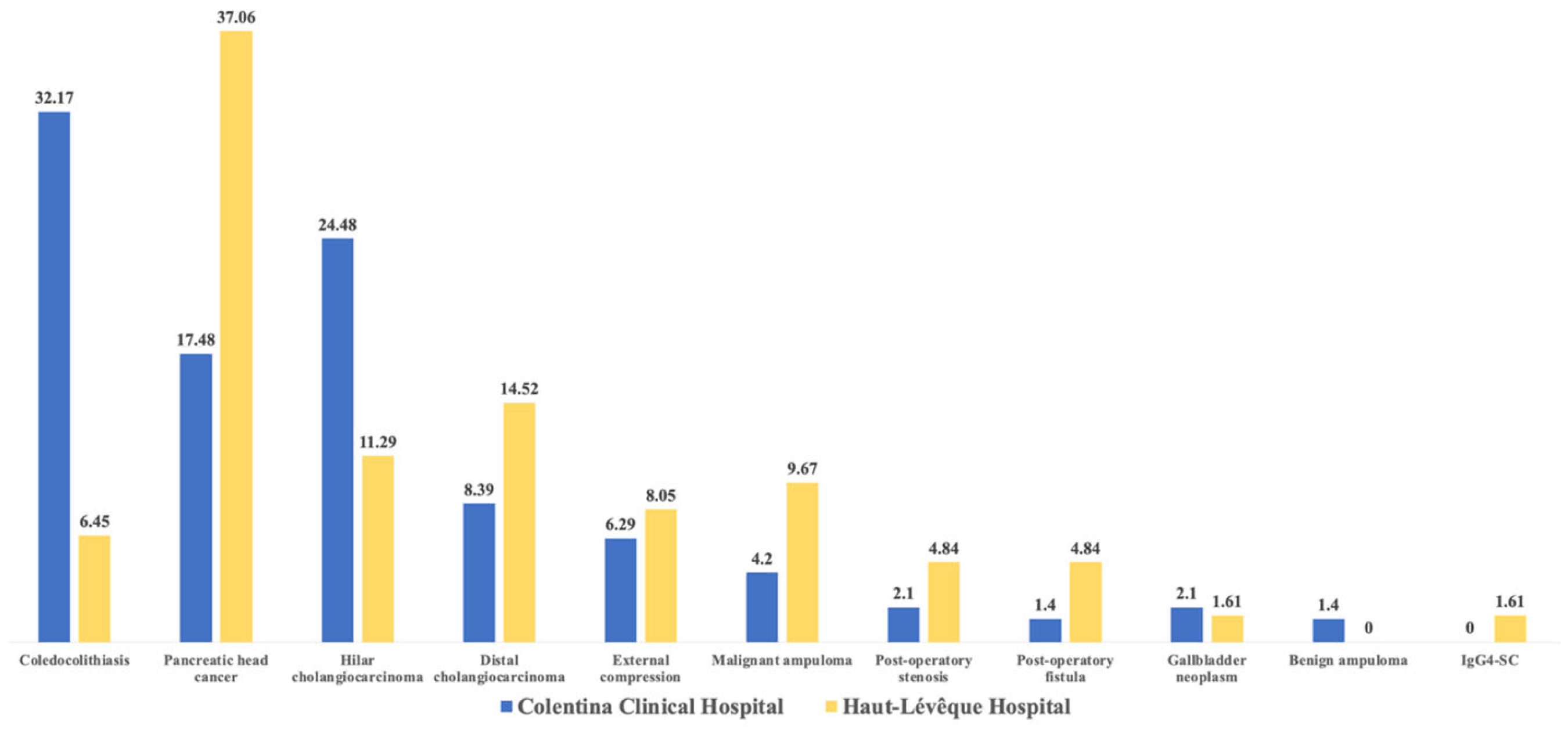
2.2. Etiologies of Acute Cholangitis

Malignant stenosis was the most common etiology of AC overall (58.04% and 75.80%), as well as within the subgroups of patients with biliary stents (77.92% and 76.19%). After performing a comparative analysis using the chi-squared test, we observed a significant difference between the groups of patients with naive papilla (p < 0.001), between the subgroups of patients from the CCH (p < 0.001), and between the total number of patients (p = 0.017) from the two medical units.
The most common etiology of AC in the CCH group was represented by choledocholithiasis (32.17%), followed by hilar cholangiocarcinoma (24.48%) and pancreatic head cancer (17.48%). Other etiologies were represented by distal cholangiocarcinoma and extrinsic compression (liver or lymph node metastases, chronic pancreatitis, or Mirizzi syndrome −6.29%). In the HLH patient group, the most common etiologies were pancreatic head cancer (41.46%), distal cholangiocarcinoma (14.52%), and hilar cholangiocarcinoma (11.29%). Other significant etiologies included malignant ampuloma (9.67%) and extrinsic compression (8.05%) (illustrated in Figure 2).

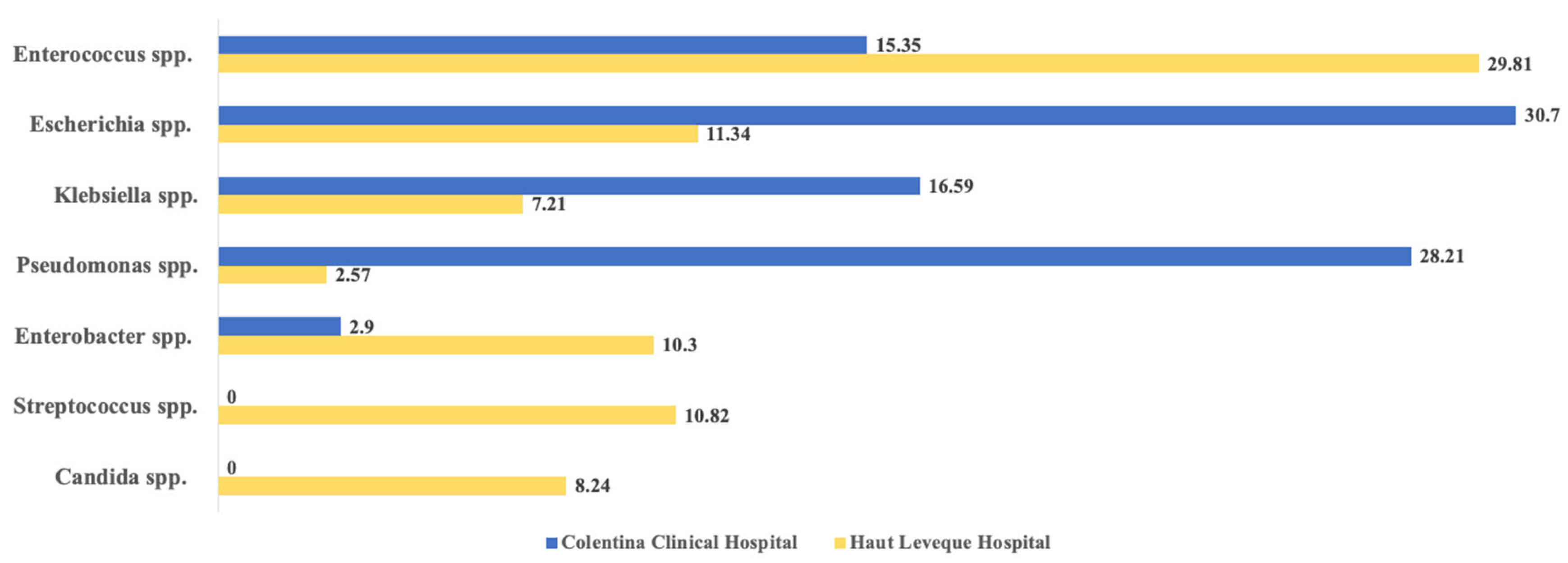
2.3. Microbial Agents Associated with AC

The majority of positive bile cultures were taken from patients with a stented CBD in both medical units (p = 0.0027). A significant difference between the two groups was in terms of the number of bacterial strains identified in each culture. While in the CCH group, the vast majority of cultures were monomicrobial (142 cases, 74.73%), in the HLH group, polymicrobial cultures (at least four distinct bacterial isolates) were more common (18 cases, 26.86%), with no such cases recorded in the CCH group. A detailed classification is shown in Table 2.
Gram-negative microorganisms represented the vast majority of bacteria identified in both study groups (detailed in Table 3 and illustrated in Figure 3). Escherichia coli (74, 30.70%), Pseudomonas spp. (68, 28.21%), Klebsiella spp. (40, 16.59%), and Enterococcus spp. (37, 15.35%) were the most common pathogens identified in the CCH study group. At the same time, less commonly identified species in this group included Achromobacter denitrificans (n = 1), Cronobacter sakazakii (n = 1), Raoultella ornithinolytica (n = 1), and Proteus mirabilis (n = 3). In the HLH study group, the most frequently identified bacteria were Enterococcus spp. (59, 30.41%), Escherichia coli (22, 11.34%), and Streptococcus spp. (21, 10.82%). Less frequently identified microbial strains included Candida spp. (n = 16), Clostridium perfringens (n = 3), Serratia marcescens (n = 5), and Veillonella parvula (n = 1).
In the comparative analysis using Fisher’s exact test, we observed significant differences when comparing all three subgroups from both medical units (“Naive Papilla” CCH vs. HLH, “Stented CBD” CCH vs. HLH, and “Total” CCH vs. HLH) for Enterococcus spp. (more frequent in the HLH group; p = 0.0112, p = 0.0122 and p = 0.0002), Escherichia spp. (more frequent in the CCH group; p = 0.0143, p = 0.0001 and p < 0.0001) and for Pseudomonas spp. (also more frequent in the CCH group; p < 0.0001 for all three subgroup comparisons). A further comparative analysis between the total number of patients can also be seen in Table 3 and Figure 3.

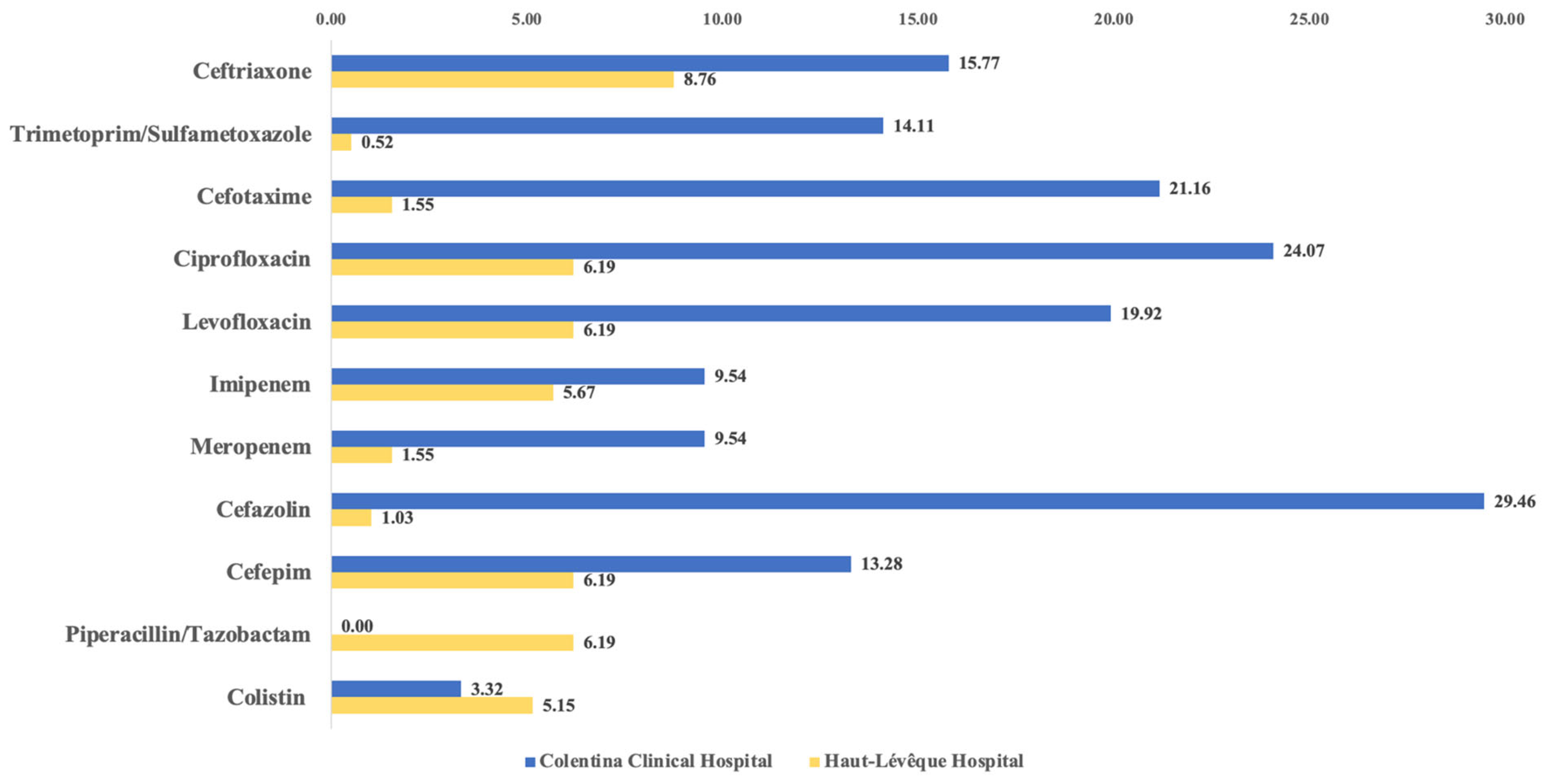
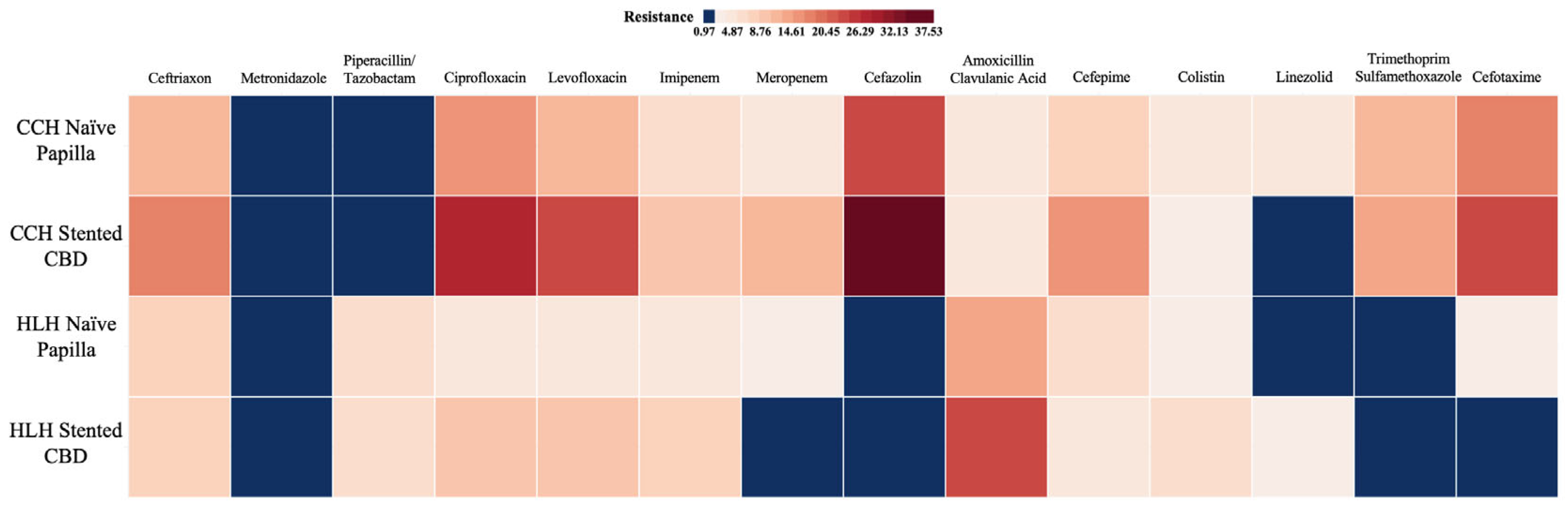
2.4. Resistance Patterns

As depicted in Table 4 and Figure 4, the resistance rates to most antibiotics were higher in the CCH group compared to the HLH group, with few exceptions seen for metronidazole, piperacillin/tazobactam, amoxicillin/clavulanic acid, and colistin. Moreover, a comparative statistical analysis using Fisher’s exact test demonstrated significant differences for several antibiotics. For example, significant resistance differences were observed for ceftriaxone (p = 0.03), ciprofloxacin (p < 0.0001), levofloxacin (p < 0.0001), meropenem (p = 0.0003), and cefazolin (p < 0.0001), to which the majority of the bacteria in the CCH group showed resistance. Conversely, significantly higher resistance was noted in the HLH group for piperacillin/tazobactam (p < 0.0001) and amoxicillin/clavulanic acid (p < 0.0001).
Table 5 and Figure 5 further detail the AR rates for the etiologic agents in the two groups, stratified by endoscopic history. Data shows that, in both groups, resistance rates were higher in the patients with biliary stents, with exceptions such as colistin, linezolid, and cefotaxime for the CCH group, and ceftriaxone, meropenem, and amoxicillin/clavulanic acid for the HLH group. Significant inter-subgroup differences were identified in the CCH group for ciprofloxacin (p = 0.045), levofloxacin (p = 0.012), cefazolin (p = 0.042), and linezolid (p = 0.034). However, no significant differences were detected among the HLH subgroups.
Furthermore, we opted for a comparative analysis of the same subgroups across the two hospitals (Naive Papilla–CCH vs. Naive Papilla–HLH and Stented CBD–CCH vs. Stented CBD–HLH). The comparison of the “Naive Papilla” subgroups revealed significant differences for piperacillin/tazobactam (p = 0.022), ciprofloxacin (p = 0.002), and levofloxacin (p = 0.043). For the “stented CBD” subgroups, significant differences were noted for piperacillin/tazobactam (p = 0.0059), ciprofloxacin (p = 0.0056), levofloxacin (p = 0.021), meropenem (p = 0.002), amoxicillin/clavulanic acid (p = 0.017), cefepime (p = 0.038), and trimethoprim/sulfamethoxazole (p = 0.0056).

2.5. Multidrug-Resistant Bacteria Involvement

Of the total bacteria identified in both patient groups, 51 (21.16%) of 241 in the CCH group and 15 (7.73%) of 194 in the HLH group were classified as MDR bacteria (p < 0.0001). Table 6 provides a detailed breakdown of the types and categories of these bacteria. In the HLH group, the vast majority of MDR bacteria (14 of 15) were of the ESBL (extended-spectrum beta-lactamase) or MLS (macrolide–lincosamide–streptogramin) types.
Conversely, in the CCH group, the bacterial spectrum was more diverse, including resistance types such as ESBL, CPE (carbapenemase-producing Enterobacterales), VRE (vancomycin-resistant Enterococci), and HLAR (high-level aminoglycoside resistance). Due to the distinct number of MDR bacteria observed between the two groups, a bivariate comparative analysis could not identify significant differences in the overall bacterial characteristics. However, in both groups, the majority of the MDR bacteria were isolated from patients with biliary stents. This association was statistically significant in the CCH group (p < 0.0001).
A particularity of our study was the ability to observe some of the included patients over multiple episodes of AC, thus allowing for the dynamic evaluation of their bile culture outcomes. Therefore, during the study period, in the CCH study group, 22 of the 32 patients with multiple episodes of AC had at least one recurrence of the same bacterial species previously isolated. Of these, nine were represented by Escherichia coli (of which eight developed resistances to at least one class of antibiotic) and ten by Pseudomonas spp. (of which four developed resistances to at least one antibiotic class of interest). In the case of the HLH group, the only bacterium identified over two episodes was E. faecium, which developed resistance to imipenem.

2.6. Antimicrobial Treatment

The most common antibiotics used empirically in the CCH group (detailed in Table 7) were the ceftriaxone–metronidazole association (in 121 episodes of AC, 64.02%), followed by ceftriaxone alone (22, 11.64%), ciprofloxacin (13, 6.87%), meropenem (9, 4.76%) and piperacillin/tazobactam (5, 2.89%). Within the HLH group, piperacillin/tazobactam was the most commonly used initial antibiotic therapy (30, 44.77%), followed by amoxicillin/clavulanic acid (9, 13.43%), the combination ceftriaxone/amoxicillin/clavulanic acid, trimethoprim/sulfamethoxazole, and the piperacillin/tazobactam–amikacin combination (each used in 3 AC episodes or 4.47%). Moreover, the initial antibiotic regimen was changed in 38 (20.10%) cases of AC for the CCH group and 7 cases (10.44%) for the HLH group (p = 0.0925).
During hospitalization, the initial antibiotic regimen was changed for 38 (20.10%) of the AC cases in the CCH group and for 7 (10.44%) cases in the HLH group (p = 0.0925). A total of 26 of the 38 (68.42%–CCH) and 3 of the 7 (42.85%–HLH) antibiotic changes were made for patients who had a biliary stent already in place. The most frequently used second line antibiotic in the CCH group was meropenem (n = 12), imipenem/cilastatin (n = 10), and the meropenem–metronidazole combination (n = 3), while in the HLH group, the most commonly used second-line antibiotics were piperacillin/tazobactam (n = 2), piperacillin/tazobactam–meropenem (n = 1), and the piperacillin/tazobactam–linezolid combination (n = 1).

2.7. Risk Factors Associated with MDR Bacteria Infections

After performing a logistic regression analysis (Table 8) for each study group (as previously described in Section 2.4), we identified several RFs associated with an MDR bacterial infection. In the CCH group, the identified RFs were, in order of strength of association, resistance to at least two fluoroquinolones, with an odds ratio (OR) of 6.25 (confidence interval (CI) between 2.817 and 13.869), p < 0.001, resistance to at least two III or IV generation cephalosporins, OR = 6.009 (CI between 3.084 and 11.708), p < 0.001, resistance to carbapenems with an OR of 4.45 (CI between 1.803 and 10.982), p = 0.001, biliary stent presence, OR of 2.847 (CI between 1.377 and 5.888), p = 0.005, and a positive blood culture, OR = 0.646 (CI between 0.212 and 1.967), p = 0.042.
In the HLH study group, the only RF proven to be associated with the presence of MDR bacterial infection were resistance to at least two fluoroquinolones, OR = 19.444 (CI between 4.752 and 79.566), p < 0.001 resistance to piperacillin/tazobactam, OR = 3.508 (CI between 1.136 and 10.831), p = 0.022, resistance to amoxicillin/clavulanic acid, OR = 3.508 (CI between 1.136 and 10.831), p = 0.029, and resistance to at least two third or fourth generation cephalosporins, OR = 3.107 (CI between 0.013 and 9.533), p = 0.047.

3. Discussion

Our study analyzed the microbiological profile and the AR patterns from routine bile aspirates in the AC patients based on data from two reference centers in Romania and France. The research was based on two main hypotheses. First, the AR rates would be higher in a country like Romania, where antibiotic consumption is significantly higher and current health policies are not yet sufficiently implemented. The second assumption was that resistance rates would be predominantly higher in patients with a history of endoscopic sphincterotomy, considering the altered anatomy and the potential presence of a biliary stent, both of which can alter the local biliary microbiota.
A number of programs aimed at limiting antibiotic consumption, both at home and in hospitals, have been adopted in both countries in recent years. Since 2006, Romania has prohibited the purchase of antibiotics without prescription, and since 2023, has launched the National Strategy for Limiting Antimicrobial Resistance and Preventing Healthcare Associated Infections, which includes such measures as limiting the number of emergency antibiotics available without prescription, introduction of rapid testing for common bacterial infections, including acute pharyngeal streptococcal infection and mandatory daily reporting, and centralization of antibiotic sales by pharmacists [23,24,25]. Similarly, France has introduced a series of measures to reduce the national antibiotic consumption by 25% by 2025, such as prescribing first-line medications and avoiding broad-spectrum antibiotics, and reimbursement of antibiotics only in the presence of a positive rapid test [26,27,28].

3.1. Risk Factors Associate with MDR Bacteria Infections

Our findings suggest that E. coli, Pseudomonas spp., Klebsiella spp., and Enterococcus were the most common microbial agents in Romania. By contrast, the most prevalent pathogens in France were Enterococcus, E. coli, and Streptococcus.
Similarly, a German investigation also reported Gram-negative bacteria as the most common agents in AC, in over 60% of bile samples. In their assessment, blood culture positivity was 32% [29]. Another investigation, which involved 208 individuals, revealed that ~85% of bile samples were positive for at least one microbial agent, with E. faecalis (~33%), E. faecium (~29%), E. coli (~23%), and Klebsiella spp. (~10%) as the most common. A polymicrobial cultures analysis revealed the prevalence of Gram-positive bacteria at ~73%, Gram-negative at ~53%, and fungi at ~24% (Candida albicans ~20%) [30].
Another US-based study on 615 subjects suspected of having AC also demonstrated a positivity of over 90% for bile aspirate samples, of which over 80% were polymicrobial, and for approximately 40% of blood cultures. Interestingly, Gram-positive agents were more commonly detected compared to our assessment, with Enterococcus spp. affecting nearly 70% and Streptococcus viridans over 35% of cases. Prior biliary endoscopic sphincterotomy was associated with a 4.5 times higher rate of bile culture positivity, as well as elevated rates of Enterococcus spp. (OR: 6.0), Pseudomonas aeruginosa (OR: 4.8), Enterobacter spp. (OR: 2.9), VRE (OR: 2.5), and Klebsiella spp. (OR: 2.1), respectively. Similarly, a history of stent placement was linked to elevated rates of Enterococcus spp. (OR: 9.9), Pseudomonas aeruginosa (OR: 5.2), Enterobacter spp. (OR: 3.9), VRE (OR: 2.8), and Klebsiella spp. (OR: 2.7) [9].
Other studies conducted on AC cases from Romania have reported similar results, but also contrasting findings. In an investigation conducted on 488 AC patients, blood culture positivity was less than 40%, whereas bile cultures were positive mainly in patients with a history of gallbladder removal (81.4%) [3]. In another Romanian research, which recruited 262 AC patients, bile cultures were positive for one microbial strain in 46%, two strains in 24%, and three strains in 3%, respectively. Gram-negative bacteria were also notably prevalent, the authors reporting the presence of E. coli predominantly in patients with benign vs. malignant biliary obstruction (56.1% vs. 37.6%). Klebsiella, Pseudomonas, and Citrobacter were identified in 24–29%, 10–14%, and 4.7–7% of cases, whereas Enterococcus was depicted in 18.7–24.7% of the AC subjects [1].
Similarly, E. coli and Klebsiella spp. strains were also the most detected Gram-negative agents in a cohort study conducted in Iran. In terms of Gram-positive strains, Staphylococcus aureus was the most commonly identified germ, as opposed to Enterococcus in our assessment [31]. Interestingly, an investigation performed in South Korea identified monomicrobial infections in 98.1% of the AC cases, revealing an increasing trend in the detection of Gram-negative agents, particularly Enterobacterales, and a decreasing trend of Gram-positive agents, namely Enterococci, over time [32].
Another study conducted in China on 277 patients with biliary tract infections reported that Gram-negative bacteria represented over 75% of the microorganisms identified in bile samples, with E. coli and K. pneumoniae comprising 26% and 15.7% of the microbial isolates, respectively. In addition, as opposed to our study, they identified a nearly 5% prevalence of Pseudomonas and Enterobacter each, whereas in our assessment Pseudomonas was identified in nearly 30% of the samples in Romania and less than 3% in France, and Enterobacter was reported in approximately 3% of the bile samples collected in the Romanian hospital and 8% of those collected in the French one. Related to Gram-positive agents, Enterococcus was the most frequently detected pathogen in both the Chinese investigation (12%) and ours, followed distantly by Staphylococcus spp. and Streptococcus spp. (approximately 4% and 2%, respectively) [14].

3.2. Antimicrobial Resistance Rates

In terms of AMR, resistance to multiple antibiotic classes was most commonly detected in the AC subjects from Romania who displayed resistance to multiple antibiotics (ceftriaxone, cefazoline, ciprofloxacin, or levofloxacin), whereas microbial strains in France were more likely resistant to amoxicillin/clavulanate and piperacillin/tazobactam. Resistance to at least two fluoroquinolones, III/IV generation cephalosporins, and carbapenems, as well as stent placement and blood cultures positivity, emerged as RFs for MDR AC, according to the data collected from the Romanian hospital. Similarly, the detection of MDR AC in the French hospital was predicted by the presence of resistance to fluoroquinolones, III/IV generation cephalosporins, amoxicillin/clavulanate, and piperacillin/tazobactam.
The same previously mentioned German study showed similar AR rates to the French reference center we examined, but significantly lower than in Romania, where bacterial strains seem resistant to multiple broad-spectrum antibiotics. The AR of Enterobacterales to ampicillin/sulbactam was over 50%, 34% to piperacillin/tazobactam, and between 20% and 27% to ciprofloxacin and cefotaxime. In addition, male sex, a high comorbidity burden, and percutaneous procedure emerged as RFs for the detection of MDR gram-negative bacteria [29].
In the Romanian cohort, more than 44% of the patients who had undergone cholecystectomy displayed ESBL-bacteria in the bile, 16.3% carbapenem-resistant Enterobacteriaceae, and 14.0% VRE, with MDR bacteria detected in nearly 75% of these subjects as opposed to 31.5% in the AC patients with intact gallbladders. In terms of microbial strains, E. coli and Klebsiella spp. were represented in percentages similar to our Romanian hospital; however, we detected a higher incidence of Pseudomonas (28% versus <7%) and a lower incidence of Enterococcus (15% versus 23.6%). AMR displayed a similar pattern, with 20–30% of the cases resistant to ampicillin/sulbactam, piperacillin/tazobactam, II and IV generation cephalosporins, and 15–20% resistant to fluoroquinolones and carbapenems. In another study, AC patients with biliary obstruction of malignant etiology displayed significantly higher rates of resistance to imipenem, ceftazidime, cefepime, and meropenem versus those with a benign etiology [2].
An assessment conducted in Turkey pointed out that E. coli (28.2%) and Pseudomonas (17.3%) are the most commonly identified pathogens in AC; however, they also revealed a high prevalence of Stenotrophomonas maltophilia at 15.2%. AMR patterns were commonly detected in both Gram-negative (P. aeruginosa and E. coli) as well as Gram-positive (E. faecium), with many strains showing below or around 50% susceptibility to ampicillin/sulbactam, cefotaxime, ampicillin, fluoroquinolones, or gentamicin [13].
In our study, bile cultures showed a positivity rate of 52%. This proportion is slightly lower than other reports in the literature, with rates that range between 60% and 87% [28,29,30]. There are some possible factors associated with this phenomenon. (1) Prior antibiotic treatment, which is negatively correlated with bile culture positivity, as it reduces bacterial load and may lead to false-negative cultures. (2) Technical limitation, such as insufficient bile aspirate, or delays in transport and processing can ultimately compromise culture viability—particularly for anaerobic or fastidious organisms. While sample processing protocols were strictly followed, these factors cannot be neglected. (3) Host and disease-specific factors, including microbial concentrations which may be below detection threshold for standard cultures in immunocompetent patients, subclinical infection, or, in mild stages of AC, may not yet have reached the threshold. Similarly, malignant and benign obstructions show different colonization patterns and culture yields, which further influence the culture results. (4) Lack of advanced diagnostic methods, such as 16S rRNA sequencing or polymerase chain reaction, which have superior sensitivity compared to standard cultures. However, these technologies are not routinely available or require further clinical validation; hence, they are primarily used in research facilities and not clinical hospitals [1,13,33,34,35,36,37,38].
Our study has several strengths and limitations. Firstly, to our knowledge, it is the first comparative study between two reference gastroenterology centers in Eastern Europe (Romania) and Western Europe (France), both with significant expertise in the management of AC. Moreover, our study was prospective in design, and thus causality can be established based on our findings. However, it only includes two centers and thus, a limited number of patients. Moreover, since it was conducted in university reference centers, our assessment might also be subject to selection bias and does not generally reflect the reality if superposed to other local or regional hospitals. However, our investigation raises awareness regarding the elevated AMR rates in AC, particularly in Romania, and can serve as documentation for policymakers in the healthcare field to establish regional or national guidelines to guide the selection of empiric antibiotic regimens in AC.

4. Materials and Methods

4.1. Study Design and Ethical Considerations

We conducted a prospective, multicenter, observational cohort study in which we included patients diagnosed with AC with a positive bile sample culture and subsequent antibiogram from two tertiary healthcare facilities from Romania and France: Gastroenterology Department of Colentina Clinical Hospital (CCH) in Bucharest, Romania and Gastroenterology, Hepatology and Digestive Oncology Department of Haut-Lévêque Hospital (HLH) in Bordeaux, France between April 2022 and October 2023. The study protocol conformed to the ethical guidelines of the 1975 Declaration of Helsinki and was approved by the internal review board of both medical centers. We obtained informed consent from all the included patients before data collection, thus ensuring strict confidentiality and privacy of their information. This study was approved by the Hospital’s Ethics Committee under registration number 10/12.09.2022.

4.2. Patient Selection and Bile Samples Collection

All of the patients included in this study had a positive diagnosis of AC according to the latest 2018 Tokyo Guidelines (TG18) criteria, and a positive bile culture sample obtained during an ERCP or an ultrasonographic-guided percutaneous transhepatic cholangiography (PTC) [39]. The exclusion criteria included patients who did not sign the informed consent, patients aged <18 years, and patients who were enrolled in another randomized clinical trial. Some eligible patients experienced multiple episodes of AC during the inclusion period; therefore, all episodes were considered. Patients were also categorized according to their endoscopic history.
We used the Olympus 190 series duodenoscopes (Olympus Corp., Tokyo, Japan), which were disinfected according to the manufacturer’s instructions, and according to the latest European Society of Gastrointestinal Endoscopy (ESGE)/European Society of Gastroenterology Nurses and Associates (ESGENA) position statement on the prevention of multidrug-resistant infections from contaminated duodenoscopes and to the latest guidelines of the French Ministry of Health and the French Society of Gastrointestinal Endoscopy [40,41,42,43]. Contamination was systematically excluded through standardized weekly microbiological tests according to local epidemiological health standards and protocols. Bile samples were collected after selective cannulation of the CBD with a standard single-use 4.4F TRUEtomeTM sphincterotome and a guide-wire (Boston Scientific, Marlborough, MA, USA). Approximately 5–10 mL of bile was aspirated before the injection of the contrast agent, and then placed in sterile containers suited for both anaerobic and aerobic bacterial cultures. Samples were transferred to the microbiology department and then cultured under aerobic and anaerobic conditions at 37 °C on 5% Columbia sheep blood agar (Becton Dickinson GmbH, Heidelberg, Germany), and Sabouraud dextrose agar (Thermo Fisher Scientific™, Waltham, MA, USA) with a first reading after 24 h, and the second reading at 48 h. Aerobic microorganisms were routinely identified and tested for antibiotic susceptibility by determining the minimum inhibitory concentration using the VITEK 2® System (bioMérieux, Lyon, France), while the identified fungi were tested using either the VITEK 2 YST system (bioMérieux, France) or the BD Phoenix™ yeast ID system (Becton, Dickinson and Company, Franklin Lakes, NJ, USA), all according to the latest European Committee on Antimicrobial Susceptibility Testing (EUCAST—https://www.eucast.org, accessed on 19 January 2025) guidelines available at the time.

4.3. Data Acquisition and Study Variables

Eligible patients were asked to sign the informed consent after each endoscopic procedure and only after being thoroughly informed about the purpose and implications of this study. Data was collected using a standardized data collection form from the electronic hospital database, patients’ charts, discharge reports, as well as imaging and laboratory files, during the hospital stay or after their discharge. The following variables were included: demographics (age, sex), clinical history (previous hospitalizations, comorbidities, medications, previous endoscopic interventions, history of cholecystectomy), presenting symptoms, laboratory data (blood count, C-reactive protein (CRP), total and direct bilirubin, aspartate aminotransferase (AST) and alanine aminotransferase (ALT)—all reported to laboratory reference values), ERCP timing, hospital stay duration, Tokyo severity score, the etiology of AC, admission rate to the intensive care unit (ICU), empiric and guided antibiotic regimes, bile culture results, and the subsequent AR pattern and MDR status assessment. Complications were also noted and further categorized according to the latest Adverse Events for Gastrointestinal Endoscopy (AGREE) classification [44].

4.4. Statistical Analysis

Statistical analyses were performed using the SPSS software, version 29.0 (IBM, Endicott, New York, NY, USA) and GraphPad Prism v9.2.0, graphical software (GraphPad, San Diego, CA, USA). Continuous variables were presented as a mean (with standard deviation), while categorical variables were reported as the number of subjects (n) and the percentage (%). Descriptive statistics showed the main demographic and clinical characteristics. The microbial species distribution and the AR patterns between the groups and sub-groups were compared using the chi-squared test or Fisher’s exact test, with a p-value < 0.05 considered to be statistically significant. Continuous variables’ normality was assessed using the D’Agostino–Pearson test, which indicated a non-normal distribution. Hence, comparisons were performed using the nonparametric Mann–Whitney U-test. In the end, we performed a logistic regression analysis in order to assess which of the RFs postulated in the literature, such as age > 75 years old, male sex, malignant stenosis, or stented CBD (independent variables), are associated with an MDR bacterial infection (dependent variable). All results were cross-verified to guarantee data accuracy.

5. Conclusions

In conclusion, AC remains a potentially fatal infectious disease without prompt antibiotic and interventional treatment. Our study demonstrates both significant variability in the microbiological spectrum and in the AR patterns between geographical regions that differ culturally, and in terms of health regulations. Furthermore, it highlights the influence of previous endoscopic interventions and stent placement on AR patterns and MDR bacteria incidence, thus pointing to the importance of bile sample cultures in guiding antibiotic treatment. While this study alone may not be sufficient for data extrapolation due to the small number of patients involved and the regional character of the study groups, it can serve as a basis for future extended multicenter epidemiological studies whose “real-life” results may modify international recommendations in this area.

Author Contributions

Conceptualization, R.B.M., F.Z., and C.C.D.; methodology, M.-A.C., A.B., M.-A.G., and C.C.D.; software, M.-A.C. and M.-A.G.; validation, R.B.M., F.Z., A.B., and C.C.D.; formal analysis, M.-A.C. and M.-A.G.; investigation, M.-A.C. and M.-A.G.; resources, M.-A.C. and M.-A.G.; data curation, M.-A.C. and M.-A.G.; writing—original draft preparation, M.-A.C., M.-A.G., and A.B.; writing—review and editing, R.B.M., F.Z., A.B., and C.C.D.; visualization, R.B.M., F.Z., and C.C.D.; supervision, R.B.M., F.Z., A.B., and C.C.D.; project administration, R.B.M., F.Z., A.B., and C.C.D.; funding acquisition, R.B.M. and C.C.D. All authors have read and agreed to the published version of the manuscript.

Funding

The publication of this paper was supported by the University of Medicine and Pharmacy “Carol Davila” through the institutional program “Publish not Perish”.

Institutional Review Board Statement

This study was approved by The Ethics Committee of Colentina Clinical Hospital under the registration number 10/12.09.2022 and by the Centre Ethique et Recherche en Santé Bordeaux.

Informed Consent Statement

Informed consent was obtained from all patients included in this study.

Data Availability Statement

Data is contained within the article.

Conflicts of Interest

The authors declare no conflicts of interest.

Abbreviations

The following abbreviations are used in this manuscript:
ARantibiotic resistance
ACacute cholangitis
CBDcommon bile duct
ERCPendoscopic retrograde cholangiopancreatography
AMRantimicrobial resistance
RFrisk factors
TG182018 Tokyo Guidelines
PTCultrasonographic-guided percutaneous transhepatic cholangiography
ESGEEuropean Society of Gastrointestinal Endoscopy
ESGENAEuropean Society of Gastroenterology Nurses and Associates
AGREEAdverse Events for Gastrointestinal Endoscopy Classification
CCHColentina Clinical Hospital
HLHHaut-Lévêque Hospital
CRPC-reactive protein
ASTaspartate aminotransferase
ALTalanine aminotransferase
ICUintensive care unit

References

  1. Miuțescu, B.; Vuletici, D.; Burciu, C.; Turcu-Stiolica, A.; Bende, F.; Rațiu, I.; Moga, T.; Sabuni, O.; Anjary, A.; Dalati, S.; et al. Identification of Microbial Species and Analysis of Antimicrobial Resistance Patterns in Acute Cholangitis Patients with Malignant and Benign Biliary Obstructions: A Comparative Study. Medicina 2023, 59, 721. [Google Scholar] [CrossRef]
  2. Miutescu, B.; Vuletici, D.; Burciu, C.; Bende, F.; Ratiu, I.; Moga, T.; Gadour, E.; Bratosin, F.; Tummala, D.; Sandru, V.; et al. Comparative Analysis of Antibiotic Resistance in Acute Cholangitis Patients with Stent Placement and Sphincterotomy Interventions. Life 2023, 13, 2205. [Google Scholar] [CrossRef]
  3. Miutescu, B.; Vuletici, D.; Burciu, C.; Bende, F.; Ratiu, I.; Moga, T.; Gadour, E.; Reddy, S.; Sandru, V.; Balan, G.; et al. Comparative Analysis of Microbial Species and Multidrug Resistance Patterns in Acute Cholangitis Patients with Cholecystectomy: A Single-Center Study. Diseases 2024, 12, 19. [Google Scholar] [CrossRef]
  4. Melzer, M.; Toner, R.; Lacey, S.; Bettany, E.; Rait, G. Biliary tract infection and bacteraemia: Presentation, structural abnormalities, causative organisms and clinical outcomes. Postgrad. Med. J. 2007, 83, 773–776. [Google Scholar] [CrossRef]
  5. Kasparian, K.; Christou, C.D.; Petidis, K.; Doumas, M.; Giouleme, O. Short vs. long-course antibiotic therapy in adults with acute cholangitis: A systematic review, meta-analysis, and evidence quality assessment. World J. Gastroenterol. 2023, 29, 3027–3039. [Google Scholar] [CrossRef]
  6. Englesbe, M.J.; Dawes, L.G. Resistant pathogens in biliary obstruction: Importance of cultures to guide antibiotic therapy. HPB 2005, 7, 144–148. [Google Scholar] [CrossRef]
  7. Schneider, J.; De Waha, P.; Hapfelmeier, A.; Feihl, S.; Rommler, F.; Schlag, C.; Algul, H.; Schmid, R.M.; Wantia, N.; Huber, W.; et al. Risk factors for increased antimicrobial resistance: A retrospective analysis of 309 acute cholangitis episodes. J. Antimicrob. Chemother. 2014, 69, 519–525. [Google Scholar] [CrossRef]
  8. Miura, F.; Okamoto, K.; Takada, T.; Strasberg, S.M.; Asbun, H.J.; Pitt, H.A.; Gomi, H.; Solomkin, J.S.; Schlossberg, D.; Han, H.; et al. Tokyo Guidelines 2018: Initial management of acute biliary infection and flowchart for acute cholangitis. J. Hepato-Biliary-Pancreat. Sci. 2018, 25, 31–40. [Google Scholar] [CrossRef]
  9. Gromski, M.A.; Gutta, A.; Lehman, G.A.; Tong, Y.; Fogel, E.L.; Watkins, J.L.; Easler, J.J.; Bick, B.L.; McHenry, L.; Beeler, C.; et al. Microbiology of bile aspirates obtained at ERCP in patients with suspected acute cholangitis. Endoscopy 2022, 54, 1045–1052. [Google Scholar] [CrossRef]
  10. Lee, C.C.; Chang, I.J.; Lai, Y.C.; Chen, S.Y.; Chen, S.C. Epidemiology and Prognostic Determinants of Patients with Bacteremic Cholecystitis or Cholangitis. Off. J. Am. Coll. Gastroenterol. 2007, 102, 563–569. [Google Scholar] [CrossRef]
  11. Mosler, P. Diagnosis and Management of Acute Cholangitis. Curr. Gastroenterol. Rep. 2011, 13, 166–172. [Google Scholar] [CrossRef]
  12. Gomi, H.; Solomkin, J.S.; Schlossberg, D.; Okamoto, K.; Takada, T.; Strasberg, S.M.; Ukai, T.; Endo, I.; Iwashita, Y.; Hibi, T.; et al. Tokyo Guidelines 2018: Antimicrobial therapy for acute cholangitis and cholecystitis. J. Hepato-Biliary-Pancreat. Sci. 2018, 25, 3–16. [Google Scholar] [CrossRef]
  13. Kaya, M. Microbial profile and antibiotic sensitivity pattern in bile cultures from endoscopic retrograde cholangiography patients. WJG 2012, 18, 3585. [Google Scholar] [CrossRef]
  14. Zhao, C.; Liu, S.; Bai, X.; Song, J.; Fan, Q.; Chen, J. A Retrospective Study on Bile Culture and Antibiotic Susceptibility Patterns of Patients with Biliary Tract Infections. Muzammil, S., editor. Evid.-Based Complement. Altern. Med. 2022, 2022, 9255444. [Google Scholar]
  15. Otani, T.; Ichiba, T.; Seo, K.; Naito, H. Blood cultures should be collected for acute cholangitis regardless of severity. J. Infect. Chemother. 2022, 28, 181–186. [Google Scholar] [CrossRef]
  16. World Health Organization. Global Action Plan on Antimicrobial Resistance [Internet]; World Health Organization: Geneva, Switzerland, 2015; 28p, Available online: https://iris.who.int/handle/10665/193736 (accessed on 19 January 2025).
  17. Centers for Disease Control and Prevention (U.S.). Antibiotic Resistance Threats in the United States, 2019 [Internet]; Centers for Disease Control and Prevention (U.S.): Atlanta, GA, USA, 2019. Available online: https://stacks.cdc.gov/view/cdc/82532 (accessed on 19 January 2025).
  18. Global Antimicrobial Resistance and Use Surveillance System (GLASS) Report 2022, 1st ed.; World Health Organization: Geneva, Switzerland, 2022; 1p.
  19. Reuken, P.A.; Torres, D.; Baier, M.; Löffler, B.; Lübbert, C.; Lippmann, N.; Stallmach, A.; Bruns, T. Risk Factors for Multi-Drug Resistant Pathogens and Failure of Empiric First-Line Therapy in Acute Cholangitis. Galdiero, M., editor. PLoS ONE 2017, 12, e0169900. [Google Scholar]
  20. Kwon, J.S.; Han, J.; Kim, T.W.; Oh, J.-H.; Kwon, H.H.; Jung, J.T.; Kwon, J.G.; Kim, E.Y.; Kim, H.G. Changes in Causative Pathogens of Acute Cholangitis and Their Antimicrobial Susceptibility over a Period of 6 Years. Korean J. Gastroenterol. 2014, 63, 299. [Google Scholar] [CrossRef]
  21. Rahbe, E.; Watier, L.; Guillemot, D.; Glaser, P.; Opatowski, L. Determinants of worldwide antibiotic resistance dynamics across drug-bacterium pairs: A multivariable spatial-temporal analysis using ATLAS. Lancet Planet. Health 2023, 7, e547–e557. [Google Scholar] [CrossRef]
  22. Aslam, B.; Wang, W.; Arshad, M.I.; Khurshid, M.; Muzammil, S.; Rasool, M.H.; Nisar, M.A.; Alvi, R.F.; Aslam, M.A.; Qamar, M.U.; et al. Antibiotic resistance: A rundown of a global crisis. IDR 2018, 11, 1645–1658. [Google Scholar] [CrossRef]
  23. World Health Organization. Strengthening Romania’s Health System to Address Antimicrobial Resistance; WHO Regional Office: Copenhagen, Denmark, 2020. [Google Scholar]
  24. Rafila, A.; Streinu-Cercel, A.; Pistol, A.; Florea, D.; Valeriu, G. National Action Plan on Antimicrobial Resistance in Romania. 2021. Available online: https://mateibals.ro/wp-content/uploads/2022/05/3_plan_actiune.pdf (accessed on 19 January 2025).
  25. Hîncu, S.; Apetroaei, M.M.; Negulescu, M.C.; Blidaru, A.; Ghica, M.; Udeanu, D.I. Implementation of an Antibiotic Restriction Formulary and the Impact on Consumption in a Romanian Hospital: A Three-Year Retrospective Study. Farmacia 2025, 73, 376–392. [Google Scholar]
  26. Maugat, S.; Berger-Carbonne, A.; Nion-Huang, M.; Ben Hmidene, G.; Cavalié, P. Antibiotic resistance prevention: A “One Health” Approach [Internet]. 2022. Available online: https://www.santepubliquefrance.fr/maladies-et-traumatismes/infections-associees-aux-soins-et-resistance-aux-antibiotiques/resistance-aux-antibiotiques/documents/rapport-synthese/prevention-de-la-resistance-aux-antibiotiques-une-demarche-une-seule-sante (accessed on 19 January 2025).
  27. French Ministry of Solidarity and Health. 2022–2025 National Strategy for Preventing Infections and Antibiotic Resistance [Internet]. 2022. Available online: https://www.who.int/publications/m/item/france-national-strategy-for-preventing-infections-and-antibiotic-resistance (accessed on 19 January 2025).
  28. Pulcini, C.; Berger-Carbonne, A.; Coignard, B.; Salomon, J. National Antibiotic Resistance Strategy for Human Health in France. China CDC Wkly. 2022, 4, 1097–1100. [Google Scholar] [CrossRef]
  29. Kruis, T.; Güse-Jaschuck, S.; Siegmund, B.; Adam, T.; Epple, H.-J. Use of microbiological and patient data for choice of empirical antibiotic therapy in acute cholangitis. BMC Gastroenterol. 2020, 20, 65. [Google Scholar] [CrossRef]
  30. Reiter, F.P.; Obermeier, W.; Jung, J.; Denk, G.; Mahajan, U.M.; De Toni, E.N.; Schirra, J.; Mayerle, J.; Schulz, C. Prevalence, Resistance Rates, and Risk Factors of Pathogens in Routine Bile Cultures Obtained during Endoscopic Retrograde Cholangiography. Dig. Dis. 2021, 39, 42–51. [Google Scholar] [CrossRef]
  31. Shafagh, S.; Rohani, S.H.; Hajian, A. Biliary infection; distribution of species and antibiogram study. Ann. Med. Surg. 2021, 70, 102822. [Google Scholar] [CrossRef]
  32. Suh, S.-W.; Choi, Y.S.; Choi, S.-H.; Do, J.H.; Oh, H.-C.; Kim, H.J.; Lee, S.E. Antibiotic selection based on microbiology and resistance profiles of bile from gallbladder of patients with acute cholecystitis. Sci. Rep. 2021, 11, 2969. [Google Scholar] [CrossRef]
  33. Krupik, Y.; Ariam, E.; Cohen, D.L.; Bermont, A.; Vosko, S.; Shirin, H.; Matalon, S. Do the Results of Bile Cultures Affect the Outcomes of Patients with Mild-to-Moderate Ascending Cholangitis? A Single Center Prospective Study. Diagnostics 2025, 15, 695. [Google Scholar] [CrossRef]
  34. Rehman, S.; Ahmed, S.; Metry, M.; Canelo, R. Significance of Bile Culture and Biliary Tract Pathology in Determining Severity of Cholangitis: Review of Current Literature. Ann. Emerg. Surg. 2017, 2, 1009. Available online: https://www.jscimedcentral.com/jounal-article-info/Annals-of-Emergency-Surgery/Significance-of-Bile-Culture--and-Biliary-Tract-Pathology--in-Determining-Severity-of--Cholangitis%3B-Review-of-Current--Literature-2897 (accessed on 19 January 2025).
  35. Stathopoulos, P.; Lerner, P.; Astheimer, P.; Breitling, L.P.; Zumblick, M.; Pararas, M.; Lohoff, M.; Gress, T.M.; Denzer, U.W. Endoscopic retrograde cholangiopancreatography-obtained bile culture in acute cholangitis: Retrospective analysis of bile cultures and risk factors in a tertiary care center. J. Gastroenterol. Hepatol. 2024, 39, 935–941. [Google Scholar] [CrossRef]
  36. Jahn, M.; Özçürümez, M.K.; Dolff, S.; Rohn, H.; Heider, D.; Dechêne, A.; Canbay, A.; Rath, P.M.; Katsounas, A. A Multipathogen Bile Sample-based PCR Assay Can Guide Empirical Antimicrobial Strategies in Cholestatic Liver Diseases. J. Clin. Transl. Hepatol. 2022, 10, 788–795. [Google Scholar] [CrossRef]
  37. Wang, J.; Wang, Y.; Cao, J.; Chen, Y.; Yu, J.; Sun, N. Identification and characterization of bile microbiota in patients with biliary obstructive diseases using next-generation sequencing of 16S rRNA and ITS. Front. Cell. Infect. Microbiol. 2025, 15, 1575824. [Google Scholar] [CrossRef]
  38. Zhao, J.; Wang, B.; Zhao, M.; Pan, X. Clinical and biochemical factors for bacteria in bile among patients with acute cholangitis. Eur. J. Gastroenterol. Hepatol. 2025, 37, 33–38. [Google Scholar] [CrossRef]
  39. Yokoe, M.; Hata, J.; Takada, T.; Strasberg, S.M.; Asbun, H.J.; Wakabayashi, G.; Kozaka, K.; Endo, I.; Deziel, D.J.; Miura, F.; et al. Tokyo Guidelines 2018: Diagnostic criteria and severity grading of acute cholecystitis (with videos). J. Hepato-Biliary-Pancreat. Sci. 2018, 25, 41–54. [Google Scholar] [CrossRef]
  40. Marchetti, B.; Boustière, C.; Chapuis, C.; Systchenko, R.; Arpurt, J.-P.; Barrioz, T.; Barthet, M.; Cellier, C.; Croguennec, B.; Dalbiès, P.-A.; et al. La désinfection du matériel en endoscopie digestive. Acta Endosc. 2007, 37, 699–704. [Google Scholar] [CrossRef]
  41. Beilenhoff, U.; Biering, H.; Blum, R.; Brljak, J.; Cimbro, M.; Dumonceau, J.-M.; Hassan, C.; Jung, M.; Neumann, C.; Pietsch, M.; et al. Prevention of multidrug-resistant infections from contaminated duodenoscopes: Position Statement of the European Society of Gastrointestinal Endoscopy (ESGE) and European Society of Gastroenterology Nurses and Associates (ESGENA). Endoscopy 2017, 49, 1098–1106. [Google Scholar] [CrossRef]
  42. OLYMPUS EVIS EXERA III TJF-Q190V Duodenoscope Reprocessiing Manual. [Internet]. Available online: https://www.olympus-europa.com/medical/en/Contact-and-Support (accessed on 19 January 2025).
  43. Société Française d’Hygiène Hospitalière (SF2H). Guide de Bonnes Pratiques de Traitement des Dispositifs Médicaux Réutilisables; SF2H: Paris, France, 2022; Available online: https://www.sf2h.net/k-stock/data/uploads/2022/11/Guide_DM_22_SF2H.pdf (accessed on 19 January 2025).
  44. Nass, K.J.; Zwager, L.W.; Van Der Vlugt, M.; Dekker, E.; Bossuyt, P.M.M.; Ravindran, S.; Thomas-Gibson, S.; Fockens, P. Novel classification for adverse events in GI endoscopy: The AGREE classification. Gastrointest. Endosc. 2022, 95, 1078–1085.e8. [Google Scholar] [CrossRef]
Figure 1. Patient selection flowchart for this study.
Antibiotics 14 00679 g001
Figure 2. The main etiologies of acute cholangitis (%). IgG4-SC/IgG4-related sclerosing cholangitis.
Antibiotics 14 00679 g002
Figure 3. Main causative agent illustrated as percentages for each medical unit (%).
Antibiotics 14 00679 g003
Figure 4. Resistance rates to the most common antibiotics.
Antibiotics 14 00679 g004
Figure 5. Heatmap of antibiotic resistance according to endoscopic history and clinical center.
Antibiotics 14 00679 g005
Table 1. Detailed clinical and paraclinical characteristics of the patients included in this study and the related episodes of AC.
VariablesColentina Clinical Hp.Haut-Lévêque Hp.Comparison Between Totals (p-Value)
Total
(n = 143)
Naive
Papilla
(n = 66)
Stented CBD (n = 77)Total
(n = 62)
Naive Papilla
(n = 41)
Stented CBD (n = 21)
Male sex (%)83 (58.04)38 (57.57)45 (58.44)40 (64.51)23 (56.09)17 (80.95)0.5355
Mean age (SD)68.49 (10.40)67.71 (10.49)69.15 (10.34)70.34 (9.02)70.24 (9.52)70.58 (8.16)0.7243
Malignant Stenosis (%)83 (58.04%)7 (10.60)60 (77.92)47 (75.80)31 (75.60)16 (76.19)0.0179
Cholecystectomy (%)411427271890.0521
Episodes of AC
VariablesColentina Clinical Hp.Haut-Lévêque Hp.
Total
n = 189
Naive Papilla
n = 86
Stent
N = 103
Total
N = 67
Naive Papilla
N = 42
Stent
N = 25
Hospitalization days. (mean)5.645.595.673.353.383.32<0.0001
Complications. yes57 (30.15%)26 (30.23%)31 (30.09%)13 (19.40%)7 (16.66%)6 (24%)0.1107
Tokyo Severity score
I140 (74.07%)68 (79.06%)72 (69.90%)45 (67.16%)28 (66.66%)17 (68%)0.3406
II33 (17.46%)14 (16.27%)19 (18.44%)15 (22.38%)10 (23.80%)5 (20%)0.3686
III16 (8.46%)4 (4.65%)12 (11.65%)7 (10.44%)4 (9.52%)3 (12%)0.6237
Need for reintervention in the same hospitalization22 (11.64%)9 (10.46%)13 (12.62%)2 (2.98%)1 (2.38%)1 (4%)0.0484
ICU admission6 (3.17%)1 (1.16%)5 (4.85%)4 (5.97%)2 (4.76%)2 (8%)0.2941
Need for readmission < 72 h after initial discharge4 (2.11%)1 (1.16%)3 (2.91%)0000.5755
Initial neg culture in previous episode44242No dataNot possible
Positive/Negative blood cultures19/1810/79/1122/914/48/5
WBC (mean value) (/mm3)12,38812,10012,63011,86111,51912,437>0.9999
TB (mean value) (mg/dL)8.189.397.177.076.927.31>0.9999
CRP (mean value)91.9474.51106.5185.8783.2690.240.8821
Abbreviations: WBC—white blood cells; CRP—C-reactive protein; TB—total bilirubin; ICU—intensive care unit; SD—standard deviation.
Table 2. Number of bacteria identified in each bile culture.
CCHHLHComparison Between Totals (p Value)
VariablesTotal (n = 190)Naive
Papilla
(n = 78)
Stented CBD
(n = 112)
Total (n = 67)Naive
Papilla
(n = 42)
Stented CBD
(n = 25)
1 bact142637918810<0.0001
2 bact451431131030.5026
3 bact3121257<0.0001
≥4 bact00024195<0.0001
Table 3. The main causative agents isolated in bile cultures.
Causative AgentsColentina Clinical Hp.Haut-Lévêque Hp.Comparison Between Totals (p Value)
Total
(n = 241)
Naive Papilla
(n = 94)
Stented CBD
(n = 147)
Total
(n = 194)
Naive Papilla
(n = 135)
Stented CBD
(n = 59)
Enterococcus spp.37 (15.35%)14 (14.89%)23 (15.64%)58 (29.81%)40 (29.62%)18 (30.50%)0.0003
Escherichia spp.74 (30.70%)24 (25.53%)50 (34.01%)22 (11.34%)17 (12.59%)5 (8.47%)<0.0001
Klebsiella spp.40 (16.59%)11 (11.70%)29 (19.72%)14 (7.21%)11 (8.14%)3 (5.08%)0.0033
Enterobacter spp.7 (2.90%)5 (5.32%)2 (1.36%)20 (10.30%)12 (8.88%)8 (13.55%)0.0022
Streptococcus spp.0 (0%)0 (0.0%)0 (0.0%)21 (10.82%)12 (8.88%)9 (15.25%)<0.0001
Candida spp.0 (0%)0 (0.0%)0 (0.0%)16 (8.54%)12 (8.88%)4 (6.77%)<0.0001
Pseudomonas spp.68 (28.21%)33 (35.10%)35 (23.80%)5 (2.57%)4 (2.96%)1 (1.69%)<0.0001
Table 4. Resistance rates to the most common antibiotics.
CCH (n = 241)HLH (n = 194)p-Value
Ceftriaxone38 (15.77%)17 (8.76%)0.030
Piperacillin/Tazobactam0 (0.00%)12 (6.19%)<0.0001
Ciprofloxacin58 (24.07%)12 (6.19%)<0.0001
Levofloxacin48 (19.92%)12 (6.19%)<0.0001
Meropenem23 (9.54%)3 (1.55%)0.0003
Cefazolin71 (29.46%)2 (1.03%)<0.0001
Amoxicillin/Clavulanic Acid11 (4.56%)42 (21.65%)<0.0001
Colistin 8 (3.32%)10 (5.15%)0.346
Linezolid6 (2.49%)3 (1.55%)0.737
Trimethoprim/Sulfamethoxazole34 (14.11%)1 (0.52%)<0.0001
Cefotaxime51 (21.16%)3 (1.55%)<0.0001
Table 5. Resistance rates to the most common antibiotics based on prior endoscopic history in the Colentina Clinical Hospital, the Haut-Lévêque Hospital, and compared between the two centers.
Colentina Clinical Hospital (n = 241)Haut-Lévêque Hospital
(n = 194)
p-Value N-CCH vs. N-HLHp-Value S-CCH vs. S-HLH
Naïve
Papilla
(n = 94)
Stented CBD
(n = 147)
p-Value
N-CCH vs. S-CCH
Naïve
Papilla
(n = 136)
Stented CBD
(n = 58)
p-Value
N-HLH vs. S-HLH
Ceftriaxone12 (12.77%)26 (17.69%)0.36612 (8.82%)5 (8.62%)>0.99990.3830.130
Piperacillin/Tazobactam0 (0.0%)0 (0.0%)>0.99998 (5.88%)4 (6.9%)0.7530.0220.0059
Ciprofloxacin16 (17.02%)42 (28.57%)0.0456 (4.41%)6 (10.34%)0.1880.0020.0056
Levofloxacin11 (11.7%)37 (25.17%)0.0126 (4.41%)6 (10.34%)0.1880.0430.021
Imipenem7 (7.45%)16 (10.88%)0.5016 (4.41%)5 (8.62%)0.3090.3890.799
Meropenem5 (5.32%)18 (12.24%)0.1133 (2.21%)0 (0.0%)0.5550.2760.002
Cefazolin35 (37.23%)36 (24.49%)0.0422 (1.47%)0 (0.0%)>0.9999>0.9999>0.9999
Amoxicillin/Clavulanic Acid4 (4.26%)7 (4.76%)>0.999933 (24.26%)9 (15.52%)0.189>0.99990.017
Cefepime8 (8.51%)24 (16.33%)0.1189 (6.62%)3 (5.17%)>0.99990.6150.038
Colistin 5 (5.32%)3 (2.04%)0.2678 (5.88%)2 (3.45%)0.726>0.99990.622
Linezolid5 (5.32%)1 (0.68%)0.0343 (2.21%)0 (0.0%)0.5550.276>0.9999
Trimethoprim/Sulfamethoxazole12 (12.77%)22 (14.97%)0.7060 (0.0%)1 (1.72%)0.298>0.99990.0056
Cefotaxime23 (24.47%)28 (19.05%)0.3353 (2.21%)0 (0.0%)0.555>0.9999>0.9999
Abbreviations: N-CCH—Naïve papilla Colentina Hospital; N-HLH—Naïve papilla Haut-Lévêque Hospital; S-CCH—Stented CBD Colentina Hospital; S-HLH—Stented CBD Haut-Lévêque Hospital, CBD—common bile duct. Significance was calculated using the chi-squared test and Fisher’s exact test, respectively, to evaluate resistance rates to different antibiotics between patients with a naïve papilla and with a stented common bile duct at the Colentina Clinical Hospital, Romania, and at the Haut-Lévêque Hospital, France, as well as between patients with a naïve papilla in Romania vs. France, as well as patients with a stented common bile duct in Romania vs. France.
Table 6. The main types of MDR bacteria.
MDR AgentsColentina Clinical Hp.Haut-Lévêque Hp.
Total
51
Naive Papilla
11
Stent
40
Total
15
Naive
Papilla
7
Stent
8
Extended-spectrum beta-lactamase (ESBL)28424642
Carbapenemase-producing Enterobacterales (CPE)716000
Carbapenemase-producing Enterobacterales New Delhi metallo-β-lactamase (CPE-NDM)202000
Vancomycin-resistant Enterococci (VRE)532000
Macrolide–lincosamide–streptogramin B antibiotics resistant (MLS)000826
High level aminoglycoside resistant Enterococci (HLAR)725000
Others *211110
* Others—Citrobacter freundii MDR; Acinetobacter baumannii XDR.
Table 7. The main antibiotic used as a first-line treatment.
Antibiotics Used as First-line TreatmentColentina Clinical Hp.Haut-Lévêque Hp.
Total Naïve PapillaStented CBDTotalNaive PapillaStented CBD
Ceftriaxone + Metronidazole1214378202
Ceftriaxone22913000
Ciprofloxacin1394000
Meropenem927220
Amoxicillin/Clavulanic Acid541954
Piperacillin/Tazobactam000301911
Table 8. Risk factors involved in MDR bacteria involvement.
VariablesOdds Ratio
(Lower CI–Upper CI)
p-ValueOdds Ratio
(Lower CI–Upper CI)
p-Value
CCHHLH
Age > 75 years old1.232 (0.642–2.356)0.5311.057 (0.34–3.292)0.923
Male sex1.137 (0.606–2.136)0.6890.643 (0.194–2.132)0.471
Polymicrobial bile cultures1.786 (0.954–3.343)0.7001.356 (0.167–11.01)0.776
Positive blood culture2.646 (0.212–1.967)0.0420.231 (0.029–1.814)0.163
Complications0.678 (0.331–1.388)0.2883.148 (0.98–10.116)0.054
Malignant stenosis1.664 (0.815–3.397)0.1620.537 (0.139–2.071)0.367
Resistance to ≥2 Fluoroquinolones6.25 (2.817–13.869)<0.00119.444 (4.752–79.566)<0.001
Resistance to Carbapenems4.45 (1.803–10.982)0.001Not significant
Resistance to ≥2 III/IV Cephalosporins6.009 (3.084–11.708)<0.0013.107 (0.013–9.533)0.047
Resistance to Piperacillin/Tazobactam1.985 (0.894–4.405)0.0923.99 (1.225–12.994)0.022
Resistance to Amoxicillin/Clavulanic Acid2.213 (0.622–7.876)0.223.508 (1.136–10.831)0.029
Hospital stay0.969 (0.881–1.065)0.511.24 (0.939–1.636)0.129
ICU admission0.61 (0.072–5.185)0.651Not significant
Need for reintervention0.87 (0.311–2.432)0.79Not significant
Stent placement2.847 (1.377–5.888)0.0052.462 (0.823–7.366)0.107
Tokyo score of III0.861 (0.277–2.682)0.059Not significant
Significance was obtained using multivariate logistic regression to evaluate the risk factors for the development of multidrug-resistant bacteria (MDR) in two gastroenterology reference centers, the Colentina Clinical Hospital (CCH) in Romania, and the Haut-Lévêque Hospital (HLH) in France. p-values equal to or less than 0.05 were considered statistically significant and reported using the green color.
Disclaimer/Publisher’s Note: The statements, opinions and data contained in all publications are solely those of the individual author(s) and contributor(s) and not of MDPI and/or the editor(s). MDPI and/or the editor(s) disclaim responsibility for any injury to people or property resulting from any ideas, methods, instructions or products referred to in the content.

