Article

Outbreak of NDM-5-Producing Proteus mirabilis During the COVID-19 Pandemic in an Argentine Hospital

by
Barbara Ghiglione
1,2,†,
Ana Paula Rodriguez
3,†,
María Sol Haim
1,4,
Laura Esther Friedman
1,
Nilton Lincopan
5,
María Eugenia Ochiuzzi
3 and
José Alejandro Di Conza
1,2,*
1
Instituto de Investigaciones en Bacteriología y Virología Molecular (IBaViM), Facultad de Farmacia y Bioquímica, Universidad de Buenos Aires, Buenos Aires 1113, Argentina
2
Consejo Nacional de Investigaciones Científicas y Técnicas (CONICET), Buenos Aires 1414, Argentina
3
Hospital General de Agudos “Carlos G. Durand”, Buenos Aires 1405, Argentina
4
Unidad Operativa Centro Nacional de Genómica y Bioinformática, ANLIS Dr. Carlos G. Malbrán, Buenos Aires 1282, Argentina
5
Department of Microbiology, Instituto de Ciencias Biomedicas, Universidade de Sao Paulo, Sao Paulo 05508-900, Brazil
*
Author to whom correspondence should be addressed.
These authors contributed equally to this work.
Antibiotics 2025, 14(6), 557; https://doi.org/10.3390/antibiotics14060557
Submission received: 11 April 2025 / Revised: 19 May 2025 / Accepted: 26 May 2025 / Published: 29 May 2025
(This article belongs to the Special Issue Multidrug-Resistance Patterns in Infectious Pathogens)

Abstract

Background: During the COVID-19 pandemic, the emergence of multidrug-resistant (MDR) pathogens, driven by heightened antibiotic usage and device-associated infections, has posed significant challenges to healthcare. This study reports an outbreak of Proteus mirabilis producing NDM-5 and CTX-M-15 β-lactamases in a hospital in Buenos Aires, Argentina, from October 2020 to April 2021. To our knowledge, this represents the first documented outbreak of NDM-5-producing P. mirabilis in the country. Methods: A total of 82 isolates were recovered from 40 patients, with 41.5% from blood cultures and 18.3% from respiratory and urinary samples, among others. Antimicrobial susceptibility testing, PCR-based methods, and MALDI-TOF MS cluster analysis were conducted. Whole genome sequencing (WGS) was performed to characterize the MLST, resistome and plasmid content. Biofilm formation assays and in vitro rifampicin susceptibility tests were also conducted. Result: Most isolates exhibited resistance to carbapenems, cephalosporins, aminoglycosides, and fluoroquinolones, while retaining susceptibility to aztreonam. Genetic analysis confirmed the co-presence of the blaNDM-5 and blaCTX-M-15 genes. Clonal relationships was supported by PCR-based typing and MALDI-TOF MS cluster analysis. WGS revealed a resistome comprising 25 resistance genes, including rmtB and both β-lactamases, as well as the presence of an incomplete IncQ1 replicon associated with multiple resistance determinants. MLST classified this clone as belonging to ST135. Despite the biofilm-forming capacity observed across strains, rifampicin demonstrated potential for disrupting established biofilms at concentrations ≥32 µg/mL in vitro. The MDR profile of the outbreak strain significantly limited therapeutic options. Conclusions: This study highlights the growing threat of NDM-producing P. mirabilis in Argentina. The absence of surveillance cultures from the index case limits insights into the outbreak’s origin. These findings underscore the importance of integrating genomic surveillance into infection control protocols to mitigate the spread of MDR pathogens.

1. Introduction

Proteus mirabilis, a member of the Enterobacterales order, exhibits a widespread presence in both natural environments and the gastrointestinal tracts of humans and animals [1]. It is considered an opportunistic pathogen capable of causing nosocomial infections, particularly of the urinary tract, wounds, and respiratory tract, along with catheter-related infections and, less frequently, infections in the blood [2].
P. mirabilis naturally exhibits resistance to polymyxins, tetracyclines, tigecycline, and nitrofurantoin. Thereby, the acquisition of resistance genes against other antibiotic families such as β-lactams, aminoglycosides, and fluoroquinolones becomes a therapeutic challenge [3].
Beyond its role in acute infections, P. mirabilis exhibits a remarkable ability to form biofilms. Biofilm formation significantly enhances the bacterium’s resistance to antimicrobial agents and host immune responses, making infections caused by P. mirabilis challenging to treat. In the healthcare setting, biofilm-related infections are of particular concern, as they contribute to prolonged hospital stays, increased healthcare costs, and higher morbidity rates [4].
Moreover, P. mirabilis is able to produce urease, leading to increased urine alkalinity and the formation of crystalline biofilms. These biofilms often result in the encrustation of medical devices and the development of struvite and apatite stones, further complicating clinical management [5]. The presence of P. mirabilis biofilms on catheters poses significant clinical challenges. Catheter encrustation and blockage necessitate frequent replacements, increasing patient discomfort and healthcare costs. Furthermore, biofilm-associated bacteria can serve as a persistent source of infection, potentially leading to severe complications such as pyelonephritis and septicemia.
During the COVID-19 pandemic, the emergence of extremely resistant microorganisms was generally observed, and an increase in carbapenem resistance incidence was also documented, possibly linked to the heightened use of broad-spectrum antibiotics in the treatment of COVID-19 patients [6]. Concurrently, there has been an observed rise in the rate of device-associated infections in intensive care units, mainly in central vascular catheters and mechanical ventilation systems.
In recent years, there has been a significant increase in the prevalence of carbapenem-resistant Enterobacterales, with P. mirabilis isolates emerging as a particularly challenging concern within clinical environments [7]. This trend has significantly restricted the range of effective therapeutic strategies available. Resistance to carbapenems is mostly mediated through the production of carbapenemases such as KPC-2 and NDM-1 [8,9], although to a lesser extent, enzymes such as VIM or IMP contribute to this resistance phenotype [10].
Although different species of NDM-producing Enterobacterales were reported in Argentina, its documentation in P. mirabilis isolates was scarcely described. The NDM-5 variant was initially detected in an E. coli clinical isolate in 2018, and its prevalence has increased to date [11,12].
Local epidemiological data from the hospital indicated that, until August 2020, P. mirabilis isolates were predominantly susceptible to antibiotics, with only a small proportion producing extended-spectrum β-lactamases (ESBLs). Carbapenemase production had been confined to other Enterobacterales species and had not been detected in members of the Proteae tribe (Ochiuzzi, M.E., personal communication).
This study describes the first documented hospital outbreak in Argentina caused by a multidrug-resistant NDM-5-producing Proteus mirabilis clonal strain during the COVID-19 pandemic.

2. Results

2.1. Epidemiological and Clinical Characteristics of the P. mirabilis Outbreak

The outbreak lasted from October 2020 to April 2021 and involved 82 MDR P. mirabilis isolates from 40 hospitalized patients. The first strain was isolated on 29 September 2020, from a 61-year-old male patient with bacteremia. The demographic and clinical characteristics of the affected patients are summarized in Table 1. During the outbreak, P. mirabilis isolates were recovered from different clinical samples, including blood cultures (n = 34; 41.5%), tracheal aspirates/bronchoalveolar lavage fluid (n = 15; 18.3%), urine cultures (n = 15; 18.3%), catheters (n = 10; 12.2%), retrocultures (n = 5; 6.1%), and miscellaneous materials (n = 3; 3.6%). Surveillance isolates from anal swabs were not included. A total of 46.2% of the isolates were from patients hospitalized for COVID-19, while 26.8% were associated with other pathologies. The remaining patients were admitted for unknown reasons. The mean age was 52 years (range: 34–90 years), with 65% being male and 35% female.

2.2. Antimicrobial Susceptibility Testing (AST) and Carbapenemase Detection

The AST revealed that all 82 isolates were non-susceptible to imipenem (IMI, MIC ≥ 16 mg/L), meropenem (MEM, MIC ≥ 16 mg/L), cefotaxime (CTX, MIC ≥ 64 mg/L), ceftazidime (CAZ, MIC ≥ 64 mg/L), cefepime (FEP, MIC range 8 to ≥16 mg/L), piperacillin/tazobactam (PTZ, MIC range 16/4 to ≥124/4 mg/L), amikacin (AMK, MIC range 32 to ≥64 mg/L), gentamicin (GEN, MIC ≥ 16 mg/L), ciprofloxacin (CIP, MIC ≥ 4 mg/L), and trimethoprim/sulfamethoxazole (TMS, MIC ≥ 4/76 mg/L) but remained susceptible to aztreonam (AZT). In this study, “non-susceptible” refers to isolates categorized as either resistant or intermediate, according to CLSI 2020 breakpoints. Although the isolates were only susceptible to aztreonam, the XDR classification was not formally applied, as not all antimicrobial categories required for such designation were tested. Additionally, all P. mirabilis isolates displayed positive results for metallo-β-lactamase (MBL) production via synergy testing with EDTA. Molecular analysis via PCR confirmed the co-occurrence of blaNDM, encoding a carbapenemase, and blaCTX-M-G1, encoding an extended-spectrum beta-lactamase (ESBL). Subsequent sequencing of selected amplicons identified the presence of blaNDM-5 and blaCTX-M-15 variants.

2.3. Clonal Relationship

A subset of 28 isolates (one per patient) was examined to investigate the genetic relationship. These isolates were manually selected to represent the full duration of the outbreak and to include different types of clinical samples. Selection avoided repeated isolates from the same patient. Given that 40 patients were affected in total, this subset covers 70% of the cases, providing a representative sample for genetic relatedness analysis. Our results indicate minimal variation in amplification band profiles when performing REP- and ERIC-PCR, suggesting a close relationship among the strains (Figure S1). Consistent with the REP and ERIC results, most of these isolates (25/28) were grouped into a single MALDI-TOF cluster when a similarity cutoff of 85% was applied (Figure S2).

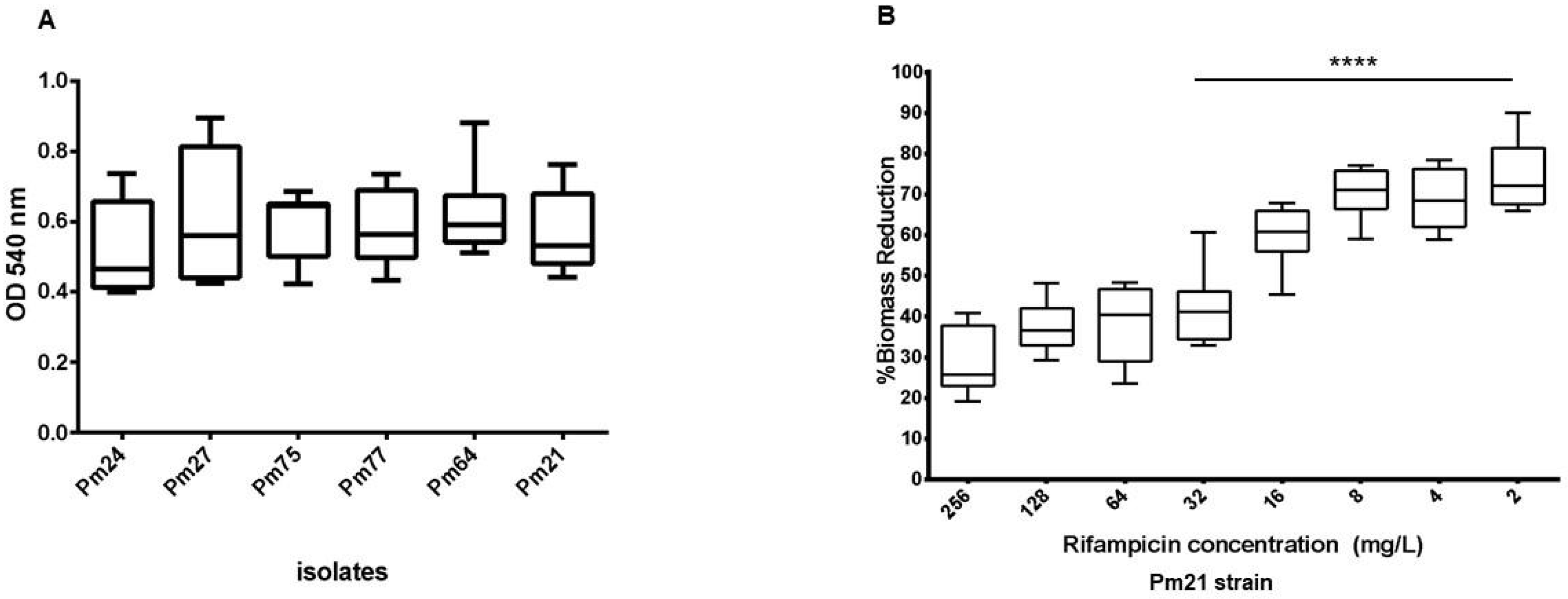
2.4. Biofilm Formation and Substrate-Specific Growth

A subset of six P. mirabilis isolates was selected from the 28 non-duplicate isolates analyzed for clonal relatedness (Section 2.3) to evaluate biofilm formation. Quantitative assays demonstrated that all isolates formed moderate biofilms, as evidenced by biomass measurements. The ODs of the 48 h biofilms stained with CV ranged from 0.398 to 0.894 (Figure 1A, Table S1). The data analysis showed no significant statistical differences in biofilm formation among the isolates. Furthermore, biofilm formation ability was assessed on Foley urinary catheter materials, revealing that biofilms were readily established on latex catheters, whereas silicone catheters showed minimal biofilm development (Figure S3), highlighting the material-dependent nature of biofilm formation in P. mirabilis.

2.5. Impact of Rifampicin on Established Biofilms

The MIC against rifampicin of the six P. mirabilis isolates was 16 mg/L. The effect of rifampicin on preformed P. mirabilis biofilms was investigated using the microplates model against the Pm21 isolate. Following treatment with rifampicin at concentrations ranging from 256 to 2 mg/l, the minimum biofilm inhibitory concentration (MIC-b) was determined to be 128 mg/L. The minimum regrowth concentration (MRC), the lowest concentration preventing microbial recovery, was ≥256 mg/L. However, biomass quantification after rifampicin exposure revealed a significant reduction starting at 32 mg/L when compared to levels of the untreated controls, as confirmed by statistical analysis (Figure 1B, Table S2). These findings underscore the potential of rifampicin to disrupt P. mirabilis biofilms, albeit at relatively high concentrations.

2.6. Genome Analysis

Whole genome sequencing (WGS) analysis of isolate Pm21, recovered from a urine sample, revealed a genome size of 4,265,609 bp, with a GC content of 39.07%, 81 contigs (>1000 bp), and an N50 value of 140,806 bp. Pm21 displayed a multilocus sequence typing (MLST) profile consistent with sequence type (ST) 135 and a virulence profile corresponding to vST138. The resistome analysis predicted multiple acquired antimicrobial resistance genes, including blaNDM-5 and blaCTX-M-15 (β-lactam resistance), rmtB, aadA1, aadA2, aadA5, aac(3)-IV, aac(6′)-Ib3, ant(3″)-Ia, aph(3′)-Ia, aph(6)-Id (aminoglycoside resistance), dfrA1, dfrA17, dfrA32 (trimethoprim resistance), sul1 and sul2 (sulfonamide resistance), ere(A) (macrolide resistance), catA1 (chloramphenicol), and tet(C), tet(J) (tetracycline resistance), among others (Table 2). Chromosomal point mutations were not detected since the PointFinder database is curated for species other than Proteus sp.

2.7. Genetic Analysis of Resistance Marker Environments

PlasmidFinder identified the IncQ1 replicon type as the only typable plasmid incompatibility group. The IncQ1 replicon was partially detected at the 5′ end of contig 49 (12,303 bp) with 100% identity, covering the first 524 bp of its total 796 bp length. The remaining portion of the replicon was not identified in any other contig within the Pm21 assembly or by mapping Illumina reads against its complete coding sequence. This suggests that contig 49 contains a truncated and likely non-functional IncQ1 replicon. BLAST (v1.4.0) analysis confirmed 100% identity and coverage of contig 49 with the chromosomes of P. mirabilis RGF134-1 (CP066833.1) and MPE0346 (CP053719.1), among others. In both animal-origin strains, this sequence is part of a chromosomal multidrug resistance genomic island (GI) containing sul2, aac(3)-IId, and aph(6)-Id, all located within contig 49 (Figure S4).
A bleomycin resistance protein coded by bleMBL was identified in contig 53 (6902 bp), downstream of the blaNDM-5 gene. A new assembly performed with plasmidSPAdes resulted in the generation of a longer contig (contig1_PlasmidSPAdes, 8258 bp, https://ri.conicet.gov.ar/handle/11336/252337, accessed on 25 May 2025). This allowed for the elucidation of the upstream region of blaNDM-5, where a truncated sequence of ISAba125 was identified (Figure S5). The blaNDM-5 genetic context in Pm21 was as follows: (5′–3′: ISAba125, partial sequence–blaNDM-5bleMBLtrpFdsbD–IS91 family transposase–qacEdelta1–sul1). The IS91 transposase coding gene was interrupted by ISKpn18, exactly as observed in the IncFIB-IncHI1B plasmids, resulting in the novel genetic platform variant described by González-Espinosa et al. in plasmids pM366-NDM-5 and pM40-NDM-5 (Figure 2) [12].
BLAST analysis of contig 1_plasmidSPAdes revealed that the top three hits, with 100% coverage and identity, correspond to three Klebsiella pneumoniae recently deposited plasmids in Argentina: pM387-NDM5 (accession number CP168953.1), pM40-NDM5 (accession number PQ247031.1), and pM366-NDM5 (accession number PQ247032.1). The last two plasmids were fully characterized [12] (https://doi.org/10.1016/j.jgar.2024.10.258). All the remaining 97 hits listed after the BLAST analysis showed 100% identity but only 84% coverage, including Escherichia coli strain Ec265 plasmid pEco265-NDM5 (PQ241462.1) and K. pneumoniae plasmid pM144-NDM-5 (PQ241463), which have also been reported in the country (Figure 2).
The whole genome Pm21 assembly was compared to reference sequences using PLSDB with the mash screen search strategy to identify plasmids contained within the sample. The analysis yielded 32 entries, of which 12 contained the blaNDM-5 gene exclusively in E. coli. From these 12 plasmids, only those sequenced both by Illumina and Oxford Nanopore Technologies were further analyzed (10/12) (Table S3). Among the 10 remaining plasmids, 8 had a length of 10,494 bp. BLAST analysis of these plasmids showed 100% coverage and >99.98% similarity among them. Of the remaining two plasmids, one (LC744474.1, 10,687 bp) displayed 95% coverage and >99.98% similarity with the others, while the second plasmid (LC744490.1, 13,652 bp) showed 83% coverage and >99.95% nucleotide similarity (Figure S6). Of the 10 analyzed plasmids, only NZ_CP048374.1, with a length of 10,494 bp, had been previously published in a study assessing the occurrence of carbapenemase-producing Enterobacteriaceae (CPE) in freshwater samples from rivers, inland canals, and streams across Switzerland. This plasmid, named pC-F-163_C, was identified in extraintestinal pathogenic E. coli ST167, which was nontypeable by incompatibility group [13]. It showed 100% coverage and nucleotide similarity to blaNDM-5-carrying contig- from the Pm21 assembly generated by PlasmidSPAdes.
Additionally, the Pm21 strain also harbored the ESBL blaCTX-M-15 and the 16S ribosomal RNA methyltransferase rmtB, which were present in different contigs, 57 (4058 bp) and 67 (1961 bp), respectively. blaCTX-M-15 was found to have an upstream IS1380-like element belonging to the ISEcp1 family of transposases, while the rmtB gene was found in association with two downstream genes encoding a proton antiporter (cdu2) and the chaperonin GroEL, identical to other rmtB-containing Enterobacterales isolates from clinical samples (GenBank accession numbers CP050367, MN061455, MN007141) [11].

2.8. Phylogenetic Tree

To explore the diversity of ST135 and provide a broader geographic context to Pm21, a core genome SNP phylogenetic tree was generated using all genomic sequences of ST135 P. mirabilis available at PubMLST (October 2024). The full dataset containing 123 P. mirabilis ST135 isolates is available at Google Spreadsheet-Proteus mirabilis ST135 dataset. The resulting phylogenetic tree is available at Microreact (https://microreact.org/project/7rKjP28HQQkktYCjLbNbJu-proteuspm21st135, accessed on 25 May 2025). Most isolates are of clinical origin, but some were isolated from food and unknown sources. Besides Pm21, only one isolate carried blaNDM-5 (ABJNEB000000000.3), which was isolated from a human rectal swab. Our phylogenetic analysis revealed a high degree of dispersion among the Proteus mirabilis ST135 isolates. Pm21 did not cluster closely to any of the analyzed genomes. The closest related genome in this analysis was JZ9, which belongs to a Chinese isolate recovered from chicken meat. The prevalence of genes related to bacterial virulence factors, including urease (ureC), flagella genes (flhA, fliF, fliG, fliP, fliL, flgN), fimbriae (mrpA, mrpH, ucaA, pmfA, pmpA, papC, papD, papF, papG, papH), hemolysin (hpmAB), biofilm formation (pstC, rcsD), autotransporters (ptaA, aipA), proteases (zapA), and siderophore-related (nrpR, ireA), was greater than 69% in all cases (nrpR showed the lowest prevalence 85/123). These results underscore the contribution of virulence determinants to the pathogenicity of ST135 P. mirabilis.

3. Discussion

This study documents an outbreak in an Argentine adult hospital attributed to P. mirabilis isolates co-producing NDM-5 MBL and CTX-M-15 ESBL. To date, reports of outbreaks caused by MDR P. mirabilis are scarce in the literature. Previously documented outbreaks have primarily involved P. mirabilis strains harboring carbapenemases such as NDM-1 [14] or VIM-4 [15], as well as ESBLs of the CTX-M-2, VEB-1, and TEM types [16,17,18].
While blaNDM-5 has been previously reported in P. mirabilis isolates from other countries, including China [19] and Austria [20], to our knowledge, this represents the initial documentation of blaNDM-5 in P. mirabilis in Argentina, and the first documented hospital outbreak caused by an NDM-producing P. mirabilis strain in the country.
Treatment of infections caused by such pathogens is often problematic due to their extensive drug resistance. The resistance pattern observed in these MDR P. mirabilis isolates generally reflects the hydrolysis spectrum of NDM-type enzymes, along with the presence of multiple aminoglycoside resistance markers and other antimicrobial resistance genes.
Despite carrying the CTX-M-15 ESBL, these isolates remained susceptible to aztreonam. Notably, this aztreonam susceptibility profile has also been observed in other clinical P. mirabilis isolates harboring ESBLs, AmpC, or carbapenemases, as previously reported by Shaaban et al. [21]. This antimicrobial agent, together with meropenem and piperacillin/tazobactam (when feasible), represented the main treatment options available for infections caused by this MDR P. mirabilis strain during the COVID-19 pandemic, a period marked by significant challenges in patient management. Following outbreak detection, the hospital’s infection control committee reinforced infection prevention measures, particularly hand hygiene practices and the change of personal protective equipment between patients. These strategies, already in place due to the COVID-19 pandemic, were intensified and played a key role in limiting further spread of the outbreak strain.
Since no surveillance cultures were performed on the index case at the time of hospital admission, it was not possible to determine whether this patient introduced the outbreak strain. Moreover, the fact that the first isolate was detected 50 days after hospitalization suggests that the strain was not acquired in the patient’s household or workplace but rather within the hospital from an unidentified source. In summary, the absence of surveillance cultures from the index case hampers the ability to trace the outbreak’s origins and fully characterize transmission dynamics. In addition, the absence of detailed clinical follow-up data, such as microbiological cure rates or patient outcomes, limited our ability to assess the full impact of the outbreak and the effectiveness of the treatments administered.
Given the high transmissibility and persistence of P. mirabilis-carrying NDM-5, effective infection control strategies are crucial for limiting further dissemination. Our findings suggest that, in line with repetitive sequence-based genomic amplification analyses, MALDI-TOF MS can aid in identifying clonally related groups of the opportunistic pathogen P. mirabilis. Supporting our results, previous studies have demonstrated the utility of MALDI-TOF MS as a first-line subtyping tool for the sensitive detection of potential dissemination events in hospital settings involving other opportunistic pathogens, such as Serratia marcescens, Citrobacter freundii [22], and Enterobacter species [23]. Although our clonal analysis relied primarily on REP-PCR, ERIC-PCR, and MALDI-TOF MS clustering, which are less discriminatory than whole genome sequencing-based approaches such as SNP analysis or cgMLST, the epidemiological data, including the unusually high frequency of carbapenem-resistant P. mirabilis isolates over a short time frame, support the occurrence of a clonal outbreak.
As observed in this study, P. mirabilis can cause catheter-associated urinary tract infections, largely due to its ability to form biofilms on catheter surfaces [24]. All six tested strains involved in this outbreak were classified as moderate biofilm producers in vitro. Notably, P. mirabilis is among the leading bacterial species responsible for biofilm-related infections associated with medical devices [25].
Interestingly, rifampicin exhibited potential for biofilm disruption at concentrations ≥32 µg/mL, although its clinical efficacy remains uncertain. Nwabor et al. reported that combining rifampicin with carbapenems enhanced its antibacterial activity and successfully eradicated established Acinetobacter baumannii biofilms [26]. Similarly, Amengol et al. demonstrated that colistin–rifampicin combinations effectively eradicated biofilms of both colistin-resistant and colistin-susceptible Pseudomonas aeruginosa [27]. These effects are likely related to the ability of rifampicin to penetrate the biofilm matrix (due to its lipophilic nature), inhibit RNA synthesis in metabolically active cells, and downregulate the expression of biofilm-associated genes [28]. However, to the best of our knowledge, no studies have specifically evaluated the effect of rifampicin on P. mirabilis biofilms.
We identified blaNDM-5 within a novel variant of the genetic platform recently described on multi-replicon IncFIB-IncHI1B plasmids in K. pneumoniae from Argentina. However, the antimicrobial resistance island containing blaNDM-5 in Pm21 could not be linked to any specific plasmid. BLAST analysis revealed that no more than 20% of pECO-265-NDM-5 and less than 15% of p366-NDM-5 were covered with Pm21 contigs (Figure S7A,B). pEco265-NDM-5 and pM144-NDM-5 belonged to the IncFII replicon type, encoding a MOB-F relaxase, whereas pM40-NDM-5 and pM366-NDM-5 were associated with multi-replicon IncFIB-IncHI1B plasmids, encoding a MOB-H relaxase [11,12]. Interestingly, neither the IncFII nor the IncHI1B replicons were detected in Pm21; however, the MOB-H relaxase was identified. blaNDM-5 has mainly been described in IncX3 plasmids as well as other Inc plasmids such as IncFII and IncI1, and in multi-replicon plasmids [29,30]. These findings indicate limited genetic similarity between circulating local plasmids and Pm21, raising the question of whether small, untypeable plasmids like pC-F-163 have already been circulating and integrating into local genetic platforms. This plasmid was determined to be highly similar at the nucleotide level (99–100%) to plasmids pM309-NDM5 and pM217_FII. Both plasmids were detected in nosocomial E. coli ST167 strains from a hematology ward in Myanmar between 2015 and 2016 [31]; (GenBank accession numbers AP018833.1 and AP018147.1, respectively). Additionally, pC-F-163_C showed 100% coverage and nucleotide similarity to plasmids circulating in Argentina, as described by González Espinosa et al. [12], including pM144-NDM-5, pEco265-NDM-5, pM40-NDM-5, and pM366-NDM-5, as well as to a blaNDM-5-carrying contig from a Pm21 assembly created by PlasmidSPAdes (Figure S8).
Pm21 also harbored the 16S ribosomal RNA methyltransferase rmtB, which was present in all plasmids described by González-Espinosa et al., except for pM144-NDM-5. Additionally, blaCTX-M-15 was detected in Pm21, as well as in the multi-replicon plasmids pM40-NDM-5 and pM366-NDM-5 [12].
The IncQ1 replicon was partially detected at the 5′ end of contig 49. We found that contig 49 displayed 100% identity and coverage with chromosomal multidrug resistance genomic islands (GI) in P. mirabilis of animal origin. According to the authors, they found six additional similar genomic islands, suggesting that the presence and spread of this IncQ1-harboring GI in P. mirabilis is not uncommon [32]. Interestingly, a BLAST search using the complete 796 bp repA sequence of IncQ1, filtered for P. mirabilis, yielded a total of 45 hits. Notably, all sequences with 66% coverage (~529 bp) were found in chromosomal DNA.
Our phylogenetic analysis demonstrated widespread dispersion among the P. mirabilis ST135 isolates, suggesting that MLST may not be a highly discriminatory tool for strain differentiation in this species. Given these findings, a more robust approach, such as core genome MLST (cgMLST), may be required for more precise strain typing, as proposed by Chen et al. [33]. However, despite its advantages, cgMLST has not yet been established as a standardized reference method for P. mirabilis, unlike in Klebsiella spp. This phylogenetic dispersion suggests the emergence of diverse NDM-producing P. mirabilis clones, reflecting a growing resistance threat. The global spread of multidrug-resistant strains, first reported in Italy and later in Poland, China, and Japan, underscores the need for effective prevention and control strategies [33].

4. Materials and Methods

4.1. Hospital Setting and Bacterial Isolates

This study was conducted at the Hospital General de Agudos “Carlos G. Durand” in Buenos Aires, Argentina, a 398-bed facility with intensive care units for adults and pediatric patients, intermediate care, a coronary unit, and general hospitalization wards. During the COVID-19 pandemic, the government implemented measures to optimize healthcare resources. Patient care was prioritized for SARS-CoV-2-infected individuals, as well as for emergency and critical cases. During periods of high bed occupancy, non-urgent medical interventions were postponed, based on medical assessment.
All bacterial isolates included in this study were recovered from clinical samples collected between October 2020 and April 2021 as part of routine diagnostic procedures. Samples were obtained by healthcare personnel in the corresponding inpatient services (e.g., ICU, medical clinic, surgery), following standard institutional protocols. Only clinical isolates were included in the analysis; no rectal swabs or surveillance cultures were used. For clonal analysis and further characterization, one isolate per patient was selected.
Following the initial detection of carbapenem-resistant P. mirabilis, a prospective survey was conducted to identify similar cases. Patient demographic data were saved from the laboratory database.

4.2. Bacterial Identification and Antimicrobial Susceptibility Testing (AST)

All clinical samples were processed at the hospital’s microbiology laboratory, following standard microbiological procedures. Proteus mirabilis identification was carried out using MALDI-TOF mass spectrometry (Vitek-MS®, bioMérieux, Marcy-l’Étoile, France), according to the manufacturer’s instructions. Antimicrobial susceptibility testing (AST) was performed using the Vitek 2C automated system (bioMérieux, Marcy-l’Étoile, France), also following the manufacturer’s protocols. MIC results were interpreted in accordance with the Clinical and Laboratory Standards Institute (CLSI) guidelines [34].
The minimum inhibitory concentration (MIC) of rifampicin was determined using the broth microdilution method in cation-adjusted Mueller–Hinton broth (MHB) [34], only for the six isolates included in the biofilm assays. Rifampicin was not assessed for therapeutic purposes, but rather to explore its potential activity against established biofilms.

4.3. Resistance Mechanisms

The production of carbapenemases was performed using the diffusion and synergy test, with meropenem (MEM), 10 µg, and imipenem (IPM), 10 µg discs (Laboratorio Britania, Buenos Aires, Argentina), interspersed with phenylboronic acid (PBA), 300 µg, and ethylenediaminetetraacetic acid (EDTA), 1 µmol discs, for assessing KPC and MBL, respectively [35].
Genotypic characterization of β-lactamase was carried out at Laboratorio de Resistencia Bacteriana (Facultad de Farmacia y Bioquímica, Universidad de Buenos Aires) using PCR amplification performed on the total DNA using specific primers for detection of metallo-carbapenemases (VIM, IMP and NDM) and extended spectrum β-lactamases (ESBLs) (CTX-M-G1, CTX-M-G2 and CTX-M-G9), under the conditions described previously [36,37]. Amplicons were sequenced on both strands using an ABI3730XL DNA Sequencer (Macrogen, Seoul, Republic of Korea).

4.4. Molecular Typing

The clonality of the isolates was determined by the homology relationships among fragments amplified by ERIC- (Enterobacterial Repetitive Intergenic Consensus) and REP- (Repetitive Extragenic Palindromic) PCR, according to the methods of Versalovic et al. [38].
Cluster analysis was performed using MALDI-TOF MS spectra with at least 110 peaks. A peak range from 3000 to 20,000 m/z was chosen for this clustering. Peaks were defined to be identical by applying a mass accuracy of 0.08% as the SARAMIS standard setting. Spectra were analyzed with a single link agglomerative clustering algorithm, applying the relative taxonomy analysis tool of SARAMIS premium software (v 4.1.1.0), to show the resulting dendrogram, with differences and similarities, in relative terms (percent matching masses). As a standard setting, the mass signal intensity was not considered in the cluster analysis [22].

4.5. Biofilm Formation Assays

A crystal violet (CV) assay was performed to evaluate the biofilm-forming ability of selected P. mirabilis isolates identified during the study period, using 96-well tissue-treated microplates [39]. Six isolates were selected as a representative subset of the outbreak strains, aiming to include the most common clinical sample sources: blood culture (three isolates, Pm24, Pm75, and Pm77), tracheal aspirate (Pm27), catheter (Pm64), and urine (Pm21, the sequenced isolate). Within these categories, the isolates were randomly chosen from different patients.
Briefly, isolates were subcultured on tryptic soy agar (TSA) for 24 h at 35 ± 2 °C, and a single colony from each was inoculated into lysogeny broth (LB) and grown overnight. The cultures were adjusted to a concentration of 106 CFU/mL and resuspended in fresh LB. Aliquots (200 µL) of the standardized inoculum were dispensed into sterile, flat-bottom 96-well polystyrene microplates and incubated at 35 °C for 48 h. Following incubation, the wells were washed with phosphate-buffered saline (PBS) to remove non-adherent cells and stained with 0.01% (w/v) CV for 30 min. The stained biofilms were then washed with distilled water to remove excess dye. To quantify biofilm biomass, the bound CV was solubilized with 95% (v/v) ethanol, and absorbance was measured at 540 nm using a multimode plate reader. Each isolate was tested in octuplicate.
The biofilm-forming ability of the isolates was classified into four categories based on OD values, following the methods of Stepanovic et al. [40]:
Non-biofilm formers: OD ≤ ODcut;
Weak biofilm formers: ODcut < OD ≤ 2 × ODcut;
Moderate biofilm formers: 2 × ODcut < OD ≤ 4 × ODcut;
Strong biofilm formers: OD > 4 × ODcut.
ODcut was defined as the mean OD of the negative control plus its standard deviation.
To assess biofilm formation on a medical device, sections of latex and silicone Foley urinary catheter (2 cm in length) were cut under sterile conditions and incubated statically at 35 °C in 4 mL of LB containing 106 CFU/mL of the Pm21 isolate. After 24 h, the catheter segments were removed, washed with PBS to eliminate non-adherent bacteria, and stained with CV, as described by Passerini de Rossi et al. (2012) [41]. The stained segments were then visually inspected for biofilm formation.

4.6. Biofilm Antimicrobial Susceptibility

Biofilm susceptibility testing of the Pm21 isolate was performed as described previously [39,42], with minor modifications. Briefly, aliquots (150 µL) of the standardized inoculum, as previously described, were added to the wells of a 96-well microplate and incubated at 35 °C for 48 h to allow for biofilm formation. After incubation, the medium was aseptically removed, the wells were washed with PBS, and 200 µL of rifampicin at two-fold serial dilutions (ranging from 256 mg/L to 2 mg/L) prepared in LB was added to the preformed biofilms.
Following overnight incubation, MIC-b was determined as the lowest antibiotic concentration that prevented the establishment of a planktonic bacterial population from the biofilm (i.e., no visible planktonic growth). The antibiotic solutions were then removed, the wells were washed, and 200 µL of LB was added. After 24 h of incubation at 35 °C, biofilm viability was assessed visually. The minimum regrowth concentration (MRC) was defined as the lowest antibiotic concentration at which bacteria failed to regrow [39,42]. Sterility and antibiotic-free controls were included in all experiments. Each condition was tested in octuplicate.
Additionally, the effect of rifampicin on biofilm biomass was assessed. Biofilms of Pm21 exposed to rifampicin (256 mg/L to 2 mg/L) were stained with CV, as previously described. Biomass reduction at each antibiotic concentration (eight wells per dilution) was quantified by calculating the ratio between the OD540 values of the treated and untreated biofilms.

4.7. Statistical Analysis

Differences in biofilm biomass between untreated and rifampicin-treated samples were assessed using the Kruskal–Wallis test, followed by Dunn’s multiple comparison test. Given the small sample size and uncertainty about the underlying distribution of the data, a non-parametric approach was selected as a conservative alternative to ANOVA. All p-values were calculated using one-tailed tests, with a significance level of 0.01. Statistical analyses and graphical representations were performed using GraphPad Software 6.01.

4.8. Whole Genome Sequencing Analysis

Whole genome sequencing (WGS) was performed on isolate Pm21, selected as a representative strain among the group of MDR P. mirabilis isolates recovered during the study period. This isolate was obtained from a urine sample. WGS of isolate Pm21 was performed through short reads on the Illumina NextSeq platform (Department of Microbiology of the Institute of Biomedical Sciences, Universidad de São Paulo, Brazil). Paired-end reads were de novo assembled into contigs using Unicycler (0.5.0+galaxy1) on the Galaxy platform (https://usegalaxy.eu/, accessed on 25 May 2025). Automated annotation was achieved with Prokka v.1.14.6, and the results were manually curated. The whole genome shotgun project of P. mirabilis Pm21 has been deposited in GenBank (SRA) under accession no. PRJNA1236692. The contig containing blaNDM-5 (contig 53) was subsequently annotated with BAKTA software (v 1.9.4) [43] to determine its genetic context. A new assembly was performed using plasmidSPAdes (v3.15.5) to obtain a longer contig. Sequence comparisons were performed using BLAST analysis. Strain typing was conducted using the Public Database for Molecular Typing and Microbial Genome Diversity (PubMLST; https://pubmlst.org/organisms/proteus-spp, accessed on 25 May 2025). The resistome was identified through ResFinder (22 March 2024) hosted on the Center for Genomic Epidemiology web page (http://genepi.food.dtu.dk/resfinder, accessed on 25 May 2025). Plasmid detection was carried out with PlasmidFinder v2.0.1, integrated into the staramr tool (v 0.11.0) [44]. Additionally, PLSDB, using the Mash screen search strategy, was employed to identify potential plasmid-related sequences and assess their similarity to those of previously reported plasmids [45,46,47]. To characterize genomic islands, in silico detection was performed using chromosomes deposited in NCBI and the IslandViewer 4 web server (http://www.pathogenomics.sfu.ca/islandviewer/, accessed on 25 May 2025) [48]. The graphical comparison of genomic islands was visualized with Clinker (https://cagecat.bioinformatics.nl/tools/clinker, accessed on 25 May 2025).
To explore ST135 diversity, a phylogenetic tree was built including Pm21 and all genomic sequences of ST135 P. mirabilis available at PubMLST (https://pubmlst.org/bigsdb?db=pubmlst_proteus_isolates&page=query&genomes=1, accessed on 25 May 2025). Briefly, core genome SNPs for ST135 genomes were determined using snippy (v4.6.0) [49], with the oldest available P. mirabilis genome (GB08, GCF_001617295.1) as a reference. Recombinant regions were removed using Gubbins (v3.3) [50]. Afterwards, an snp-only alignment was generated from the recombination-free Gubbins output using SNP-sites (v2.5.1) [51], which was used as the input for IQ-TREE (v1.6.12) [52]. The tree was built using the best fit model, determined with the function ‘-m TEST’ (TVM+F+ASC) and 1000 bootstraps. The tree was visualized with Microreact (https://microreact.org/, accessed on 25 May 2025) [53]. For all ST135 genomes, plasmid replicons, virulence, and antimicrobial resistance genes were detected using ABRicate, with different databases and thresholds: PlasmidFinder (default settings); a custom database with P. mirabilis virulence-related reference sequences; the Virulence Factor Database (VFDB) with an identity threshold of 70% and coverage of 60%; and ResFinder (default settings), respectively.

5. Conclusions

This study reports the first hospital outbreak in Argentina caused by P. mirabilis producing NDM-5, involving 40 patients during the COVID-19 pandemic. The outbreak strain exhibited an MDR profile, including blaCTX-M-15 and rmtB genes, and was confirmed as clonal by REP-/ERIC-PCR and MALDI-TOF MS analysis. All tested isolates formed moderate biofilms, and rifampicin reduced biofilm biomass in vitro at high concentrations. Genomic analysis revealed blaNDM-5 associated with the same genetic environment recently identified in K. pneumoniae in Argentina. No plasmid incompatibility groups other than partial IncQ1 were detected in this isolate. However, IncQ1 could not be linked to blaNDM-5.
These findings underscore the value of local epidemiological surveillance for early detection of unusual resistance profiles, which may indicate the beginning of an outbreak. Molecular characterization is essential to define such events and guide effective control measures. Although the transmission route was not determined, it may be related to the pandemic context, characterized by increased patient condition severity, antibiotic use, and invasive procedures. Strengthening genomic surveillance and infection control remains critical to limit the spread of high-risk MDR clones.

Supplementary Materials

The following supporting information can be downloaded at: https://www.mdpi.com/article/10.3390/antibiotics14060557/s1, Figure S1: Electrophoretic profiles of NDM-5-producing P. mirabilis isolates. Figure S2: Dendrogram of the MALDI-TOF spectra of P. mirabilis isolates using SARAMIS. Figure S3: Biofilm formation by Proteus mirabilis on different catheter materials. Figure S4: Linear comparison diagram of multi-drug resistance genomic island of RGF134-1, MPE0346 and contig 49 of Pm21. Figure S5. Comparison of blaNDM-5 genetic context in Proteus mirabilis Pm21. Figure S6. Nucleotide similarity and coverage of plasmids from PLSDB that displayed high identity with P. mirabilis Pm21 assembly and contained blaNDM-5. Figure S7. (A). Comparison among pECO-265-NDM-5 (used as a template), pM144-NDM-5, and the Pm21 assembly. BLAST analysis revealed no more than 20% coverage between Pm21 and pECO-265-NDM-5. Graphics were generated using Proksee, with relevant features annotated using CARD and mobileOG-DB. (B). Comparison among pM40-NDM-5 (used as a template), pM366-NDM-5, pECO-265-NDM-5 and the Pm21 assembly. BLAST analysis revealed less than 15% with pM40-NDM-5 and p366-NDM-5. Only CDSs identified by gene names using Bakta were included in the graphic to simplify comprehension. Figure S8. Nucleotide similarity and coverage of plasmid NZ_CP048374.1 (10,494 bp) with plasmids circulating in Argentina, including pM144-NDM-5, pEco265-NDM-5, pM40-NDM-5, and pM366-NDM-5, as well as with the Pm21 contig assembled using PlasmidSPAdes, all showing 100% coverage and nucleotide similarity. Table S1: Biofilm formation (OD540 values) of Proteus mirabilis isolates obtained from different patients and different types of samples (Pm24, Pm75* and Pm77* from blood cultures, Pm27 from tracheal aspirate, Pm64* from catheter and Pm21 from urine). Table S2: Effect of rifampicin on biofilm biomass (OD540 values) of Proteus mirabilis Pm21 strain. Table S3: Plasmids from PLSDB that displayed high identity with Proteus mirabilis Pm21 assembly and contained blaNDM-5.

Author Contributions

B.G.: Conceptualization, methodology, data curation, validation, formal analysis, investigation, writing—original draft preparation, writing—review and editing. A.P.R.: conceptualization, methodology, data curation, validation, formal analysis, investigation, writing—review and editing. M.S.H. and L.E.F.: methodology, data curation, validation, formal analysis, writing—review and editing. N.L.: methodology, validation. M.E.O.: conceptualization, data curation, validation, investigation, writing—review and editing. J.A.D.C.: conceptualization, methodology, data curation, validation, formal analysis, investigation, resources, writing—original draft preparation, writing—review and editing, supervision, funding acquisition. All authors have read and agreed to the published version of the manuscript.

Funding

This research was funded by Agencia Nacional de Promoción Científica y Tecnológica (PICT 2019-1879) and University of Buenos Aires: UBACyT 2023 (20020220300012BA) to J.A.D.C.

Institutional Review Board Statement

The study was approved by the Ethics Committee of the Facultad de Farmacia y Bioquímica (Universidad de Buenos Aires) (RESCD-2020-134-E-UBA-DCT_FFYB, 20 August 2020) for studies involving bacteria recovered from human clinical samples.

Informed Consent Statement

Not applicable.

Data Availability Statement

The whole genome shotgun project of P. mirabilis Pm21 has been deposited in GenBank under the Sequence Read Archive (SRA) accession no. submission: PRJNA1236692. The genome assembly (in Fasta format) used in this paper was deposited in the CONICET Digital Institutional Repository (https://ri.conicet.gov.ar/handle/11336/252337, accessed on 25 May 2025). Other data presented in this study are available upon request from the corresponding author.

Acknowledgments

The authors would like to thank María del Valle Cruz and Florencia Neskovic for their technical support and BRITANIA for the donation of supplies.

Conflicts of Interest

The authors declare no conflicts of interest.

Abbreviations

The following abbreviations are used in this manuscript:
AMKamikacin
ASTantimicrobial susceptibility testing
AZTaztreonam
CAZceftazidime
CPEcarbapenemase-producing Enterobacteriaceae
CFUcolony forming unit
CIPciprofloxacin
CLSIClinical and Laboratory Standards Institute
CTXcefotaxime
CVcrystal violet
EDTAethylenediaminetetraacetic Acid
ERICEnterobacterial Repetitive Intergenic Consensus
ESBLextended spectrum β-lactamase
FEPcefepime
GENgentamicin
GIgenomic island
IPMImipenem
LBlysogeny broth
MALDI-TOF MSmatrix-assisted laser desorption/ionization time-of-flight mass spectrometry
MBminimum biofilm MIC (assumed: MIC-b)
MBLmetallo-β-lactamase
MDRmultidrug resistant
MEMmeropenem
MICminimum inhibitory concentration
MIC-bminimum inhibitory concentration in the biofilm plate
MLSTmultilocus sequence typing
MRCminimum regrowth concentration
ODoptical density
PBAphenylboronic acid
PBSphosphate-buffered saline
PTZpiperacillin/tazobactam
REP-PCRrepetitive extragenic palindromic PCR
STsequence type
TMStrimethoprim/sulfamethoxazole
TSAtryptic soy agar
TSBtryptic soy broth
WGSwhole genome sequencing
cgMLSTcore genome multilocus sequence typing

References

  1. Zhu, X.; Zhang, Y.; Shen, Z.; Xia, L.; Wang, J.; Zhao, L.; Wang, K.; Wang, W.; Hao, Z.; Liu, Z. Characterization of NDM-1-Producing Carbapenemase in Proteus Mirabilis among Broilers in China. Microorganisms 2021, 9, 2443. [Google Scholar] [CrossRef] [PubMed]
  2. Liu, M.; Li, D.; Jia, W.; Ma, J.; Zhao, X. Study of the Molecular Characteristics and Homology of Carbapenem-Resistant Proteus Mirabilis by Whole Genome Sequencing. J. Med. Microbiol. 2023, 72, 001648. [Google Scholar] [CrossRef]
  3. Bitar, I.; Mattioni Marchetti, V.; Mercato, A.; Nucleo, E.; Anesi, A.; Bracco, S.; Rognoni, V.; Hrabak, J.; Migliavacca, R. Complete Genome and Plasmids Sequences of a Clinical Proteus Mirabilis Isolate Producing Plasmid Mediated NDM-1 From Italy. Microorganisms 2020, 8, 339. [Google Scholar] [CrossRef] [PubMed]
  4. Fusco, A.; Coretti, L.; Savio, V.; Buommino, E.; Lembo, F.; Donnarumma, G. Biofilm Formation and Immunomodulatory Activity of Proteus Mirabilis Clinically Isolated Strains. Int. J. Mol. Sci. 2017, 18, 414. [Google Scholar] [CrossRef]
  5. Wilks, S.A.; Fader, M.J.; Keevil, C.W. Novel Insights into the Proteus Mirabilis Crystalline Biofilm Using Real-Time Imaging. PLoS ONE 2015, 10, e0141711. [Google Scholar] [CrossRef]
  6. Ficik, J.; Andrezál, M.; Drahovská, H.; Böhmer, M.; Szemes, T.; Liptáková, A.; Slobodníková, L. Carbapenem-Resistant Klebsiella Pneumoniae in COVID-19 Era—Challenges and Solutions. Antibiotics 2023, 12, 1285. [Google Scholar] [CrossRef]
  7. Li, Y.; Yin, M.; Fang, C.; Fu, Y.; Dai, X.; Zeng, W.; Zhang, L. Genetic Analysis of Resistance and Virulence Characteristics of Clinical Multidrug-Resistant Proteus Mirabilis Isolates. Front. Cell. Infect. Microbiol. 2023, 13, 1229194. [Google Scholar] [CrossRef] [PubMed]
  8. Hua, X.; Zhang, L.; Moran, R.A.; Xu, Q.; Sun, L.; van Schaik, W.; Yu, Y. Cointegration as a Mechanism for the Evolution of a KPC-Producing Multidrug Resistance Plasmid in Proteus Mirabilis. Emerg. Microbes Infect. 2020, 9, 1206–1218. [Google Scholar] [CrossRef]
  9. He, J.; Sun, L.; Zhang, L.; Leptihn, S.; Yu, Y.; Hua, X. A Novel SXT/R391 Integrative and Conjugative Element Carries Two Copies of the blaNDM-1 Gene in Proteus Mirabilis. mSphere 2021, 6, e0058821. [Google Scholar] [CrossRef]
  10. Fritzenwanker, M.; Falgenhauer, J.; Hain, T.; Imirzalioglu, C.; Chakraborty, T.; Yao, Y. The Detection of Extensively Drug-Resistant Proteus Mirabilis Strains Harboring Both VIM-4 and VIM-75 Metallo-β-Lactamases from Patients in Germany. Microorganisms 2025, 13, 266. [Google Scholar] [CrossRef]
  11. Costa, A.; Figueroa-Espinosa, R.; Gaudenzi, F.; Lincopan, N.; Fuga, B.; Ghiglione, B.; Gutkind, G.; Di Conza, J. Co-Occurrence of NDM-5 and RmtB in a Clinical Isolate of Escherichia Coli Belonging to CC354 in Latin America. Front. Cell. Infect. Microbiol. 2021, 11, 654852. [Google Scholar] [CrossRef] [PubMed]
  12. González-Espinosa, F.; Di Pilato, V.; Calabrese, L.; Costa, E.; Costa, A.; Gutkind, G.; Cejas, D.; Radice, M. Integral Genomic Description of BlaNDM-5-Harbouring Plasmids Recovered from Enterobacterales in Argentina. J. Glob. Antimicrob. Resist. 2024, 39, 224–226. [Google Scholar] [CrossRef] [PubMed]
  13. Bleichenbacher, S.; Stevens, M.J.A.; Zurfluh, K.; Perreten, V.; Endimiani, A.; Stephan, R.; Nüesch-Inderbinen, M. Environmental Dissemination of Carbapenemase-Producing Enterobacteriaceae in Rivers in Switzerland. Environ. Pollut. 2020, 265, 115081. [Google Scholar] [CrossRef]
  14. Yang, L.; He, H.; Chen, Q.; Wang, K.; Lin, Y.; Li, P.; Li, J.; Liu, X.; Jia, L.; Song, H.; et al. Nosocomial Outbreak of Carbapenemase-Producing Proteus Mirabilis With Two Novel Salmonella Genomic Island 1 Variants Carrying Different blaNDM–1 Gene Copies in China. Front. Microbiol. 2022, 12, 800938. [Google Scholar] [CrossRef]
  15. Protonotariou, E.; Poulou, A.; Politi, L.; Meletis, G.; Chatzopoulou, F.; Malousi, A.; Metallidis, S.; Tsakris, A.; Skoura, L. Clonal Outbreak Caused by VIM-4-Producing Proteus Mirabilis in a Greek Tertiary-Care Hospital. Int. J. Antimicrob. Agents 2020, 56, 106060. [Google Scholar] [CrossRef]
  16. Nakano, R.; Nakano, A.; Abe, M.; Inoue, M.; Okamoto, R. Regional Outbreak of CTX-M-2 β-Lactamase-Producing Proteus Mirabilis in Japan. J. Med. Microbiol. 2012, 61, 1727–1735. [Google Scholar] [CrossRef]
  17. Cremet, L.; Bemer, P.; Rome, J.; Juvin, M.-E.; Navas, D.; Bourigault, C.; Guillouzouic, A.; Caroff, N.; Lepelletier, D.; Asseray, N.; et al. Outbreak Caused by Proteus Mirabilis Isolates Producing Weakly Expressed TEM-Derived Extended-Spectrum β-Lactamase in Spinal Cord Injury Patients with Recurrent Bacteriuria. Scand. J. Infect. Dis. 2011, 43, 957–961. [Google Scholar] [CrossRef] [PubMed]
  18. Jain, S.; Gaind, R.; Kothari, C.; Sehgal, R.; Shamweel, A.; Thukral, S.S.; Chellani, H.K. VEB-1 Extended-Spectrum β-Lactamase-Producing Multidrug-Resistant Proteus Mirabilis Sepsis Outbreak in a Neonatal Intensive Care Unit in India: Clinical and Diagnostic Implications. JMM Case Rep. 2016, 3, e005056. [Google Scholar] [CrossRef]
  19. Sun, L.; Xu, J.; He, F. Genomic Characterisation of a Proteus mirabilis Clinical Isolate from China Carrying blaNDM-5 on an IncX3 Plasmid. J. Glob. Antimicrob. Resist. 2019, 19, 317–319. [Google Scholar] [CrossRef]
  20. Valentin, T.; Feierl, G.; Masoud-Landgraf, L.; Kohek, P.; Luxner, J.; Zarfel, G. Proteus mirabilis Harboring Carbapenemase NDM-5 and ESBL VEB-6 Detected in Austria. Diagn. Microbiol. Infect. Dis. 2018, 91, 284–286. [Google Scholar] [CrossRef]
  21. Shaaban, M.; Elshaer, S.L.; Abd El-Rahman, O.A. Prevalence of Extended-Spectrum β-Lactamases, AmpC, and Carbapenemases in Proteus mirabilis Clinical Isolates. BMC Microbiol. 2022, 22, 247. [Google Scholar] [CrossRef] [PubMed]
  22. Rödel, J.; Mellmann, A.; Stein, C.; Alexi, M.; Kipp, F.; Edel, B.; Dawczynski, K.; Brandt, C.; Seidel, L.; Pfister, W.; et al. Use of MALDI-TOF Mass Spectrometry to Detect Nosocomial Outbreaks of Serratia Marcescens and Citrobacter Freundii. Eur. J. Clin. Microbiol. Infect. Dis. 2019, 38, 581–591. [Google Scholar] [CrossRef] [PubMed]
  23. De Florio, L.; Riva, E.; Giona, A.; Dedej, E.; Fogolari, M.; Cella, E.; Spoto, S.; Lai, A.; Zehender, G.; Ciccozzi, M.; et al. MALDI-TOF MS Identification and Clustering Applied to Enterobacter Species in Nosocomial Setting. Front. Microbiol. 2018, 9, 1885. [Google Scholar] [CrossRef]
  24. Wasfi, R.; Hamed, S.M.; Amer, M.A.; Fahmy, L.I. Proteus Mirabilis Biofilm: Development and Therapeutic Strategies. Front. Cell Infect. Microbiol. 2020, 10, 414. [Google Scholar] [CrossRef]
  25. Bouhrour, N.; Nibbering, P.H.; Bendali, F. Medical Device-Associated Biofilm Infections and Multidrug-Resistant Pathogens. Pathogens 2024, 13, 393. [Google Scholar] [CrossRef]
  26. Nwabor, L.C.; Chukamnerd, A.; Nwabor, O.F.; Pomwised, R.; Voravuthikunchai, S.P.; Chusri, S. Rifampicin Enhanced Carbapenem Activity with Improved Antibacterial Effects and Eradicates Established Acinetobacter Baumannii Biofilms. Pharmaceuticals 2023, 16, 477. [Google Scholar] [CrossRef] [PubMed]
  27. Armengol, E.; Kragh, K.N.; Tolker-Nielsen, T.; Sierra, J.M.; Higazy, D.; Ciofu, O.; Viñas, M.; Høiby, N. Colistin Enhances Rifampicin’s Antimicrobial Action in Colistin-Resistant Pseudomonas Aeruginosa Biofilms. Antimicrob. Agents Chemother. 2023, 67, e01641-22. [Google Scholar] [CrossRef]
  28. Ferreira, L.; Pos, E.; Nogueira, D.R.; Ferreira, F.P.; Sousa, R.; Abreu, M.A. Antibiotics with Antibiofilm Activity—Rifampicin and Beyond. Front. Microbiol. 2024, 15, 1435720. [Google Scholar] [CrossRef]
  29. Marchetti, V.M.; Bitar, I.; Mercato, A.; Nucleo, E.; Bonomini, A.; Pedroni, P.; Hrabak, J.; Migliavacca, R. Complete Nucleotide Sequence of Plasmids of Two Escherichia Coli Strains Carrying blaNDM–5 and blaNDM–5 and blaOXA–181 From the Same Patient. Front. Microbiol. 2020, 10, 3095. [Google Scholar] [CrossRef]
  30. Hornsey, M.; Phee, L.; Wareham, D.W. A Novel Variant, NDM-5, of the New Delhi Metallo-β-Lactamase in a Multidrug-Resistant Escherichia Coli ST648 Isolate Recovered from a Patient in the United Kingdom. Antimicrob. Agents Chemother. 2011, 55, 5952–5954. [Google Scholar] [CrossRef]
  31. Sugawara, Y.; Akeda, Y.; Hagiya, H.; Sakamoto, N.; Takeuchi, D.; Shanmugakani, R.K.; Motooka, D.; Nishi, I.; Zin, K.N.; Aye, M.M.; et al. Spreading Patterns of NDM-Producing Enterobacteriaceae in Clinical and Environmental Settings in Yangon, Myanmar. Antimicrob. Agents Chemother. 2019, 63, e01924-18. [Google Scholar] [CrossRef] [PubMed]
  32. Wang, Q.; Peng, K.; Liu, Y.; Xiao, X.; Wang, Z.; Li, R. Characterization of TMexCD3-TOprJ3, an RND-Type Efflux System Conferring Resistance to Tigecycline in Proteus Mirabilis, and Its Associated Integrative Conjugative Element. Antimicrob. Agents Chemother. 2021, 65, 10–1128. [Google Scholar] [CrossRef]
  33. Chen, S.L.; Kang, Y.T.; Liang, Y.H.; Qiu, X.T.; Li, Z.J. A Core Genome Multilocus Sequence Typing Scheme for Proteus Mirabilis. Biomed. Environ. Sci. 2023, 36, 343–352. [Google Scholar] [CrossRef]
  34. CLSI M100 Performance Standards for Antimicrobial Susceptibility Testing, 30th ed.; CLSI Supplement M100 Clinical and Laboratory Standards Institute: Wayne, PA, USA, 2020.
  35. Cordeiro-Moura, J.R.; Fehlberg, L.C.C.; Nodari, C.S.; de Matos, A.P.; de Alves, V.O.; Cayô, R.; Gales, A.C. Performance of Distinct Phenotypic Methods for Carbapenemase Detection: The Influence of Culture Media. Diagn. Microbiol. Infect. Dis. 2020, 96, 114912. [Google Scholar] [CrossRef] [PubMed]
  36. Dominguez, J.E.; Redondo, L.M.; Figueroa Espinosa, R.A.; Cejas, D.; Gutkind, G.O.; Chacana, P.A.; Di Conza, J.A.; Fernández Miyakawa, M.E. Simultaneous Carriage of Mcr-1 and Other Antimicrobial Resistance Determinants in Escherichia Coli From Poultry. Front. Microbiol. 2018, 9, 1679. [Google Scholar] [CrossRef] [PubMed]
  37. Marchisio, M.L.; Liebrenz, K.I.; Méndez, E.d.l.A.; Di Conza, J.A. Molecular Epidemiology of Cefotaxime-Resistant but Ceftazidime-Susceptible Enterobacterales and Evaluation of the in Vitro Bactericidal Activity of Ceftazidime and Cefepime. Braz. J. Microbiol. 2021, 52, 1853–1863. [Google Scholar] [CrossRef]
  38. Versalovic, J.; Koeuth, T.; Lupski, R. Distribution of Repetitive DNA Sequences in Eubacteria and Application to Finerpriting of Bacterial Enomes. Nucleic Acids Res. 1991, 19, 6823–6831. [Google Scholar] [CrossRef]
  39. Passerini de Rossi, B.; García, C.; Calenda, M.; Vay, C.; Franco, M. Activity of Levofloxacin and Ciprofloxacin on Biofilms and Planktonic Cells of Stenotrophomonas Maltophilia Isolates from Patients with Device-Associated Infections. Int. J. Antimicrob. Agents 2009, 34, 260–264. [Google Scholar] [CrossRef]
  40. Stepanovic, S.; Vukovic, D.; Dakic, I.; Savic, B.; Svabic-Vlahovic, M. A Modified Microtiter-Plate Test for Quantification of Staphylococcal Biofilm Formation. J. Microbiol. Methods 2000, 40, 175–179. [Google Scholar] [CrossRef]
  41. Passerini de Rossi, B.; Feldman, L.; Pineda, M.S.; Vay, C.; Franco, M. Comparative in Vitro Efficacies of Ethanol-, EDTA- and Levofloxacin-Based Catheter Lock Solutions on Eradication of Stenotrophomonas Maltophilia Biofilms. J. Med. Microbiol. 2012, 61, 1248–1253. [Google Scholar] [CrossRef]
  42. Cernohorská, L.; Votava, M. Determination of Minimal Regrowth Concentration (MRC) in Clinical Isolates of Various Biofilm-Forming Bacteria. Folia Microbiol. 2004, 49, 75–78. [Google Scholar] [CrossRef] [PubMed]
  43. Schwengers, O.; Jelonek, L.; Dieckmann, M.A.; Beyvers, S.; Blom, J.; Goesmann, A. Bakta: Rapid and Standardized Annotation of Bacterial Genomes via Alignment-Free Sequence Identification. Microb. Genom. 2021, 7, 000685. [Google Scholar] [CrossRef]
  44. Bharat, A.; Petkau, A.; Avery, B.P.; Chen, J.C.; Folster, J.P.; Carson, C.A.; Kearney, A.; Nadon, C.; Mabon, P.; Thiessen, J.; et al. Correlation between Phenotypic and In Silico Detection of Antimicrobial Resistance in Salmonella Enterica in Canada Using Staramr. Microorganisms 2022, 10, 292. [Google Scholar] [CrossRef] [PubMed]
  45. Galata, V.; Fehlmann, T.; Backes, C.; Keller, A. PLSDB: A Resource of Complete Bacterial Plasmids. Nucleic Acids Res. 2019, 47, D195–D202. [Google Scholar] [CrossRef]
  46. Molano, L.-A.G.; Hirsch, P.; Hannig, M.; Müller, R.; Keller, A. The PLSDB 2025 Update: Enhanced Annotations and Improved Functionality for Comprehensive Plasmid Research. Nucleic Acids Res. 2025, 53, D189–D196. [Google Scholar] [CrossRef]
  47. Schmartz, G.P.; Hartung, A.; Hirsch, P.; Kern, F.; Fehlmann, T.; Müller, R.; Keller, A. PLSDB: Advancing a Comprehensive Database of Bacterial Plasmids. Nucleic Acids Res. 2022, 50, D273–D278. [Google Scholar] [CrossRef]
  48. Siebor, E.; Neuwirth, C. Proteus Genomic Island 1 (PGI1), a New Resistance Genomic Island from Two Proteus Mirabilis French Clinical Isolates. J. Antimicrob. Chemother. 2014, 69, 3216–3220. [Google Scholar] [CrossRef]
  49. Seemann, T. Snippy: Rapid Haploid Variant Calling and Core Genome Alignment. Available online: https://github.com/tseemann/snippy (accessed on 1 March 2025).
  50. Croucher, N.J.; Page, A.J.; Connor, T.R.; Delaney, A.J.; Keane, J.A.; Bentley, S.D.; Parkhill, J.; Harris, S.R. Rapid Phylogenetic Analysis of Large Samples of Recombinant Bacterial Whole Genome Sequences Using Gubbins. Nucleic Acids Res. 2015, 43, e15. [Google Scholar] [CrossRef] [PubMed]
  51. Page, A.J.; Taylor, B.; Delaney, A.J.; Soares, J.; Seemann, T.; Keane, J.A.; Harris, S.R. SNP-Sites: Rapid Efficient Extraction of SNPs from Multi-FASTA Alignments. Microb. Genom. 2016, 2, e000056. [Google Scholar] [CrossRef]
  52. Nguyen, L.-T.; Schmidt, H.A.; von Haeseler, A.; Minh, B.Q. IQ-TREE: A Fast and Effective Stochastic Algorithm for Estimating Maximum-Likelihood Phylogenies. Mol. Biol. Evol. 2015, 32, 268–274. [Google Scholar] [CrossRef]
  53. Argimón, S.; Abudahab, K.; Goater, R.J.E.; Fedosejev, A.; Bhai, J.; Glasner, C.; Feil, E.J.; Holden, M.T.G.; Yeats, C.A.; Grundmann, H.; et al. Microreact: Visualizing and Sharing Data for Genomic Epidemiology and Phylogeography. Microb. Genom. 2016, 2, e000093. [Google Scholar] [CrossRef] [PubMed]
Figure 1. (A) Biofilm formation of P. mirabilis isolates obtained from different types of samples. (B) Effect of rifampicin on biofilm biomass of P. mirabilis Pm21 strain. Asterisks (****) indicate p < 0.0001 for biomass reduction measurement.
Antibiotics 14 00557 g001
Figure 2. Comparison of the Pm21 contig and its genetic context with IncFII and multi-replicon IncFIB-HI1B plasmids, recently reported in Argentina, carrying blaNDM-5. The visualization was generated using Clinker (v 0.0.31). Arrows represent coding sequences, with homologous genes depicted in the same color and aligned based on sequence similarity.
Antibiotics 14 00557 g002
Table 1. Demographic and clinical characteristics of 40 patients involved in the NDM-5-producing Proteus mirabilis outbreak.
PatientAgeSexCOVID StatusSample Origin *Date #Sample Source (n)
161MpositiveICU29 September 2020blood culture (1), urine (1)
242MpositiveMCU1 October 2020catheter (2), tracheal aspirate (2)
352MunknownICU8 October 2020catheter (1)
445MpositiveICU20 October 2020urine (1)
551MpositiveICU23 October 2020urine (1), tracheal aspirate (1)
637FnegativeICU27 October 2020catheter (1), blood culture (1)
760MpositiveICU30 October 2020blood culture (2)
846MpositiveICU7 November 2020catheter (1)
953FpositiveICU8 November 2020blood culture (2), tracheal aspirate (2)
1057MpositiveICU11 November 2020blood culture (1), tracheal aspirate (1), urine (1)
1145MnegativeMCU22 November 2020urine (1), blood culture (1), catheter (1)
1266FnegativeGER25 November 2020blood culture (1)
1347FunknownMCU25 November 2020blood culture (1)
1450FunknownICU27 November 2020blood culture (1)
1553FnegativeGS2 December 2020tracheal aspirate (1), miscellaneous (1)
1651MnegativeMCU5 December 2020blood culture (1)
1755FunknownMCU6 December 2020miscellaneous (1)
1845MpositiveMCU20 December 2020blood culture (1)
1957MunknownGER7 January 2021blood culture (1), urine (2)
2065FnegativeICU13 January 2021tracheal aspirate (1)
2160MnegativeMCU17 January 2021urine (1)
2243MnegativeICU21 January 2021urine (1)
2348FpositiveICU6 February 2021blood culture (1), catheter (1)
2490MnegativeMCU9 February 2021miscellaneous (1)
2545MunknownICU10 February 2021urine (1)
2650MunknownICU13 February 2021blood culture (4), retroculture (1), catheter (1)
2760FunknownICU5 March 2021blood culture (2)
2871MunknownMCU10 March 2021urine (1)
2945MpositiveICU12 March 2021catheter (2)
3044FnegativeGS24 March 2021blood culture (3), retroculture (2)
3154MpositiveICU27 March 2021tracheal aspirate (1), blood culture (2), urine (2)
3257MpositiveICU14 April 2021tracheal aspirate (1)
3364MnegativeICU17 April 2021blood culture (2), tracheal aspirate (1)
3434FpositiveICU18 April 2021tracheal aspirate (1)
3556FunknownICU24 April 2021blood culture (2), retroculture (2)
3654MpositiveMCU26 April 2021blood culture (2)
3752MpositiveICU27 April 2021tracheal aspirate (1)
3855FpositiveICU27 April 2021tracheal aspirate (1)
3940MpositiveICU29 April 2021tracheal aspirate (1)
4051MpositiveICU30 April 2021blood culture (2), urine (2)
* ICU: intensive care unit; MCU: medical clinic unit; GS: general surgery; GER: geriatrics. Samples labeled as “tracheal aspirate” may also include bronchoalveolar lavage fluid. Date # = date of the first isolate; (n) = number of isolates. The “miscellaneous” category includes isolates recovered from abdominal fluid, bone, and skin biopsy samples.
Table 2. Genomic characteristics of Pm21 isolate.
Proteus mirabilis Pm21 Assembly Metrics
CharacteristicsDetails
Genome size (bp)4,265,609
% GC content39.07
N50 (bp)140,806
Resistome
GenePredicted Phenotype%Identity%CoverageHSP Length/Total Length
aac(3)-IIdgentamicin99.88100861/861
aac(3)-IVgentamicin, tobramycin100100777/777
aadA1streptomycin100100789/789
aadA2streptomycin99.8897.92802/819
aadA5streptomycin99.87100789/789
aph(3″)-Ibstreptomycin100100804/804
aph(3′)-Iakanamycin100100816/816
aph(4)-Iahygromycin1001001026/1026
aph(6)-Idkanamycin100100837/837
blaCTX-M-15ampicillin, ceftriaxone100100876/876
blaNDM-5ampicillin, amoxicillin/clavulanic acid, cefoxitin, ceftriaxone, meropenem100100813/813
blaOXA-1ampicillin100100831/831
blaOXA-2ampicillin100100828/828
blaTEM-1Bampicillin100100861/861
catchloramphenicol98.17100.15655/654
catA1chloramphenicol99.85100660/660
catB3chloramphenicol10069.83442/633
dfrA1trimethoprim100100474/474
dfrA17-like *trimethoprim10086.92412/474
dfrA32-like *trimethoprim10086.92412/474
ere(A)erythromycin99.841001221/1221
qacEdelta1resistance to antiseptics10084.68282/333
rmtBamikacin, gentamicin, kanamycin, streptomycin100100756/756
sul1sulfisoxazole100100840/840
sul2sulfisoxazole100100816/816
tet(C)tetracycline99.661001191/1191
tet(J)tetracycline99.081001197/1197
PlasmidsIncQ110065.83524/796
GenBank accession numberSequence Read Archive submission: PRJNA1236692.
Genome assemblyhttps://ri.conicet.gov.ar/handle/11336/252337 (accessed on 25 May 2025)
* Located on different contigs in the assembly.
Disclaimer/Publisher’s Note: The statements, opinions and data contained in all publications are solely those of the individual author(s) and contributor(s) and not of MDPI and/or the editor(s). MDPI and/or the editor(s) disclaim responsibility for any injury to people or property resulting from any ideas, methods, instructions or products referred to in the content.

