Article

Dissemination of Tylosin Residues in the Poultry Environment: Evaluating Litter and Droppings as Sources of Risk

1
Laboratory of Food Safety, Department of Preventive Animal Medicine, Faculty of Veterinary and Animal Sciences, University of Chile, Santiago 8820808, Chile
2
Doctorate Program of Forestry, Agriculture, and Veterinary Sciences (DCSAV), University of Chile, Santiago 8820808, Chile
3
Laboratory of Veterinary Pharmacology (FARMAVET), Faculty of Veterinary and Animal Sciences, University of Chile, Santiago 8820808, Chile
4
Laboratory of Bacterial Pathogens Diagnostic and Antimicrobial Resistance, Department of Preventive Animal Medicine, Faculty of Veterinary and Animal Sciences, University of Chile, Santiago 8820808, Chile
*
Author to whom correspondence should be addressed.
Antibiotics 2025, 14(5), 477; https://doi.org/10.3390/antibiotics14050477
Submission received: 28 March 2025 / Revised: 5 May 2025 / Accepted: 6 May 2025 / Published: 8 May 2025
(This article belongs to the Section Antibiotics Use and Antimicrobial Stewardship)

Abstract

:
Introduction: Tylosin, a veterinary antimicrobial belonging to the macrolide family, is commonly used in the poultry industry. Residues generated from its use can be present in the litter and droppings of treated birds. Due to the diverse uses of poultry byproducts, such as fertilizing agricultural soils or incorporation into the diets of other animal species, there is a risk to public health, as the presence of antimicrobial residues favors the development of antimicrobial resistance, which is a global problem. Objective: This study aimed to evaluate the dissemination of tylosin residues from the litter and droppings of treated birds and untreated birds in a controlled broiler environment. Methods: Bird droppings and litter samples were collected and analyzed using HPLC-MS/MS to detect and quantify tylosin residues. Results: The residue concentrations detected in the dropping matrix only exceeded the Limits of Quantification (LOQ = 4 µg kg−1) in the treated group. The litter matrix had statistically significant differences between the study groups. The persistence of tylosin residues in the litter of birds at day 42 was 290.16 µg kg−1 in the treated group (A) and 9.35 µg kg−1 in the adjacent untreated group (B.1). Conclusions: The results indicate that exposure distance influences tylosin residue dissemination.

1. Introduction

Antibiotics are considered contaminants of emerging concern (CEC) due to their cumulative toxic effects on various organisms. Also, the persistence of these contaminants has been linked to the development of antibiotic-resistant bacteria, especially the impact of low concentrations of antimicrobial residues on the microbial diversity of the ecosystem [1,2]. As a result of increased food production, agricultural activities have become one of the most important drivers in releasing emerging contaminants into the environment, with antibiotic residues from livestock waste being a major contaminant with cumulative capabilities, potentially polluting the environment [2,3]. In poultry, antimicrobials (AMs) have been widely used to treat infectious diseases. In some cases, they have been prescribed preventively and as growth promoters, often administered through feed or drinking water [4]. However, this practice risks reintroducing antimicrobial residues into the food chain, contaminating poultry products, byproducts, and the surrounding environment [5,6].
Tylosin, a macrolide antimicrobial widely used in poultry production, is a key therapeutic agent for treating infections caused by Mycoplasma gallisepticum, Clostridium spp. and Streptococcus pyogenes. Despite its veterinary importance, research on tylosin persistence in the environment and its contribution to antimicrobial resistance remains limited compared to other AM classes. This gap is concerning because tylosin residues can accumulate in poultry litter composed of droppings, feed waste, feathers, wastewater, and bedding materials, which are highly susceptible to AM contamination [7,8,9]. Several studies have been reported on the detection of residues of this drug in edible and non-edible products of the poultry industry, such as muscle, liver, feathers, litter, and droppings, among others [9,10,11]. This is the case of the studies by Pokrant et al. [12] and Paranhos et al. [9], where they validated a methodology to detect residues of this AM in poultry litter and droppings, respectively. They subsequently tested the method in different samples and found residues in these matrices. Critically, 17–90% of administered AMs are excreted in the original form or as active metabolites, depending on the compound and species [13]. Once excreted, AM residues can permeate the broader production environment, including housing facilities, slaughterhouses, and waste management systems. Such contamination is of critical concern, as these components are intricately linked to the food production chain, creating pathways for residues to re-enter human and ecological systems [14,15,16,17].
Beyond direct contamination pathways, airborne particulate matter and bioaerosols from poultry facilities have been documented to carry AM residues, bacteria, and resistance genes. McEachran et al. [18] detected tylosin residues in particulate matter at concentrations of approximately 340 µg kg−1 at distances of 10–20 m. Pollutant concentrations in bioaerosols, including AM residues, tend to decrease with increasing distance from the emission source [8]. Recent controlled studies also revealed that antimicrobial residues, such as oxytetracycline and sulfachloropyridazine, can disseminate into untreated pens under certain conditions [19,20]. Although the extent of this dissemination appears limited, the phenomenon requires further investigation to elucidate the full implications. In the case of tylosin, although the depletion of this macrolide in chicken droppings has been studied [19], its ability to disseminate in a controlled poultry environment has not been studied; the distance of exposure is a variable to be considered to understand the behavior of antimicrobial residues in productive environments.
Environmental risks are further amplified by the agricultural reuse of poultry litter and manure, commonly employed as fertilizers or feed supplements [21,22]. These matrices, excluded from regulatory oversight due to their non-human consumption status, have been shown to introduce AM residues into soil. Rashid et al. [23] identified macrolide residues in soil amended with poultry manure, with tylosin detected in 85% of manure samples and 50% of soil samples. Concentrations ranged from 1.65 to 72.8 µg kg−1 in manure to 1.65–9.9 µg kg−1 in soil, demonstrating the transfer of AMs into environmental matrices and their persistence at ecologically relevant levels. These findings underscore the potential for the agricultural reuse of poultry waste to perpetuate antimicrobial resistance by maintaining selective pressure on soil microbial communities. Therefore, the potential dissemination of antimicrobial residues within productive environments reinforces their accumulation in non-edible by-products such as poultry litter, introducing these emerging contaminants into the food chain, affecting the environment as well as human and animal health, but mainly contributing to the development of antimicrobial resistance by microorganisms.
Although the existing literature demonstrates the prolonged persistence of veterinary antimicrobial residues in the environment, the role of broiler droppings, litter [24,25,26], and exposure distance in dissemination within poultry systems remains poorly understood. Moreover, not all classes of antimicrobials used in poultry production, such as tylosin, have been thoroughly studied in this context. This knowledge gap limits the development of effective strategies to mitigate the risks associated with antimicrobial use. To address this, the present study aimed to evaluate the effect of exposure distance on the spread of tylosin residues from the litter and droppings of therapeutically treated broiler chickens to untreated birds, providing insights into the role of poultry matrices in dissemination and contributing to the control of associated risks.

2. Results

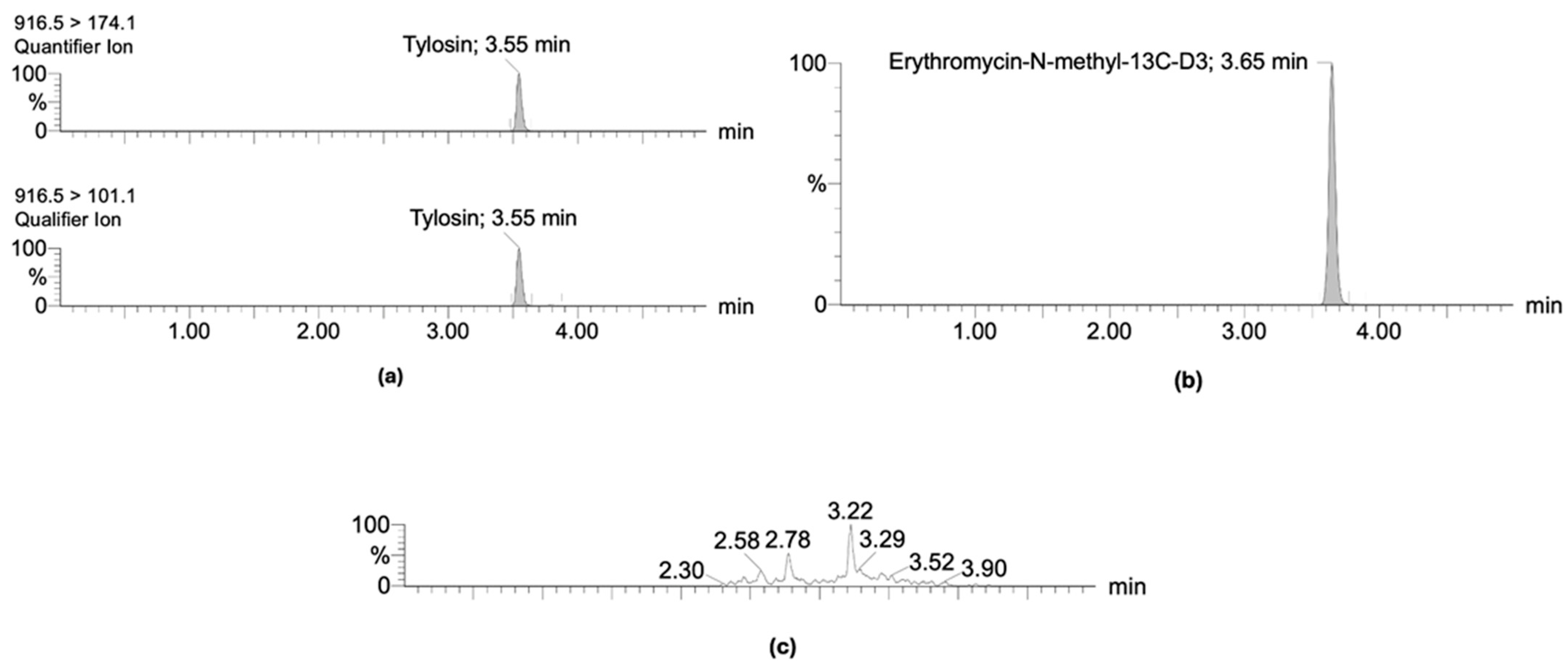
2.1. Validation and Optimization of Chemical Analytical Method

The HPLC-MS/MS method for the detection of tylosin in litter and droppings complied with all parameters established by the Commission Implementing Regulation (European Union) 2021/808/EC [27] and VICH GL2 [28] guidelines. No interferences were observed at the retention time of tylosin, corresponding to 3.55 min for the quantifier and qualifier ion and 3.65 min for the internal standard erythromycin-N-methyl-13C-D3 (Figure 1). The limit of detection (LOD) and quantification (LOQ) of the analytical method were 1 and 4 µg kg⁻1, respectively. Additionally, the method demonstrated precision, with a relative standard deviation (RSD) below 25% for reproducibility and 16.6% for repeatability, with a recovery rate close to 100%, as detailed in Table 1. The method also proved to be linear and robust, with an R2 ≥ 0.96 and >0.05 p-value using the Mandel test, obtaining a standard deviation expressed as the mass/mass lower than that of precision (0.04 µg kg⁻1 versus 2.03 µg kg⁻1 for litter; 0.0001 µg kg⁻1 versus 0.0010 µg kg⁻1 for droppings). Finally, the matrix effect showed an RSD of 11.59% in litter and 10.48% for droppings.
The analyte in litter ranged between 41 and 46 µg kg⁻1 during the first and second weeks. However, by the third-week post-fortification, the concentration decreased to 35.14 µg kg⁻1. In droppings, the concentrations were consistent between 38 and 40 µg kg⁻1 during the first and second weeks but declined to 36.40 µg kg⁻1 by the third week.

2.2. Detection and Quantification of Tylosin Residues in Litter and Droppings in Poultry

The tylosin concentration from the bird litter of the treated group (A) started with an average of 1313.88 μg kg−1 on day 3 post-treatment. Concentrations above the limit of quantification (LOQ = 4 µg kg−1) were identified throughout the whole production cycle 42 days). In the untreated group directly next to the treated group (B.1), the concentrations started at 25.89 µg kg−1 on day 3 post-treatment, decreasing gradually but still exceeding the LOQ through day 18 post-treatment. In the sentinel group (B.2), separated by a 30 cm distance from the treated group, concentrations were 11.41 µg kg−1 on day 3 post-treatment. However, at the end of the productive cycle, the concentrations were lower than the limit of detection (LOD = 1 µg kg−1), which meant that no concentrations were detected in these matrices (Table 2).
Tylosin concentrations in droppings from group A averaged at 8.67 μg kg−1 on the third day after treatment, with trace concentrations found between day 6 and 9 post-treatment. Concentrations decreased below the LOD during the production cycle. For both sentinel groups, B.1 and B.2, concentrations were not quantified during the production cycle (Table 2).
For the control group (C), no concentrations were detected for both litter and droppings.

2.3. Determination of the Dissemination of Tylosin Residues in the Poultry Environment

The non-parametric Kruskal–Wallis test was used to evaluate the differences between the medians of the groups under study, determining statistically significant differences between the tylosin concentrations detected in the litter of the treated group (A), the adjacent group (B.1), and the group distanced 30 cm from the treated group (B.2) (p-value = 2.2 × 10⁻¹⁶). The eta-squared estimate was 0.8 with a 95% confidence level, demonstrating a moderate effect of distance on detected concentrations of tylosin residues, identifying that the shorter the distance of exposure to animals treated with tylosin, the higher the concentrations of residues disseminated in the production environment. Subsequently, Dunn’s test was used to evaluate which groups showed these differences, making the following three comparisons: group A–group B.1 (p-value = 1.241669 × 10⁻⁸), group A–group B.2 (p-value = 7.517277 × 10⁻¹⁸), and group B.1–group B.2 (p-value = 7.010577 × 10⁻⁴), indicating that all the medians are different from each other (Figure 2).

3. Discussion

This study detected tylosin residue concentrations in broiler litter and droppings using HPLC-MS/MS, which complies with the requirements of the Commission Decision 2021/808/EC [27], the Food and Drug Administration (FDA), and VICH GL2 [28] guidelines, confirming the accuracy and reliability for the detection and quantification of tylosin residues in broiler chicken droppings and litter.
After antimicrobial treatment, we detected tylosin residue concentrations in broiler litter and droppings and the dissemination of these residues to the untreated sentinel groups. Group A presented higher concentrations for both matrices, followed by group B.1 and then group B.2. These findings could be explained by the proximity of B.1 to the treated group, which likely resulted in increased exposure to contaminated dust and litter particles, emphasizing the critical role of physical separation or barriers in mitigating the risk of cross-contamination.
In droppings, residues were quantified only in the first sampling in group A (8.67 µg kg−1). These concentrations decreased during the study, falling below the detection limit by the end of the experiment in every group. These low concentrations could be explained by tylosin’s low oral bioavailability (13.7%) and short half-life (1.5–2 h) [29], suggesting that the most excretion occurred during the treatment and before the first sampling point. Previous studies reported variable tylosin concentrations in manure (1.65 to 13740 µg kg−1), though in field conditions with uncontrolled environments and different methodologies that may explain the variability in results [23,30]. Pokrant et al. [12] performed a study using a similar method and reported 104.66 µg kg−1 in droppings 5 days post-treatment. This difference could be explained by variations in the treatment duration (7 versus 5 days) and sampling method, where combined samples from different groups could have led to cross-contamination.
Litter showed higher concentrations due to the accumulative effect of different elements and the organic composition of the litter, which facilitates macrolide accumulation [8,31]. These findings align with Pokrant et al. [19], who reported ten times higher oxytetracycline concentrations in litter compared to droppings. Additionally, antimicrobial residues may also be found in other litter components, such as feathers, where tylosin concentrations of 9246.53 µg kg−1 have been reported 4 days post-treatment, decreasing to 356.59 µg kg−1 by day 15 [32].
Limited controlled studies exist on tylosin residues in poultry litter; however, reported concentrations have ranged from 135 to 597 µg kg−1 [9,11]. Our study detected concentrations from 1313.88 to 290.16 µg kg−1 in litter between days 3 and 18 post-treatment. These results exceeded those reports in field studies on the first sampling points, likely due to differences in sampling conditions and animal management.
Tylosin’s residue dissemination to sentinel groups was detected, suggesting that bird movement, such as dust bathing, facilitates the dispersion of litter components [33]. Considering the above, chicken litter plays a fundamental role in the persistence of these residues. This matrix is composed of droppings, feed, shavings, and feathers, and it has been reported that the prevalence of antibiotic-resistant bacteria can exceed 60% for specific microorganisms. These include Escherichia coli and Enterococci, which show multi-resistance to different families of antimicrobials [34]. Kruskal–Wallis and Dunn’s tests showed significant differences between the groups, with higher tylosin concentrations in the adjacent group (B.1) compared to the 30 cm distant group (B.2). This suggests that distance affects dissemination, with residues decreasing as separation increases. Although our study distances do not reflect commercial settings, they provide insight into future research considering larger-scale production systems. It is relevant to continue investigating both the persistence and dissemination of residues in larger-scale production conditions that are more representative of the industry, considering other factors such as greater distances of exposure, a greater number of animals, and the administration of antimicrobials in feed, among other factors.
Despite group differences, all concentrations decreased over time, which was confirmed through linear regression analysis. Group A (treatment) exhibited the steepest decline, likely due to the cessation of excretion. At the same time, sentinel group B.1 may have experienced slower reductions due to their continuous exposure to group A. Pokrant et al. [19] observed a 43% oxytetracycline reduction in litter, compared to 78% for tylosin in this study, likely due to this lower environmental persistence of antimicrobials [33,35]. Future studies should include more frequent sampling and individual fecal collections to minimize dilution effects and evaluate the excretion of tylosin. Also, post-euthanasia litter sampling could help determine the long-term persistence of tylosin in the matrix.
The persistence of antimicrobial residues can be a risk for both humans and animals that are exposed to them. Mainly because of the development of antimicrobial resistance, as pathogens are continuously exposed to these drugs, where selection pressure favors the expression and transmission of resistance genes, this causes difficulties in the treatment of infectious diseases in both humans and animals. On the other hand, exposure to residues of these drugs can also cause hypersensitivity reactions, carcinogenic effects, and imbalance in the intestinal microbiota, among other results [8,15].
Tylosin residues in poultry waste pose environmental concerns, as residues have been detected in fertilized soil [23], potentially contributing to antimicrobial resistance [36]. Tylosin may also induce cross-resistance to other macrolides, such as erythromycin [37].
This study shows that these byproducts are a potential source for disseminating antimicrobials in the production system. Further research under commercial conditions is necessary to assess tylosin’s persistence and dissemination, incorporating factors such as greater exposure distances, larger flock sizes, and feed-based antimicrobial administration.

4. Materials and Methods

The experimental design was carried out at the Animal Management Unit (UMA) located at the Faculty of Veterinary and Livestock Sciences of the University of Chile (FAVET), Santiago, Chile. Male Ross 308 broiler chickens were used as the study model. These were obtained from “Agrícola Chorombo” on their first day of life and were immediately transported to the facility where the study was conducted. The birds were received on the first day of life under controlled environmental conditions (ambient temperature: 27 °C; litter temperature: 30 °C). Conditions such as temperature (25 ± 5 °C) and humidity (50–60%) were regulated using heating and ventilation systems to maintain appropriate ranges according to the birds’ age requirements. The floor was covered with about 30 cm of sanitized wood shavings with a 5 to 20 mm particle size. The animals had ad libitum access to water and feed. The study was conducted in an Animal Handling Unit at the Faculty of Veterinary and Animal Sciences of the University of Chile.
Biosecurity measures for working with animals were implemented as suggested in the “Biosecurity and Associated Risks Standards Manual—Fondecyt (National Fund for Scientific and Technological Development)—CONICYT (National Commission for Scientific and Technological Research)” [38]. The handling and euthanasia of the animals were conducted in compliance with the guidelines of Law No. 20.380 on Animal Protection [39], Directive 2010/63/EU [40], and Regulation (EC) No. 1099/2009 [41]. The study was conducted under Certificate No. 22551–VET–UCH issued by the Institutional Committee for the Care and Use of Animals (CICUA) of the University of Chile.

4.1. Description of Experimental Animals

The study was conducted in two separate rooms: one designated as the control and the other as experimental. The first room contained a single pen for the control group (C), while the second room had three pens for experimental groups A, B.1, and B.2. Each pen measured 1 m2, and 40 animals were used, with 10 birds assigned to each group. The number of birds was determined based on the fact that broiler chickens can weigh up to 3.2 kg, and the “Poultry Industry Manual” of the United States Department of Agriculture (USDA) recommends a maximum live weight density of 34.372 kg/m2 [42]. The experimental design is shown in Figure 3.
The antibiotic tylosin phosphate 10% was used for the treatment, administered at a dose of 35 mg kg1 of tylosin base every 24 h for five days, as indicated on the product label. Treatment began on day 20 of life and was administered orally using a Levin No. 6 orogastric tube (Cranberry International Sdn. Bhd., Shah Alam, Malaysia) to ensure the full dose was delivered to each animal. The vehicle used for the administration of antimicrobials was distilled water.

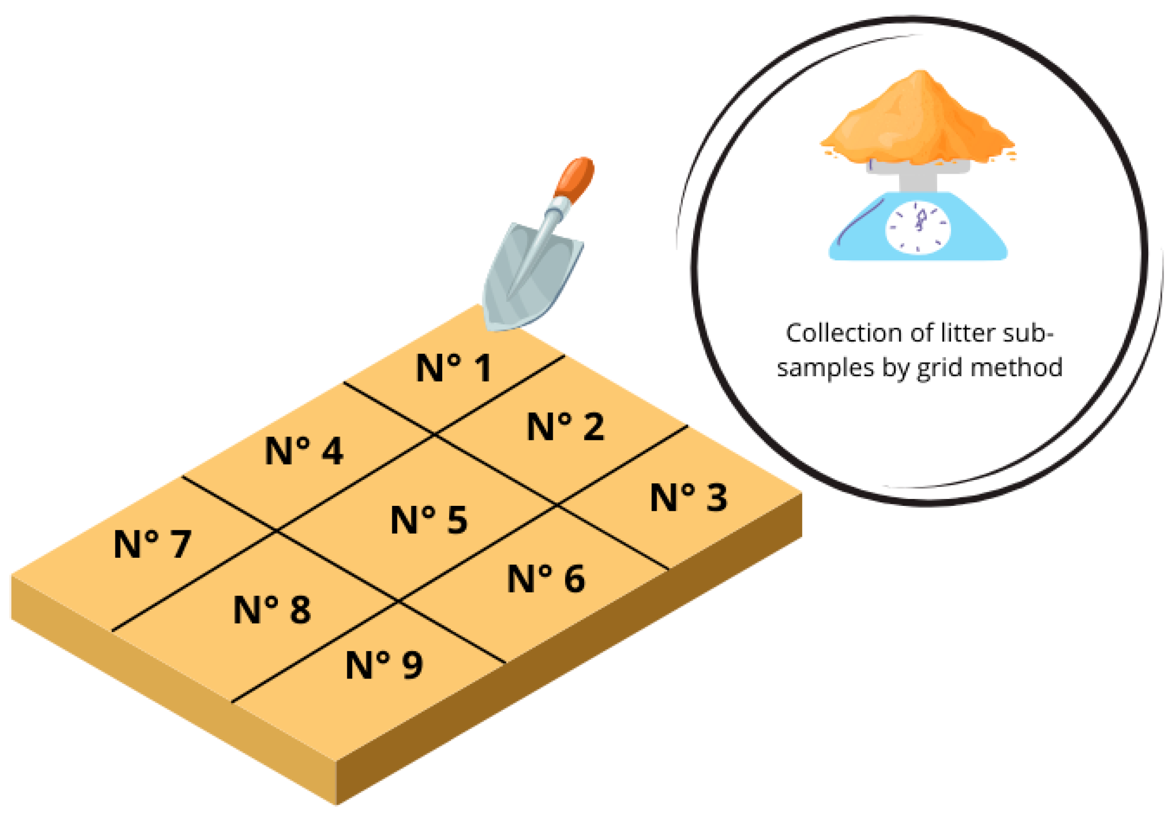
Sampling of Litter and Droppings

Samples of litter and droppings were collected from each group. A total of six samples were conducted on days 3, 6, 9, 12, 15, and 18 post-treatment. The pen was divided into nine quadrants using the grid method for the litter samples, as described in the IAEA report “Soil Sampling for Environmental Contaminants” (Figure 4) [43]. Approximately 20 g of litter was collected from each quadrant, resulting in a final 180 to 200 g sample per experimental group. The sampling order was as follows: group C, group B.2, group B.1, and group A to prevent the cross-contamination of untreated groups.
Dropping samples were collected in 50 mL polypropylene tubes. Cloacal stimulation was performed on each individual using sterile swabs to collect the samples. The samples from each group were pooled into a single tube, as the focus of this study was to evaluate the group-level concentrations rather than individual variations. All samples were obtained in triplicate and stored at −20 °C until processing.

4.2. Chemical Analysis

4.2.1. Tylosin Standards

Tylosin tartrate (98% purity; CAS:1401-69-0) and erythromycin-N-methyl-13C-D3 (95% purity; CAS: 2378755-50-9) were used in this study, with the latter employed as the internal standard of the sample analysis. These standards were purchased from Toronto Research Chemicals (Toronto, ON, Canada). Stock solutions were prepared in methanol at 1000 µg mL⁻1, and intermediate or working solutions were diluted to 1000 ng mL⁻1 in the same solvent. These solutions were stored at −80 °C.

4.2.2. Chemicals and Solvents

HPLC-grade water, acetonitrile, and methanol were purchased from LiChrosolv® (MERCK KGaA, Darmstadt, Germany). Citric acid monohydrate (EMSURE® ACS, Reag. Ph Eur, MERCK KGaA, Darmstadt, Germany), dibasic sodium phosphate (Na2HPO4, EMSURE® ACS, Reag. Ph Eur, MERCK KGaA, Darmstadt, Germany), and disodium salt dihydrate of ethylenediaminetetraacetic acid (Na2EDTA, Titriplex® III ACS, ISO, Reag. Ph Eur, MERCK KGaA, Darmstadt, Germany).
The 0.1 M EDTA/McIlvaine buffer (pH 4.0 ± 0.1) was used for tylosin extraction from matrices. Extracts were prepared by mixing 0.1 M citric acid, 0.2 M disodium phosphate, and Na2EDTA, followed by sonication and pH adjustment with citric acid or phosphate solutions.
Two solutions were prepared for chromatographic analysis. Eluent A was prepared by dissolving 2.0 mM of ammonium formate and 0.16% formic acid in water. Eluent B was prepared with the same concentration of ammonium formate and formic acid in methanol.

4.2.3. Sample Preparation

The extraction method implemented by Yévenes et al. [44] and Pokrant et al. [12] was followed. Bedding and droppings were homogenized using a blender and a sterile wooden stick. A 1 ± 0.01 g sample was weighed, spiked with intermediate solution standards, and subsequently subjected to extraction. The samples were enriched with an 8 mL McIlvaine-EDTA buffer (pH 4.0 ± 0.1) and 2 mL acetonitrile, followed by 10 min of agitation, 5 min of sonication, and 10 min of centrifugation at 4000–5000 rpm. Filtration was performed using glass microfiber Whatman™ filter paper of grade GF/A (1.6 µm) (MERCK) and SPE Oasis HLB columns (Supelco, MERCK KGaA, Darmstadt, Germany). Solvents were evaporated under nitrogen flow in a water bath at 40–50 °C, and the sample was reconstituted with 200 µL of methanol and 300 µL of water. Finally, the samples were agitated for 5 min, sonicated for 5 min, centrifuged for 5 min at 1700 rpm, and transferred to an amber glass vial using a syringe with Millipore filters (Millex®, Merck KGaA, Burlington, MA, USA).
This chemical analysis was performed at the Laboratory of Veterinary Pharmacology (FARMAVET) of the Faculty of Veterinary and Animal Sciences at the University of Chile, which is accredited under normative standards.

4.2.4. LC-MS/MS Analysis

For tylosin detection, the ACQUITY UPLC® I-Class System (Waters™, Milford, MA, USA), coupled with a Xevo TQ-S micro triple quadrupole mass spectrometer (Waters™, Milford, MA, USA) were used. Chromatographic separation was performed using an ACQUITY UPLC® HSS T3 analytical column (1.8 µm, 2.1 × 100 mm; Waters™ Corp, USA), and the resulting data were processed with MassLynx™ software version 4.2 and TargetLynx® xs software version 4.2.
The chromatographic conditions consisted of a sample injection volume of 10 µL, a mobile phase gradient flow rate of 300 µL/min, and a chromatographic column temperature of 40 °C. The percentage of each mobile phase injected over time is detailed in Table 3. The mass spectrometer operated in the positive ion mode with the following instrument conditions: a capillary voltage of 3.00 kV, source temperature at 150 °C, desolvation gas flow of 1000 L/h with a desolvation temperature of 600 °C, and a sample cone gas flow of 10 L/h. Specific mass spectrometric settings for the analytes are detailed in Table 4.
Tylosin was confirmed in the experimental samples by meeting the following criteria: retention time and relative retention time with a variation not exceeding ±2.5%, a signal-to-noise ratio of at least 3:1, and a relative deviation of the ion ratio within ±40%. Once confirmed, quantification was performed using calibration curves derived from fortified blank samples spiked at different, equidistant levels and processed alongside the experimental samples.

4.2.5. Analytical Method Validation/Verification

The analytical method was validated for poultry litter and droppings according to the recommendations from Commission Decision 2021/808/EC [27] of the Food and Drug Administration (FDA) and VICH GL2 [28]. For this purpose, the following parameters were evaluated:
  • Limit of detection (LOD): Twenty fortified samples were analyzed at the selected LODs, with a signal-to-noise ratio of ≥3:1. A coefficient of variation (CV%) of <25% was accepted.
  • Limit of quantification (LOQ): Twenty fortified samples at the LOD were analyzed, and the standard deviation was determined, which was then multiplied by 1.64 and added to the LOD. The signal-to-noise ratio for this parameter must be ≥10:1 to be accepted.
  • Retention time (TR): Six samples with a tylosin standard were analyzed to evaluate the TR of each, with an accepted coefficient of variation of <1%.
  • Specificity: Twenty blank samples were evaluated to determine if there were any interferents in the TR.
  • Linearity: Three calibration curves were created using five different concentrations, with the lowest concentration at LOD. A determination coefficient (R2) ≥ 0.95 and a p-value >0.05 using the Mandel test was required for acceptance.
  • Precision and recovery: To achieve precision, two parameters were evaluated: intra-laboratory reproducibility and repeatability. Six calibration curves at 25, 50, and 75 µg kg−1 were analyzed for each parameter on three days. Repeatability was assessed by performing all analyses under identical conditions by the same analyst, while intra-laboratory reproducibility involved analyses conducted by different analysts under varying conditions. Recovery was evaluated based on these samples, and this was achieved when the mean recovery percentage was between −20 and 20% on each level.
  • Matrix effect: A chemical extraction was performed on 20 blank samples fortified at the end of the extraction process. The signal obtained from these samples was compared to pure standard injections.
  • Ruggedness: This was evaluated by modifying three conditions: the amount of EDTA/McIlvaine buffer added to the sample initially (8 mL to 4 mL of buffer), the centrifugation time at the beginning of the extraction process (10 to 5 min), and the volume used to wash the extraction column (5 mL to 2 mL of HPLC water). The method was considered robust when the standard deviation of the modified samples was equal to or lower than intra-laboratory reproducibility.
  • Stability: fifteen chicken litter and dropping samples were fortified at 50 µg kg−1 and stored at −20 °C. Five samples were analyzed after 1 week, another five samples were analyzed after 2 weeks, and the remaining five were analyzed after 3 weeks.

4.2.6. Statistical Analysis

To assess the effect of distance on the residue concentrations observed across different groups, the normality of the data was first evaluated using the Shapiro–Wilk test. Homoscedasticity was assessed using Levene’s test. As the data did not meet these assumptions, the non-parametric Kruskal–Wallis test was applied, considering a p-value < 0.05 and a 95% confidence interval to determine if there were statistically significant differences between the medians of tylosin concentrations within the study groups. This aimed to evaluate whether the exposure distance affected the antimicrobial residue concentration in broiler litter and droppings. Subsequently, Dunn’s test was performed to assess the differences between the medians of concentrations for each group. The matrices were evaluated individually. Statistical analyses were carried out using R version 4.3.2 and RStudio version 2023.09.1+494.

5. Conclusions

Tylosin is an antimicrobial that can spread within the production environment through the matrices studied. Litter presented the highest concentrations in the samples taken, and the presence of residues decreased as the exposure distance from the pens increased. This implies that these matrices are a potential route for disseminating AM residues, posing a hazard to public health. Broiler chicken droppings and litter are byproducts of the poultry production process and serve as indicators of AM residues in the poultry farm. These matrices are easily accessible, and their collection is non-invasive for the animals, allowing for the continuous assessment of the presence of AM residues in the production environment.

Author Contributions

Conceptualization, M.B.V., A.M., L.L. and J.C.; methodology, M.B.V., I.S., F.M., A.F., M.M. and A.M.; software, M.B.V. and I.S.; validation, I.S., M.M., A.F., P.C. and F.M.; formal analysis, M.B.V., E.P. and A.M.; investigation, M.B.V. and I.S.; resources, E.P. and J.C.; data curation, P.C., L.T. and M.B.V.; writing—original draft preparation, M.B.V., I.S. and P.C.; writing—review and editing, E.P., L.T., A.M. and L.L.; visualization, L.L. A.M. and J.C.; supervision, L.L., A.M. and J.C.; project administration, J.C.; funding acquisition, J.C. All authors have read and agreed to the published version of the manuscript.

Funding

This work was supported by The National Agency of Research and Development (ANID, by its Spanish acronym) and FONDECYT Regular Project N°1220520.

Institutional Review Board Statement

The animal study protocol was approved by the Institutional Animal Care and Use Committee (CICUA by its Spanish acronym) of the University of Chile (Protocol code: 22551-VET-UCH, approval date: 22 April 2022).

Informed Consent Statement

Not applicable.

Data Availability Statement

The data presented in this study are available in the article.

Conflicts of Interest

The authors declare no conflicts of interest.

References

  1. Baralla, E.; Demontis, M.P.; Dessi, F.; Varoni, M.V. An Overview of Antibiotics as Emerging Contaminants: Occurrence in Bivalves as Biomonitoring Organisms. Animals 2021, 11, 3239. [Google Scholar] [CrossRef] [PubMed]
  2. Wang, F.; Xiang, L.; Leung, K.S.; Elsner, M.; Zhang, Y.; Guo, Y.; Pan, B.; Sun, H.; An, T.; Ying, G.; et al. Emerging contaminants: A One Health perspective. Innovation 2024, 5, 100612. [Google Scholar] [CrossRef]
  3. Nguyen, M.; Lin, C.; Nguyen, H.; Hung, N.T.Q.; La, D.D.; Nguyen, X.H.; Chang, S.W.; Chung, W.J.; Nguyen, D.D. Occurrence, fate, and potential risk of pharmaceutical pollutants in agriculture: Challenges and environmentally friendly solutions. Sci. Total Environ. 2023, 899, 165323. [Google Scholar] [CrossRef] [PubMed]
  4. Islam, M.A.; Bose, P.; Rahman, M.Z.; Muktaruzzaman, M.; Sultana, P.; Ahamed, T.; Khatun, M.M. A review of antimicrobial usage practice in livestock and poultry production and its consequences on human and animal health. J. Adv. Vet. Anim. Res. 2024, 11, 675–685. [Google Scholar] [CrossRef] [PubMed]
  5. Chen, J.; Ying, G.-G.; Deng, W.-J. Antibiotic Residues in Food: Extraction, Analysis, and Human Health Concerns. J. Agric. Food Chem. 2019, 67, 7569–7586. [Google Scholar] [CrossRef]
  6. Ghimpețeanu, O.M.; Pogurschi, E.N.; Popa, D.C.; Dragomir, N.; Drăgotoiu, T.; Mihai, O.D.; Petcu, C.D. Antibiotic Use in Livestock and Residues in Food—A Public Health Threat: A Review. Foods 2022, 11, 1430. [Google Scholar] [CrossRef]
  7. Ashworth, A.J.; Chastain, J.P.; Moore, P.A. Nutrient Characteristics of Poultry Manure and Litter. In Animal Manure: Production, Characteristics, Environmental Concerns, and Management; ASA Special Publications; John Wiley & Sons: Hoboken, NJ, USA, 2020; Volume 67, pp. 63–87. [Google Scholar] [CrossRef]
  8. Gržinić, G.; Piotrowicz-Cieślak, A.; Klimkowicz-Pawlas, A.; Górny, R.L.; Ławniczek-Wałczyk, A.; Piechowicz, L.; Olkowska, E.; Potrykus, M.; Tankiewicz, M.; Krupka, M.; et al. Intensive poultry farming: A review of the impact on the environment and human health. Sci. Total Environ. 2023, 858, 160014. [Google Scholar] [CrossRef]
  9. Paranhos, A.G.O.; Pereira, A.R.; da Fonseca, I.C.; Sanson, A.L.; Afonso, R.J.C.F.; Aquino, S.F. Analysis of tylosin in poultry litter by HPLC-UV and HPLC-MS/MS after LTPE. Int. J. Environ. Anal. Chem. 2020, 101, 2568–2585. [Google Scholar] [CrossRef]
  10. Monir, H.H.; Fayez, Y.M.; Nessim, C.K.; Michael, A.M. When is it safe to eat different broiler chicken tissues after administration of doxycycline and Tylosin mixture? J. Food Sci. 2021, 86, 1162–1171. [Google Scholar] [CrossRef]
  11. Alonso, L.L.; Podio, N.S.; Marino, D.J.G.; Almada, N.S.; Gange, J.M.; Bernigaud, I.; Mórtola, N.; Wunderlin, D.A. Evaluating antibiotic occurrence, degradation, and environmental risks in poultry litter within Argentina’s agricultural hub. Sci. Total Environ. 2024, 920, 170993. [Google Scholar] [CrossRef]
  12. Pokrant, E.; Trincado, L.; Yévenes, K.; Terraza, G.; Maddaleno, A.; Martín, B.S.; Zavala, S.; Hidalgo, H.; Lapierre, L.; Cornejo, J. Determination of five antimicrobial families in droppings of therapeutically treated broiler chicken by high-performance liquid chromatography-tandem mass spectrometry. Poult. Sci. 2021, 100, 101313. [Google Scholar] [CrossRef] [PubMed]
  13. Zubair, M.; Li, Z.; Zhu, R.; Wang, J.; Liu, X.; Liu, X. The Antibiotics Degradation and Its Mechanisms during the Livestock Manure Anaerobic Digestion. Molecules 2023, 28, 4090. [Google Scholar] [CrossRef] [PubMed]
  14. Bos, B.; Groot Koerkamp, P.W.G.; Groenestein, K. A novel design approach for livestock housing based on recursive control—With examples to reduce environmental pollution. Livest. Prod. Sci. 2003, 84, 157–170. [Google Scholar] [CrossRef]
  15. EFSA Panel on Biological Hazard (BIOHAZ). Role played by the environment in the emergence and spread of antimicrobial resistance (AMR) through the food chain. EFSA J. 2021, 19, e06651. [Google Scholar] [CrossRef]
  16. Ghirardini, A.; Grillini, V.; Verlicchi, P. A review of the occurrence of selected micropollutants and microorganisms in different raw and treated manure—Environmental risk due to antibiotics after application to soil. Sci. Total Environ. 2020, 707, 136118. [Google Scholar] [CrossRef]
  17. Marutescu, L.G.; Jaga, M.; Postolache, C.; Barbuceanu, F.; Milita, N.M.; Romascu, L.M.; Schmitt, H.; de Roda Husman, A.M.; Sefeedpari, P.; Glaeser, S.; et al. Insights into the impact of manure on the environmental antibiotic residues and resistance pool. Front. Microbiol. 2022, 13, 965132. [Google Scholar] [CrossRef]
  18. McEachran, A.D.; Blackwell, B.R.; Hanson, J.D.; Wooten, K.J.; Mayer, G.D.; Cox, S.B.; Smith, P.N. Antibiotics, Bacteria, and Antibiotic Resistance Genes: Aerial Transport from Cattle Feed Yards via Particulate Matter. Environ. Health Perspect. 2015, 123, 337–343. [Google Scholar] [CrossRef] [PubMed]
  19. Pokrant, E.; Yévenes, K.; Trincado, L.; Terraza, G.; Galarce, N.; Maddaleno, A.; Martín, B.S.; Lapierre, L.; Cornejo, J. Evaluation of Antibiotic Dissemination into the Environment and Untreated Animals, by Analysis of Oxytetracycline in Poultry Droppings and Litter. Animals 2021, 11, 853. [Google Scholar] [CrossRef]
  20. Vargas, M.B.; Pokrant, E.; García, I.; Cadena, R.; Mena, F.; Yévenes, K.; Fuentes, C.; Zavala, S.; Flores, A.; Maturana, M.; et al. Assessing the spread of sulfachloropyridazine in poultry environment and its impact on Escherichia coli resistance. Prev. Vet. Med. 2024, 233, 106362. [Google Scholar] [CrossRef]
  21. Bolan, N.S.; Szogi, A.A.; Chuasavathi, T.; Seshadri, B.; Rothrock, M.J.; Panneerselvam, P. Uses and management of poultrylitter. World’s Poult. Sci. J. 2010, 66, 673–698. [Google Scholar] [CrossRef]
  22. Kyakuwaire, M.; Olupot, G.; Amoding, A.; Nkedi-Kizza, P.; Basamba, T.A. How Safe is Chicken Litter for Land Application as an Organic Fertilizer?: A Review. Int. J. Environ. Res. Public Health 2019, 16, 3521. [Google Scholar] [CrossRef]
  23. Rashid, A.; Muhammad, J.; Khan, S.; Kanwal, A.; Sun, Q. Poultry manure gleaned antibiotic residues in soil environment: A perspective of spatial variability and influencing factors. Chemosphere 2023, 317, 137907. [Google Scholar] [CrossRef] [PubMed]
  24. Albero, B.; Tadeo, J.L.; Escario, M.; Miguel, E.; Pérez, R.A. Persistence and availability of veterinary antibiotics in soil and soil-manure systems. Sci. Total Environ. 2018, 643, 1562–1570. [Google Scholar] [CrossRef] [PubMed]
  25. Berendsen, B.; Lahr, J.; Nibbeling, C.; Jansen, L.; Bongers, I.; Wipfler, E.; Van de Schans, M. The persistence of a broad range of antibiotics during calve, pig and broiler manure storage. Chemosphere 2018, 204, 267–276. [Google Scholar] [CrossRef]
  26. Cortés, P.; Pokrant, E.; Yévenes, K.; Maddaleno, A.; Flores, A.; Vargas, M.B.; Trincado, L.; Maturana, M.; Lapierre, L.; Cornejo, J. Antimicrobial Residues in Poultry Litter: Assessing the Association of Antimicrobial Persistence with Resistant Escherichia coli Strains. Antibiotics 2025, 14, 89. [Google Scholar] [CrossRef]
  27. European Commission. Council Regulation (EC) No 2021/808 of 22 March 2021 on the Performance of Analytical Methods for Residues of Pharmacologically Active Substances Used in Food-Producing Animals and on the Interpretation of Results as well as on the Methods to Be Used for Sampling and Repealing Decisions 2002/657/EC and 98/179/EC. Available online: https://eur-lex.europa.eu/eli/reg_impl/2021/808/oj/eng (accessed on 23 March 2025).
  28. European Medicines Agency (EMA). VICH GL2: Validation of Analytical Procedures: Methodology-Scientific Guideline. 1998. Available online: https://www.ema.europa.eu/en/vich-gl2-validation-analytical-procedures-methodology-scientific-guideline (accessed on 23 March 2025).
  29. Ji, L.; Dong, L.; Ji, H.; Feng, X.; Li, D.; Ding, R.; Jiang, S. Comparative pharmacokinetics and bioavailability of tylosin tartrate and tylosin phosphate after a single oral and i.v. administration in chickens. J. Vet. Pharmacol. Ther. 2013, 37, 312–315. [Google Scholar] [CrossRef]
  30. Ho, Y.B.; Zakaria, M.P.; Latif, P.A.; Saari, N. Simultaneous determination of veterinary antibiotics and hormone in broiler manure, soil and manure compost by liquid chromatography–tandem mass spectrometry. J. Chromatogr. A 2012, 1262, 160–168. [Google Scholar] [CrossRef] [PubMed]
  31. Pan, M.; Chu, L.M. Fate of antibiotics in soil and their uptake by edible crops. Sci. Total Environ. 2017, 599–600, 500–512. [Google Scholar] [CrossRef]
  32. Cornejo, J.; Pokrant, E.; Carvallo, C.; Maddaleno, A.; San Martín, B. Depletion of tylosin residues in feathers, muscle and liver from broiler chickens after completion of antimicrobial therapy. Food Addit. Contam. Part A 2018, 35, 448–457. [Google Scholar] [CrossRef]
  33. Derakhshani, S.M.; Ogink, N.W.M.; Bos, B.A.P.; Groot Koerkamp, P.W.G. Sensitivity analysis of fine dust spreading from litter in poultry houses. Biosyst. Eng. 2021, 208, 272–286. [Google Scholar] [CrossRef]
  34. Chen, Z.; Jiang, X. Microbiological Safety of Chicken Litter or Chicken Litter-Based Organic Fertilizers: A Review. Agriculture 2014, 4, 1–29. [Google Scholar] [CrossRef]
  35. Cycoń, M.; Mrozik, A.; Piotrowska-Seget, Z. Antibiotics in the Soil Environment—Degradation and Their Impact on Microbial Activity and Diversity. Front. Microbiol. 2019, 10, 338. [Google Scholar] [CrossRef] [PubMed]
  36. Larsson, D.G.J.; Flach, C.-F. Antibiotic resistance in the environment. Nat. Rev. Microbiol. 2022, 20, 257–269. [Google Scholar] [CrossRef] [PubMed]
  37. Aronson, J.K. (Ed.) Tylosin. In Meyler’s Side Effects of Drugs (Sixteenth Edition); Elsevier: Amsterdam, The Netherlands, 2016; p. 233. [Google Scholar] [CrossRef]
  38. CONICYT. Manual de Normas de Bioseguridad y Riesgos Asociados-Fondecyt-CONICYT; Versión 2018; CONICYT: Santiago, Chile, 2018. [Google Scholar]
  39. Ministerio de Salud Subsecretaría de Salud Pública Ley Núm. 20.380 Sobre Protección de Animales. 2009. Available online: https://www.bcn.cl/leychile/navegar?idNorma=1006858 (accessed on 10 March 2025).
  40. Directive 2010/63/EU of the European Parliament and of the Council of 22 September 2010 on the Protection of Animals Used for Scientific PurposesText with EEA Relevance. 47p. Available online: https://eur-lex.europa.eu/LexUriServ/LexUriServ.do?uri=OJ:L:2010:276:0033:0079:en:PDF (accessed on 10 March 2025).
  41. Council Regulation (EC) No 1099/2009 of 24 September 2009 on the Protection of Animals at the Time of KillingText with EEA Relevance. 30p. Available online: https://eur-lex.europa.eu/LexUriServ/LexUriServ.do?uri=OJ:L:2009:303:0001:0030:EN:PDF (accessed on 10 March 2025).
  42. US Department of Agriculture (USDA). Poultry Industry Manual; US Department of Agriculture (USDA): Washington, DC, USA, 2013. [Google Scholar]
  43. IAEA International Atomic Energy Agency. Soil Sampling for Environmental Contaminants; IAEA International Atomic Energy Agency: Vienna, Austria, 2004. [Google Scholar]
  44. Yévenes, K.; Pokrant, E.; Trincado, L.; Lapierre, L.; Galarce, N.; Martín, B.S.; Maddaleno, A.; Hidalgo, H.; Cornejo, J. Detection of Antimicrobial Residues in Poultry Litter: Monitoring a Risk through a Selective and Sensitive HPLC–MS/MS Method. Animals 2021, 11, 1399. [Google Scholar] [CrossRef]
Figure 1. (a) Chromatograms of tylosin quantifier and qualifier ion. (b) Chromatogram of internal standard erythromycin-N-methyl-13C-D3. (c) Chromatogram of litter sample free of tylosin residues.
Antibiotics 14 00477 g001
Figure 2. The boxplot of tylosin concentrations by experimental groups transformed into a natural logarithm in the broiler chicken litter matrix. There is evidence of differences between medians (Kruskal–Wallis; p-value < 0.05).
Antibiotics 14 00477 g002
Figure 3. The experimental design of the study. Each group consisted of 10 broiler chickens. The control group was housed separately, while group A received a therapeutic dose of tylosin. Groups B.1 and B.2 served as sentinel groups, with group B.2 placed at 30 cm.
Antibiotics 14 00477 g003
Figure 4. The sampling of poultry litter using the grid method. Equal samples were obtained by the sampling point and subsequently homogenized.
Antibiotics 14 00477 g004
Table 1. Precision and recovery results of the validation method for the detection of tylosin in chicken droppings and litter.
MatrixFortified
Concentration (µg kg−1)
Repeatability (RSD %) *Reproducibility (RSD %)Recovery (RSD%)
Droppings253.014.6798.34
502.884.52101.66
750.991.5499.45
Litter254.174.6197.11
503.964.35102.89
751.371.5199.04
* Relative standard deviation expressed as percentage.
Table 2. Tylosin concentrations detected in the litter and droppings of treated and untreated birds from each experimental group by the number of post-treatment days.
Tylosin Concentrations (µg·kg−1 ww)
GroupMatrixDay 3Day 6Day 9Day 12Day 15Day 18
ALitter1313.881284.941015.09955.42351.61290.16
Droppings8.67<LOQ **<LOQ<LOD *<LOD<LOD
B.1Litter25.8924.9521.389.719.519.35
Droppings<LOQ<LOD<LOD<LOD<LOD<LOD
B.2Litter11.41<LOQ<LOQ<LOQ<LOD<LOD
Droppings<LOD<LOD<LOD<LOD<LOD<LOD
ww: wet weight; * <LOD: below the limit of detection for the technique (droppings and litter 1 µg kg−1); ** LOQ: below the limit of quantification for the technique (droppings and litter 4 µg kg−1).
Table 3. Mobile phase gradient flow.
Time (Minutes)Solution A (%)Solution B (%)
0.00982
1.00982
2.005545
3.102575
3.30982
5.50982
7.00982
Table 4. Specific mass spectrometric conditions.
AnalytePrecursor Ion (m/z)Productor Ion (m/z)Dwell Time (s)Cone VoltageCollision Energy
Tylosin916.50174.10 10.00945.0040.00
101.10 20.00945.0045.00
Erythromycin-N-methyl-13C-D3738.40162.100.00920.0030.00
1 Quantifier ion; 2 qualifier ion.
Disclaimer/Publisher’s Note: The statements, opinions and data contained in all publications are solely those of the individual author(s) and contributor(s) and not of MDPI and/or the editor(s). MDPI and/or the editor(s) disclaim responsibility for any injury to people or property resulting from any ideas, methods, instructions or products referred to in the content.

