Article

Antimicrobial Susceptibility Determination of Less Frequently Isolated Legionella Species by Broth and Agar Dilution

1
Department of Medical Microbiology, Division of Infection and Immunity, Cardiff University, University Hospital of Wales, Cardiff CF14 4XN, UK
2
Ineos Oxford Institute for Antimicrobial Research, Department of Biology, University of Oxford, Oxford OX1 3RE, UK
3
Respiratory and Vaccine Preventable Bacteria Reference Unit (RVPBRU), United Kingdom Health Security Agency, London NW9 5EQ, UK
4
NHS England, Wellington House, 133-155 Waterloo Rd., London SE1 8UG, UK
*
Author to whom correspondence should be addressed.
Antibiotics 2025, 14(11), 1165; https://doi.org/10.3390/antibiotics14111165
Submission received: 3 October 2025 / Revised: 11 November 2025 / Accepted: 14 November 2025 / Published: 17 November 2025

Abstract

Background/Objectives: Infections caused by Legionella species are primarily associated with Legionella pneumophila, but non-pneumophila species are increasingly implicated in human disease. Despite this, antimicrobial susceptibility testing (AST) data for non-pneumophila species remain scarce, and standardised testing protocols or resistance thresholds have not been established. This study aimed to address this gap by evaluating and comparing AST performance for non-pneumophila Legionella species relative to L. pneumophila using three methodologies. Methods: AST was conducted on 89 Legionella isolates using LASARUS agar dilution, buffered yeast extract broth microdilution (BYE-BMD), and BCYE-α agar dilution, against ampicillin, azithromycin, chloramphenicol, doxycycline, levofloxacin, and rifampicin. Growth performance and minimum inhibitory concentrations (MICs) were assessed after a 96 h incubation. Results: MIC profiles were obtained using LASARUS and BYE-BMD for 53.9% and 93.3% of isolates, respectively. While L. pneumophila reached sufficient turbidity in BYE-BMD after a 48 h incubation, non-pneumophila species required an extended incubation (72–96 h). Non-pneumophila species displayed broader MIC ranges against azithromycin (0.016–1 mg/L) and levofloxacin (0.016–0.25 mg/L), but a narrower rifampicin range (≤0.0005–0.032 mg/L) relative to L. pneumophila. L. longbeachae exhibited a higher MIC50 for rifampicin despite overlapping susceptibility ranges across all species (0.001–0.016 mg/L). Conclusions: This study demonstrates species-specific differences in Legionella susceptibility and highlights the limitations in extrapolating L. pneumophila-based AST data. Azithromycin MICs in non-pneumophila species exceeded those of L. pneumophila, raising clinical concern. While BYE-BMD was the most effective method for MIC determination, three species required BCYE-α due to poor growth. These findings support developing standardised, species-specific AST protocols and thresholds amid rising macrolide resistance and the increasing detection of non-pneumophila infections.

1. Introduction

To date, the Legionella genus comprises 67 Legionella species, 30 of which have been identified as human pathogens [1,2]. Legionella are fastidious obligate aerobe Gram-negative bacteria and the causative agent of Legionnaires’ Disease (LD), an atypical form of pneumonia, accounting for ~2–9% of all community acquired pneumonia cases worldwide [1,3]. The dominant cause of LD in Europe and the United States is L. pneumophila, accounting for approximately 90% of reported cases [1]. In contrast, in Australia and New Zealand, 60–70% of LD cases are caused by L. longbeachae [1]. By comparison, L. longbeachae accounts for 1.1% and 1.8% of reported cases in the United Kingdom (2017–2023) and the United States (2018–2019), respectively [4,5]. Examples of Legionella species less frequently identified as disease contributors include L. anisa, L. bozemanae, and L. micdadei, with clinical manifestations encompassing Pontiac fever, bacterial endocarditis, skin and soft tissue infections, and septic arthritis [1,6,7,8]. Legionella species replicate within macrophages, so effective therapy requires antibiotics that penetrate host cells, such as tetracyclines, macrolides, and fluoroquinolones; agents that cannot enter host cells, such as aminoglycosides and β-lactams, are ineffective [3].
Legionella species are found predominantly within natural and man-made water systems, including lakes, reservoirs, and air-conditioning cooling towers, ubiquitously across the globe [9]. L. longbeachae is an exception to this, as it is primarily found in soil and the bark of pine trees [6]. Within the environment, Legionella can adopt different lifestyles as free-living biofilm-associated bacteria or associated with a host [1]. Association with their natural amoebae host species provides protection, increasing the resistance of Legionella to antibiotics, biocides, and acids, and provides an environment for replication and increased transmission via the release of air-borne vesicles [10,11].
Legionella is increasingly recognised as an important pathogen due to its disease potential, global significance, and the increasing threat it poses with the expansion of man-made water systems [12,13,14]. An increase in diagnostic techniques, awareness, and a movement towards the active monitoring of water sources has resulted from this, including the implementation of the Drinking Water Directive (DWD) law in Europe [12,14]. The gold standard for the detection and quantification of Legionella species, for environmental screening and clinical diagnosis, remains culture-based screening, which takes several days for the detection of most Legionella species [9,12]. For environmental screening, non-culture-based methods, including qPCR (ISO 12869), have been accepted for the risk-based monitoring of Legionella, as these results can be achieved in a shorter time [12]. Clinically, legionellosis is diagnosed using clinical and/or radiological symptoms and laboratory results, with 90% of cases in Europe being diagnosed with a urine antigen test (UAT) [10]. However, as the UAT is biased towards only detecting antigens for serogroup 1 L. pneumophila, microbial culture remains the only detection method for other clinically relevant Legionella species [15].
Despite an increase in the monitoring and awareness of Legionella, there remains a lack of internationally standardised methodology, susceptibility cut-offs, or control strains for the antimicrobial susceptibility testing (AST) of Legionella species [16]. Currently, the minimum inhibitory concentrations (MICs) of Legionella species have been determined using broth microdilution (BMD), E-test gradient strips on BCYE (Buffered Charcoal Yeast Extract), BCYE agar dilution, and Legionella Antimicrobial Susceptibility and Resistance Universal Screening medium (LASARUS) agar dilution [16,17,18]. However, there is a paucity of investigations that benchmark ASTs for other clinically relevant Legionella species against L. pneumophila, and the limited comparisons that do exist utilise BCYE, an activated charcoal-containing medium that has been shown to be variable and unreliable for AST [16,19,20].
This study aims to provide a foundation for AST comparisons of non-pneumophila Legionella species relative to L. pneumophila, with the objective of identifying species-suitable methodologies and associated limitations. As no previous investigation have utilised the BMD method for species-level comparison in this context, we hereby employed a proposed standardised BMD protocol alongside both non-charcoal-containing and charcoal-containing agar dilution methodologies.

2. Results

2.1. Legionella Species Growth by Method of MIC Determination

AST of 89 Legionella isolates was conducted using both the LASARUS agar dilution and buffered yeast extract broth microdilution (BYE-BMD). Using LASARUS, complete MICs were collected for 53.9% (n = 48) of the isolates after 96 h. This included all tested L. pneumophila and L. longbeachae isolates. After 72 h, BYE-BMD obtained complete MIC profiles for 93.3% (n = 83) of the isolates. All L. pneumophila MICs could be obtained after 48 h. Six isolates had MICs obtained with BCYE agar dilution, having failed to obtain complete AST profiles with LASARUS or BMD. Table 1 outlines the methods used to obtain the MICs for each of the 34 Legionella species investigated.

2.2. Antibiotic Susceptibility

2.2.1. LASARUS Agar Dilution

MIC profiles were available for forty-eight isolates, comprising sixteen of the tested Legionella species, after 96 h of incubation (Table 1). Inconsistent growth on LASARUS across biological replicates for five additional species (L. cherrii, L. donaldsonii, L. dresdenensis, L. moravica, and L. nautarum) was recorded but not included here due to incomplete MIC profiles (see Supplementary Materials). Isolates have been grouped by species where ten or more representative isolates were available. For at least one species group (L. pneumophila (n = 13), L. longbeachae (n = 10), or other Legionella species (n = 25)), a significantly different median MIC was identified across all antibiotics, excluding levofloxacin (Figure 1).
Thirteen Legionella species had MIC values ± 3 dilutions from the mean MIC of L. pneumophila for all antibiotics, excluding levofloxacin. MICs > 3 dilutions higher than the mean MIC of L. pneumophila with ampicillin, azithromycin, or rifampicin were observed in L. anisa, L. longbeachae, L. quinlivanii, and L. rubrilucens. A > 3 dilutions lower MIC than the mean of L. pneumophila was observed in the species L. anisa, L. dumoffii, L. feeleii, L. gormanii, L. israelensis, L. jordanis, L. londiniensis, L. longbeachae, L. micdadei, L. parisiensis, L. rubrilucens, and L. shakespearei against ampicillin, chloramphenicol, or doxycycline. Table 2 outlines the MIC ranges, MIC50, and MIC90 concentrations of isolates within each of the species’ groups.

2.2.2. Broth Microdilution

The growth of 33/34 tested Legionella spp. was supported to obtain complete MIC profiles for 83 isolates after 72 h utilising BYE-BMD (Table 1). The only species to not grow in the broth was L. wadsworthii. To compare directly with the incubation time requirements of LASARUS and BCYE, the results shown here for BYE-BMD were collected after 96 h of incubation (Figure 2). See Supplementary Materials for the MICs obtained after 72 h. Isolates have been grouped by species where ten or more representative isolates were available. Across at least one of the species groups (L. pneumophila (n = 13), L. longbeachae (n = 10), L. anisa (n = 10), or other Legionella species (n = 50)), a significantly different median MIC was observed against ampicillin, azithromycin, doxycycline, and levofloxacin.
Twenty-six Legionella spp. had MIC values 3 dilutions lower than the mean MIC of L. pneumophila for doxycycline and rifampicin. A relative MIC difference was also seen for nine of these species (L. anisa, L. beliardensis, L. cherrii, L. fallonii, L. jordanis, L. londiniensis, L. longbeachae, L. oakridgensis, and L. quinlivanii) against azithromycin and two species (L. dresdenensis and L. jordanis) against ampicillin. Table 3 outlines the MIC ranges, MIC50, and MIC90 concentrations of isolates within each of the species groups.

2.2.3. BCYE Agar Dilution

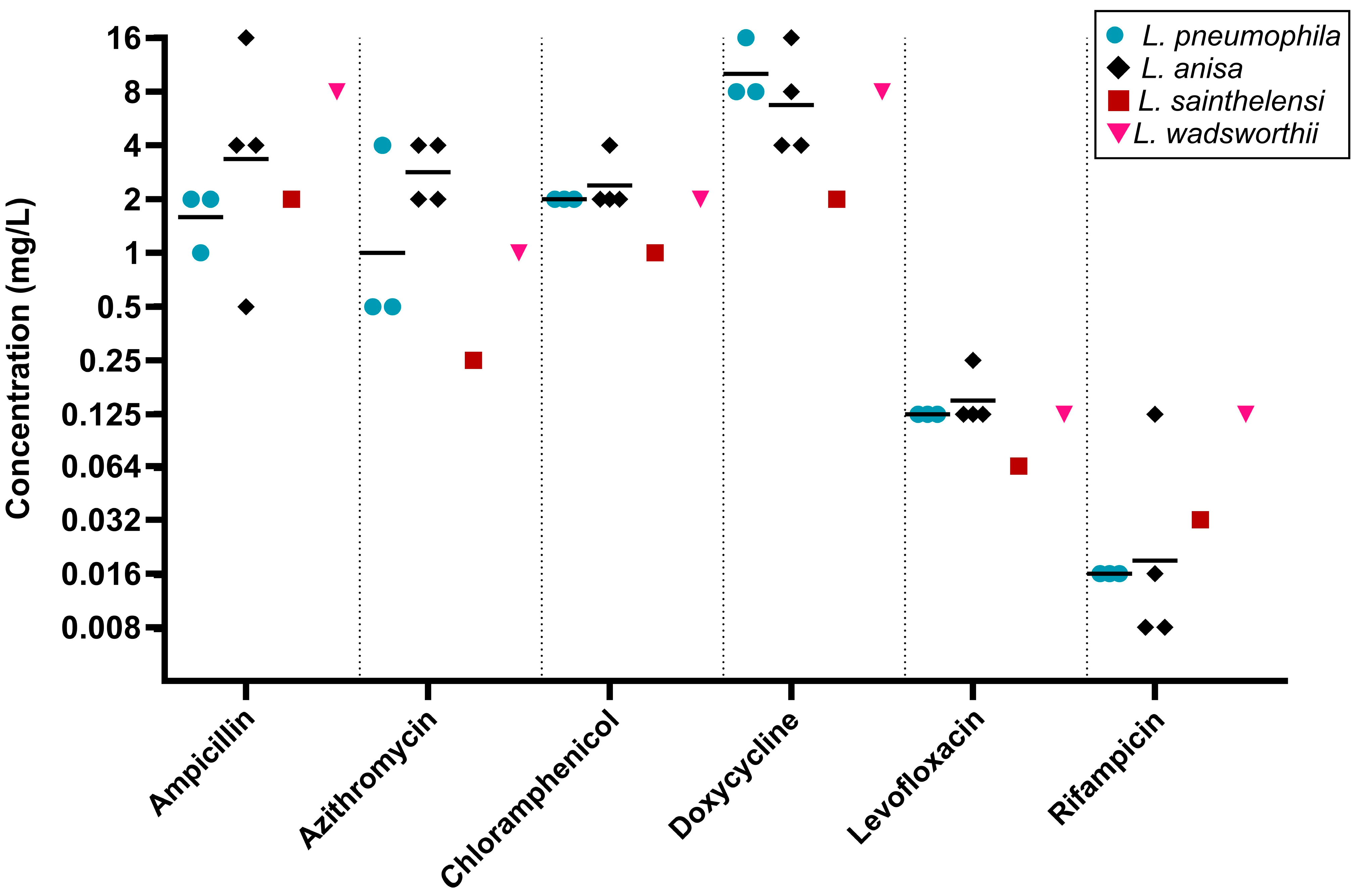
Only six isolates, representing L. anisa (n = 4), L. sainthelensi (n = 1), and L. wadsworthii (n = 1), were tested using BCYE agar dilution, having failed to complete susceptibility testing in both the LASARUS agar dilution and BYE-BMD. Three L. pneumophila strains were included for species comparisons. Figure 3 outlines the MIC distributions across all six tested antibiotics. For five of the six tested antibiotics, L. anisa had a greater range of MICs compared to L. pneumophila.

2.3. Differences Between Legionella Species

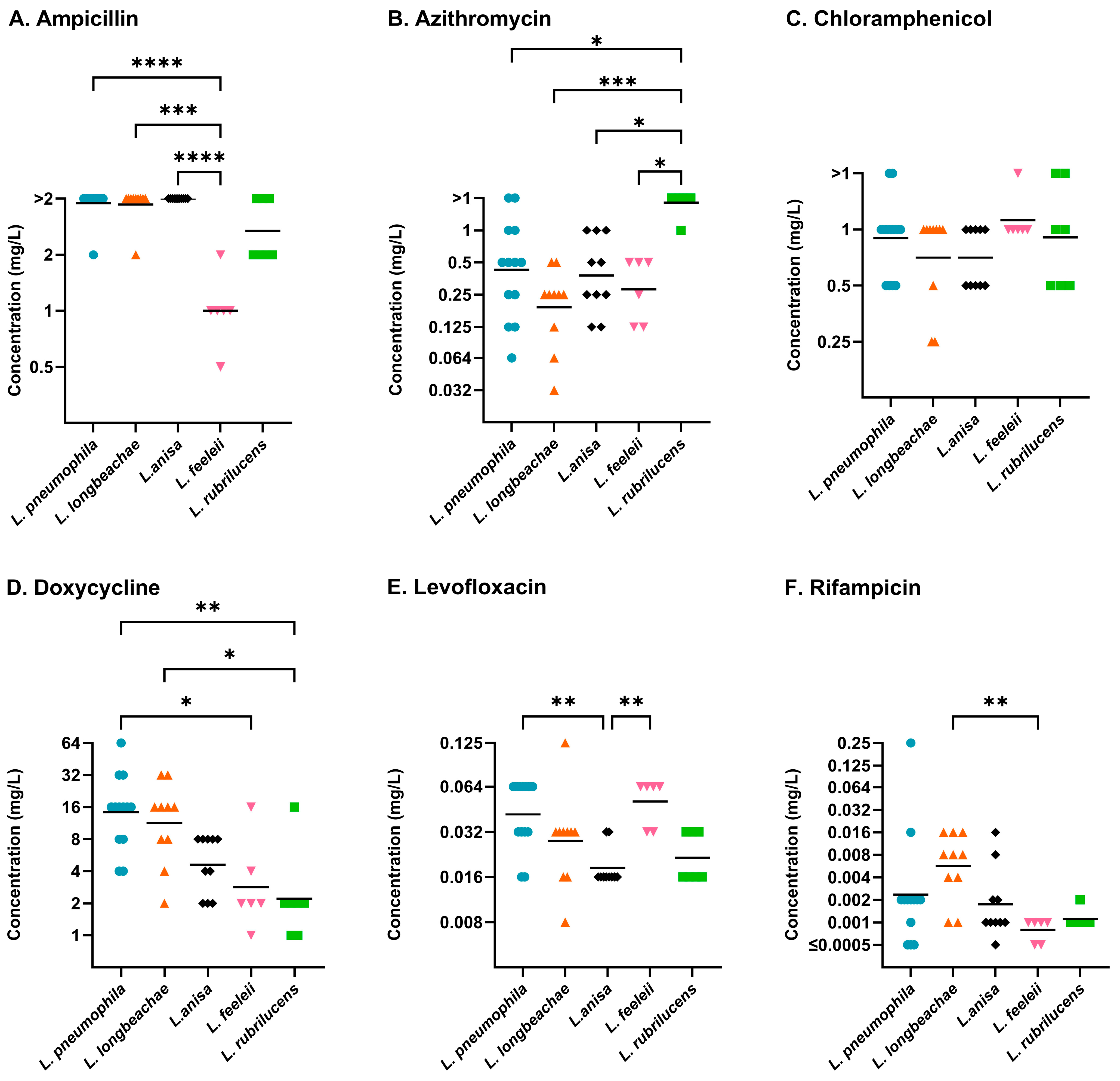
Legionella species, with more than five representative isolates, were compared using the proposed standardised BMD method of Sewell et al. (Figure 4) [21]. This included L. anisa (n = 10), L. feeleii (n = 6), L. longbeachae (n = 10), L. pneumophila (n = 13), and L. rubrilucens (n = 7). The overall comparisons of the five species using the Kruskal–Wallis test were significant for ampicillin (p ≤ 0.0001), azithromycin (p ≤ 0.001), doxycycline (p ≤ 0.001), levofloxacin (p ≤ 0.001), and rifampicin (p ≤ 0.01). Dunn’s multiple comparisons identified L. feeleii with ampicillin and doxycycline, L. rubrilucens with azithromycin and doxycycline, and L. anisa with levofloxacin to have significantly different susceptibilities compared to L. pneumophila.

2.4. Agreement of Legionella Antibiotic Susceptibility Testing Methods

The reproducibility of MICs obtained across LASARUS, BYE-BMD, and BCYE agar dilutions was evaluated by using three serogroup 1 L. pneumophila reference strains (NCTC 11192; NCTC 12008; NCTC 12024). Figure 5 summarises the MICs obtained for these three strains, across the three methods, for all tested antibiotics after 96 h of incubation. The results for BYE-BMD have been included for a 72 h incubation, as it was the earliest time point for completed MICs across the tested Legionella species. Significant differences in MICs obtained across the three methods were observed for ampicillin (p ≤ 0.01), chloramphenicol (p ≤ 0.05), and levofloxacin (p ≤ 0.01) using the Friedman test. The susceptibilities determined using LASARUS and BYE-BMD were not found to be significantly different.
Additionally, comparisons for the MICs of other Legionella species could be made for those successfully growing in broth and on LASARUS (Table 1). For all non-pneumophila species, the MICs obtained using BYE-BMD after 72- and 96 h and LASARUS after 96 h were found to be overall significantly different using the Friedman test (Table 4). For L. longbeachae, BYE-BMD MICs recorded after 96 h were found to be significantly higher than LASARUS after 96 h for ampicillin and azithromycin (Figure 6A). A significant increase in doxycycline MICs was observed between 72- and 96 h in BYE-BMD, and, for rifampicin BYE-BMD after 72 h, the MIC was significantly lower than in both LASARUS and BYE-BMD after 96 h. A greater lack of MIC reproducibility was observed in the mixed collection of Legionella species (Figure 6B).

3. Discussion

Official guidelines for standardised methods of antibiotic susceptibility testing, resistance thresholds, and ECOFF (Epidemiological Cut-Off) values for Legionella species remain undefined [22]. The designation of such criteria would provide a basis for identifying potential antibiotic resistance, or reduced susceptibility, emerging in Legionella spp. to clinically important antibiotics. Antibiotics which are specifically important for the treatment of Legionella infections are those which can act intracellularly and can accumulate at therapeutic concentrations within alveolar macrophages [23]. Currently, Legionella is widely regarded as susceptible to all antibiotics used to treat legionellosis [24]. However, the recent identification of L. pneumophila strains with a high macrolide MIC (erythromycin and azithromycin MICs ≥ 1024 mg/L) indicates that the few effective antibiotics for this species will need systematic surveillance [25,26].
Here, we report the antibiotic susceptibility data for thirty-three non-pneumophila Legionella species utilising a multi-site validated broth microdilution method for L. pneumophila [21]. Additionally, the susceptibility data for sixteen Legionella species have been presented through an implementation of the solid medium LASARUS [16]. Prior AST publications on Legionella species have utilised a variety of methods, including BYE-BMD, BCYE agar dilution, and E-test gradient strips on BCYE agar [19,27,28,29]. Historically, all Legionella required activated charcoal to absorb toxins for growth on solid media. Most of our tested isolates yielded a successful MIC determination utilising non-charcoal-based media, including LASARUS agar dilution (53.9%) or BYE-BMD (93.3%). However, six isolates required AST on BCYE agar for growth. The results collected from growth on charcoal-based media have been widely acknowledged to cause elevated MICs due to the chelating effect of activated charcoal [16,30,31]. As these samples were tested alongside L. pneumophila type strains, the MIC values, while not directly interpretable, showed no significant differences to L. pneumophila. Furthermore, some non-pneumophila Legionella species can require up to 2 weeks to achieve measurable growth [32]. Both LASARUS and the BYE-BMD protocols were refined using L. pneumophila, not other Legionella species. It is possible that longer incubation times may have generated more results, but the loss of antibiotic potency with longer incubation times is a significant concern to result validity.
Whilst L. pneumophila is regarded as the most clinically relevant Legionella species, the increased rate of L. longbeachae-related legionellosis in parts of the southern hemisphere, and increasing rates globally, emphasise the clinical importance of non-pneumophila species [6]. Isenman et al. evaluated the MICs of 61 clinical L. longbeachae against six antibiotics using both BYE-BMD and E-test gradient strips on BCYE [33]. To date, and as far as we are aware, no other study has exclusively published AST data focused on clinically sourced L. longbeachae. Other papers have published AST for L. longbeachae; however, they have incorporated only select type strains and environmentally sourced isolates, much like the work we have presented [34,35]. The ranges for azithromycin and rifampicin MICs have been reported as 0.032–0.25 mg/L, 0.06–0.25 mg/L, 0.06–0.5 mg/L and ≤0.008–0.064, ≤0.004–0.03, and ≤0.002–0.06, respectively, by Ienman et al., Gómez-Lus et al., and Nimmo and Bull [33,34,35]. Ienman et al. and Gómez-Lus et al. conducted AST with BYE-BMD, whereas Nimmo and Bull obtained MIC values using disc diffusion on BCYE agar. These ranges overlap with our ranges of 0.032–0.5 mg/L for azithromycin and 0.001–0.016 mg/L for rifampicin. Gómez-Lus et al. additionally reported MICs for levofloxacin, with a range of 0.008–0.016 mg/L, and an MIC50 and MIC90 of 0.016 mg/L. Our study found L. longbeachae to have a two-fold increase in MIC50 and MIC90 at 0.032 mg/L, and an elevated MIC range of 0.032–0.125 mg/L.
Levofloxacin MICs have been reported by Stout et al. for L. pneumophila, L. micdadei, L. bozemanae, and L. jordanis, obtained using BYE-BMD [36]. Our study identified respective MICs of 0.032 mg/L and 0.016 mg/L for clinically and environmentally sourced strains of L. micdadei and 0.016 mg/L for L. bozemanae, comparable to the 0.015 mg/L previously reported for each species. Bopp et al. have additionally reported an MIC of 0.015 mg/L for L. micdadei, utilising BYE macrotube dilution [37]. A two-fold higher MIC was observed by Stout et al. for L. jordanis, with an MIC of 0.06 mg/L, similar to our 0.032 mg/L. We observed variable levofloxacin MICs in L. pneumophila, ranging from 0.016 to 0.064 mg/L, correlating with prior published BYE-BMD ranges [24,34,36,38,39]. Gómez-Lus et al. have previously reported data on L. dumoffii and L. gormanii for eight antibiotics, including azithromycin, levofloxacin, and rifampicin [34]. The results collected in this study were within the published MIC ranges for azithromycin (0.125–0.5 mg/L) and rifampicin (<0.004–0.008 mg/L). However, for L. dumoffii, we identified MICs of 0.032 mg/L and for L. gormanii an MIC of 0.125 mg/L with levofloxacin, 2-fold and 8-fold higher than the respective maximum range Gómez-Lus et al. published.
Whilst we identified multiple, statistically significant differences between the MICs obtained in LASARUS agar dilution or BYE-BMD, as discussed above, our findings have aligned with previously published data. For our L. pneumophila control strains, ampicillin was the only antibiotic identified to differ significantly between the two methods, with MICs being notably higher (range ≥ 2 mg/L) in BMD compared to LASARUS agar dilution (range 0.25–1 mg/L) for this species. However, as our MIC range was capped at 2 mg/L, it is possible we were only observing a 2–4-fold increase in the maximum MIC against ampicillin in BMD. Our ampicillin MICs in LASARUS are comparable to those reported in broth by Wilson et al. (range 0.125–1 mg/L) [40]. Across the non-pneumophila Legionella species, there was a reduced concordance between the two methods. This may be due to the optimisation of both AST methodologies for L. pneumophila and highlights a need to re-evaluate the existing methods to support a greater testing of non-pneumophila species.
Despite the reduced concordance in non-pneumophila ASTs between methods, performing biological replicates alongside L. pneumophila type strains has provided an important basis for comparison. We have observed across clinically relevant non-pneumophila Legionella species that MICs can differ significantly from those of L. pneumophila. This includes the elevated MICs against azithromycin, the first-line macrolide therapeutic for the treatment of LD, of six Legionella species in LASARUS agar dilution (L. erythra, L. gormanii, L. londiniensis, L. longbeachae, L. quinlivanii, and L. rubrilucens) and nine in BYE-BMD (L. bozemanae, L. cherrii, L. erythra, L. fairfieldensis, L. gormanii, L. maceachernii, L. micdadei, L. rubrilucens, and L. spiritensis) which shared an MIC of >1 mg/L with one L. pneumophila strain. Such differences emphasise the importance of improved diagnostics for non-pneumophila Legionella, with microbial culture remaining the most accurate or available method, and the availability of MIC data [41,42,43].
Whilst there are currently no defined antibiotic resistance breakpoints for Legionella, guidance published by The European Committee on Antimicrobial Susceptibility Testing (EUCAST) has advised on methods for detecting antibiotic resistance mechanisms in L. pneumophila by comparing MIC data to defined ECOFF values [26]. This has been supported by the identification of antibiotic resistance genes in L. pneumophila, including gyrA (point mutation resulting in ciprofloxacin resistance) and lpeAB (efflux pump associated with macrolide resistance) [24,44]. Additionally, a resistance to fluoroquinolones has been reported in association with a gyrA83 mutation [45]. Investigations into L. pneumophila have since attributed elevated MICs to these defined genes [46,47,48,49]. However, numerous studies have also reported elevated azithromycin MICs in samples negative for the presence of the lpeAB gene, highlighting the need for continued investigations into currently unidentified antibiotic resistance mechanisms within Legionella species [16,18,46,50].
Many research gaps remain in our understanding of emerging or undefined antibiotic resistance in Legionella species. This is particularly true for non-pneumophila species, which, as demonstrated in this study, have the potential to exhibit a reduced susceptibility to antibiotics that are otherwise effective in treating Legionella infections. The potential emergence of reduced macrolide or fluoroquinolone susceptibility in environmental or clinical Legionella strains poses a significant public health concern, particularly among vulnerable populations and during healthcare-associated outbreaks [51,52]. Strengthening AMR surveillance and establishing standardised AST protocols for Legionella are therefore critical for maintaining effective treatment options and informing global antimicrobial stewardship efforts [22].
This study has also highlighted the current limitations faced when trying to establish a standardised AST protocol for all Legionella species. All samples grew on BCYE; however, AST of all samples was not conducted on BCYE due to the previously mentioned limitations of the method. The successful growth of the whole isolate panel could not be obtained using LASARUS or BYE media, potentially relating to nutrient and charcoal differences compared to BCYE. An inconsistent growth was observed for five Legionella species on the LASARUS medium, and these were excluded to ensure data consistency and prevent misinterpretation, as incomplete or unreliable MIC profiles could bias comparative analyses. Consequently, MIC data for these five species were not presented in the figures. This represents a limitation of the study and underscores the challenges of achieving consistent growth conditions across diverse Legionella species. Despite this, the published BYE-BMD method obtained non-pneumophila MICs within 72 h, and L. pneumophila within 48 h, providing a successful AST alternative for thirty-three Legionella species faster than the tested agar-based methods [21]. It is standard practice to perform single determinations for measuring MICs in microbiology; we have performed all measurements as biological replicates (with triplicates where results differed by 2-fold dilution); however, as we were developing this as a comparative method, not performing all measurements in triplicate could be viewed as a limitation to this study.
Although LASARUS agar dilution performed less consistently, it remains advantageous for its high-throughput testing capacity with multipoint inoculation, improved safety compared to BCYE due to the translucency of the agar (negating the requirement of opening plate lids due to condensation), and subsequent potential for result collection using automated optical systems [16]. BYE-BMD, while effective, is limited by its labour-intensive set up and contamination checks, and BCYE by its charcoal chelation and opacity. Overall, our findings provide a foundation for future AST standardisation and comparative analyses across 34 Legionella species, addressing a key gap in the characterisation of Legionella antimicrobial susceptibility.

4. Materials and Methods

4.1. Study Design

A total of 89 Legionella isolates, comprising 34 Legionella species and 36 registered NCTC strains, were supplied by the UK Health Security Agency (UKHSA; London, UK) (Table A1 and Table A2). Samples were collected from clinical and environmental sources (1977–2021).
Biological replicates were performed to obtain an n = 2. If MICs were ±2 dilutions apart, a third replicate was performed to correct discrepancies and identify outliers. Initially, AST of all isolates was conducted using LASARUS agar dilution and BYE-BMD. Isolates with no MICs determined in either method were put forward for agar dilution using BCYE. Three L. pneumophila NCTC strains (NCTC 11192; NCTC 12008; NCTC 12024) were included for testing on BCYE.

4.2. Microbial Culturing

All isolates were cultured on BCYE media from glycerol bead archived (stored −80 °C on microbank beads (Technical Service Consultants, Heywood, UK)) and incubated at 36 °C (±1 °C) for 72 to 96 h in a humidified atmosphere. These conditions were replicated for the incubation of LASARUS and BCYE agar dilution plates. BMD plates were incubated on a shaking incubator (100 rpm) (Luckham R100 rotatest shaker, Burgess Hill, UK) at 37 °C for 96 h.

4.3. Antimicrobial Susceptibility Testing

AST was conducted against six antibiotics: ampicillin, doxycycline, levofloxacin, rifampicin (Sigma-Aldrich, Poole, UK), azithromycin (Aspire Pharma, Petersfield, UK), and chloramphenicol (Sigma-Aldrich, Poole, UK). Working stocks were prepared fresh at concentrations of 2560 mg/L, 80 mg/L, and 2.5 mg/L and final concentration ranges for AST between 0.0005 and 128 mg/L.
The solid culture medium LASARUS was prepared in-house based on the previously available formulation (Instant Test Ltd., Blackwood, UK). BCYE (Sigma–Aldrich, Poole, UK) was prepared according to the manufacturer’s instructions. Antimicrobial susceptibility testing (AST) was performed on both LASARUS and BCYE as previously described, with the adaptation of using a 1:10 dilution of a 1 McFarland suspension [16]. BYE-BMD was prepared and conducted as previously reported [21].

4.4. Statistical Analysis

Statistical analysis was performed using GraphPad Prism version 10.5.0. All datasets were assessed for normality using the Shapiro–Wilk test. As most datasets did not meet the assumptions of normality, non-parametric methods were used. The Kruskal–Wallis test was applied for comparisons between species (independent groups), and the Friedman test for comparisons between methods (repeated measures). Where appropriate, post hoc analysis was conducted using Dunn’s multiple comparisons test. Log2-transformed MIC data were also assessed, but transformation did not result in a normal distribution, and thus non-parametric methods remained appropriate. All graphs were additionally generated in GraphPad Prism. Calculations of MIC50 and MIC90, the concentrations at which 50% and 90% of the isolate panel growth is inhibited, respectively, along with MIC ranges, were performed independently.

4.5. Ethical Approval

Ethical approval was not required due to the anonymisation of patient details by UKHSA prior to the receival of isolates at Cardiff University. No ethical approval is required for NCTC strains.

5. Conclusions

While significant efforts to standardise AST internationally for L. pneumophila using BYE-BMD are currently underway, L. pneumophila is not the only cause of legionellosis, and antimicrobial resistance thresholds may not be directly applicable to other Legionella species. While BYE-BMD was able to establish consistent MICs for all species tested, 4/10 L. anisa isolates (an emerging pathogen of concern) would not grow on any medium other than BCYE; furthermore, longer incubations were often required for non-pneumophila species to achieve turbidity (BMD) or colony formation (agars), which impacts direct comparisons. Ampicillin cannot be used to treat legionellosis; however, a majority of Legionella species had MICs >2 mg/L in BMD, which is a generalised pK/pD therapeutic efficacy cut-off. Regarding clinically relevant antimicrobials, L. pneumophila had higher or equivalent MIC distributions relative to all other species except for rifampicin, where L. longbeachae was significantly higher, and there are a number of Legionella species isolates with higher MICs for azithromycin. Given that azithromycin is a first-line therapeutic, resistance thresholds may have to be established for each species individually in the future.

Supplementary Materials

The following supporting information can be downloaded at https://www.mdpi.com/article/10.3390/antibiotics14111165/s1. MIC values for individual isolates used in figures and partial MIC profiles for five species with inconsistent growth on LASARUS which have not been included in LASARUS analysis.

Author Contributions

Conceptualization, C.F., V.J.C. and O.B.S.; methodology, M.S. and E.A.R.P.; validation, A.P. and R.B.; formal analysis, C.F.; investigation, C.F., A.P. and M.S.; resources, B.A., J.D. and I.B.; data curation, C.F. and M.S.; writing—original draft preparation, C.F.; writing—review and editing, all authors; visualisation, A.P. and R.B.; supervision, V.J.C. and O.B.S.; project administration, O.B.S. All authors have read and agreed to the published version of the manuscript.

Funding

This research received no external funding.

Institutional Review Board Statement

Not applicable.

Data Availability Statement

Data available on request from the authors.

Conflicts of Interest

The authors declare no conflicts of interest.

Abbreviations

The following abbreviations are used in this manuscript:
ASTAntimicrobial Susceptibility Testing
BCYEBuffered Charcoal Yeast Extract
BMDBroth Microdilution
BYEBuffered Yeast Extract
DWDDrinking Water Directive
ECOFFEpidemiological Cut-Off Value
EUCASTThe European Committee on Antimicrobial Susceptibility Testing
LASARUSLegionella Antimicrobial Susceptibility and Resistance Universal Screening Medium
LDLegionnaire’s Disease
MICMinimum Inhibitory Concentration
NCTCNational Collection of Type Cultures
UATUrine Antigen Test
UKHSAUk Health Security Agency

Appendix A

Appendix A.1

All tested Legionella samples were supplied by UKHSA from type strain collections or clinical and environmental origins.
Table A1. Sample origin: type strain, clinical, or environmental.
Legionella
Species
Sample OriginIsolates
(n)
Type StrainClinicalEnvironmental
L. anisa--1414
L. beliardensis1--1
L. birminghamensis1--1
L. bozemanae--11
L. brunensis1--1
L. busanensis1--1
L. cherrii1-23
L. cincinnatiensis1--1
L. donaldsonii1--1
L. dresdenensis1--1
L. dumoffii--22
L. erythra1-12
L. fairfieldensis1--1
L. fallonii--22
L. feeleii2136
L. gormanii--11
L. hackeliae2--2
L. israelensis1--1
L. jamestowniensis--11
L. jordanis--11
L. londiniensis1-12
L. longbeachae-10-10
L. maceachernii1--1
L. micdadei-112
L. nautarum1--1
L. oakridgensis1--1
L. parisiensis1--1
L. pneumophila13--13
L. quinlivanii1-12
L. rubrilucens1-67
L. sainthelensi-112
L. shakespearei1--1
L. spiritensis--11
L. wadsworthii-1-1

Appendix A.2

Thirty-six NCTC type strains, from twenty-two Legionella species, were supplied by UKHSA.
Table A2. Cohort of NCTC Legionella type strains.
Legionella SpeciesSerogroupNCTCATCC
L. beliardensis-13315700512
L. birminghamensis-1243743702
L. brunensis-1224043878
L. busanensis-13316BAA-518
L. cherrii-1197635252
L. cincinnatiensis-1243843753
L. donaldsonii---
L. dresdenensis-13409-
L. erythra-1197735303
L. fairfieldensis-1248849588
L. feeleii11202235072
L. feeleii21197835849
L. hackeliae11197935250
L. hackeliae21198035999
L. israelensis-1201043119
L. londiniensis-1293149505
L. maceachernii-1198235300
L. nautarum-1293249506
L. oakridgensis-1153133761
L. parisiensis-1198335299
L. pneumophila (Philadelphia 1)11119233152
L. pneumophila (Knoxville-1)11128633153
L. pneumophila (OLDA)11200843109
L. pneumophila (Allentown 1)11202443106
L. pneumophila (Togus 1)21123033154
L. pneumophila (Bloomington-2)31123233155
L. pneumophila (Los Angeles-1)41123333156
L. pneumophila (Chicago-2)61140633215
L. pneumophila (Chicago 8)71198433823
L. pneumophila (Concorde 3)81198535096
L. pneumophila (570-CO-H)121218043290
L. pneumophila (82A3105)131218143736
L. pneumophila (1169-MN-H)141217443703
L. quinlivanii-1243343830
L. rubrilucens-1198735304
L. shakespearei-1282949655

References

  1. Mondino, S.; Schmidt, S.; Rolando, M.; Escoll, P.; Gomez-Valero, L.; Buchrieser, C. Legionnaires’ Disease: State of the Art Knowledge of Pathogenesis Mechanisms of Legionella. Annu. Rev. Pathol. Mech. Dis. 2020, 15, 439–466. [Google Scholar] [CrossRef] [PubMed]
  2. Legionella—LPSN (List of Prokaryotic Names with Standing in Nomenclature). Available online: https://lpsn.dsmz.de/genus/legionella (accessed on 20 October 2025).
  3. Cunha, B.A.; Burillo, A.; Bouza, E. Legionnaires’ Disease. Lancet 2016, 387, 376–385. [Google Scholar] [CrossRef] [PubMed]
  4. Legionellosis in Residents of England and Wales: 2017 to 2023 Report. Available online: https://www.gov.uk/government/statistics/legionellosis-in-residents-of-england-and-wales-2017-to-2023/legionellosis-in-residents-of-england-and-wales-2017-to-2023-report (accessed on 20 October 2025).
  5. CDC Surveillance Report 2018–2019. Available online: https://www.cdc.gov/legionella/php/surveillance/surveillance-report-2018-2019.html (accessed on 20 October 2025).
  6. Chambers, S.T.; Slow, S.; Scott-Thomas, A.; Murdoch, D.R. Legionellosis Caused by Non-Legionella pneumophila Species, with a Focus on Legionella longbeachae. Microorganisms 2021, 9, 291. [Google Scholar] [CrossRef] [PubMed]
  7. Roussotte, M.; Massy, E. Case Report of Arthritis Caused by Legionella Anisa and Review of the Literature. BMC Infect. Dis. 2022, 22, 633. [Google Scholar] [CrossRef]
  8. Gattuso, G.; Rizzo, R.; Lavoro, A.; Spoto, V.; Porciello, G.; Montagnese, C.; Cinà, D.; Cosentino, A.; Lombardo, C.; Mezzatesta, M.L.; et al. Overview of the Clinical and Molecular Features of Legionella pneumophila: Focus on Novel Surveillance and Diagnostic Strategies. Antibiotics 2022, 11, 370. [Google Scholar] [CrossRef]
  9. Viasus, D.; Gaia, V.; Manzur-Barbur, C.; Carratalà, J. Legionnaires’ Disease: Update on Diagnosis and Treatment. Infect. Dis. Ther. 2022, 11, 973–986. [Google Scholar] [CrossRef]
  10. Chauhan, D.; Shames, S.R. Pathogenicity and Virulence of Legionella: Intracellular Replication and Host Response. Virulence 2021, 12, 1122–1144. [Google Scholar] [CrossRef]
  11. Newton, H.J.; Ang, D.K.; van Driel, I.R.; Hartland, E.L. Molecular Pathogenesis of Infections Caused by Legionella pneumophila. Clin. Microbiol. Rev. 2010, 23, 274–298. [Google Scholar] [CrossRef]
  12. Delaney, S.; Arcari, T.; O’Connor, O. Legionella Water Testing and the EU Drinking Water Directive: Could Potentially Harmful Legionella Bacteria Slip through the Gaps? BioTechniques 2022, 72, 229–231. [Google Scholar] [CrossRef]
  13. Khodr, A.; Kay, E.; Gomez-Valero, L.; Ginevra, C.; Doublet, P.; Buchrieser, C.; Jarraud, S. Molecular Epidemiology, Phylogeny and Evolution of Legionella. Infect. Genet. Evol. 2016, 43, 108–122. [Google Scholar] [CrossRef]
  14. European Union. Directive (EU) 2020/2184 of the European Parliament and of the Council of 16 December 2020 on the quality of water intended for human consumption (recast) (Text with EEA relevance). Off. J. Eur. Union 2020, L 435, 1–62. [Google Scholar]
  15. Ha, R.; Heilmann, A.; Lother, S.A.; Turenne, C.; Alexander, D.; Keynan, Y.; Rueda, Z.V. The Adequacy of Current Legionnaires’ Disease Diagnostic Practices in Capturing the Epidemiology of Clinically Relevant Legionella: A Scoping Review. Pathogens 2024, 13, 857. [Google Scholar] [CrossRef]
  16. Portal, E.; Sands, K.; Portnojs, A.; Chalker, V.J.; Spiller, O.B. Legionella Antimicrobial Sensitivity Testing: Comparison of Microbroth Dilution with BCYE and LASARUS Solid Media. J. Antimicrob. Chemother. 2021, 76, 1197–1204. [Google Scholar] [CrossRef]
  17. Sharaby, Y.; Nitzan, O.; Brettar, I.; Höfle, M.G.; Peretz, A.; Halpern, M. Antimicrobial Agent Susceptibilities of Legionella pneumophila MLVA-8 Genotypes. Sci. Rep. 2019, 9, 6138. [Google Scholar] [CrossRef]
  18. Cocuzza, C.E.; Martinelli, M.; Perdoni, F.; Giubbi, C.; Vinetti, M.E.A.; Calaresu, E.; Frugoni, S.; Scaturro, M.; Ricci, M.L.; Musumeci, R. Antibiotic Susceptibility of Environmental Legionella pneumophila Strains Isolated in Northern Italy. Int. J. Environ. Res. Public Health 2021, 18, 9352. [Google Scholar] [CrossRef] [PubMed]
  19. Pendland, S.L.; Martin, S.J.; Chen, C.; Schreckenberger, P.C.; Danziger, L.H. Comparison of Charcoal- and Starch-Based Media for Testing Susceptibilities of Legionella Species to Macrolides, Azalides, and Fluoroquinolones. J. Clin. Microbiol. 1997, 35, 3004–3006. [Google Scholar] [CrossRef]
  20. Chen, S.C.; Paul, M.L.; Gilbert, G.L. Susceptibility of Legionella Species to Antimicrobial Agents. Pathology 1993, 25, 180–183. [Google Scholar] [CrossRef]
  21. Sewell, M.; Farley, C.; Portal, E.A.R.; Lindsay, D.; Ricci, M.L.; Jarraud, S.; Scaturro, M.; Descours, G.; Krøvel, A.V.; Barton, R.; et al. Broth Microdilution Protocol for Determining Antimicrobial Susceptibility of Legionella pneumophila to Clinically Relevant Antimicrobials. J. Microbiol. Methods 2025, 228, 107071. [Google Scholar] [CrossRef]
  22. Portal, E.; Descours, G.; Ginevra, C.; Mentasti, M.; Afshar, B.; Chand, M.; Day, J.; Echahidi, F.; Franzin, L.; Gaia, V.; et al. Legionella Antibiotic Susceptibility Testing: Is It Time for International Standardization and Evidence-Based Guidance? J. Antimicrob. Chemother. 2021, 76, 1113–1116. [Google Scholar] [CrossRef]
  23. Bongers, S.; Hellebrekers, P.; Leenen, L.P.H.; Koenderman, L.; Hietbrink, F. Intracellular Penetration and Effects of Antibiotics on Staphylococcus Aureus Inside Human Neutrophils: A Comprehensive Review. Antibiotics 2019, 8, 54. [Google Scholar] [CrossRef]
  24. Vandewalle-Capo, M.; Massip, C.; Descours, G.; Charavit, J.; Chastang, J.; Billy, P.A.; Boisset, S.; Lina, G.; Gilbert, C.; Maurin, M.; et al. Minimum Inhibitory Concentration (MIC) Distribution among Wild-Type Strains of Legionella pneumophila Identifies a Subpopulation with Reduced Susceptibility to Macrolides Owing to Efflux Pump Genes. Int. J. Antimicrob. Agents 2017, 50, 684–689. [Google Scholar] [CrossRef]
  25. Ginevra, C.; Beraud, L.; Pionnier, I.; Sallabery, K.; Bentayeb, H.; Simon, B.; Allam, C.; Chastang, J.; Ibranosyan, M.; Decroix, V.; et al. Detection of Highly Macrolide-Resistant Legionella pneumophila Strains from a Hotel Water Network Using Systematic Whole-Genome Sequencing. J. Antimicrob. Chemother. 2022, 77, 2167–2170. [Google Scholar] [CrossRef]
  26. EUCAST. Guidance Document on Antimicrobial Susceptibility Testing of Legionella pneumophila; European Committee on Antimicrobial Susceptibility Testing (EUCAST): Växjö, Sweden, 2021. [Google Scholar]
  27. Nielsen, K.; Bangsborg, J.M.; Høiby, N. Susceptibility of Legionella Species to Five Antibiotics and Development of Resistance by Exposure to Erythromycin, Ciprofloxacin, and Rifampicin. Diagn. Microbiol. Infect. Dis. 2000, 36, 43–48. [Google Scholar] [CrossRef] [PubMed]
  28. Sikora, A.; Gładysz, I.; Kozioł-Montewka, M.; Wójtowicz-Bobin, M.; Stańczak, T.; Matuszewska, R.; Krogulska, B. Assessment of Antibiotic Susceptibility of Legionella pneumophila Isolated from Water Systems in Poland. Ann. Agric. Environ. Med. 2017, 24, 66–69. [Google Scholar] [CrossRef]
  29. Schülin, T.; Wennersten, C.B.; Ferraro, M.J.; Moellering, R.C.; Eliopoulos, G.M. Susceptibilities of Legionella Spp. to Newer Antimicrobials In Vitro. Antimicrob. Agents Chemother. 1998, 42, 1520–1523. [Google Scholar] [CrossRef] [PubMed]
  30. García, M.T.; Pelaz, C.; Giménez, M.J.; Aguilar, L. In Vitro Activities of Gemifloxacin versus Five Quinolones and Two Macrolides against 271 Spanish Isolates of Legionella pneumophila: Influence of Charcoal on Susceptibility Test Results. Antimicrob. Agents Chemother. 2000, 44, 2176–2178. [Google Scholar] [CrossRef] [PubMed]
  31. Pendland, S.L.; Losnedahl, K.J.; Schriever, C.A. In-Vitro Activity of Gatifloxacin, a Novel Fluoroquinolone, Compared with That of Ciprofloxacin against Legionella spp. J. Antimicrob. Chemother. 1999, 44, 295–297. [Google Scholar] [CrossRef]
  32. Mercante, J.W.; Winchell, J.M. Current and Emerging Legionella Diagnostics for Laboratory and Outbreak Investigations. Clin. Microbiol. Rev. 2015, 28, 95–133. [Google Scholar] [CrossRef]
  33. Isenman, H.; Anderson, T.; Chambers, S.T.; Podmore, R.G.; Murdoch, D.R. Antimicrobial Susceptibilities of Clinical Legionella longbeachae Isolates. J. Antimicrob. Chemother. 2018, 73, 1102–1104. [Google Scholar] [CrossRef]
  34. Gómez-Lus, R.; Adrián, F.; del Campo, R.; Gómez-Lus, P.; Sánchez, S.; García, C.; Rubio, M.C. Comparative in Vitro Bacteriostatic and Bactericidal Activity of Trovafloxacin, Levofloxacin and Moxifloxacin against Clinical and Environmental Isolates of Legionella spp. Int. J. Antimicrob. Agents 2001, 18, 49–54. [Google Scholar] [CrossRef]
  35. Nimmo, G.R.; Bull, J.Z. Comparative Susceptibility of Legionella pneumophila and Legionella longbeachae to 12 Antimicrobial Agents. J. Antimicrob. Chemother. 1995, 36, 219–223. [Google Scholar] [CrossRef]
  36. Stout, J.E.; Arnold, B.; Yu, V.L. Comparative Activity of Ciprofloxacin, Ofloxacin, Levofloxacin, and Erythromycin Against Legionella Species by Broth Microdilution and Intracellular Susceptibility Testing in HL-60 Cells. Diagn. Microbiol. Infect. Dis. 1998, 30, 37–43. [Google Scholar] [CrossRef] [PubMed]
  37. Bopp, L.H.; Baltch, A.L.; Ritz, W.J.; Michelsen, P.B.; Smith, R.P. Activities of Tigecycline and Comparators against Legionella pneumophila and Legionella Micdadei Extracellularly and in Human Monocyte-Derived Macrophages. Diagn. Microbiol. Infect. Dis. 2011, 69, 86–93. [Google Scholar] [CrossRef]
  38. Cruz, C.; Rodrigues, L.; Fernandes, F.; Santos, R.; Paixão, P.; Chasqueira, M.J. Antibiotic Susceptibility Pattern of Portuguese Environmental Legionella Isolates. Front. Cell. Infect. Microbiol. 2023, 13, 1141115. [Google Scholar] [CrossRef] [PubMed]
  39. Stout, J.E.; Sens, K.; Mietzner, S.; Obman, A.; Yu, V.L. Comparative Activity of Quinolones, Macrolides and Ketolides against Legionella Species Using in Vitro Broth Dilution and Intracellular Susceptibility Testing. Int. J. Antimicrob. Agents 2005, 25, 302–307. [Google Scholar] [CrossRef] [PubMed]
  40. Wilson, R.E.; Hill, R.L.R.; Chalker, V.J.; Mentasti, M.; Ready, D. Antibiotic Susceptibility of Legionella pneumophila Strains Isolated in England and Wales 2007–17. J. Antimicrob. Chemother. 2018, 73, 2757–2761. [Google Scholar] [CrossRef]
  41. Avni, T.; Bieber, A.; Green, H.; Steinmetz, T.; Leibovici, L.; Paul, M. Diagnostic Accuracy of PCR Alone and Compared to Urinary Antigen Testing for Detection of Legionella spp.: A Systematic Review. J. Clin. Microbiol. 2016, 54, 401–411. [Google Scholar] [CrossRef]
  42. CDC Laboratory Testing for Legionella. Available online: https://www.cdc.gov/legionella/php/laboratories/index.html (accessed on 20 October 2025).
  43. Pascale, M.R.; Salaris, S.; Mazzotta, M.; Girolamini, L.; Fregni Serpini, G.; Manni, L.; Grottola, A.; Cristino, S. New Insight Regarding Legionella Non-Pneumophila Species Identification: Comparison between the Traditional Mip Gene Classification Scheme and a Newly Proposed Scheme Targeting the rpoB Gene. Microbiol. Spectr. 2021, 9, e01161-21. [Google Scholar] [CrossRef]
  44. Bruin, J.P.; Koshkolda, T.; IJzerman, E.P.F.; Lück, C.; Diederen, B.M.W.; Den Boer, J.W.; Mouton, J.W. Isolation of Ciprofloxacin-Resistant Legionella pneumophila in a Patient with Severe Pneumonia. J. Antimicrob. Chemother. 2014, 69, 2869–2871. [Google Scholar] [CrossRef]
  45. Shadoud, L.; Almahmoud, I.; Jarraud, S.; Etienne, J.; Larrat, S.; Schwebel, C.; Timsit, J.-F.; Schneider, D.; Maurin, M. Hidden Selection of Bacterial Resistance to Fluoroquinolones In Vivo: The Case of Legionella pneumophila and Humans. eBioMedicine 2015, 2, 1179–1185. [Google Scholar] [CrossRef]
  46. Minetti, C.; Barton, R.; Farley, C.; Spiller, O.B.; Rodrigues, R.; Gonçalves, P. Antimicrobial Susceptibility Testing Reveals Reduced Susceptibility to Azithromycin and Other Antibiotics in Legionella pneumophila Serogroup 1 Isolates from Portugal. Eur. J. Clin. Microbiol. Infect. Dis. 2024, 43, 1297–1308. [Google Scholar] [CrossRef]
  47. Scaturro, M.; Lanni, A.; Mancini, F.; Girolamo, A.; Fillo, S.; Ciammaruconi, A.; Lista, F.; Cocuzza, C.E.; Musumeci, R.; Ginevra, C.; et al. Antimicrobial Susceptibility and Epidemiological Types of Legionella pneumophila Human Isolates from Italy (1987–2020). J. Glob. Antimicrob. Resist. 2025, 41, 181–188. [Google Scholar] [CrossRef] [PubMed]
  48. Pereira, B.B.; Marrafa, M.; Cruz, C.; Rodrigues, L.; Nunes, F.; Monteiro, S.; Santos, R.; Carneiro, R.N.; Neto, C.; Aguilar, J.; et al. Antimicrobial Resistance Genes in Legionella from Artificial Water Systems: Findings from a Two-Year Study. Antibiotics 2024, 13, 1121. [Google Scholar] [CrossRef] [PubMed]
  49. Michel, C.; Echahidi, F.; De Muylder, G.; Sewell, M.; Boostrom, I.; Denis, O.; Spiller, O.B.; Pierard, D. Occurrence of Macrolides Resistance in Legionella pneumophila ST188: Results of the Belgian Epidemiology and Resistome Investigation of Clinical Isolates. Int. J. Infect. Dis. 2025, 153, 107786. [Google Scholar] [CrossRef] [PubMed]
  50. Yang, J.-L.; Sun, H.; Zhou, X.; Yang, M.; Zhan, X.-Y. Antimicrobial Susceptibility Profiles and Tentative Epidemiological Cutoff Values of Legionella pneumophila from Environmental Water and Soil Sources in China. Front. Microbiol. 2022, 13, 924709. [Google Scholar] [CrossRef]
  51. Pappa, O.; Chochlakis, D.; Sandalakis, V.; Dioli, C.; Psaroulaki, A.; Mavridou, A. Antibiotic Resistance of Legionella pneumophila in Clinical and Water Isolates—A Systematic Review. Int. J. Environ. Res. Public Health 2020, 17, 5809. [Google Scholar] [CrossRef]
  52. Samuelsson, J.; Hallström, L.P.; Marrone, G.; Dias, J.G. Legionnaires’ Disease in the EU/EEA*: Increasing Trend from 2017 to 2019. Eurosurveillance 2023, 28, 2200114. [Google Scholar] [CrossRef]
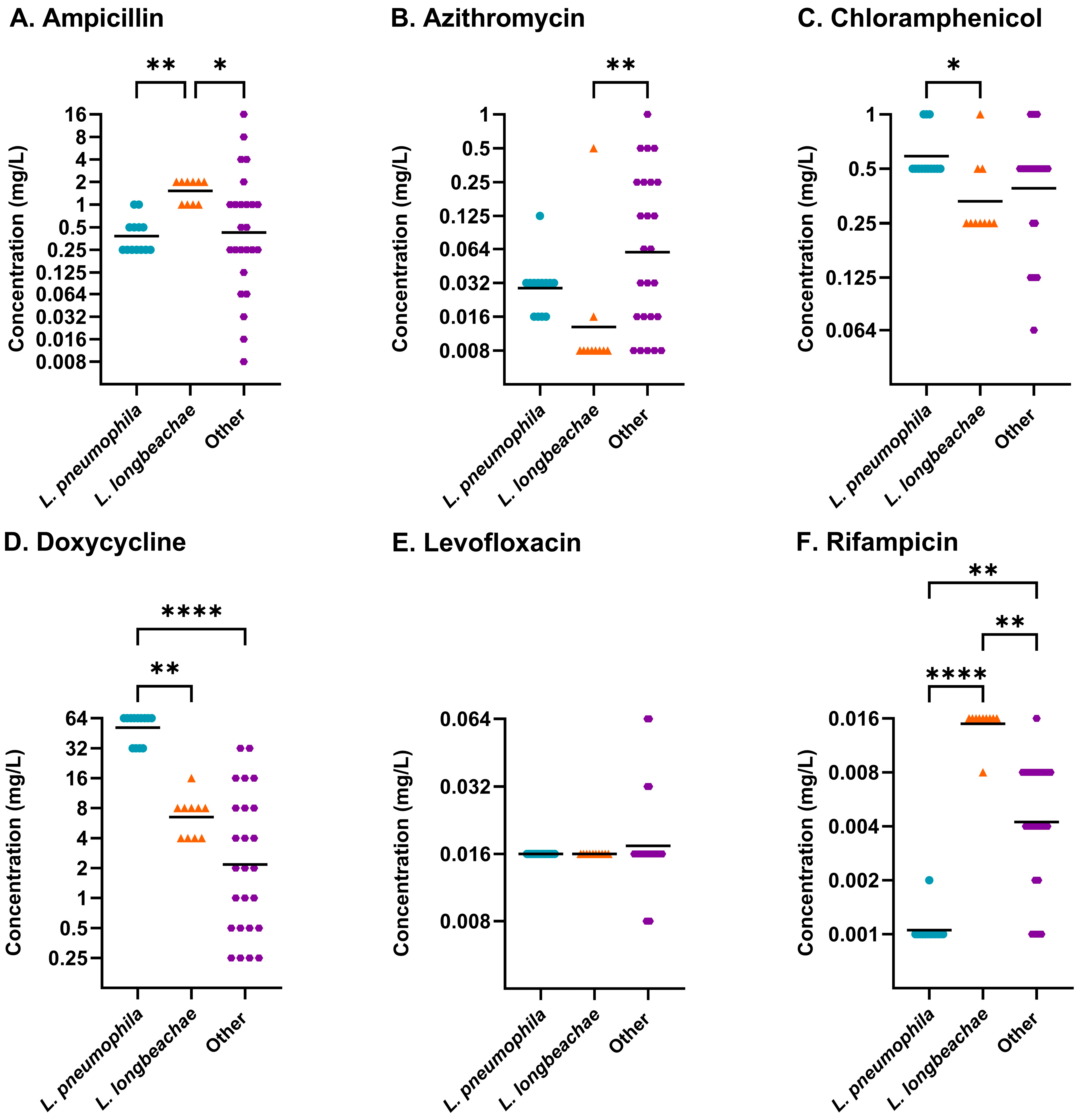
Figure 1. Distribution of antibiotic susceptibilities determined using LASARUS agar dilution at 96 h incubation for (A) ampicillin, (B) azithromycin, (C) chloramphenicol, (D) doxycycline, (E) levofloxacin, and (F) rifampicin. L. pneumophila (blue circle), L. longbeachae (orange triangle), and other Legionella species (purple hexagon) were identified to have overall significantly different MICs through utilisation of non-parametric Kruskal–Wallis analysis for five antibiotics (ampicillin p ≤ 0.01; azithromycin p ≤ 0.01; chloramphenicol p ≤ 0.05; doxycycline p ≤ 0.0001; rifampicin p ≤ 0.0001). The geometric mean (bar) and significant differences between species collections, determined using post hoc Dunn’s multiple comparisons test, are shown. MICs (mg/L) are shown on a Log2 scale to reflect doubling dilutions. (Abbreviations: p ≤ 0.05, *; p ≤ 0.01, **; p ≤ 0.0001, ****).
Antibiotics 14 01165 g001
Figure 2. Distribution of antibiotic susceptibilities determined using BYE-BMD after 96 h shaking incubation for (A) ampicillin, (B) azithromycin, (C) chloramphenicol, (D) doxycycline, (E) levofloxacin, and (F) rifampicin. Individual Legionella species are shown separately where data was available for >10 isolates. L. pneumophila (blue circle), L. longbeachae (orange triangle), L. anisa (black diamond), and other Legionella species (purple hexagon) were identified to have overall significantly different MICs through utilisation of non-parametric Kruskal–Wallis analysis for four antibiotics (ampicillin p ≤ 0.01; azithromycin p ≤ 0.05; doxycycline p ≤ 0.001; levofloxacin p ≤ 0.01). The geometric mean (bar) and significant differences between species collections, determined using post hoc Dunn’s multiple comparisons test, are shown. MICs (mg/L) are shown on a Log2 scale to reflect doubling dilutions. (Abbreviations: p ≤ 0.05, *; p ≤ 0.01, **).
Antibiotics 14 01165 g002
Figure 3. Distribution of antibiotic susceptibilities determined using BCYE agar dilution after 96 h. For L. pneumophila (blue circle) and L. anisa (black diamond), the geometric mean is shown (bar). Susceptibility ranges of L. pneumophila, L. anisa, and L. wadsworthii (pink inverted triangle) overlapped for all six antibiotics. L. sainthelensi (red square) demonstrated reduced susceptibility against azithromycin, chloramphenicol, doxycycline, and levofloxacin. MICs (mg/L) are shown on a Log2 scale to reflect doubling dilutions.
Antibiotics 14 01165 g003
Figure 4. Comparative susceptibility of L. anisa (black diamond), L. feeleii (pink inverted triangle), L. longbeachae (orange triangle), and L. rubrilucens (green square) benchmarked against L. pneumophila (blue circle), using BYE-BMD for clinically relevant antibiotics (ampicillin (A), azithromycin (B), chloramphenicol (C), doxycycline (D), levofloxacin (E), and rifampicin (F)). The geometric mean (bar) and significant differences between species collections, determined using post hoc Dunn’s multiple comparisons test, are shown. MICs (mg/L) are shown on a Log2 scale to reflect doubling dilutions. (Abbreviations: p ≤ 0.05, *; p ≤ 0.01, **; p ≤ 0.001, ***; p ≤ 0.0001, ****).
Antibiotics 14 01165 g004
Figure 5. Three serogroup 1 L. pneumophila strains had MICs determined across LASARUS agar dilution (purple circle), BYE-BMD (pink triangle), and BCYE agar dilution (grey diamond) for six antibiotics after 96 h incubation. MICs obtained using BYE-BMD after 72 h (orange square) have been included as a comparison for the earliest read time. The geometric mean (bar) and significant differences between MICs for single antibiotics, identified using post hoc Dunn’s multiple comparisons test, are shown. Antibiotic ranges on the broth axis in brackets represent capped test ranges for azithromycin (>1 mg/L), chloramphenicol (>1 mg/L), and ampicillin (>2 mg/L). MICs (mg/L) are shown on a Log2 scale to reflect doubling dilutions. (Abbreviations: p ≤ 0.05, *).
Antibiotics 14 01165 g005
Figure 6. After 72 h incubation in BYE-BMD (orange square) and 96 h incubation in LASARUS agar dilution (blue circle) and BYE-BMD (pink triangle), MICs could be obtained for L. longbeachae (A) and a collection of other Legionella species (B). The geometric mean (bar) and significant differences between MICs obtained for single antibiotics, identified using post hoc Dunn’s multiple comparisons test, are shown. Antibiotic ranges on the broth axis in brackets represent capped test ranges for azithromycin (>1 mg/L), chloramphenicol (>1 mg/L), and ampicillin (>2 mg/L). MICs (mg/L) are shown on a Log2 scale to reflect doubling dilutions. (Abbreviations: p ≤ 0.05, *; p ≤ 0.01, **; p ≤ 0.001, ***; p ≤ 0.0001, ****).
Antibiotics 14 01165 g006
Table 1. Method of Legionella species antibiotic susceptibility testing.
Legionella SpeciesIsolates
(n)
Susceptibility Profiles 1 (n (%))
LASARUSBrothBCYE
L. anisa141 (7.14)10 (83.33)4 (100)
L. beliardensis1-1 (100)n/a
L. birminghamensis1-1 (100)n/a
L. bozemanae1-1 (100)n/a
L. brunensis1-1 (100)n/a
L. busanensis1-1 (100)n/a
L. cherrii3-3 (100)n/a
L. cincinnatiensis11 (100)1 (100)n/a
L. donaldsonii1-1 (100)n/a
L. dresdenensis1-1 (100)n/a
L. dumoffii22 (100)2 (100)n/a
L. erythra21 (50)2 (100)n/a
L. fairfieldensis1-1 (100)n/a
L. fallonii2-1 (100)n/a
L. feeleii64 (66.67)6 (100)n/a
L. gormanii11 (100)1 (100)n/a
L. hackeliae2-2 (100)n/a
L. israelensis11 (100)1 (100)n/a
L. jamestowniensis1-1 (100)n/a
L. jordanis11 (100)1 (100)n/a
L. londiniensis22 (100)2 (100)n/a
L. longbeachae1010 (100)10 (100)n/a
L. maceachernii1-1 (100)n/a
L. micdadei21 (50)2 (100)n/a
L. nautarum1-1 (100)n/a
L. oakridgensis1-1 (100)n/a
L. parisiensis11 (100)1 (100)n/a
L. pneumophila1313 (100)13 (100)3 (100)
L. quinlivanii21 (50)2 (100)n/a
L. rubrilucens77 (100)7 (100)n/a
L. sainthelensi2-1 (100)1 (100)
L. shakespearei11 (100)1 (100)n/a
L. spiritensis1-1 (100)n/a
L. wadsworthii1--1 (100)
8948 (53.93)83 (93.26)9 (100) *
1 For each method and species, the number and percentage of isolates with completed minimum inhibitory concentration profiles has been shown. All Legionella species were tested using LASARUS agar dilution and BYE broth microdilution. BCYE agar dilution only included isolates with no successful growth in the other methods and three L. pneumophila reference strains for comparison (NCTC 11192; NCTC 12008; NCTC 12024). Isolates where no testing was performed on BCYE are shown as n/a. (* the percentage isolate growth is displayed for the number of isolates which were put forward for testing and not representative of the whole species collection).
Table 2. Antibiotic susceptibility of Legionella in LASARUS agar dilution.
Organism (n)AntibioticMIC (mg/L)
MIC50MIC90Range
L. pneumophila
(13)
Ampicillin0.2510.25–1
Azithromycin0.0320.0320.016–0.125
Chloramphenicol0.510.5–1
Doxycycline646432–64
Levofloxacin0.0160.0160.016
Rifampicin0.0010.0010.001–0.002
L. longbeachae
(10)
Ampicillin221–2
Azithromycin0.0080.0160.008–0.5
Chloramphenicol0.250.50.25–1
Doxycycline884–16
Levofloxacin0.0160.0160.016
Rifampicin0.0160.0160.008–0.016
Other Legionella species
(25)
Ampicillin0.540.008–16
Azithromycin0.0640.50.008–1
Chloramphenicol0.510.064–1
Doxycycline2160.25–32
Levofloxacin0.0160.0320.008–0.064
Rifampicin0.0040.0080.001–0.016
Isolates have been grouped by species where ten or more representative isolates were available. For each of the six tested antibiotics, the range of MICs and concentrations of antibiotic required to inhibit growth of 50% (MIC50) and 90% (MIC90) of the isolate panel have been presented.
Table 3. Antibiotic susceptibility in BYE broth microdilution.
Organism (n)AntibioticMIC (mg/L)
MIC50MIC90Range
L. pneumophila
(13)
Ampicillin>2>22–>2
Azithromycin0.5>10.064–>1
Chloramphenicol1>10.5–>1
Doxycycline16324–64
Levofloxacin0.0640.0640.016–0.064
Rifampicin0.0020.016≤0.0005–0.25
L. longbeachae
(10)
Ampicillin>2>22–>2
Azithromycin0.250.50.032–0.5
Chloramphenicol110.25–1
Doxycycline16322–32
Levofloxacin0.0320.0320.008–0.125
Rifampicin0.0080.0160.001–0.016
L. anisa
(10)
Ampicillin>2>2>2
Azithromycin0.2510.125–1
Chloramphenicol0.510.5–1
Doxycycline482–8
Levofloxacin0.0160.0320.016–0.032
Rifampicin0.0010.008≤0.0005–0.016
Other Legionella species
(50)
Ampicillin>2>20.016–>2
Azithromycin0.5>10.016–>1
Chloramphenicol1>10.25–>1
Doxycycline4160.5–32
Levofloxacin0.0320.1250.016–0.25
Rifampicin0.0020.008≤0.0005–0.032
Isolates have been grouped by species where ten or more representative isolates were available. For each of the six tested antibiotics, the range of MICs and concentrations of antibiotic required to inhibit growth of 50% (MIC50) and 90% (MIC90) of the isolate panel have been presented.
Table 4. Non-pneumophila significant differences in MICs across methods.
Legionella GroupAntibioticFriedman TestDunn’s Multiple Comparisons Test
L. longbeachaeAmpicillin***LASARUS—BYE-BMD 96 h**
Azithromycin***LASARUS—BYE-BMD 96 h**
Chloramphenicol* ns
Doxycycline*BYE-BMD 72 h—BYE-BMD 96 h*
Levofloxacin* ns
Rifampicin**LASARUS—BYE-BMD 72 h*
Legionella spp.Ampicillin****LASARUS—BYE-BMD 72 h**
LASARUS—BYE-BMD 96 h****
Azithromycin****LASARUS—BYE-BMD 72 h***
LASARUS—BYE-BMD 96 h****
Chloramphenicol****LASARUS—BYE-BMD 96 h***
Doxycycline***LASARUS—BYE-BMD 96 h*
BYE-BMD 72 h—BYE-BMD 96 h***
Levofloxacin****LASARUS—BYE-BMD 72 h**
LASARUS—BYE-BMD 96 h***
Rifampicin****LASARUS—BYE-BMD 72 h***
BYE-BMD 72 h—BYE-BMD 96 h*
Collections of non-pneumophila Legionella species that had successful MIC determination in LASARUS agar dilution and BYE-BMD were compared. Significant differences identified using Friedman test and post hoc Dunn’s multiple comparisons test are shown. (Abbreviations: ns, not significant, p ≤ 0.05, *; p ≤ 0.01, **; p ≤ 0.001, ***; p ≤ 0.0001, ****).
Disclaimer/Publisher’s Note: The statements, opinions and data contained in all publications are solely those of the individual author(s) and contributor(s) and not of MDPI and/or the editor(s). MDPI and/or the editor(s) disclaim responsibility for any injury to people or property resulting from any ideas, methods, instructions or products referred to in the content.