Article

Exploring the Molecular Mechanisms of Macrolide Resistance in Laboratory Mutant Helicobacter pylori

by
Meltem Ayaş
1,2,*,
Sinem Oktem-Okullu
3,
Orhan Özcan
4,
Tanıl Kocagöz
2,3 and
Yeşim Gürol
3
1
Department of Medical Laboratory Techniques, Vocational School of Health Services, Acibadem Mehmet Ali Aydinlar University, 34752 Istanbul, Turkey
2
Department of Medical Biotechnology, Institute of Health Sciences, Acibadem Mehmet Ali Aydinlar University, 34752 Istanbul, Turkey
3
Department of Medical Microbiology, Faculty of Medicine, Acibadem Mehmet Ali Aydinlar University, 34752 Istanbul, Turkey
4
TrioScience Biotechnology, 34000 Istanbul, Turkey
*
Author to whom correspondence should be addressed.
Antibiotics 2024, 13(5), 396; https://doi.org/10.3390/antibiotics13050396
Submission received: 29 February 2024 / Revised: 10 April 2024 / Accepted: 18 April 2024 / Published: 26 April 2024

Abstract

:
Resistance to clarithromycin, a macrolide antibiotic used in the first-line treatment of Helicobacter pylori infection, is the most important cause of treatment failure. Although most cases of clarithromycin resistance in H. pylori are associated with point mutations in 23S ribosomal RNA (rRNA), the relationships of other mutations with resistance remain unclear. We examined possible new macrolide resistance mechanisms in resistant strains using next-generation sequencing. Two resistant strains were obtained from clarithromycin-susceptible H. pylori following exposure to low clarithromycin concentrations using the agar dilution method. Sanger sequencing and whole-genome sequencing were performed to detect resistance-related mutations. Both strains carried the A2142G mutation in 23S rRNA. Candidate mutations (T1495A, T1494A, T1490A, T1476A, and G1472T) for clarithromycin resistance were detected in the Mutant-1 strain. Furthermore, a novel mutation in the gene encoding for the sulfite exporter TauE/SafE family protein was considered to be linked to clarithromycin resistance or cross-resistance, being identified as a target for further investigations. In the Mutant-2 strain, a novel mutation in the gene that encodes DUF874 family protein that can be considered as relevant with antibiotic resistance was detected. These mutations were revealed in the H. pylori genome for the first time, emphasizing their potential as targets for advanced studies.

1. Introduction

Approximately 50% of the world’s population is colonized by Helicobacter pylori, a Gram-negative, spiral-shaped bacterium that can survive in the acidic environment of the stomach. The International Agency for Research on Cancer has identified H. pylori as a Group 1 carcinogen that is capable of causing gastric cancer [1,2]. Furthermore, it has been reported that eradicating H. pylori is an effective strategy for treating peptic ulcers and gastric mucosa-associated lymphoid tissue lymphoma, as well as for preventing gastric cancer [3].
Several factors, such as socioeconomic status and environmental conditions, have been linked to the risk of H. pylori infection at early ages [4,5,6,7]. Therefore, although the incidence of H. pylori infection is decreasing in industrialized countries, it remains stable in developing countries [8].
H. pylori is typically acquired early in childhood. Although the prevalence of H. pylori infection is higher in the first five years of life in developing countries, there have been reports of decreased infection rates due to improved hygiene and sanitation [9,10]. Conversely, the risk of infection is low in adulthood [8,11,12]. Current estimates suggest that approximately 4.4 billion individuals worldwide are infected with H. pylori. In European countries, the prevalence of H. pylori has decreased since 2000; however, it remains unchanged in Asia. Reports indicate that Africa has the highest prevalence (70.1%), whereas Switzerland has the lowest (18.9%). Among Southern Asian countries, Pakistan and India exhibit the highest prevalence of H. pylori (81% and 63.5%, respectively). In Western Asia, Turkey has the highest prevalence (77.2%) [13].
The presence of H. pylori in saliva, vomit, and stool samples has been observed, but there is no evidence supporting these bodily fluids as the predominant mode of transmission. Person-to-person transmission through gastro–oral, oral–oral, or fecal–oral exposure appears likely. Because H. pylori has a limited host range, new infections arise via person-to-person transmission or environmental contamination. It remains unclear which of the aforementioned transmission routes is the most frequent [14,15,16,17]. Hereditary susceptibility to H. pylori infection has also been considered, but it has not been proven [18].
According to current international guidelines on H. pylori treatment, first-line therapy includes proton pump inhibitors (PPIs), clarithromycin, and amoxicillin/metronidazole. Although this therapy is considered standard, treatment failure due to increased antibiotic resistance, particularly to clarithromycin, has been observed. Consequently, guidelines do not recommend the standard therapy in regions where clarithromycin resistance rates exceed 15% [19,20,21,22,23,24]. With increasing resistance rates, recommending standard triple therapies as empiric therapy may no longer be feasible. Given the high level of resistance to clarithromycin and metronidazole, along with varied resistance patterns across populations, it is crucial to design standard triple therapies to address local resistance patterns. Wherever possible, treatment decisions should be based on strain culture susceptibility information. Various alternative treatment strategies are currently employed in clinical practice to manage antibiotic-resistant H. pylori strains. Consequently, novel effective treatments with greater efficacy, including probiotics, have been developed and utilized to enhance eradication regimens and mitigate antibiotic-associated side effects [25].
Clarithromycin, a macrolide antibiotic, is the most potent bacteriostatic antibiotic used to treat H. pylori infection. Because many international guidelines recommend clarithromycin-containing bismuth quadruple therapies for H. pylori treatment, clarithromycin remains important in treating H. pylori infections [19,20,21]. In recent years, clarithromycin resistance has become more prevalent worldwide. The resistance rate in Europe increased from 9% to 17.6% between 1998 and 2008, while that in Japan increased from 7% to 27.7% between 2000 and 2006 [12]. Clarithromycin resistance rates in Turkey have been estimated to have reached approximately 40% [26]. The World Health Organization recently published a list of bacteria requiring new antibiotics urgently, and clarithromycin-resistant H. pylori was included in the high-priority list [27].
Inhibiting protein synthesis is the main mechanism of action of clarithromycin, which binds to receptors located in the 23S ribosomal subunit of the 50S ribosome. Point mutations in the peptidyl transferase encoded in domain V of 23S ribosomal RNA (rRNA) are responsible for clarithromycin resistance [28]. Several point mutations in the 23S rRNA gene have been reported to decrease the drug’s affinity for ribosomes, causing the bacteria to become resistant. More than 90% of clarithromycin-resistant strains harbor three point mutations in the peptidyl transferase region of domain V of 23S rRNA. These mutations include a substitution from adenine to guanine at position 2143 (A2143G) and one from adenine to guanine or cytosine at position 2142 (A2142G or A2142C) [29,30,31,32,33]. Several other point mutations in the region contribute to clarithromycin resistance; however, their significance remains unclear [31,32,33]. Another mechanism of clarithromycin resistance involves five efflux pump systems. Current findings suggest that four groups of genes (HP0605–HP0607, HP0971–HP0969, HP1327–HP1329, and HP1489–HP1487) can function as efflux pumps in H. pylori [34,35,36]. Despite the major molecular mechanisms of clarithromycin resistance demonstrated in previous studies, it remains unclear whether other gene mutations outside 23S rRNA are associated with resistance in H. pylori. The presented study aimed to identify new clarithromycin resistance mechanisms in H. pylori. Because there is a need for more information about the clarithromycin resistance mechanism in H. pylori, this study provides a key contribution to the literature. The tests currently used in laboratories are designed to detect clarithromycin resistance in H. pylori by identifying only the most common mutations (A2143G, A2142G, and A2142C) associated with resistance. Therefore, we predict that the candidate mutations identified in the study could also contribute to the development of molecular diagnostic tests by highlighting new resistance mechanisms in the target regions.

2. Results

2.1. Clarithromycin Minimum Inhibitory Concentration (MIC) Value for Helicobacter pylori G27

The agar dilution test results were evaluated visually. Bacterial growth was observed at clarithromycin concentrations of 0.015 and 0.03 µg/mL and on the growth control medium. However, no growth was noted at clarithromycin concentrations of ≥0.06 µg/mL. Thus, the MIC of clarithromycin was determined to be 0.06 μg/mL (Figure 1).

2.2. Establishment of Resistant Strains

The reference strain H. pylori G27, which is susceptible to clarithromycin, was exposed to low concentrations of clarithromycin, and two resistant mutant strains were obtained. Accordingly, Mutant-1 and Mutant-2 exhibited clarithromycin MICs of 128 and >256 μg/mL, respectively (Figure 1).

2.3. Sanger Sequencing

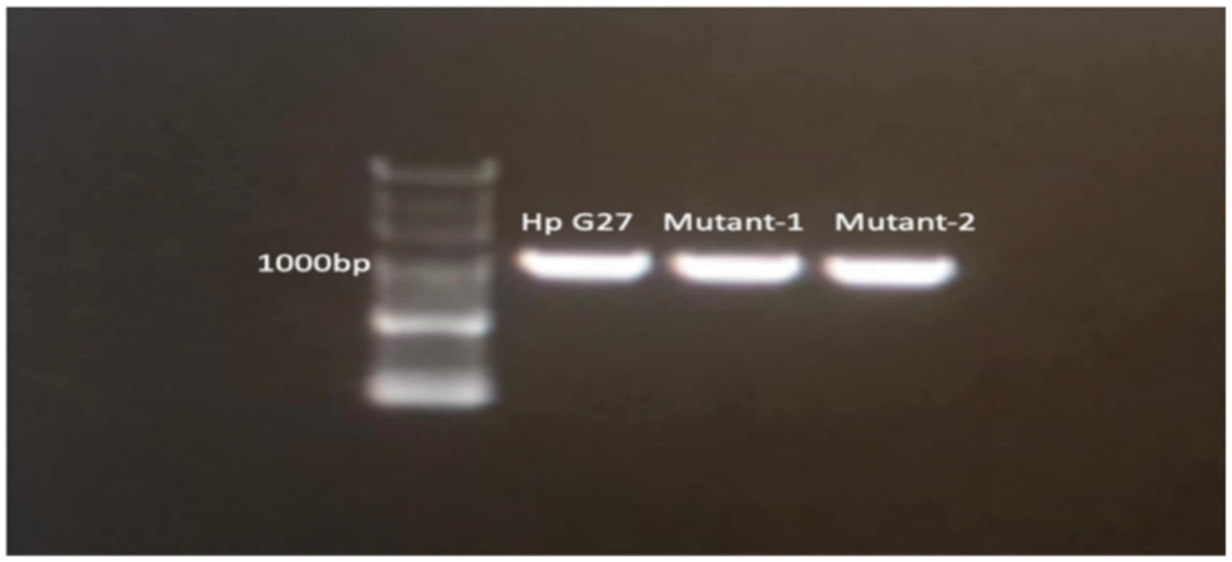
Polymerase chain reaction (PCR) results associated with the amplicons are presented in Figure 2. The A2142G mutation was detected in both the Mutant-1 and Mutant-2 strains (Figure 3).

2.4. Next-Generation Sequencing (NGS) for Detecting Mutations in the Resistant Strains

The DNAs of H. pylori G27 and the mutant strains were sequenced. The resulting sequences were compared between G27 and the mutants. In the Mutant-1 strain, shifts from thymine to adenine at positions 1476, 1490, 1494, and 1495, as well as a shift from guanine to thymine at position 1472 were observed in the 23S rRNA region. Additionally, the A2142G mutation was detected in Mutant-1. Furthermore, a single-nucleotide mutation in a gene encoding the sulfite exporter TauE/SafE family protein was observed. Because of the leucine-to-proline mutation, structural changes occurred in the proteins. In Mutant-2, the A2142G was detected. In addition, mutations were also detected in genes that encodes the DUF874 Family protein in Mutant 2. As a result of these mutations, some aminoacidic changes were observed (Table 1). Bioinformatic analysis data are presented as the Supplementary Data (Supplementary Data, Tables S1 and S2, Figure S1).

3. Discussion

The discovery of H. pylori infection and its role in various diseases, including gastric cancer, has completely changed the treatment landscape for infected patients. Currently, antibiotics represent the only available effective therapy for H. pylori infection. However, few antibiotics can act against this bacterium, which resides in acidic stomach conditions. The standard triple therapy, including a PPI, clarithromycin, and either amoxicillin or metronidazole, has been used as the first-line treatment for eradicating H. pylori in regions where clarithromycin resistance rates are lower than 15%. The success of the treatment relies on factors such as smoking, treatment adherence, and antibiotic resistance. Resistance to clarithromycin has been reported to be the most crucial factor that leads to treatment failure [37,38].
Clarithromycin is a bacteriostatic antibiotic that reversibly binds to the peptidyl transferase loop of domain V of 23S rRNA. In most Gram-negative bacteria, the mechanism of resistance to clarithromycin or other macrolides involves modification of the target region (nucleotide mutations and methylation of 23S rRNA) or overexpression of efflux pumps [39].
Macrolides exert bacteriostatic effects by inhibiting protein synthesis via reversible binding to the peptidyl transferase loop of domain V of 23S rRNA in the 50S subunit [40]. Clarithromycin is the most preferred macrolide for H. pylori treatment because of its pharmacokinetic advantages, including stability under acidic conditions and better absorption in the gastric mucus layer [41]. The World Health Organization has reported clarithromycin-resistant H. pylori as a high-priority pathogen that requires attention in treatment [42]. Generally, Gram-negative bacteria can develop resistance to macrolides through three mechanisms: methylation of the target cells or mutations of critical genes, efflux mechanisms, and enzymatic deactivation of the drug. In H. pylori, specific mutations in the 23S rRNA region have been linked to clarithromycin resistance. Three primary 23S rRNA mutations have been reported, namely A2142G, A2143G, and A2142C, with prevalence rates of 11.7%, 69.8%, and 2.6%, respectively. Numerous other point mutations have also been identified, such as A2115G, T2117C, G2141A, T2182C, G2224A, C2245T, T2289C, C2611A, and T2717C. Despite their low frequencies, the clinical relevance of these mutations is unproven. However, correlations of T2182C, C2611A, and T2717C with low resistance levels have been reported [30,43]. Furthermore, recent studies have found that additional resistance mutations affecting the ribosomal protein L22p and the translation initiation factor IF2 are observed in experimentally induced clarithromycin resistance phenotypes. However, these mutations have not been reported in clinical isolates [44,45].
In the present study, two clarithromycin-resistant strains were successfully produced through exposure to low concentrations of clarithromycin, and novel candidate genes related to clarithromycin resistance were identified using NGS. The A2142G mutation, which is frequently reported to be associated with high clarithromycin MICs in the literature [37,44], was detected in the 23S rRNA region in both mutant strains.
In the Mutant-1 strain, six single-nucleotide mutations at different positions in 23S rRNA were detected via NGS, including A2142G. Sanger sequencing of the clarithromycin target region was performed to confirm the known mutation in mutant strains. The other mutations (T1476A, T1490A, T1494A, T1495A, and G1472T) were detected outside the peptidyl transferase region via whole-genome sequencing (WGS). To the best of our knowledge, these mutations have not been reported previously in the literature. Ultimately, all five candidate mutations should also be confirmed by Sanger sequencing. Furthermore, the relationships of these mutations with clinical clarithromycin resistance should be studied using clinical strains or samples in future studies. Clarithromycin-resistant strains with no 23S rRNA mutation have also been reported, indicating that unknown genes outside 23S rRNA are associated with clarithromycin resistance in H. pylori. Binh et al. discovered two novel gene mutations (infB and rpl22) related to clarithromycin resistance in H. pylori that exert synergistic effects with 23S rRNA mutations, resulting in higher MICs [37]. However, such mutations were not detected in the present study. A gene mutation as a novel candidate mechanism was observed in one clarithromycin-resistant mutant strain in the present study. This gene encodes the sulfite exporter TauE/SafE family protein. The mutation resulted in an altered amino acid sequence of the protein, with leucine being changed to proline. Weinitschke et al. reported that TauE/SafE proteins act as sulfite/organosulfonate exporters in the metabolism of C2 sulfonates [45]. Furthermore, Rakitin et al. compared the genomes of clinical and permafrost strains of Acinetobacter lwoffii and detected two copies of the gene encoding the TauE/SafE family sulfite exporter in environmental isolates whereas clinical strains lacked this gene duplication [46]. Considering the widespread occurrence of organosulfonates in nature, the researchers likely hypothesized that permafrost strains may utilize organosulfonates [46]. To the best of our knowledge, the relationship between sulfite exporter TauE/SafE family proteins and antibiotic resistance has not been reported for any bacterium. Based on this knowledge, if clarithromycin is found in its anionic form, it can be considered that the sulfite exporter TauE/SafE family protein might function as a drug efflux pump due to its role as an anion transporter. Consequently, mutations in this protein could lead to drug resistance. Conversely, previous studies have identified organic anion transporter family members 1B1 and 1B3 as targets of clarithromycin in mammalian cells. It has been reported that clarithromycin inhibits these proteins, leading to interactions with substrate drugs [47]. Hence, it can also be predicted that inhibition of the sulfite exporter TauE/SafE family protein by a high concentration of clarithromycin may result in cross-resistance to substrate anionic antibiotics such as fluoroquinolones, tetracyclines, ampicillin, and rifampicin, which are commonly used to treat H. pylori infection. Future studies assessing the antibiotic susceptibility of H. pylori G27 and its mutants to anionic antibiotics are needed to confirm this hypothesis.
In the Mutant-2 strain, single point mutations were identified in two consecutive genes in different positions that encode the DUF874 family protein in the genome. These mutations resulted in changes in amino acid structures. It is worth noting that these mutations, particularly due to amino acid alterations, can induce changes in the three-dimensional structure of the protein, signifying their significance (Supplementary Materials, Table S2).While studies in the literature have indicated an unclear understanding of the function of the DUF874 family protein in H. pylori, recent research suggests that this protein may play a crucial role in H. pylori’s adaptation to stress conditions within host cells by synthesizing DNA-binding proteins. In a study using mouse models, these proteins have been associated with the survival of H. pylori bacteria in the host mouse [48]. To the best of our knowledge, no study demonstrated that clarithromycin resistance was associated with DUF874 family protein mutations in H. pylori. We anticipate that our findings will pave the way for further studies via verification in other studies.
Additionally, during the bioinformatics analysis of the sequencing results, some rRNA strand breaks were detected, and, surprisingly, these breaks occurred in the same regions. They were predicted as hypothetically methylated sites and likely cause resistance to antibiotics by decreasing clarithromycin affinity and changing the ribosomal structure (Supplementary Materials, Figure S2). In the literature, resistance to clarithromycin and other macrolides in Gram-positive and Gram-negative bacteria has been linked to post-transcriptional methylation of 23S rRNA [32,39]. However, to the best of our knowledge, no study has revealed the relationship between RNA methylation and clarithromycin resistance in H. pylori. Further research is required to investigate whether the 23S rRNA region in H. pylori exhibits differential methylation patterns to confirm this claim.
In conclusion, this study identified novel clarithromycin resistance mechanisms in H. pylori. The newly discovered mutations in the 23S rRNA region could serve as targets for molecular diagnosis of clarithromycin resistance pending validation studies. Additionally, hypothetically methylated regions in 23S rRNA were demonstrated in the mutant genome for the first time, underscoring the need for further studies to validate these findings. Identifying these alternative genetic mechanisms of reduced susceptibility could facilitate the development of accurate and reliable sequence-based diagnostics for predicting clarithromycin susceptibility and aid in the design of novel therapeutics.

4. Materials and Methods

4.1. Bacteria

H. pylori G27, which has been extensively used in research because of its relatively straightforward genetics and reliable growth, was used for this study. The bacterium was stored at −80 °C in Brucella broth containing 25% (v/v) glycerol.

4.2. Subculture of H. pylori

First, H. pylori was subcultured in Colombia blood agar (Oxoid, Hampshire, United Kingdom) to recover the bacteria from the frozen environment. The agar included 5% defibrinated horse blood and antibiotic cocktails. The agar was dissolved in distilled water according to the manufacturer’s instructions and then autoclaved at 121 °C for 15 min in the liquid sterilization mode. Thereafter, the agar was kept in a water bath at 50 °C for 1 h to achieve a suitable temperature for adding antibiotics and blood. Antibiotic cocktails and freshly prepared β-cyclodextrin were added to the medium to prevent contamination and contribute to bacterial growth. Finally, horse blood was added, and 20 mL of agar was poured into each Petri dish. H. pylori was streaked onto the prepared agar plates and incubated at 37 °C for 3 days under microaerophilic conditions (5% O2, 10% CO2, and 85% N2) provided by an automated closed system. After incubation, Gram staining was performed to eliminate the possibility of contamination. All processes using H. pylori were conducted in a biosafety level II cabinet in the microbiology laboratory.

4.3. Agar Dilution Susceptibility Tests

Agar dilution susceptibility tests were performed according to the Clinical and Laboratory Standards Institute (CLSI) guidelines [49]. According to these guidelines, the resistance breakpoint for clarithromycin is >0.5 µg/mL [49]. First, 2-fold serial dilutions of clarithromycin were prepared in 2 mL of broth. Then, 1 mL of sterile defibrinated sheep blood was added to each tube (to obtain 5% horse blood in 20 mL). Finally, 17 mL of Mueller–Hinton agar (Becton Dickinson and Company, Franklin Lake, NJ, USA) was added. Therefore, the initial concentration of clarithromycin was diluted 10-fold, with final concentrations of 0.015–256 μg/mL achieved on Petri dishes.

4.4. Inoculation of Bacteria and Interpretations of Results

A 2.0 McFarland bacterial suspension was prepared in sterile saline, and 3 mL of the suspension was inoculated onto each plate at different antibiotic concentrations. After 3 days of incubation under microaerophilic conditions, the lowest concentration of the related antibiotic that prevented visible growth of the bacterium was defined as the MIC. According to the European Committee on Antimicrobial Susceptibility Testing and CLSI guidelines, the resistance breakpoint for clarithromycin is >0.5 mg/mL [49,50]. Agar dilution susceptibility tests were performed thrice.

4.5. In Vitro Selection of Clarithromycin-Resistant Strains

H. pylori G27, which is susceptible to clarithromycin, was used as the parent strain. Clarithromycin-resistant strains were generated from this strain via exposure to low concentrations of clarithromycin in vitro using the agar dilution method. A single colony of G27 was inoculated onto Mueller–Hinton agar supplemented with 5% defibrinated sheep blood without antibiotics. The plate was incubated at 37 °C under microaerophilic conditions (10% O2, 5% CO2, and 85% N2) for 72 h. Colonies on agar plates were harvested, placed in sterile saline, and exposed to serially doubling concentrations of clarithromycin (0.03–256 µg/mL) using the agar dilution method. Then, colonies from the highest concentrations under which bacteria could grow were obtained and repeatedly transferred to the same clarithromycin concentration five times before being exposed to a higher concentration. Individual colonies on the plates containing a clarithromycin concentration greater than the MIC were isolated [37]. In other words, clarithromycin MIC determination and mutant selection were performed simultaneously.

4.6. DNA Isolation from H. pylori G27 and Mutant Strains

Using the High Pure PCR template preparation kit (Roche, Basel, Switzerland), the genomic DNA of H. pylori was isolated according to the manufacturer’s instructions. DNA quantification and quality control were completed immediately after isolation using a NanoDrop One/OneC Spectrophotometer (Thermo Fisher Scientific, Waltham, MA, USA). The DNA samples’ ratio of absorbance at 260/280 nm (OD 1.8–2.0) and 260/230 nm (OD 2–2.2) was considered for DNA sequencing analysis.

4.7. Sanger Sequencing

Specific primers covering all putative mutations associated with clarithromycin resistance were used to target regions. The primers and PCR conditions are described in Table 2 [51]. PCR amplicons were sent to Eurofins Scientific (Luxembourg, Germany) for Sanger sequencing. The DNA sequences were edited using BioEdit version 7.2.5 and aligned using BLAST [52,53].

4.8. WGS and Bioinformatics Analysis

WGS was performed using Illumina NGS Platforms (Illumina, San Diego, CA, USA) by Eurofins Scientific (Luxembourg). Candidate mutations associated with clarithromycin resistance were obtained by comparing the reconstructed genomes of the mutant strains with that of the H. pylori G27 strain. Bioinformatics analysis was performed using the tools listed in Table 3.

Supplementary Materials

The following supporting information can be downloaded at: https://www.mdpi.com/article/10.3390/antibiotics13050396/s1, Table S1: The combined bioinformatics analysis results of H. pylori G27 and Mutant-1 whole genome data; Table S2: The combined bioinformatics analysis results of H. pylori G27 and Mutant-2 whole genome data. Figure S1: Position of SNPs detected (T1495A, T1494A, T1490A, T1476A, G1472T) via WGS in Mutant 1 strain. Figure S2: Signal decreases observed in Sanger sequence analysis are thought to be due to potential methylations occurring in the 23S rRNA region, leading to breaks.

Author Contributions

Conceptualization, M.A. and S.O.-O.; methodology, M.A. and T.K.; software, O.Ö.; validation, M.A., S.O.-O. and T.K.; formal analysis, O.Ö.; investigation, M.A.; resources, T.K.; data curation, O.Ö.; writing—original draft preparation, M.A.; writing—review and editing, M.A., S.O.-O. and Y.G.; visualization, M.A.; supervision, T.K. and Y.G.; project administration, Y.G. All authors have read and agreed to the published version of the manuscript.

Funding

This study was funded by Acibadem Mehmet Ali Aydinlar University Scientific Research Projects Coordination Unit, grant number 2022/02-25. The article processing charge has not been funded.

Institutional Review Board Statement

This study was approved by the Acıbadem University Ethics Committee (dated 21 November 2019, protocol number 2019/18-31).

Data Availability Statement

The data presented in this article are included as Supplementary Material.

Acknowledgments

We would like to express our gratitude to, İbrahim Sertdemir for contributing by sharing the necessary programs for analysing some of the bioinformatics data.

Conflicts of Interest

Author Orhan Özcan was employed by the company TrioScience Biotechnology. The remaining authors declare that the research was conducted in the absence of any commercial or financial relationships that could be construed as a potential conflict of interest.

References

  1. Schistosomes, liver flukes and Helicobacter pylori. IARC Monogr. Eval. Carcinog. Risks Hum. 1994, 61, 1–241.
  2. Abrams, J.A.; Wang, T.C. Adenocarcinoma and other tumors of the stomach. In Sleisenger and Fordtran’s Gastrointestinal and Liver Disease Pathophysiology/Diagnosis/Management, 9th ed.; Feldman, M., Friedman, L.S., Brandt, L.J., Eds.; Elsevier Saunders: Philadelphia, PA, USA, 2010. [Google Scholar]
  3. Ford, A.C.; Forman, D.; Hunt, R.H.; Yuan, Y.; Moayyedi, P. Helicobacter pylori eradication therapy to prevent gastric cancer in healthy asymptomatic infected individuals: Systematic review and meta-analysis of randomised controlled trials. BMJ 2014, 348, g3174. [Google Scholar] [CrossRef] [PubMed]
  4. Perez-Perez, G.I.; Rothenbacher, D.; Brenner, H. Epidemiology of Helicobacter pylori infection. Helicobacter 2004, 9 (Suppl. S1), 1–6. [Google Scholar] [CrossRef] [PubMed]
  5. Pounder, R.E.; Ng, D. The prevalence of Helicobacter pylori infection in different countries. Aliment. Pharmacol. Ther. 1995, 9 (Suppl. S2), 33–39. [Google Scholar] [PubMed]
  6. Kivi, M.; Johansson, A.L.; Reilly, M.; Tindberg, Y. Helicobacter pylori status in family members as risk factors for infection in children. Epidemiol. Infect. 2005, 133, 645–652. [Google Scholar] [CrossRef] [PubMed]
  7. Nouraie, M.; Latifi-Navid, S.; Rezvan, H.; Radmard, A.R.; Maghsudlu, M.; Zaer-Rezaii, H.; Amini, S.; Siavoshi, F.; Malekzadeh, R. Childhood hygienic practice and family education status determine the prevalence of Helicobacter pylori infection in Iran. Helicobacter 2009, 14, 40–46. [Google Scholar] [CrossRef] [PubMed]
  8. Khalifa, M.M.; Sharaf, R.R.; Aziz, R.K. Helicobacter pylori: A poor man’s gut pathogen? Gut Pathog. 2010, 2, 1–12. [Google Scholar] [CrossRef] [PubMed]
  9. Liou, J.M.; Malfertheiner, P.; Lee, Y.C.; Sheu, B.S.; Sugano, K.; Cheng, H.C.; Yeoh, K.-G.; Hsu, P.-I.; Goh, K.-L.; Mahachai, V.; et al. Asian Pacific Alliance on Helicobacter and Microbiota (APAHAM). Screening and eradication of Helicobacter pylori for gastric cancer prevention: The Taipei global consensus. Gut 2020, 69, 2093–2112. [Google Scholar] [CrossRef] [PubMed]
  10. Yuan, C.; Adeloye, D.; Luk, T.T.; Huang, L.; He, Y.; Xu, Y.; Ye, X.; Yi, Q.; Song, P.; Rudan, I. The global prevalence of and factors associated with Helicobacter pylori infection in children: A systematic review and meta-analysis. Lancet Child Adolesc. Heal. 2022, 6, 185–194. [Google Scholar] [CrossRef]
  11. Fiedorek, S.C.; Malaty, H.M.; Evans, D.L.; Pumphrey, C.L.; Casteel, H.B.; Evans, D.J., Jr.; Graham, D.Y. Factors influencing the epidemiology of Helicobacter pylori infection in children. Pediatrics 1991, 88, 578–582. [Google Scholar] [CrossRef]
  12. Laarej, K.; Alami, R.; Bentaleb, L.; Jbilou, M.; El Kabbaj, S.; Elmaouardi, M.; Bouklouze, A. Epidemiological study of Helicobacter pylori infection in a population in the Rabat-sale-Zamour-Zaer region. Am. J. Epidemiol. 2020, 4, 6–9. [Google Scholar]
  13. Hooi, J.K.Y.; Lai, W.Y.; Ng, W.K.; Suen, M.M.Y.; Underwood, F.E.; Tanyingoh, D.; Malfertheiner, P.; Graham, D.Y.; Wong, V.W.S.; Wu, J.C.Y.; et al. Global prevalence of Helicobacter pylori infection: Systematic review and meta-analysis. Gastroenterology 2017, 153, 420–429. [Google Scholar] [CrossRef] [PubMed]
  14. Cave, D.R. Transmission and epidemiology of Helicobacter pylori. Am. J. Med. 1996, 100, 12S–17S; discussion 17S. [Google Scholar] [CrossRef]
  15. Mégraud, F. Transmission of Helicobacter pylori: Faecal-oral versus oral-oral route. Aliment. Pharmacol. Ther. 1995, 9 (Suppl. S2), 85–91. [Google Scholar] [PubMed]
  16. Perry, S.; de la Luz Sanchez, M.; Yang, S.; Haggerty, T.D.; Hurst, P.; Perez-Perez, G.; Parsonnet, J. Gastroenteritis and transmission of Helicobacter pylori infection in households. Emerg. Infect. Dis. 2006, 12, 1701–1708. [Google Scholar] [CrossRef] [PubMed]
  17. Kayali, S.; Manfredi, M.; Gaiani, F.; Bianchi, L.; Bizzarri, B.; Leandro, G.; Di Mario, F.; Angelis, G.L.D. Helicobacter pylori, transmission routes and recurrence of infection: State of the art. Acta Biomed. 2018, 89, 72–76. [Google Scholar] [PubMed]
  18. Graham, D.Y.; Malaty, H.M.; Evans, D.G.; Evans, D.J., Jr.; Klein, P.D.; Adam, E. Epidemiology of Helicobacter pylori in an asymptomatic population in the United States. Effect of age, race, and socioeconomic status. Gastroenterology 1991, 100, 1495–1501. [Google Scholar] [CrossRef] [PubMed]
  19. Malfertheiner, P.; Megraud, F.; O’Morain, C.A.; Gisbert, J.P.; Kuipers, E.J.; Axon, A.T.; El-Omar, E.M. Management of Helicobacter pylori infection-the Maastricht V/Florence consensus report. Gut 2017, 66, 6–30. [Google Scholar] [CrossRef] [PubMed]
  20. Fallone, C.A.; Chiba, N.; van Zanten, S.V.; Fisch-Bach, L.; Gisbert, J.P.; Hunt, R.H.; Jones, N.L.; Render, C.; Leontiadis, G.I.; Moayyedi, P.; et al. The Toronto Consensus for the treatment of Helicobacter pylori infection in adults. Gastroenterology 2016, 151, 51–69.e14. [Google Scholar] [CrossRef] [PubMed]
  21. Chey, W.D.; Leontiadis, G.I.; Howden, C.W.; Moss, S.F. ACG clinical guideline: Treatment of Helicobacter pylori infection. Am. J. Gastroenterol. 2017, 112, 212–239. [Google Scholar] [CrossRef]
  22. IARC Working Group on the Evaluation of Carcinogenic Risks to Humans. Biological agents. A review of human carcinogens. IARC Monogr. Eval. Carcinog. Risks Hum. 2012, 100, 1–441. [Google Scholar]
  23. Torre, L.A.; Bray, F.; Siegel, R.L.; Ferlay, J.; Lortet-Tieulent, J.; Jemal, A. Global cancer statistics, 2012. CA Cancer J. Clin. 2015, 65, 87–108. [Google Scholar] [CrossRef] [PubMed]
  24. Ferlay, J.; Soerjomataram, I.; Dikshit, R.; Eser, S.; Mathers, C.; Rebelo, M.; Parkin, D.M.; Forman, D.; Bray, F. Cancer incidence and mortality worldwide: Sources, methods and major patterns in GLOBOCAN 2012. Int. J. Cancer 2015, 136, E359–E386. [Google Scholar] [CrossRef] [PubMed]
  25. Goderska, K.; Agudo Pena, S.; Alarcon, T. Helicobacter pylori treatment: Antibiotics or probiotics. Appl. Microbiol. Biotechnol. 2018, 102, 1–7. [Google Scholar] [CrossRef] [PubMed]
  26. Kaplan, M.; Tanoglu, A.; Duzenli, T.; Tozun, A.N. Helicobacter pylori treatment in Turkey: Current status and rational treatment options. North. Clin. Istanb. 2020, 7, 87–94. [Google Scholar] [CrossRef] [PubMed]
  27. WHO. Global Priority List of Antibiotic-Resistant Bacteria to Guide Research, Discovery, and Development of New Antibiotics. Available online: https://www.who.int/news/item/27-02-2017-who-publishes-list-of-bacteria-for-which-new-antibiotics-are-urgently-needed (accessed on 30 March 2024).
  28. Liu, W.Z.; Xiao, S.D.; Hu, P.J.; Lu, H.; Cui, Y.; Tytgat, G.N. A new quadruple therapy for Helicobacter pylori using tripotassium dicitrato bismuthate, furazolidone, josamycin and famotidine. Aliment. Pharmacol. Ther. 2000, 14, 1519–1522. [Google Scholar] [CrossRef] [PubMed]
  29. Mégraud, F. H. pylori antibiotic resistance: Prevalence, importance, and advances in testing. Gut 2004, 53, 1374–1384. [Google Scholar] [CrossRef]
  30. Francesco, V.D.; Zullo, A.; Hassan, C.; Giorgio, F.; Rosania, R.; Ierardi, E. Mechanisms of Helicobacter pylori antibiotic resistance: An updated appraisal. World J. Gastrointest. Pathophysiol. 2011, 2, 35–41. [Google Scholar] [CrossRef]
  31. Mégraud, F.; Lehours, P. Helicobacter pylori detection and antimicrobial susceptibility testing. Clin. Microbiol. Rev. 2007, 20, 280–322. [Google Scholar] [CrossRef]
  32. Gerrits, M.M.; van Vliet, A.H.; Kuipers, E.J.; Kusters, J.G. Helicobacter pylori and antimicrobial resistance: Molecular mechanisms and clinical implications. Lancet Infect. Dis. 2006, 6, 699–709. [Google Scholar] [CrossRef]
  33. Versalovic, J.; Osato, M.S.; Spakovsky, K.; Dore, M.P.; Reddy, R.; Stone, G.G.; Shortridge, D.; Flamm, R.K.; Tanaka, S.K.; Graham, D.Y. Point mutations in the 23S rRNA gene of Helicobacter pylori associated with different levels of clarithromycin resistance. J. Antimicrob. Chemother. 1997, 40, 283–286. [Google Scholar] [CrossRef] [PubMed]
  34. Tsugawa, H.; Suzuki, H.; Muraoka, H.; Ikeda, F.; Hirata, K.; Matsuzaki, J.; Saito, Y.; Hibi, T. Enhanced bacterial efflux system is the first step to the development of metronidazole resistance in Helicobacter pylori. Biochem. Biophys. Res. Commun. 2011, 404, 656–660. [Google Scholar] [CrossRef] [PubMed]
  35. Mehrabadi, J.F.; Sirous, M.; Daryani, N.E.; Eshraghi, S.; Akbari, B.; Shirazi, M.H. Assessing the role of the RND efflux pump in metronidazole resistance of Helicobacter pylori by RT-PCR assay. J. Infect. Dev. Ctries. 2011, 5, 88–93. [Google Scholar] [CrossRef] [PubMed]
  36. Bina, J.E.; Alm, R.A.; Uria-Nickelsen, M.; Thomas, S.R.; Trust, T.J.; Hancock, R.E. Helicobacter pylori uptake and efflux: Basis for intrinsic susceptibility to antibiotics in vitro. Antimicrob. Agents Chemother. 2000, 44, 248–254. [Google Scholar] [CrossRef] [PubMed]
  37. Binh, T.T.; Shiota, S.; Suzuki, R.; Matsuda, M.; Trang, T.T.; Kwon, D.H.; Iwatani, S.; Yamaoka, Y. Discovery of novel mutations for clarithromycin resistance in Helicobacter pylori by using next-generation sequencing. J. Antimicrob. Chemother. 2014, 69, 1796–1803. [Google Scholar] [CrossRef] [PubMed]
  38. Suzuki, S.; Kusano, C.; Horii, T.; Ichijima, R.; Ikehara, H. The ideal Helicobacter pylori treatment for the present and the future. Digestion 2022, 103, 62–68. [Google Scholar] [CrossRef] [PubMed]
  39. Vester, B.; Douthwaite, S. Macrolide resistance conferred by base substitutions in 23S rRNA. Antimicrob. Agents Chemother. 2001, 45, 1–12. [Google Scholar] [CrossRef]
  40. Tomb, J.F.; White, O.; Kerlavage, A.R.; Clayton, R.A.; Sutton, G.G.; Fleischmann, R.D.; Ketchum, K.A.; Klenk, H.P.; Gill, S.; Dougherty, B.A.; et al. Erratum: The complete genome sequence of the gastric pathogen Helicobacter pylori. Nature 1997, 389, 412. [Google Scholar] [CrossRef]
  41. Tshibangu-Kabamba, E.; Yamaoka, Y. Helicobacter pylori infection and antibiotic resistance—From biology to clinical implications. Nat. Rev. Gastroenterol. Hepatol. 2021, 18, 613–629. [Google Scholar] [CrossRef]
  42. Abadi, A.T.B. Resistance to clarithromycin and gastroenterologist’s persistence roles in nomination for Helicobacter pylori as high priority pathogen by World Health Organization. World J. Gastroenterol. 2017, 23, 6379–6384. [Google Scholar] [CrossRef]
  43. Thung, I.; Aramin, H.; Vavinskaya, V.; Gupta, S.; Park, J.Y.; Crowe, S.E.; Valasek, M.A. The global emergence of Helicobacter pylori antibiotic resistance. Aliment. Pharmacol. Ther. 2016, 43, 514–533. [Google Scholar] [CrossRef] [PubMed]
  44. Tshibangu-Kabamba, E.; Ngoma-Kisoko, P.J.; Tuan, V.P.; Matsumoto, T.; Akada, J.; Kido, Y.; Tshimpi-Wola, A.; Tshiamala-Kashala, P.; Ahuka-Mundeke, S.; Ngoyi, D.M.; et al. Next-generation sequencing of the whole bacterial genome for tracking molecular insight into the broad-spectrum antimicrobial resistance of Helicobacter pylori clinical isolates from the Democratic Republic of Congo. Microorganisms 2020, 8, 887. [Google Scholar] [CrossRef]
  45. Weinitschke, S.; Denger, K.; Cook, A.M.; Smits, T.H.M. The DUF81 protein TauE in Cupriavidus necator H16, a sulfite exporter in the metabolism of C2 sulfonates. Microbiology 2007, 153, 3055–3060. [Google Scholar] [CrossRef] [PubMed]
  46. Rakitin, A.L.; Ermakova, A.Y.; Beletsky, A.V.; Petrova, M.; Mardanov, A.V.; Ravin, N.V. Genome analysis of Acinetobacter lwoffii strains isolated from permafrost soils aged from 15 thousand to 1.8 million years revealed their close relationships with present-day environmental and clinical isolates. Biology 2021, 10, 871. [Google Scholar] [CrossRef] [PubMed]
  47. Seithel, A.; Eberl, S.; Singer, K.; Auge, D.; Heinkele, G.; Wolf, N.B.; Dörje, F.; Fromm, M.F.; König, J. The influence of macrolide antibiotics on the uptake of organic anions and drugs mediated by OATP1B1 and OATP1B3. Drug Metab. Dispos. 2007, 35, 779–786. [Google Scholar] [CrossRef] [PubMed]
  48. Wang, G.; Maier, R.J. A novel DNA-binding protein plays an important role in Helicobacter pylori stress tolerance and survival in the host. J. Bacteriol. 2015, 197, 973–982. [Google Scholar] [CrossRef] [PubMed]
  49. CLSI. Methods for Antimicrobial Dilution and Disk Susceptibility Testing of Infrequently Isolated or Fastidious Bacteria, 3rd ed; CLSI guideline M45; Clinical and Laboratory Standards Institute: Wayne, PA, USA, 2015. [Google Scholar]
  50. EUCAST. Breakpoint Tables for Interpretation of MICs and Zone Diameters. 2024, Version 14.0. Available online: https://www.eucast.org/clinical_breakpoints (accessed on 8 April 2024).
  51. Alavifard, H.; Mirzaei, N.; Yadegar, A.; Baghaei, K.; Smith, S.M.; Sadeghi, A.; Zali, M.R. Investigation of clarithromycin resistance-associated mutations and virulence genotypes of Helicobacter pylori isolated from Iranian population: A cross-sectional study. Curr. Microbiol. 2021, 78, 244–254. [Google Scholar] [CrossRef] [PubMed]
  52. Hall, T.A. BioEdit: A User-Friendly Biological Sequence Alignment Editor and Analysis Program, for Windows 95/98/NT; Nucleic Acids Symposium Series; Information Retrieval Ltd.: London, UK, 1999; pp. c1979–c2000. [Google Scholar]
  53. BLAST: Basic Local Alignment Search Tool. Available online: https://blast.ncbi.nlm.nih.gov/Blast.cgi?BLAST_SPEC=blast2seq&LINK_LOC=align2seq&PAGE_TYPE=BlastSearch (accessed on 1 January 2023).
  54. Bushnell, B. Bbtools Software Package. Available online: http://sourceforge.net/projects/bbmap (accessed on 20 December 2023).
  55. Prjibelski, A.; Antipov, D.; Meleshko, D.; Lapidus, A.; Korobeynikov, A. Using SPAdes de novo assembler. Curr. Protoc. Bioinform. 2020, 70, e102. [Google Scholar] [CrossRef] [PubMed]
  56. Tanizawa, Y.; Fujisawa, T.; Nakamura, Y. DFAST: A flexible prokaryotic genome annotation pipeline for faster genome publication. Bioinformatics 2018, 34, 1037–1039. [Google Scholar] [CrossRef]
  57. Li, H.; Handsaker, B.; Wysoker, A.; Fennell, T.; Ruan, J.; Homer, N.; Marth, G.; Abecasis, G.; Durbin, R.; 1000 Genome Project Data Processing Subgroup. The sequence alignment/map format and SAMtools. Bioinformatics 2009, 25, 2078–2079. [Google Scholar] [CrossRef]
  58. Rice, P.; Longden, I.; Bleasby, A. EMBOSS: The European molecular biology open software suite. Trends Genet. 2000, 16, 276–277. [Google Scholar] [CrossRef] [PubMed]
Figure 1. Agar dilution assay for assessing the clarithromycin susceptibility of the strains. (A): Growth control, (B): Clarithromycin MIC value of the H. pylori G27 strain, (C): Clarithromycin MIC value of the Mutant-1 strain, (D): Clarithromycin MIC value of the Mutant-2 strain. Cla: Clarithromycin.
Antibiotics 13 00396 g001
Figure 2. PCR amplification of the H. pylori G27, Mutant-1, and Mutant-2 strains and 1% agarose gel electrophoresis.
Antibiotics 13 00396 g002
Figure 3. Multiple-sequence alignment of 23S rRNA gene sequences of the H. pylori G27, Mutant-1, and Mutant-2 strains compared to a reference gene.
Antibiotics 13 00396 g003
Table 1. Candidate mutations associated with clarithromycin resistance in mutant strains detected via whole-genome sequencing.
GenePosition of MutationsMutation TypeH. pylori G27MutationMutant-1Mutant-2AA Change
23SrRNA2142SNPAG++-
23SrRNA1490SNPTA+No
23SrRNA1494SNPTA+No
23SrRNA1495SNPTA+No
23SrRNA1476SNPTA+No
23SrRNA1472SNPGT+No
Sulfite exporter
TauE/SafE family protein
35SNPAG+L > P
DUF874 Family proteinGene-1589SNPTC+H > R
DUF874 Family proteinGene-1587SNPGA+No
DUF874 Family proteinGene-1572SNPGT+No
DUF874 Family proteinGene-1553SNPGA+T* > I
DUF874 Family proteinGene-1549SNPTG+T* > A
DUF874 Family proteinGene-2941SNPTC+No
DUF874 Family proteinGene-2890SNPCT+No
DUF874 Family proteinGene-2703SNPGA+A** > V
DUF874 Family proteinGene-2701SNPGA+No
SNP: single nucleotide polymorphism, A: Adenine, T: Thymine, G: Guanine, C: Cytosine, AA: Amino acids, L: Leucine, P: Proline, H: Histidine, R: Arginine, T*: Threonine, I: Isoleucine, A**: Alanine, V: Valine. +: a nucleotide mutation occurred, −: no nucleotide mutation occurred. No: No aminoacid change occurred.
Table 2. Primers and PCR conditions used for obtaining PCR amplicons.
PrimersPCR Conditions
23S-F 5′-AGCACCGTAAGTTCGCGATAAG-3′
  • Initial denaturation: 94 °C for 4 min
  • 30 cycles at 94 °C for 1 min
  • 56 °C for 45 s
  • 72 °C for 10 min
23S-R 5′-CTTTCAGCAGTTATCACATCC-3′
Table 3. Bioinformatics tools used for whole-genome sequencing.
ToolsIntended PurposeReferences
BBtools v39.01—BBDukQuality trimming and filtering[54]
Spades v3.15.4Genome assembly[55]
Dfast v1.6.0Annotation of the assembled contigs[56]
BBtools v39.01—BBmapMapping of H. pylori reads to the reference G27 genome[54]
BBtools v39.01—Call VariantsVariant calling from sam files with callvariants.sh.[57]
EMBOSS Transeq v6.6.0.0Conversion of SNP-bearing contigs and regions for phenotypic change[58]
Disclaimer/Publisher’s Note: The statements, opinions and data contained in all publications are solely those of the individual author(s) and contributor(s) and not of MDPI and/or the editor(s). MDPI and/or the editor(s) disclaim responsibility for any injury to people or property resulting from any ideas, methods, instructions or products referred to in the content.