
Antifungal and Coagulation Properties of a Copper (I) Oxide Nanopowder Produced by Out-of-Phase Pulsed Sonoelectrochemistry

 , , and
Abstract
1. Introduction
2. Results and Discussion
2.1. Nanopowder Characterization
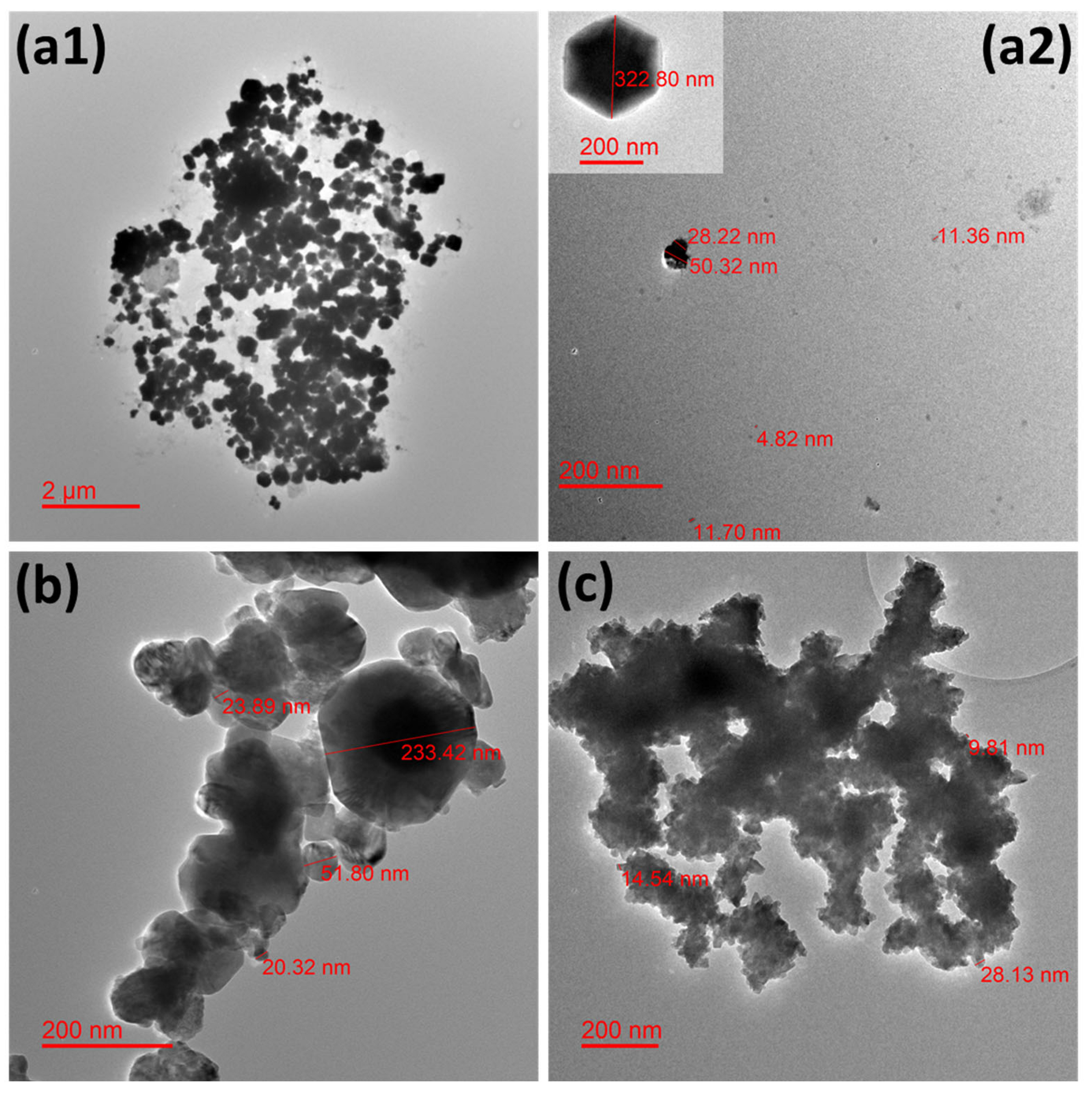
2.1.1. TEM Analysis
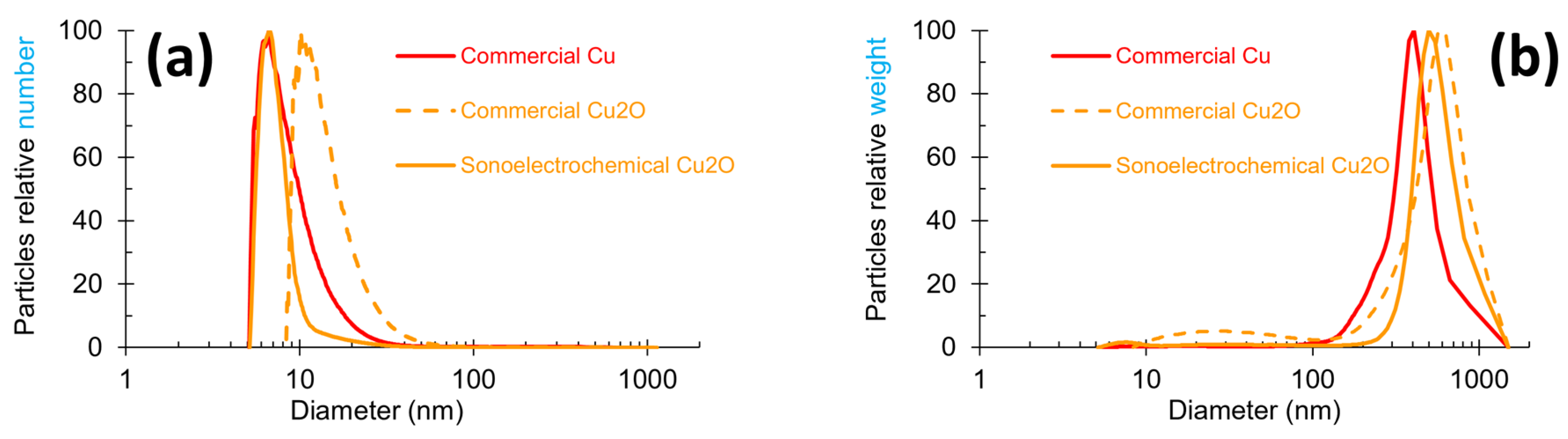
2.1.2. Particle Size Measurements
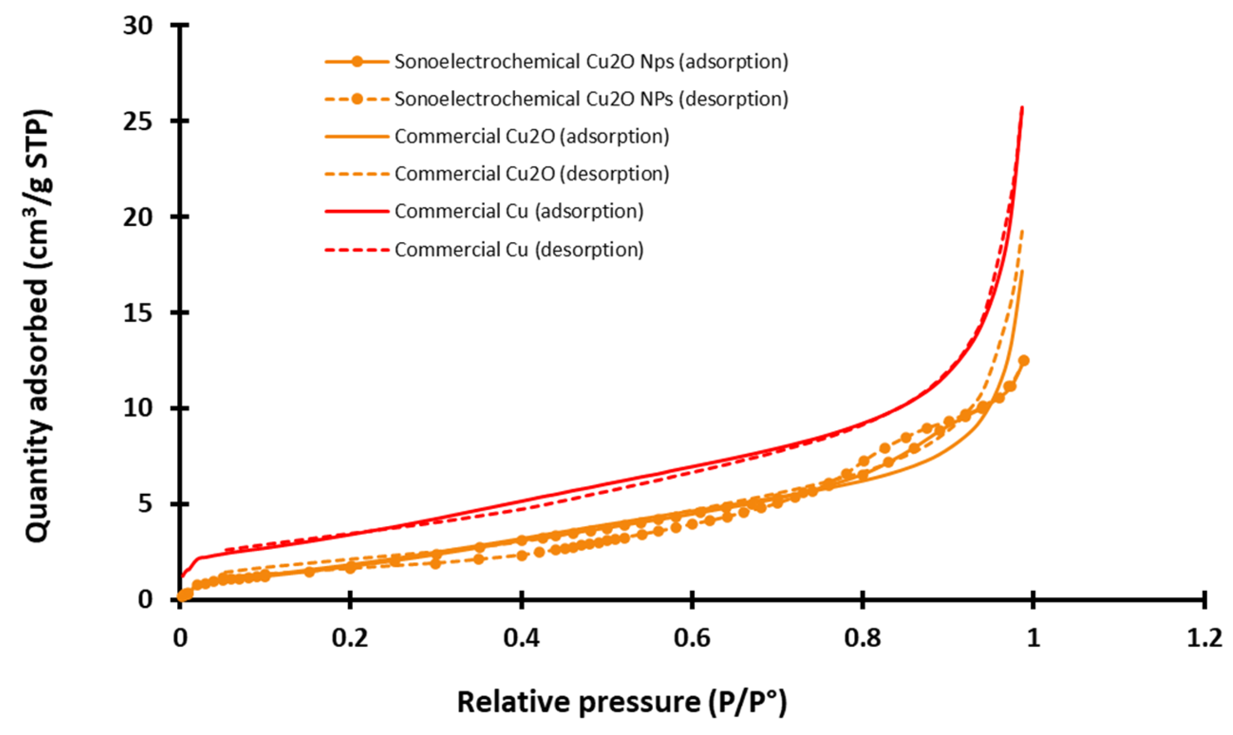
2.1.3. Specific Area Determination
2.2. Antifungal Activities of Nanopowders
2.3. Hemocompatibility
2.3.1. Hemolysis
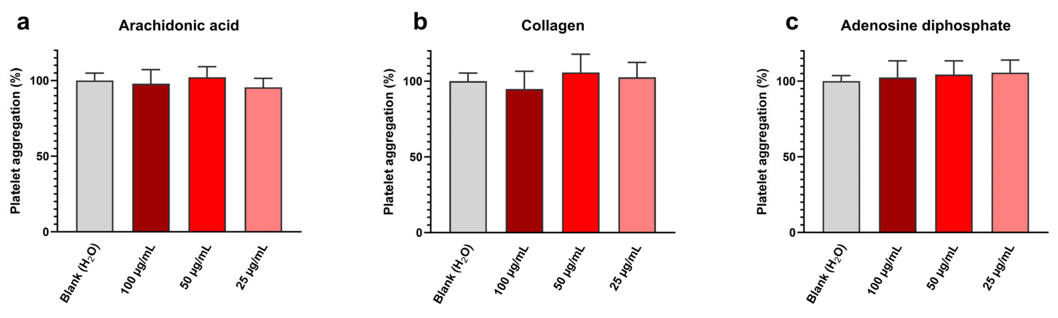
2.3.2. Platelet Aggregation
2.3.3. Coagulation
2.3.4. Global Conclusion on Hemocompatibility Results
3. Materials and Methods
3.1. Nanopowders
3.2. Nanopowder Characterization Techniques
3.3. Antifungal Assays
3.4. Hemocompatibility Testing
3.4.1. Biological Material for Platelet and Coagulation Assessment
3.4.2. Hemolysis
3.4.3. Platelet Aggregation
3.4.4. Coagulation
3.5. Statistical Analyses
4. Conclusions and Perspectives
Author Contributions
Funding
Institutional Review Board Statement
Informed Consent Statement
Data Availability Statement
Acknowledgments
Conflicts of Interest
References
- Kiseleva, M.; Omar, M.M.; Boisselier, É.; Selivanova, S.V.; Fortin, M.-A. A Three-Dimensional Printable Hydrogel Formulation for the Local Delivery of Therapeutic Nanoparticles to Cervical Cancer. ACS Biomater. Sci. Eng. 2022, 8, 1200–1214. [Google Scholar] [CrossRef] [PubMed]
- Dadi, R.; Azouani, R.; Traore, M.; Mielcarek, C.; Kanaev, A. Antibacterial Activity of ZnO and CuO Nanoparticles against Gram Positive and Gram Negative Strains. Mater. Sci. Eng. C 2019, 104, 109968. [Google Scholar] [CrossRef] [PubMed]
- Andra, S.; Balu, S.K.; Jeevanandam, J.; Muthalagu, M. Emerging Nanomaterials for Antibacterial Textile Fabrication. Naunyn-Schmiedeberg’s Arch. Pharmacol. 2021, 394, 1355–1382. [Google Scholar] [CrossRef] [PubMed]
- Mondal, A.; Chowdhury, S.; Mondal, N.K.; Shaikh, W.A.; Debnath, P.; Chakraborty, S. Insecticidal and Fungicidal Performance of Bio-Fabricated Silver and Gold Nanoparticles. Int. J. Environ. Sci. Technol. 2022, 19, 1573–1592. [Google Scholar] [CrossRef]
- Tan, E.P.; Djearamane, S.; Wong, L.S.; Rajamani, R.; Tanislaus Antony, A.C.; Subbaih, S.K.; Janakiraman, A.K.; Aminuzzaman, M.; Subramaniyan, V.; Sekar, M.; et al. An In Vitro Study of the Antifungal Efficacy of Zinc Oxide Nanoparticles against Saccharomyces cerevisiae. Coatings 2022, 12, 1988. [Google Scholar] [CrossRef]
- Gur, T.; Meydan, I.; Seckin, H.; Bekmezci, M.; Sen, F. Green Synthesis, Characterization and Bioactivity of Biogenic Zinc Oxide Nanoparticles. Environ. Res. 2022, 204, 111897. [Google Scholar] [CrossRef]
- Mohammed, A.K.; Salh, K.K.; Ali, F.A. ZnO, TiO2 and Ag Nanoparticles Impact against Some Species of Pathogenic Bacteria and Yeast. Cell. Mol. Biol. 2021, 67, 24–34. [Google Scholar] [CrossRef]
- Garcia-Marin, L.E.; Juarez-Moreno, K.; Vilchis-Nestor, A.R.; Castro-Longoria, E. Highly Antifungal Activity of Biosynthesized Copper Oxide Nanoparticles against Candida albicans. Nanomaterials 2022, 12, 3856. [Google Scholar] [CrossRef]
- De la Harpe, K.M.; Kondiah, P.P.D.; Choonara, Y.E.; Marimuthu, T.; du Toit, L.C.; Pillay, V. The Hemocompatibility of Nanoparticles: A Review of Cell-Nanoparticle Interactions and Hemostasis. Cells 2019, 8, 1209. [Google Scholar] [CrossRef]
- Yedgar, S.; Barshtein, G.; Gural, A. Hemolytic Activity of Nanoparticles as a Marker of Their Hemocompatibility. Micromachines 2022, 13, 2091. [Google Scholar] [CrossRef]
- Gomes, A.C.; Sarria, M.P. Interaction of Nanoparticles with Blood Components and Associated Pathophysiological Effects. In Unraveling the Safety Profile of Nanoscale Particles and Materials—From Biomedical to Environmental Applications; InTech: London, UK, 2018; ISBN 978-953-51-3940-9. [Google Scholar] [CrossRef]
- Mancier, V.; Daltin, A.-L.; Leclercq, D. Synthesis and Characterization of Copper Oxide (I) Nanoparticles Produced by Pulsed Sonoelectrochemistry. Ultrason. Sonochem. 2008, 15, 157–163. [Google Scholar] [CrossRef] [PubMed]
- Fattoum, S.; Chopart, J.-P.; Charpentier, E.; Mejia, J.; Gangloff, S.C.; Mancier, V. Synthesis, Physico-Chemical Characterizations and Antibacterial Properties of Copper Oxide (I) Nanopowders Elaborated by out-of-Phase Pulsed Sonoelectrochemistry. Mater. Chem. Phys. 2022, 290, 126614. [Google Scholar] [CrossRef]
- Yuan, Z.; Li, Y.; He, Y.; Qian, K.; Zhang, Y. Differential Analysis of Three Copper-Based Nanomaterials with Different Morphologies to Suppress Alternaria alternata and Safety Evaluation. Int. J. Mol. Sci. 2023, 24, 9673. [Google Scholar] [CrossRef] [PubMed]
- Chen, L.Q.; Kang, B.; Ling, J. Cytotoxicity of Cuprous Oxide Nanoparticles to Fish Blood Cells: Hemolysis and Internalization. J. Nanoparticle Res. 2013, 15, 1507. [Google Scholar] [CrossRef]
- Poland, C.A.; Hubbard, S.A.; Levy, L.; Mackie, C. Inhalation Toxicity of Copper Compounds: Results of 14-Day Range Finding Study for Copper Sulphate Pentahydrate and Dicopper Oxide and 28-Day Subacute Inhalation Exposure of Dicopper Oxide in Rats. Toxicology 2022, 474, 153221. [Google Scholar] [CrossRef] [PubMed]
- Henson, T.E.; Navratilova, J.; Tennant, A.H.; Bradham, K.D.; Rogers, K.R.; Hughes, M.F. In Vitro Intestinal Toxicity of Copper Oxide Nanoparticles in Rat and Human Cell Models. Nanotoxicology 2019, 13, 795–811. [Google Scholar] [CrossRef] [PubMed]
- Padmavathi, A.R.; Murthy, P.S.; Das, A.; Priya, A.; Sushmitha, T.J.; Pandian, S.K.; Toleti, S.R. Impediment to Growth and Yeast-to-Hyphae Transition in Candida albicans by Copper Oxide Nanoparticles. Biofouling 2020, 36, 56–72. [Google Scholar] [CrossRef] [PubMed]
- Jaswal, T.; Gupta, J. A Review on the Toxicity of Silver Nanoparticles on Human Health. Mater. Today Proc. 2023, 81, 859–863. [Google Scholar] [CrossRef]
- Kadhum, M.A.K.A. Preparation ZnO Nanoparticles with Different Concentration by Laser Ablation in Liquid and Their Use in Anti Bacterial Activity. Acad. Sci. J. 2024, 2, 79–98. [Google Scholar] [CrossRef]
- Abid, N.; Khan, A.M.; Shujait, S.; Chaudhary, K.; Ikram, M.; Imran, M.; Haider, J.; Khan, M.; Khan, Q.; Maqbool, M. Synthesis of Nanomaterials Using Various Top-Down and Bottom-Up Approaches, Influencing Factors, Advantages, and Disadvantages: A Review. Adv. Colloid Interface Sci. 2022, 300, 102597. [Google Scholar] [CrossRef]
- Reisse, J.; Delplancke, J.-L.; Winand, R. Device for the Production of Ultrafine Powders. WO Patent WO 95/33871, 3 June 1994. [Google Scholar]
- Ren, J.; Wang, W.; Sun, S.; Zhang, L.; Wang, L.; Chang, J. Crystallography Facet-Dependent Antibacterial Activity: The Case of Cu2O. Ind. Eng. Chem. Res. 2011, 50, 10366–10369. [Google Scholar] [CrossRef]
- Moyes, D.L.; Wilson, D.; Richardson, J.P.; Mogavero, S.; Tang, S.X.; Wernecke, J.; Höfs, S.; Gratacap, R.L.; Robbins, J.; Runglall, M.; et al. Candidalysin Is a Fungal Peptide Toxin Critical for Mucosal Infection. Nature 2016, 532, 64–68. [Google Scholar] [CrossRef] [PubMed]
- Alonso, M.F.; Gow, N.A.R.; Erwig, L.P.; Bain, J.M. Macrophage Migration Is Impaired within Candida albicans Biofilms. J. Fungi 2017, 3, 31. [Google Scholar] [CrossRef] [PubMed]
- Pourahmad, J.; Salami, M.; Zarei, M.H. Comparative Toxic Effect of Bulk Copper Oxide (CuO) and CuO Nanoparticles on Human Red Blood Cells. Biol. Trace Elem. Res. 2023, 201, 149–155. [Google Scholar] [CrossRef] [PubMed]
- Eldine, R.S.S.; Kader, A.M.E.; Shalaby, T.I.; Balbaa, O.A. Evaluation of Toxicity of Copper Oxide Nano Particles on Human Blood. J. Biophys. Struct. Biol. 2021, 9, 10–19. [Google Scholar] [CrossRef]
- Karlsson, H.L.; Cronholm, P.; Hedberg, Y.; Tornberg, M.; De Battice, L.; Svedhem, S.; Wallinder, I.O. Cell Membrane Damage and Protein Interaction Induced by Copper Containing Nanoparticles—Importance of the Metal Release Process. Toxicology 2013, 313, 59–69. [Google Scholar] [CrossRef] [PubMed]
- Yang, M.; Zhou, H.; Cheng, Y.; Hong, Q.; Chen, J.; Zhang, Q.; Pan, C. Incorporation of Copper and Strontium Ions in TiO2 Nanotubes via Dopamine to Enhance Hemocompatibility and Cytocompatibility. Nanotechnol. Rev. 2022, 11, 1450–1463. [Google Scholar] [CrossRef]
- Siegfried, M.J.; Choi, K.-S. Electrochemical Crystallization of Cuprous Oxide with Systematic Shape Evolution. Adv. Mater. 2004, 16, 1743–1746. [Google Scholar] [CrossRef]
- Gou, L.; Murphy, C.J. Solution-Phase Synthesis of Cu2O Nanocubes. Nano Lett. 2003, 3, 231–234. [Google Scholar] [CrossRef]
- Park, E.-J.; Khaliullin, T.O.; Shurin, M.R.; Kisin, E.R.; Yanamala, N.; Fadeel, B.; Chang, J.; Shvedova, A.A. Fibrous Nanocellulose, Crystalline Nanocellulose, Carbon Nanotubes, and Crocidolite Asbestos Elicit Disparate Immune Responses upon Pharyngeal Aspiration in Mice. J. Immunotoxicol. 2018, 15, 12–23. [Google Scholar] [CrossRef]
- Zia, F.; Kendall, M.; Watson, S.P.; Mendes, P.M. Platelet Aggregation Induced by Polystyrene and Platinum Nanoparticles Is Dependent on Surface Area. RSC Adv. 2018, 8, 37789–37794. [Google Scholar] [CrossRef] [PubMed]
- Sing, K.S.W. Reporting Physisorption Data for Gas/Solid Systems with Special Reference to the Determination of Surface Area and Porosity (Recommendations 1984). Pure Appl. Chem. 1985, 57, 603–619. [Google Scholar] [CrossRef]
- Rousse, C.; Josse, J.; Mancier, V.; Levi, S.; Gangloff, S.C.; Fricoteaux, P. Synthesis of Copper–Silver Bimetallic Nanopowders for a Biomedical Approach; Study of Their Antibacterial Properties. RSC Adv. 2016, 6, 50933–50940. [Google Scholar] [CrossRef]
- Peng, L.; Wei, H.; Tian, L.; Xu, J.; Li, M.; Yu, Q. Phospholipid/Protein Co-Mediated Assembly of Cu2O Nanoparticles for Specific Inhibition of Growth and Biofilm Formation of Pathogenic Fungi. Sci. China Mater. 2021, 64, 759–768. [Google Scholar] [CrossRef]
- Jiang, L.; Yu, Y.; Li, Y.; Yu, Y.; Duan, J.; Zou, Y.; Li, Q.; Sun, Z. Oxidative Damage and Energy Metabolism Disorder Contribute to the Hemolytic Effect of Amorphous Silica Nanoparticles. Nanoscale Res. Lett. 2016, 11, 57. [Google Scholar] [CrossRef] [PubMed]
- Kutwin, M.; Sawosz, E.; Jaworski, S.; Kurantowicz, N.; Strojny, B.; Chwalibog, A. Structural Damage of Chicken Red Blood Cells Exposed to Platinum Nanoparticles and Cisplatin. Nanoscale Res. Lett. 2014, 9, 257. [Google Scholar] [CrossRef] [PubMed]
- Krajewski, S.; Prucek, R.; Panacek, A.; Avci-Adali, M.; Nolte, A.; Straub, A.; Zboril, R.; Wendel, H.P.; Kvitek, L. Hemocompatibility Evaluation of Different Silver Nanoparticle Concentrations Employing a Modified Chandler-Loop in Vitro Assay on Human Blood. Acta Biomater. 2013, 9, 7460–7468. [Google Scholar] [CrossRef]
- Standard Practice for Assessment of Hemolytic Properties of Materials. Available online: https://www.astm.org/f0756-17.html (accessed on 16 February 2024).
- Hante, N.K.; Medina, C.; Santos-Martinez, M.J. Effect on Platelet Function of Metal-Based Nanoparticles Developed for Medical Applications. Front. Cardiovasc. Med. 2019, 6, 139. [Google Scholar] [CrossRef]
- Saikia, J.; Mohammadpour, R.; Yazdimamaghani, M.; Northrup, H.; Hlady, V.; Ghandehari, H. Silica Nanoparticle–Endothelial Interaction: Uptake and Effect on Platelet Adhesion under Flow Conditions. ACS Appl. Bio Mater. 2018, 1, 1620–1627. [Google Scholar] [CrossRef]
- Feng, L.; Yang, X.; Liang, S.; Xu, Q.; Miller, M.R.; Duan, J.; Sun, Z. Silica Nanoparticles Trigger the Vascular Endothelial Dysfunction and Prethrombotic State via miR-451 Directly Regulating the IL6R Signaling Pathway. Part. Fibre Toxicol. 2019, 16, 16. [Google Scholar] [CrossRef]
- Laloy, J.; Haguet, H.; Alpan, L.; Mancier, V.; Mejia, J.; Levi, S.; Dogné, J.-M.; Lucas, S.; Rousse, C.; Fricoteaux, P. Characterization of Core/Shell Cu/Ag Nanopowders Synthesized by Electrochemistry and Assessment of Their Impact on Hemolysis, Platelet Aggregation, and Coagulation on Human Blood for Potential Wound Dressing Use. J. Nanoparticle Res. 2017, 19, 266. [Google Scholar] [CrossRef]
- Taketomi, Y.; Kuramoto, A. Ultrastructural Studies on the Surface Coat of Human Platelet Aggregated by Polylysine and Dextran. Thromb. Haemost. 1978, 40, 11–23. [Google Scholar] [CrossRef] [PubMed]
- Simak, J.; De Paoli, S. The Effects of Nanomaterials on Blood Coagulation in Hemostasis and Thrombosis. WIREs Nanomed. Nanobiotechnol. 2017, 9, e1448. [Google Scholar] [CrossRef]
- Tran, H.D.N.; Akther, F.; Xu, Z.P.; Ta, H.T. Chapter 6—Effects of Nanoparticles on the Blood Coagulation System (Nanoparticle Interface with the Blood Coagulation System). In Nanotechnology for Hematology, Blood Transfusion, and Artificial Blood; Denizli, A., Nguyen, T.A., Rajan, M., Alam, M.F., Rahman, K., Eds.; Micro and Nano Technologies; Elsevier: Amsterdam, The Netherlands, 2022; pp. 113–140. ISBN 978-0-12-823971-1. [Google Scholar]
- Kushwaha, A.; Goswami, L.; Kim, B.S. Nanomaterial-Based Therapy for Wound Healing. Nanomaterials 2022, 12, 618. [Google Scholar] [CrossRef] [PubMed]
- Klinkajon, W.; Supaphol, P. Novel Copper (II) Alginate Hydrogels and Their Potential for Use as Anti-Bacterial Wound Dressings. Biomed. Mater. Bristol. Engl. 2014, 9, 045008. [Google Scholar] [CrossRef] [PubMed]
- Naskar, A.; Kim, K.-S. Recent Advances in Nanomaterial-Based Wound-Healing Therapeutics. Pharmaceutics 2020, 12, 499. [Google Scholar] [CrossRef]


| Sample of Nanopowder | Diameter Extracted from Relative Number Graph (Figure 2a) (nm) | Diameter Extracted from Relative Weight Graph (Figure 2b) (nm) |
|---|---|---|
| Sonoelectrochemical Cu2O | 7 | 498 |
| Commercial Cu2O | 10 | 28 and 568 |
| Commercial Cu | 7 | 407 |
| Diameter Range (nm) | Sample of Nanopowder | ||
|---|---|---|---|
| Sonoelectrochemical Cu2O | Commercial Cu2O | Commercial Cu | |
| 0–10 | 79 | 10 | 49 |
| 10–15 | 9 | 39 | 20 |
| 15–25 | 6 | 32 | 11 |
| 25–50 | 3 | 14 | 4 |
| 50–100 | 1 | 3 | 2 |
| >100 | 2 | 2 | 14 |
| Diameter Range (nm) | Sample of Nanopowder | ||
|---|---|---|---|
| Sonoelectrochemical Cu2O | Commercial Cu2O | Commercial Cu | |
| <100 | ≈0 | 1 | ≈0 |
| 100–250 | ≈0 | 2 | 6 |
| 250–500 | 29 | 20 | 61 |
| >500 | 71 | 77 | 33 |
| Sample of NPw | MRSA [13] | E. coli [13] | C. albicans | |
|---|---|---|---|---|
| Mean IZD (mm) | Mean IZD (mm) | Mean IZD (mm) | Mean DZD (mm) | |
| Sonoelectrochemical Cu2O | 14.5 ± 0.9 | 9.9 ± 0.3 | 8.5 ± 0.2 | 28.8 ± 0.3 |
| Commercial Cu2O | 13.6 ± 0.5 | 10.5 ± 0.5 | 8.5 ± 0.5 | 29.0 ± 0.3 |
| Commercial Cu | 14.8 ± 0.6 | 12.0 ± 0.3 | 10.2 ± 0.4 | 29.7 ± 3.3 |
| Platelet Aggregation (% ± Standard Deviation) | |||||
|---|---|---|---|---|---|
| Incubation Medium | Platelet Inducers Tested | Arachidonic Acid (Figure 5a) | Collagen (Figure 5b) | Adenosine Diphosphate (Figure 5c) | |
| Control | Water (blank) | 100 ± 5 | 100 ± 5 | 100 ± 2 | |
| Sonoelectrochemical Cu2O nanopowder (concentrations in µg/mL) | 100 | 98 ± 7 | 95 ± 11 | 102 ± 9 | |
| 50 | 102 ± 5 | 106 ± 10 | 104 ± 8 | ||
| 25 | 96 ± 5 | 103 ± 8 | 106 ± 8 | ||
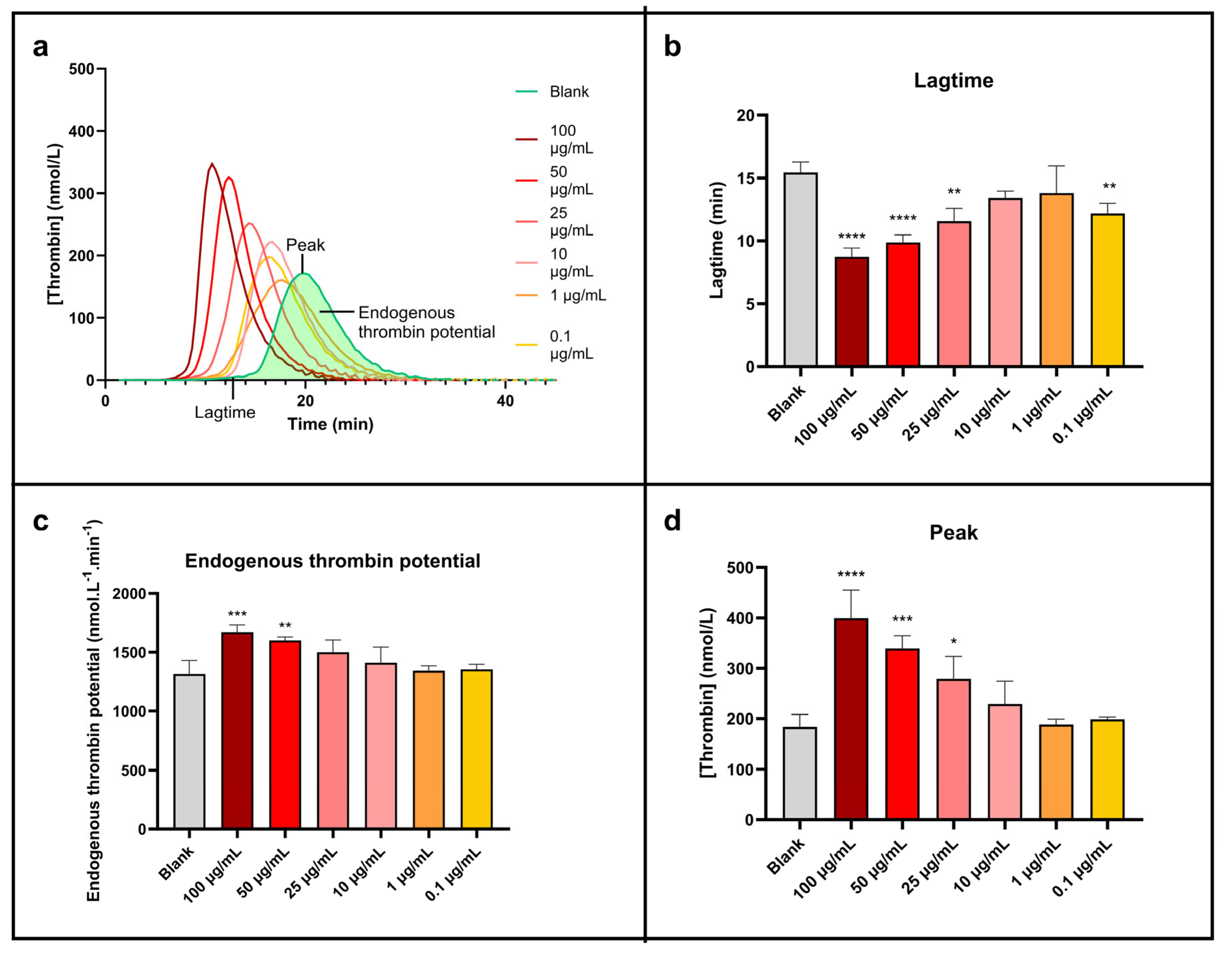
| Tested Condition | Lagtime (Figure 6b) | Endogenous Thrombin Potential (Figure 6c) | Peak (Figure 6d) | ||||
|---|---|---|---|---|---|---|---|
| Mean (min ± Standard Deviation) | % ± Standard Deviation | Mean (nmol·L−1·min ± Standard Deviation) | % ± Standard Deviation | Mean (nmol·L−1 ± Standard Deviation) | % ± Standard Deviation | ||
| Control | Water (blank) | 15.4 ± 0.9 | 100 ± 5 | 1315 ± 117 | 100 ± 9 | 184 ± 25 | 100 ± 13 |
| Sonoelectrochemical Cu2O nanopowder (concentrations in µg/mL) | 100 | 8.7 ± 0.7 | 57 ± 5 | 1671 ± 61 | 127 ± 5 | 399 ± 56 | 217 ± 30 |
| 50 | 9.9 ± 0.6 | 64 ± 4 | 1600 ± 31 | 122 ± 2 | 339 ± 26 | 184 ± 14 | |
| 25 | 11.6 ± 1.0 | 75 ± 7 | 1501 ± 104 | 114 ± 8 | 279 ± 45 | 152 ± 24 | |
| 10 | 13.4 ± 0.6 | 87 ± 4 | 1412 ± 133 | 107 ± 10 | 229 ± 46 | 124 ± 25 | |
| 1 | 13.8 ± 2.2 | 90 ± 14 | 1344 ± 42 | 102 ± 3 | 188 ± 11 | 102 ± 6 | |
| 0.1 | 12.2 ± 0.9 | 79 ± 5 | 1355 ± 44 | 103 ± 3 | 199 ± 5 | 108 ± 2 | |
Disclaimer/Publisher’s Note: The statements, opinions and data contained in all publications are solely those of the individual author(s) and contributor(s) and not of MDPI and/or the editor(s). MDPI and/or the editor(s) disclaim responsibility for any injury to people or property resulting from any ideas, methods, instructions or products referred to in the content. |
© 2024 by the authors. Licensee MDPI, Basel, Switzerland. This article is an open access article distributed under the terms and conditions of the Creative Commons Attribution (CC BY) license (https://creativecommons.org/licenses/by/4.0/).
Share and Cite
Mancier, V.; Fattoum, S.; Haguet, H.; Laloy, J.; Maillet, C.; Gangloff, S.C.; Chopart, J.-P. Antifungal and Coagulation Properties of a Copper (I) Oxide Nanopowder Produced by Out-of-Phase Pulsed Sonoelectrochemistry. Antibiotics 2024, 13, 286. https://doi.org/10.3390/antibiotics13030286
Mancier V, Fattoum S, Haguet H, Laloy J, Maillet C, Gangloff SC, Chopart J-P. Antifungal and Coagulation Properties of a Copper (I) Oxide Nanopowder Produced by Out-of-Phase Pulsed Sonoelectrochemistry. Antibiotics. 2024; 13(3):286. https://doi.org/10.3390/antibiotics13030286
Chicago/Turabian StyleMancier, Valérie, Sirine Fattoum, Hélène Haguet, Julie Laloy, Christina Maillet, Sophie C. Gangloff, and Jean-Paul Chopart. 2024. "Antifungal and Coagulation Properties of a Copper (I) Oxide Nanopowder Produced by Out-of-Phase Pulsed Sonoelectrochemistry" Antibiotics 13, no. 3: 286. https://doi.org/10.3390/antibiotics13030286
APA StyleMancier, V., Fattoum, S., Haguet, H., Laloy, J., Maillet, C., Gangloff, S. C., & Chopart, J.-P. (2024). Antifungal and Coagulation Properties of a Copper (I) Oxide Nanopowder Produced by Out-of-Phase Pulsed Sonoelectrochemistry. Antibiotics, 13(3), 286. https://doi.org/10.3390/antibiotics13030286