Article

Prevalence and Antimicrobial Resistance Profile of Diarrheagenic Escherichia coli from Fomites in Rural Households in South Africa

by
Phathutshedzo Rakhalaru
,
Lutendo Munzhedzi
,
Akebe Luther King Abia
,
Jean Pierre Kabue
,
Natasha Potgieter
and
Afsatou Ndama Traore
*
Department of Biochemistry and Microbiology, Faculty of Science, Engineering and Agriculture, University of Venda, Private Bag X5050, Thohoyandou 0950, South Africa
*
Author to whom correspondence should be addressed.
Antibiotics 2023, 12(8), 1345; https://doi.org/10.3390/antibiotics12081345
Submission received: 14 July 2023 / Revised: 8 August 2023 / Accepted: 19 August 2023 / Published: 21 August 2023
(This article belongs to the Special Issue Antimicrobial Resistance and Environmental Health)

Abstract

:
Diarrheagenic Escherichia coli (DEC) pathotypes are the leading cause of mortality and morbidity in South Asia and sub-Saharan Africa. Daily interaction between people contributes to the spreading of Escherichia coli (E. coli), and fomites are a common source of community-acquired bacterial infections. The spread of bacterial infectious diseases from inanimate objects to the surrounding environment and humans is a serious problem for public health, safety, and development. This study aimed to determine the prevalence and antibiotic resistance of diarrheagenic E. coli found in toilets and kitchen cloths in the Vhembe district, South Africa. One hundred and five samples were cultured to isolate E. coli: thirty-five samples were kitchen cloths and seventy-five samples were toilet swabs. Biochemical tests, API20E, and the VITEK®-2 automated system were used to identify E. coli. Pathotypes of E. coli were characterised using Multiplex Polymerase Chain Reaction (mPCR). Nine amplified gene fragments were sequenced using partial sequencing. A total of eight antibiotics were used for the antibiotic susceptibility testing of E. coli isolates using the Kirby–Bauer disc diffusion method. Among the collected samples, 47% were positive for E. coli. DEC prevalence was high (81%), with ETEC (51%) harboring lt and st genes being the most dominant pathotype found on both kitchen cloths and toilet surfaces. Diarrheagenic E. coli pathotypes were more prevalent in the kitchen cloths (79.6%) compared with the toilet surfaces. Notably, hybrid pathotypes were detected in 44.2% of the isolates, showcasing the co-existence of multiple pathotypes within a single E. coli strain. The antibiotic resistance testing of E. coli isolates from kitchen cloths and toilets showed high resistance to ampicillin (100%) and amoxicillin (100%). Only E. coli isolates with hybrid pathotypes were found to be resistant to more than three antibiotics. This study emphasizes the significance of fomites as potential sources of bacterial contamination in rural settings. The results highlight the importance of implementing proactive measures to improve hygiene practices and antibiotic stewardship in these communities. These measures are essential for reducing the impact of DEC infections and antibiotic resistance, ultimately safeguarding public health.

1. Introduction

Diarrheal disease remains a significant public health issue particularly in rural areas where there is limited availability of clean water and adequate sanitation facilities [1,2]. The spread of pathogens through fomites is a serious concern to human health, safety, and development. Fomites act as reservoirs and potential vectors for pathogenic bacteria, including E.coli, leading to the spread of infections within households [3]. Pathogenic bacteria can survive on fomites for an extended period, and the duration of their survival is influenced by factors such as temperature, humidity, and the availability of other microorganisms [4,5]. Previous studies have shown the presence of E. coli on various fomites, including kitchen surfaces and cloths, toilet surfaces, door handles, and bathroom surfaces. These fomites serve as source of transmission, posing a potential health risk [6,7].
E. coli is a Gram-negative bacterium that typically inhabits the lower intestines of warm-blooded animals, and certain E. coli, O157: H7, leads to severe gastrointestinal infections in humans [8,9]. Studies by Seidman et al. [10] and Potgieter et al. [11] have reported the contamination of toilet seats in rural households with total coliforms and E. coli. Research findings have also revealed that kitchen cloths exhibit bacterial contamination, with E. coli emerging as the most frequently detected microorganism [9,12,13]. E. coli is generally used as an indicator of faecal pollution and indicates the presence of other pathogenic bacteria, such as Salmonella and Shigella, which have been associated with diarrhea [14]. Apart from its role as an indicator organism, E. coli can be classified as diarrheagenic (intestinal) or extraintestinal pathotypes [15]. Diarrheagenic E. coli pathotypes are categorized into six well characterized groups harboring specific genes: enteroinvasive E. coli (EIEC), enteropathogenic E. coli (EPEC), enterotoxigenic E. coli (ETEC), enterohemorrhagic E. coli (EHEC), enteroaggregative E. coli (EAEC), and diffusely adherent E. coli [16,17]. Certain E. coli pathotypes can acquire virulence genes from other E. coli strains, resulting in what is known as a hybrid pathotype [18,19,20]. Diarrhea caused by pathogenic E. coli is a leading cause of morbidity and mortality worldwide, especially in children younger than five years [21,22].
Infections caused by E. coli are usually treated using antibiotics such as penicillin, gentamycin, ampicillin, amoxicillin, chloramphenicol, rifampicin, and tetracycline [23]. However, some studies have documented that E. coli has become resistant to some antibiotics due to their widespread and inappropriate use [24,25]. Such misuse poses a serious health problem [26]; antimicrobial resistant E. coli strains have been reported as the main carriers of antibiotic resistance genes to ampicillin, penicillin, tetracycline, and rifampicin [27,28]. Hybrid DEC pathotypes have been reported to exhibit multidrug resistance to beta-lactam antibiotics [20,29].
In South Africa, inadequate access to water supply, sanitation services, and hygiene is considered the eleventh most significant risk factor leading to illnesses [30]. About 73% of toilets in rural households in the Vhembe District are pit holes with no water taps close to the toilets [31], suggesting that most people might not wash their hands immediately after using the toilets. Even in situation where water is accessible, most people wash their hands solely with water without using detergents [30].
Thus, poor sanitation and hygiene are still serious problems in rural households in the Vhembe district. Therefore, this study aimed to determine the prevalence and antibiotic resistance of diarrheagenic E. coli contamination in household fomites, highlighting the importance of implementing effective hygiene measures to mitigate transmission risks.

2. Methods and Materials

2.1. Study Area and Period

This study was conducted in Tshamutilikwa village (−22.892981245885206, 30.600267380011015) in the Vhembe district, South Africa (Figure 1), from May to August 2021. Tshamutilikwa is a place with a population of 814 people, according to the Census conducted in 2011 (https://census2011.adrianfrith.com/place/966110) (accessed on 3 August 2023). It covers an area of 1.06 square kilometers. With a population density of 766.49 people per square kilometer, Tshamutilikwa is a relatively densely populated area. The village consists of 203 households, resulting in an average of 191.15 households per square kilometer.

2.2. Ethics

Ethical clearance was obtained from the University of Venda [SEA/21/MBY/02/1608]. Sampling was done after receiving permission from the household owners by signing a consent form and questionnaire to answer and sign.

2.3. Sample Collection

Kitchen cloths, toilet seats, and toilet door handle swabs were collected door-to-door from the selected households and 70 toilet (35 seats and 35 door handles) samples were collected from participating households in Tshamutilikwa. A specific prepared questionnaire was administered to household owners to obtain information on various water, sanitation, and hygiene [WASH] factors, including the source of water, usage of kitchen cloths, condition of the kitchen cloth, toilet condition, type of toilet, handwashing means after using the toilet, the incidence of diarrhea in the household, and the practice of sharing the toilet with neighbors.
Samples were collected from toilet surfaces using the peptone water sterile swab-rinse method described by Hurst et al. [32]. In addition, a total of 35 old and used kitchen cloths were collected. Participants were requested to place the kitchen cloths in sterile zipping lock bags in exchange for new kitchen cloths. Before sample analysis, a brief description of the quality of kitchen cloths based on aspects such as dirty/clean or wet/dry was recorded. The toilets were categorized as clean or dirty based on their appearance. Clean toilets had no visible dirt, while dirty toilets had visible dirt, stains, and feces on toilet seats. The samples were immediately transported in ice to the microbiology laboratory and analyzed within four hours of sampling.

2.4. Bacterial Isolation and Identification

2.4.1. Bacterial Isolation

In the laboratory, 5 cm by 5 cm (length × breadth) pieces were aseptically cut from each kitchen cloth sample and placed into a sterile flask containing 50 mL nutrient broth (Davies diagnostic (Pty) Ltd., Randburg, Gauteng, South Africa) for enrichment, vortexed for 5 min, and incubated at 37 °C overnight [33]. After incubation, 1 mL of the inoculated broth was transferred into a clean, sterile test tube containing 9 mL of sterile water. The diluted solution was mixed thoroughly by vortexing, and 0.5 mL of the diluted solution was then spread on sterile Eosin Methylene Blue (EMB) agar (Davies diagnostic (Pty) Ltd., Randburg, Gauteng, South Africa) plates using a sterile glass spreader and incubated for 24 h at 37 °C. Toilet seat and door handle swab samples were streaked directly on EMB agar and incubated for 24 h at 37 °C. After incubation, two distinct green metallic shiny colonies (characteristic of E. coli) were selected from each EMB plate and subcultured on a sterile nutrient agar plate to isolate pure colonies. All media used were prepared according to the manufacturer’s specifications.

2.4.2. Bacterial Identification

The colonies obtained from sub-culturing on nutrient agar plates were analyzed using various biochemical tests such as the Kligler iron agar test [34], Urease test, Simmon citrate test [35], and the API20E (bioMérieux, Marcy I’Etoile, France). Presumptive E. coli isolates were further confirmed using the VITEK 2 automated system (bioMérieux, Marcy-l’Étoile, France) as described by the manufacturer. Briefly, a bacterial suspension was created by mixing E. coli colonies with 0.85% phosphate-buffered saline (PBS) (Thermo Fisher Scientific, Randburg, South Africa), resulting in a concentration of 1 × 10⁸ CFU/mL Mcflarland standard. Subsequently, 2 mL of these suspensions were automatically loaded into the VITEK 2 ID system, utilizing the GNB cards specifically designed for E. coli identification. The cards were analyzed through kinetic fluorescence measurement, and the results were reported within 3 h.

2.5. Molecular Identification of E. coli Isolates

2.5.1. DNA Extraction

DNA extraction was performed as previously described by Omar et al. [36]. Briefly, 2 mL of nutrient broth with E. coli was aliquoted into sterile 2 mL Eppendorf tubes (Sigma-Aldrich, St Louis, MI, USA). The tubes were centrifuged at 13,000× g for 120 s to separate the cells from the supernatant. The DNA binding to celite was enhanced using lysis buffer mixed with 250 µL of 100% ethanol. Before washing, the celite-bound DNA was added to the spin columns. Qiagen elution buffer (Southern Cross Biotechnology®, Hilden, Germany) of 100 µL was used for DNA elution. Extracted DNA was then used as a template for PCR reactions.

2.5.2. Genotypic Identification and Classification of E. coli Pathotypes

Genotypic identification and classification of selected isolates into the different E. coli pathotypes were performed using an 11-gene multiplex PCR, as previously reported [37,38]. The primers used in this study are in (Table 1). A total volume of 20 µL reaction mixture consisted of 10 µL, 2X Qiagen® PCR multiplex mix (Qiagen®, Hilden, Germany), 1 µL 5× Q-solution, 2 µL of DNA template, 5 µL PCR grade water, and 2 µL of the primer mix containing 0.1 μM of lt and mdh, 0.5 μM of stx1 and st, 0.3 μM of eaeA and stx2, and 0.2 μM of astA, bfp eagg, ial, and gapdh primers. Multiplex PCR amplification was performed in a Bio-Rad MyCyclerTM Thermal cycler (Bio-Rad, Hercules, CA, USA) under the following PCR conditions: an initial activation at 95 °C for 15 min, followed by denaturation at 94 °C for 45 s, and annealing was performed at 55 °C for 45 s. Extension was done at 68 °C for 2 min (35 cycles) [38]. PCR amplifications were separated using agarose gel electrophoresis, the bacterial DNAs were loaded into pre-cast wells in the gel, and a current was applied as described by Alfinete et al. [39].

2.6. Sequencing and Phylogenetic Analysis

Sequencing and phylogenetic analysis of E. coli was performed to compare the bacterial isolates obtained from the kitchen cloths and toilets within the same household and to investigate whether similar bacterial clones existed in different households, to identify any potential spread of identical clones within the community. DNA partial sequencing was performed on ABI 3500XL Genetic Analyzer POP7TM (Thermo Scientific, Waltham, MA, USA) using the same specific primers (Table 1). The reading of the DNA sequence was done and edited on FinchTV v1.4 (Geospiza Inc., Seattle, WA, USA). Nucleotide sequences of E. coli were compared with other reference strains on GenBank by blasting on the NCBI program (available at http://www.ncbi.nlm.nih.gov/) (accessed on 19 May 2022). For constructing the phylogenetic tree, MEGA X version 10.2.6 software was used to create phylogenetic trees by the neighbor-joining method and evaluated at 1000 bootstrap replicates for each gene [41,42].

2.7. Determination of Antibiotic Susceptibility

All the E. coli isolates were tested for sensitivity to different antibiotics using the Kirby–Bauer standard disc diffusion method [46,47]. For the disc diffusion assay, bacteria were grown for 24 h on Mueller–Hinton agar (Davies Diagnostics (pty) Ltd., Randburg, Gauteng, South Africa), harvested, and then suspended in 0.85% sterile PBS solution adjusted to a 0.5 McFarland turbidity standard, equivalent to 108 CFU/mL. The standardized bacterial suspension was streaked onto Mueller–Hinton agar plates using a sterile cotton swab and exposed to commercially available antibiotic discs (Thermo Fisher Scientific, Waltham, MA, USA). The zones of inhibition were measured using a ruler after 24 h of incubation at 37 °C. The resistance patterns of the isolates to 8 different antibiotics (Table 2) were then interpreted as either Resistant (R), Intermediate resistant (I), or Sensitive (S), following the guidelines set by the Clinical Laboratory Standards Institute (CLSI, 2020) (https://clsi.org/meetings/susceptibility-testing-subcommittees/) (accessed on 3 August 2023). The antibiotics selected (Table 2) in this study are commonly used to treat diarrheal infections caused by diarrheagenic E. coli pathotypes.

3. Results

3.1. Study Characteristics

In all, 54.3% (19/35) of the kitchen cloths were dirty; among those, 54.6% (11/19) were dry and dirty, and 42.1% (8/19) were wet and dirty (Table 3). Of 35 toilets where swab samples were collected, 54.3% (19/35) were not clean, and two had feces on the seats.

3.2. Prevalence of E. coli on Kitchen Cloths and Toilets

Of the 105 samples collected, 46.7% (49/105) were positive for E. coli. All the kitchen cloths had bacterial contamination. A total of 71.4% (25/35) of the kitchen cloths (n = 35) were contaminated with E. coli. Out of the 70 samples collected from the toilets, 24 (34.3%) were contaminated with E. coli (Table 4).

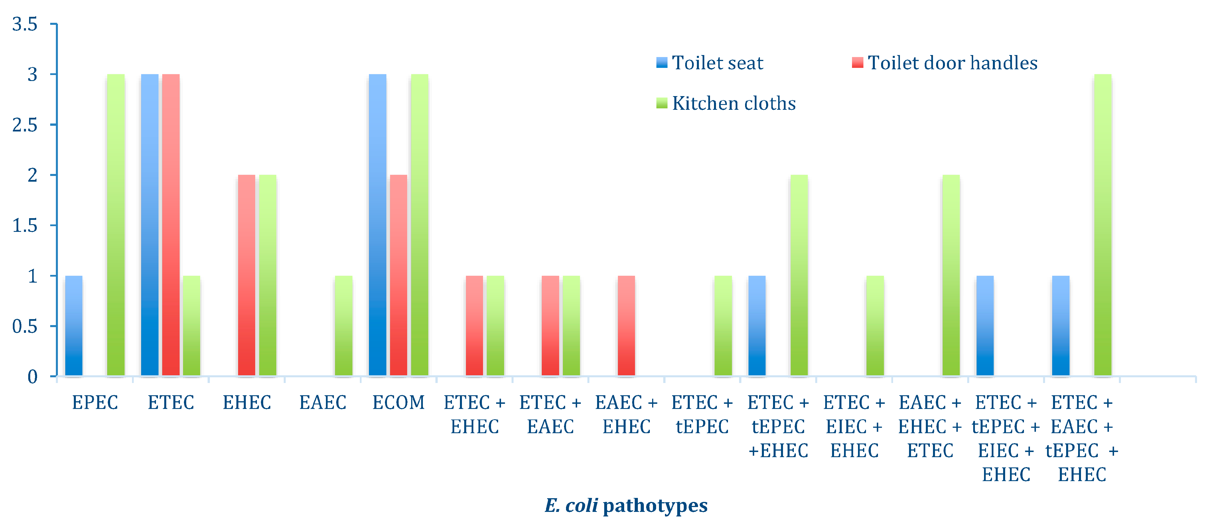
3.3. Characterization of E. coli Pathotypes

The mdh gene was used as an internal control to ensure the PCR worked for each E. coli isolate. A total of 43/49 (90%) isolates were positive for the E. coli housekeeping gene (mdh). All the E. coli isolates with mdh genes also tested positive for the gapdh gene. The m-PCR test did not show any false positives or PCR inhibition as the external control gene (gapdh) was detected in all samples.
Multiplex PCR detected five DEC pathotypes (EAEC, EHEC, EPEC, ETEC, and EIEC). The prevalence of commensal E. coli (8/43; 18.6%) was lower than that of DEC (35/43; 81%). ETEC (22/43; 51%), harboring lt and st genes, was the most dominant DEC pathotype found in kitchen cloths and on toilet surfaces.
Different hybrid pathogenic strains of E. coli were found, 24 (55.8%) non-hybrid pathotypes and 19 (44.2%) hybrid pathotypes. There was a high prevalence of hybrids with two pathotypes, making the percentage 18.7% (Figure 2). Most of the hybrid E. coli strains exhibited the presence of the Asta gene, which is known to be carried by E. coli toxins.
Based on the sample type, more diarrheagenic E. coli pathotypes were found in kitchen cloths (39/49, 79.6%) followed by toilet seats (20/49; 40.8%) and toilet door handles (11/49; 22.4%), respectively (Figure 2). In addition, the gel image illustrating the results of the Multiplex PCR is provided in Supplementary Figure S1.

3.4. Antibiotic Resistance Profile of E. coli

The E. coli isolates obtained from kitchen cloths exhibited varying levels of antibiotic resistance. E. coli isolates from the toilet surfaces and kitchen cloths displayed the highest resistance to ampicillin (24/24; 100%) and amoxicillin (24/24; 100%). (Table 5).
Some of the isolates showed resistance to more than two antibiotics; only 28.6% (14/49) of the isolates did not show resistance to three antibiotics. The multidrug resistance of E. coli isolates was found in 6.1% (3/49) of the isolates (Table 5). Only E. coli isolates with hybrid pathotypes were found to be resistant to more than three antibiotics used.

3.5. Sequence and Phylogenetic Analysis

Sequence Analysis and Phylogenetic Analysis

The study findings indicated that among the households sampled, only three households (numbers 7, 28, and 35) exhibited the consistent presence of the same pathotypes (ETEC, EHEC, and EAEC) across all three sample types: kitchen cloth, toilet seat, and toilet door handle surfaces. Specifically, household number 7 showed ETEC, 28 showed EHEC, and 35 showed EAEC detected in all sample types. Nine amplified DNA extracts were sent for partial sequencing (including three of Stx1, three of lt, and three of Eagg). Of the nine amplified E. coli isolates, only one stx1 (1/3; 33.3%) and one Eagg (1/3; 33.3%) were successfully sequenced.
The two sequences obtained were blasted on GenBank for comparison with other reference E. coli strains. The similarities with the reference strain for the Stx1 gene sequence ranged from 80 to 89.4%, and for the Eagg gene sequence ranged from 81 to 89.8% (Figure 3 and Figure 4).
The Stx1 sequence (accession no. 0N193544) from the present study was closely related to a reference strain isolated in water from Hungary (accession no. DQ44966.1) and shared a common ancestor with an E. coli strain from human feces in Bangladesh (Figure 3).
The Eagg sequence (accession no. 0N241000) obtained from the toilet seat in this study shared a common ancestor with an E. coli strain (accession no. MZ330843.1) from handwash water in the Vhembe District, South Africa (Figure 4). There were limited reference strains of Stx1 and Eagg genes in Africa.

4. Discussion

Poor sanitation and hygiene are still major problems in rural communities, especially in low- and middle-income countries (LMICs). They have been associated with increased diarrhea disease caused by enteric pathogens. Diarrhea is the leading cause of morbidity and mortality worldwide [42,48]. There are few reports on the prevalence and antibiotic resistance of diarrheagenic E. coli from household fomites in the Vhembe District. A large percentage difference was observed when comparing the bacterial rate of contamination on toilets (12/35; 34.3%) and kitchen cloths (25/35; 71.4%). This has been associated with the differences in the environmental conditions of the kitchen cloths (wet and dirty) and the toilets (dry and clean). The high frequency of E. coli on kitchen cloths can be attributed to the multipurpose use and the wet condition, which is suitable for the growth of bacteria [49,50]. The kitchen cloths examined in this study were highly contaminated with enterotoxigenic E. coli. Similar results were also reported by Chavatte et al. [12]. Furthermore, Speirs et al. [51] expressed concerns regarding the presence of enteric microorganisms in wet areas of the domestic kitchen, such as dishcloths, sink surfaces, and draining boards. These studies emphasize the potential health risks associated with dirty wet kitchen fomites that harbor bacterial contamination.
The high prevalence of DEC proves that kitchen cloths can be sources of food poisoning since ETEC, EHEC, and EPEC are pathogenic [33]. Studies have shown that the most frequently used fomites (toilets and kitchen cloths) are highly contaminated [52,53]. This study revealed a similar percentage of E. coli on both toilet seats and door handles. Similar findings have been previously reported [6,52,54,55]. Therefore, household toilets and kitchen cloths should be seen as important vehicles for transmitting diarrheagenic E. coli to humans.
Some of the presumptive E. coli isolates did not show the presence of the mdh gene, which could be due to the low DNA concentration or some PCR inhibitors and is in line with another study [40] in South Africa that reported that 15% of E. coli isolates were negative for the mdh gene. However, E. coli isolates that tested positive for mdh showed the presence of the gapdh gene, which was used as an external control. Using the gapdh gene as an external control helps ensure accurate PCR results with no false positives and no PCR inhibitors [40,56].
In this study, it was discovered that 44.2% of the E. coli isolate exhibited the combination of two or more genes from different pathotypes. Furthermore, hybrid pathotypes were more prevalent on kitchen cloths and toilet seats, respectively. Enterotoxigenic E. coli and EAEC were the most prevalent DEC in the diarrheal stool samples of young children living in the Vhembe district [22]. In addition, E. coli isolates with two or more virulence genes of DEC were found. Banda et al. [57] reported similar findings in the toilets and floor swabs from households in the Vhembe District, South Africa. The challenge concerning hybrid pathogens lies in their combination of virulence genes that leads to the development of severe diseases [58]. Several DEC strains with more virulence genes have been observed elsewhere in children’s diarrhea stool samples [48]. Previous studies reported an increase in the number of infections due to emerging DEC hybrid pathotypes [59,60,61]. Identifying a substantial proportion of diarrheagenic E. coli hybrid pathotypes on fomites highlights the need for effective hygiene measures in rural households. These findings highlight the potential for these fomites to serve as a reservoir for harmful bacteria, increasing household members’ risk of diarrheal illnesses.
A high prevalence of DEC resistance to commonly used antibiotics was found in the study area. Kitchen cloths and toilet surfaces in the rural areas of the Vhembe District were contaminated with DEC strains exhibiting high resistance to Beta-lactam antibiotics (ampicillin, amoxicillin, and penicillin). There is an increase in DEC resistance to amoxicillin and ampicillin in the current study as compared with the previous studies in Africa [60,61,62]. The inappropriate use of antibiotics has been identified as a contributing factor to antibiotic resistance in developing countries [10]. However, all the DEC isolates were susceptible to azithromycin and gentamycin. High E. coli susceptibility to gentamycin has been previously reported [63]. This study identified E. coli strains exhibiting resistance to multiple antibiotics. These findings agree with earlier reports on E. coli multidrug resistance in South Africa [64,65,66]. For example, Bolukaoto et al. [39], reported the multidrug resistance of DEC to two or more antibiotics (ampicillin, amoxicillin, cefotaxime, and others). This indicates a concerning situation where these commonly used antibiotics may not effectively treat infections caused by these resistant strains of DEC.
Sequences of Stx1 and Eagg gene fragments identified in this study were related to reference strains associated with infections such as hemolytic uremic syndrome (HUS) and diarrhea in patients from Egypt, South Africa, China, Japan, USA, and the UK [66,67,68,69,70,71,72], suggesting that Stx1 and Eagg strains found in this study may pose a threat to human health. The first outbreak of human Stx1 disease in South Africa occurred in 1992, a decade after the first outbreak in the United States of America [73]. Eagg identified in this study from the toilet seat and Eagg previously isolated from handwash water in 2019 (accession no. MZ330843.1) in the Vhembe District share a similar ancestor (Figure 4). This shows inadequate hygiene and sanitation and possible routes of transmission from the toilet to humans. This reveals the continuous spread of diarrheagenic E. coli from 2019 to 2021 in the Vhembe District. Furthermore, Ojima et al. [65], and Sharma et al. [13] demonstrated that washing dishcloths with regular detergent or soaps was insufficient in destroying pathogenic bacteria and recommended soaking the dishcloths in sodium hypochlorite for 3 to 4 min, then washing them in hot water.
Enterotoxigenic E. coli and EAEC are significantly associated with hemolytic uremic syndrome (HUS), urinary tract infection, and diarrhea worldwide [66,67]. E. coli strains have been classified based on genetic and evolutionary relationships into four main phylogroups (A, B1, B2, and D). Some studies have reported that E. coli strains with Stx1 and Eagg genes fall under phylogroup B2 and D, respectively [68,69]. The phylogenetic trees from this study revealed the relatedness of Stx1 and Eagg from South Africa with others from different countries (Figure 3 and Figure 4). However, there are few reference E. coli strains with the same genes in Africa. Therefore, E. coli sequences from this study play a vital role in providing valuable epidemiological data specific to Africa. By analyzing these sequences, researchers can gain insights into the prevalence, distribution, and potential transmission patterns of these particular E. coli strains within Africa. This information is important in understanding and addressing the region’s public health implications associated with these strains.

5. Conclusions

There is a high prevalence of pathogenic and antimicrobial-resistant E. coli on kitchen cloths and toilet surfaces in the Vhembe District, South Africa. Kitchen cloths and toilets should be seen as important fomites for transmitting DEC. Furthermore, this study highlighted the inefficiency of regular detergents or soaps in eliminating pathogenic bacteria from kitchen cloths, emphasizing the need for proper hygiene practices such as soaking the cloths in sodium hypochlorite and washing them in hot water. The findings in this study indicate the urgency of implementing effective measures to combat antibiotic resistance and improve domestic hygiene practices in rural households to mitigate the spread of DEC.

Supplementary Materials

The following supporting information can be downloaded at: https://www.mdpi.com/article/10.3390/antibiotics12081345/s1. Figure S1. Depicts gel images A and B, showcasing the multiplex PCR results for detecting different E. coli genes on household kitchen cloths and toilet surfaces.

Author Contributions

Conceptualization, A.N.T. and N.P.; methodology, P.R., L.M., A.N.T. and N.P.; validation, A.N.T. and A.L.K.A.; formal analysis, P.R., J.P.K. and A.N.T.; investigation, P.R.; data curation, P.R., L.M. and J.P.K.; writing—original draft preparation, P.R.; writing—review and editing, J.P.K., A.N.T., A.L.K.A., N.P. and P.R.; supervision, A.N.T. and N.P.; project administration, P.R. and A.N.T.; funding acquisition, A.N.T. and N.P. All authors have read and agreed to the published version of the manuscript.

Funding

This research received no external funding.

Institutional Review Board Statement

This study was conducted after obtaining the approval of the Institutional Review Board (or Ethics Committee) of University of Venda (SEA/21/MBY/02/1608).

Informed Consent Statement

Informed consent was obtained from all subjects involved in this study.

Data Availability Statement

Raw sequencing reads are available in the National Center for Biotechnology Information (NCBI) GenBank database (accession numbers 0N241000 and 0N193544).

Acknowledgments

The authors would like to thank the people of Tshamutilikwa village for their support and agreement to sample in their homes. We would also like to acknowledge the support provided to this study by the Microbiology Department.

Conflicts of Interest

The authors declare no conflict of interest.

References

  1. Keddy, K.H. Old and new challenges related to global burden of diarrhoea. Lancet Infect. Dis. 2018, 18, 1163–1164. [Google Scholar] [CrossRef]
  2. Das, R.; Palit, P.; Haque, M.A.; Ahmed, T.; Faruque, A.G. Association between Pathogenic Variants of Diarrheagenic Escherichia coli and Growth in Children under 5 Years of Age in the Global Enteric Multicenter Study. Am. J. Trop. Med. Hyg. 2022, 107, 72. [Google Scholar] [CrossRef] [PubMed]
  3. Stephens, B.; Azimi, P.; Thoemmes, M.S.; Heidarinejad, M.; Allen, J.G.; Gilbert, J.A. Microbial exchange via fomites and implications for human health. Curr. Pollut. Rep. 2019, 5, 198–213. [Google Scholar] [CrossRef]
  4. Katzenberger, R.H.; Rösel, A.; Vonberg, R.-P. Bacterial survival on inanimate surfaces: A field study. BMC Res. Notes 2021, 14, 97. [Google Scholar] [CrossRef]
  5. Van Elsas, J.D.; Semenov, A.V.; Costa, R.; Trevors, J.T. Survival of Escherichia coli in the environment: Fundamental and public health aspects. ISME J. 2011, 5, 173–183. [Google Scholar] [CrossRef] [PubMed]
  6. Alonge, O.; Auwal, B.; Aboh, M. Bacterial contamination of toilet door handles on Baze University campus Abuja Nigeria. Afr. J. Clin. Exp. Microbiol. 2019, 20, 35–41. [Google Scholar] [CrossRef]
  7. Nworie, A.; Ayeni, J.; Eze, U.; Azi, S. Bacterial contamination of door handles/knobs in selected public conveniences in Abuja metropolis, Nigeria: A public health threat. Cont. J. Med. Res. 2012, 6, 7–11. [Google Scholar]
  8. Kaur, R.; Singh, D.; Kesavan, A.K.; Kaur, R. Molecular characterization and antimicrobial susceptibility of bacterial isolates present in tap water of public toilets. Int. Health 2020, 12, 472–483. [Google Scholar] [CrossRef]
  9. Su, C.; Brandt, L.J. Escherichia coli O157: H7 infection in humans. Ann. Intern. Med. 1995, 123, 698–707. [Google Scholar] [CrossRef] [PubMed]
  10. Seidman, J.; Kanungo, R.; Bourgeois, A.; Coles, C. Risk factors for antibiotic-resistant E. coli in children in a rural area. Epidemiol. Infect. 2009, 137, 879–888. [Google Scholar] [CrossRef]
  11. Potgieter, N.; Aja-Okorie, U.; Mbedzi, R.L.; Traore-Hoffman, A.N. Bacterial contamination on household latrine surfaces: A case study in rural and Peri-urban communities in South Africa. In Current Microbiological Research in Africa: Selected Applications for Sustainable Environmental Management; Springer: Cham, Switzerland, 2020; pp. 175–183. [Google Scholar]
  12. Chavatte, N.; Baré, J.; Lambrecht, E.; Van Damme, I.; Vaerewijck, M.; Sabbe, K.; Houf, K. Co-occurrence of free-living protozoa and foodborne pathogens on dishcloths: Implications for food safety. Int. J. Food Microbiol. 2014, 191, 89–96. [Google Scholar] [CrossRef]
  13. Sharma, M.; Eastridge, J.; Mudd, C. Effective household disinfection methods of kitchen sponges. Food Control 2009, 20, 310–313. [Google Scholar] [CrossRef]
  14. Ugboko, H.U.; Nwinyi, O.C.; Oranusi, S.U.; Oyewale, J.O. Childhood diarrhoeal diseases in developing countries. Heliyon 2020, 6, e03690. [Google Scholar] [CrossRef]
  15. Keshav, V.; Kruger, C.A.; Mathee, A.; Naicker, N.; Swart, A.; Barnard, T.G. E. coli from dishcloths as an indicator of hygienic status in households. J. Water Sanit. Hyg. Dev. 2015, 5, 351–358. [Google Scholar] [CrossRef]
  16. Levine, M.M. Escherichia coli that cause diarrhea: Enterotoxigenic, enteropathogenic, enteroinvasive, enterohemorrhagic, and enteroadherent. J. Infect. Dis. 1987, 155, 377–389. [Google Scholar] [CrossRef] [PubMed]
  17. Modgil, V.; Mahindroo, J.; Narayan, C.; Kalia, M.; Yousuf, M.; Shahi, V.; Koundal, M.; Chaudhary, P.; Jain, R.; Sandha, K.S. Comparative analysis of virulence determinants, phylogroups, and antibiotic susceptibility patterns of typical versus atypical Enteroaggregative E. coli in India. PLOS Neglected Trop. Dis. 2020, 14, e0008769. [Google Scholar] [CrossRef] [PubMed]
  18. Nyholm, O.; Halkilahti, J.; Wiklund, G.; Okeke, U.; Paulin, L.; Auvinen, P.; Haukka, K.; Siitonen, A. Comparative genomics and characterization of hybrid Shigatoxigenic and enterotoxigenic Escherichia coli (STEC/ETEC) strains. PLoS ONE 2015, 10, e0135936. [Google Scholar] [CrossRef]
  19. Hazen, T.H.; Michalski, J.; Luo, Q.; Shetty, A.C.; Daugherty, S.C.; Fleckenstein, J.M.; Rasko, D.A. Comparative genomics and transcriptomics of Escherichia coli isolates carrying virulence factors of both enteropathogenic and enterotoxigenic E. coli. Sci. Rep. 2017, 7, 3513. [Google Scholar] [CrossRef]
  20. Bolukaoto, J.Y.; Singh, A.; Alfinete, N.; Barnard, T.G. Occurrence of hybrid diarrhoeagenic Escherichia coli associated with multidrug resistance in environmental water, Johannesburg, South Africa. Microorganisms 2021, 9, 2163. [Google Scholar] [CrossRef]
  21. Nguyen, T.V.; Le Van, P.; Le Huy, C.; Gia, K.N.; Weintraub, A. Detection and characterization of diarrheagenic Escherichia coli from young children in Hanoi, Vietnam. J. Clin. Microbiol. 2005, 43, 755–760. [Google Scholar] [CrossRef]
  22. Ledwaba, S.; Kabue, J.; Barnard, T.; Traore, A.; Potgieter, N. Enteric pathogen co-infections in the paediatric population from rural communities in the Vhembe District, South Africa. South Afr. J. Child Health 2018, 12, 170–174. [Google Scholar] [CrossRef]
  23. Beaber, J.W.; Hochhut, B.; Waldor, M.K. SOS response promotes horizontal dissemination of antibiotic resistance genes. Nature 2004, 427, 72–74. [Google Scholar] [CrossRef]
  24. Ochoa, T.J.; Ruiz, J.; Molina, M.; Del Valle, L.J.; Vargas, M.; Gil, A.I.; Ecker, L.; Barletta, F.; Hall, E.R.; Cleary, T.G. High frequency of antimicrobial resistance of diarrheagenic E. coli in Peruvian infants. Am. J. Trop. Med. Hyg. 2009, 81, 296. [Google Scholar] [CrossRef]
  25. Shigemura, K.; Yamashita, M.; Tanaka, K.; Arakawa, S.; Fujisawa, M.; Adachi, M. Chronological change of antibiotic use and antibiotic resistance in Escherichia coli causing urinary tract infections. J. Infect. Chemother. 2011, 17, 646–651. [Google Scholar] [CrossRef]
  26. Kapil, A. The challenge of antibiotic resistance: Need to contemplate. Indian J. Med. Res. 2005, 121, 83. [Google Scholar]
  27. Ventola, C.L. The antibiotic resistance crisis: Part 1: Causes and threats. Pharm. Ther. 2015, 40, 277. [Google Scholar]
  28. Scaletsky, I.C.; Souza, T.B.; Aranda, K.R.; Okeke, I.N. Genetic elements associated with antimicrobial resistance in enteropathogenic Escherichia coli (EPEC) from Brazil. BMC Microbiol. 2010, 10, 25. [Google Scholar] [CrossRef] [PubMed]
  29. Furlan, J.P.R.; Ramos, M.S.; Dos Santos, L.D.R.; da Silva Rosa, R.; Stehling, E.G. Multidrug-resistant Shiga toxin-producing Escherichia coli and hybrid pathogenic strains of bovine origin. Vet. Res. Commun. 2023, 1–7. [Google Scholar] [CrossRef] [PubMed]
  30. Wilkinson, M.; du Tout, A.; Mashimbye, D.; Cooligen, S. Assessment of Handwashing and Hand Hygiene Behaviour; Water Research Commission: Pretoria, South Africa, 2012. [Google Scholar]
  31. Vhembe. Vhembe District Municipality 2021/22 IDP Review; Vhembe District. 2022; pp. 88–89. Available online: http://www.vhembe.gov.za/media/content/documents/2021/2025/o_2021f2087kkc2021almpu2072g2022j2021l2027s2023vh2022k.pdf?filename=2021%202022%202020IDP%202020REVIEW.pdf (accessed on 3 August 2023).
  32. Hurst, C.J.; Crawford, R.L.; Garland, J.L.; Lipson, D.A. Manual of Environmental Microbiology; American Society for Microbiology Press: Washington, DC, USA, 2007. [Google Scholar]
  33. Nkiwane, L.; Chigo, T. Microbial Analysis of Woven Cotton Kitchen Towels. Zimb. J. Sci. Technol. 2014, 9, 47–58. [Google Scholar]
  34. Lehman, D. Triple Sugar Iron Agar Protocols; American Society for Microbiology: Washington, DC, USA, 2005; pp. 1–7. [Google Scholar]
  35. Alam, M.; Nahar, S.; Chowdhury, M.; Hasan, M.; Islam, M.; Sikdar, B. Biochemical characterization of bacteria responsible for bacterial black pit disease of lime (Citrus aurantifolia) and their control system. Eur. J. Biotechnol. Biosci. 2019, 7, 16–22. [Google Scholar]
  36. Omar, K.; Potgieter, N.; Barnard, T. Development of a rapid screening method for the detection of pathogenic Escherichia coli using a combination of Colilert® Quanti-Trays/2000 and PCR. Water Sci. Technol. Water Supply 2010, 10, 7–13. [Google Scholar] [CrossRef]
  37. Barnard, T.; Robertson, C.; Jagals, P.; Potgieter, N. A rapid and low-cost DNA extraction method for isolating Escherichia coli DNA from animal stools. Afr. J. Biotechnol. 2011, 10, 1485–1490. [Google Scholar]
  38. Boom, R.; Sol, C.; Salimans, M.; Jansen, C.; Wertheim-van Dillen, P.; Van der Noordaa, J. Rapid and simple method for purification of nucleic acids. J. Clin. Microbiol. 1990, 28, 495–503. [Google Scholar] [CrossRef] [PubMed]
  39. Alfinete, N.W.; Bolukaoto, J.Y.; Heine, L.; Potgieter, N.; Barnard, T.G. Virulence and phylogenetic analysis of enteric pathogenic Escherichia coli isolated from children with diarrhoea in South Africa. Int. J. Infect. Dis. 2022, 114, 226–232. [Google Scholar] [CrossRef]
  40. Omar, K.; Barnard, T. Detection of diarrhoeagenic Escherichia coli in clinical and environmental water sources in South Africa using single-step 11-gene m-PCR. World J. Microbiol. Biotechnol. 2014, 30, 2663–2671. [Google Scholar] [CrossRef]
  41. Pass, M.; Odedra, R.; Batt, R. Multiplex PCRs for identification of Escherichia coli virulence genes. J. Clin. Microbiol. 2000, 38, 2001–2004. [Google Scholar] [CrossRef] [PubMed]
  42. Aranda, K.R.S.; Fagundes-Neto, U.; Scaletsky, I.C.A. Evaluation of multiplex PCRs for diagnosis of infection with diarrheagenic Escherichia coli and Shigella spp. J. Clin. Microbiol. 2004, 42, 5849–5853. [Google Scholar] [CrossRef]
  43. Moses, A.; Garbati, M.; Egwu, A.; Ameh, E. Detection of E. coli 0157 and 026 serogroups in human immunodeficiency virus-infected patients with clinical manifestation of diarrhoea in Maiduguri, Nigeria. Res. J. Med. Med. Sci. 2006, 1, 140–145. [Google Scholar]
  44. Kimata, K.; Shima, T.; Shimizu, M.; Tanaka, D.; Isobe, J.; Gyobu, Y.; Watahiki, M.; Nagai, Y. Rapid categorization of pathogenic Escherichia coli by multiplex PCR. Microbiol. Immunol. 2005, 49, 485–492. [Google Scholar] [CrossRef]
  45. Mbene, A.B.; Houreld, N.N.; Abrahamse, H. DNA damage after phototherapy in wounded fibroblast cells irradiated with 16 J/cm2. J. Photochem. Photobiol. B: Biol. 2009, 94, 131–137. [Google Scholar] [CrossRef]
  46. Bauer, A.; Kirby, W.; Sherris, J.C.; Turck, M. Antibiotic susceptibility testing by a standardized single disk method. Am. J. Clin. Pathol. 1966, 45, 493–496. [Google Scholar] [CrossRef]
  47. Hudzicki, J. Kirby-Bauer disk diffusion susceptibility test protocol. Am. Soc. Microbiol. 2009, 15, 55–63. [Google Scholar]
  48. Natarajan, M.; Kumar, D.; Mandal, J.; Biswal, N.; Stephen, S. A study of virulence and antimicrobial resistance pattern in diarrhoeagenic Escherichia coli isolated from diarrhoeal stool specimens from children and adults in a tertiary hospital, Puducherry, India. J. Health Popul. Nutr. 2018, 37, 17. [Google Scholar] [CrossRef]
  49. Sibiya, J.E.; Gumbo, J.R. Knowledge, attitude and practices (KAP) survey on water, sanitation and hygiene in selected schools in Vhembe District, Limpopo, South Africa. Int. J. Environ. Res. Public Health 2013, 10, 2282–2295. [Google Scholar] [CrossRef] [PubMed]
  50. Borchgrevink, C.P.; Cha, J.; Kim, S. Hand washing practices in a college town environment. J. Environ. Health 2013, 75, 18–25. [Google Scholar]
  51. Speirs, J.; Anderton, A.; Anderson, J. A study of the microbial content of the domestic kitchen. Int. J. Environ. Health Res. 1995, 5, 109–122. [Google Scholar] [CrossRef]
  52. Barker, J.; Bloomfield, S. Survival of Salmonella in bathrooms and toilets in domestic homes following salmonellosis. J. Appl. Microbiol. 2000, 89, 137–144. [Google Scholar] [CrossRef]
  53. McGinnis, S.; Marini, D.; Amatya, P.; Murphy, H.M. Bacterial contamination on latrine surfaces in community and household latrines in Kathmandu, Nepal. Int. J. Environ. Res. Public Health 2019, 16, 257. [Google Scholar] [CrossRef] [PubMed]
  54. Ngonda, F. Assessment of bacterial contamination of toilets and bathroom doors handle/knobs at Daeyang Luke hospital. Pharm. Biol. Eval. 2017, 4, 193–197. [Google Scholar] [CrossRef]
  55. Maori, L.; Agbor, V.O.; Ahmed, W.A. The prevalence of bacterial organisms on toilet door handles in Secondary Schools in Bokkos LGA, Jos, Plateau Sate, Nigeria. IOSR J. Pharm. Biol. Sci. 2013, 8, 85–91. [Google Scholar]
  56. van Pelt-Verkuil, E.; van Belkum, A.; Hays, J.P. Ensuring PCR quality–laboratory organisation, PCR optimization and controls. In Principles and Technical Aspects of PCR Amplification; Springer: Dordrecht, The Netherlands, 2008; pp. 183–212. [Google Scholar]
  57. Banda, N.T. Characterization of E. coli Strains from Rural Communities in the Vhembe District (Limpopo South Africa). Ph.D. Thesis, University of Venda, Tohoyandu, South Africa, 2019. [Google Scholar]
  58. Lindstedt, B.-A.; Finton, M.D.; Porcellato, D.; Brandal, L.T. High frequency of hybrid Escherichia coli strains with combined Intestinal Pathogenic Escherichia coli (IPEC) and Extraintestinal Pathogenic Escherichia coli (ExPEC) virulence factors isolated from human faecal samples. BMC Infect. Dis. 2018, 18, 544. [Google Scholar] [CrossRef]
  59. Borgatta, B.; Kmet-Lunaček, N.; Rello, J. E. coli O104: H4 outbreak and haemolytic–uraemic syndrome. Med. Intensiv. (Engl. Ed.) 2012, 36, 576–583. [Google Scholar] [CrossRef]
  60. Mare, I.; Coetzee, J. The incidence of transmissible drug resistance factors among strains of Escherichia coli in the Pretoria area. S. Afr. Med. J. 1966, 40, 980–981. [Google Scholar] [PubMed]
  61. Chiyangi, H.; Muma, J.B.; Malama, S.; Manyahi, J.; Abade, A.; Kwenda, G.; Matee, M.I. Identification and antimicrobial resistance patterns of bacterial enteropathogens from children aged 0–59 months at the University Teaching Hospital, Lusaka, Zambia: A prospective cross sectional study. BMC Infect. Dis. 2017, 17, 117. [Google Scholar] [CrossRef] [PubMed]
  62. Balogun, S.; Ojo, O.; Bayode, O.; Kareem, S. High level detection of extended spectrum beta-lactamase gene encoding Enterobacteriaceae in public toilets in Abeokuta, Nigeria. Ceylon J. Sci. 2020, 49, 165–172. [Google Scholar] [CrossRef]
  63. Mirsoleymani, S.R.; Salimi, M.; Shareghi Brojeni, M.; Ranjbar, M.; Mehtarpoor, M. Bacterial pathogens and antimicrobial resistance patterns in pediatric urinary tract infections: A four-year surveillance study (2009–2012). Int. J. Pediatr. 2014, 2014, 126142. [Google Scholar] [CrossRef]
  64. Lamprecht, C.; Romanis, M.; Huisamen, N.; Carinus, A.; Schoeman, N.; Sigge, G.O.; Britz, T.J. Escherichia coli with virulence factors and multidrug resistance in the Plankenburg River. S. Afr. J. Sci. 2014, 110, 01–06. [Google Scholar] [CrossRef]
  65. Ojima, M.; Toshima, Y.; Koya, E.; Ara, K.; Tokuda, H.; Kawai, S.; Kasuga, F.; Ueda, N. Hygiene measures considering actual distributions of microorganisms in Japanese households. J. Appl. Microbiol. 2002, 93, 800–809. [Google Scholar] [CrossRef]
  66. Bielaszewska, M.; Karch, H. Non-O157: H7 Shiga toxin (verocytotoxin)-producing Escherichia coli strains: Epidemiological significance and microbiological diagnosis. World J. Microbiol. Biotechnol. 2000, 16, 711–718. [Google Scholar] [CrossRef]
  67. Friedrich, A.; Bielaszewska, M.; Karch, H. Diagnosis of Shiga Toxin-Producing Escherichia coli Infections/Die Diagnostik von Shiga Toxin-produzierenden Escherichia coli-Infektionen. LaboratoriumsMedizin 2002, 26, 183–190. [Google Scholar] [CrossRef]
  68. Wieler, L.H.; Semmler, T.; Eichhorn, I.; Antao, E.M.; Kinnemann, B.; Geue, L.; Karch, H.; Guenther, S.; Bethe, A. No evidence of the Shiga toxin-producing E. coli O104: H4 outbreak strain or enteroaggregative E. coli (EAEC) found in cattle faeces in northern Germany, the hotspot of the 2011 HUS outbreak area. Gut Pathog. 2011, 3, 17. [Google Scholar] [CrossRef] [PubMed]
  69. Escobar-Páramo, P.; Clermont, O.; Blanc-Potard, A.-B.; Bui, H.; Le Bouguénec, C.; Denamur, E. A specific genetic background is required for acquisition and expression of virulence factors in Escherichia coli. Mol. Biol. Evol. 2004, 21, 1085–1094. [Google Scholar] [CrossRef] [PubMed]
  70. Riley, L.W.; Remis, R.S.; Helgerson, S.D.; McGee, H.B.; Wells, J.G.; Davis, B.R.; Hebert, R.J.; Olcott, E.S.; Johnson, L.M.; Hargrett, N.T. Hemorrhagic colitis associated with a rare Escherichia coli serotype. N. Engl. J. Med. 1983, 308, 681–685. [Google Scholar] [CrossRef] [PubMed]
  71. Aijuka, M.; Santiago, A.E.; Girόn, J.A.; Nataro, J.P.; Buys, E.M. Enteroaggregative Escherichia coli is the predominant diarrheagenic E. coli pathotype among irrigation water and food sources in South Africa. Int. J. Food Microbiol. 2018, 278, 44–51. [Google Scholar] [CrossRef]
  72. Van Der Hooft, J.J.; Goldstone, R.J.; Harris, S.; Burgess, K.E.; Smith, D.G. Substantial extracellular metabolic differences found between phylogenetically closely related probiotic and pathogenic strains of Escherichia coli. Front. Microbiol. 2019, 19, 252. [Google Scholar] [CrossRef]
  73. Isaäcson, M.; Canter, P.H.; Effler, P.; Arntzen, L.; Bomans, P.; Heenan, R. Haemorrhagic colitis epidemic in Africa. Lancet 1993, 341, 961. [Google Scholar] [CrossRef]
Figure 1. Map of South Africa (left) indicating sample collection site in the Vhembe District (Thulamela Municipality) where Tshamutilikwa village is located. The start indicates Vhembe District https://www.mappr.co/counties/south-africa/) (accessed on 3 August 2023).
Antibiotics 12 01345 g001
Figure 2. Prevalence of non-hybrid and hybrid E. coli pathotypes on kitchen cloths, toilet seats, and toilet door handles.
Antibiotics 12 01345 g002
Figure 3. Phylogenetic tree based on 614 nucleotide sequences of the E. coli stx1 gene fragment constructed using the neighbor-joining method. The black dot indicates the sequence obtained from this study (May 2021) in the rural community of Vhembe District, South Africa. Fifteen reference E. coli strains with the same gene were selected randomly from GenBank. Bootstrap values greater than 70% for the branches were considered. The phylogenetic tree was constructed using Mega X version 10.2.6 software.
Antibiotics 12 01345 g003
Figure 4. Phylogenetic tree based on 194 nucleotide sequence of E. coli eagg gene fragment constructed using the neighbor-joining method. The black dot indicates the sequence obtained from this study (May 2021) in the rural community of Vhembe District, South Africa. Fifteen E. coli reference strains with the same gene were selected randomly from GenBank. The phylogenetic tree was constructed using Mega X version 10.2.6 software.
Antibiotics 12 01345 g004
Table 1. Primers used to identify diarrheagenic E. coli pathotype-associated genes.
PathogenPrimersSequence (5′-3′)Size (bp)Conc. (µM)Reference
E. colimdh (F)GGT ATG GAT CGT TCC GAC CT3000.1Omar et al. [40]
Mdh(R)GGC AGA ATG GTA ACA CCA GAG
EIECial (F)GGT ATG ATG ATG AGT CCA6300.2Pass et al. [41]
ial (R)GGA GGC CAA CAA TTA TTT CC
EHEC/Atypical EPECeaeA (F)GGT ATG ATG ATG ATG AGT CCA9170.3Aranda et al. [42]
eaeA(R)GGA GGC CAA CAA TTA TTT CC
Typical EPECbfpA (F)AAT GGT GCT TGC GCT TGC TGC410
EAECeagg (F)AGA CTC TGG CGA AAG ACT GTA TC1940.2Pass et al. [41]
Eagg(R)ATG GCT GTC TGT AAT AGA TGA GAA C
EHECstx1 (F)ACA CTG GAT GAT CTC AGT GG6140.5Moses et al. [43]
stx1(R)CTG AAT CCC CCT CCA TTA TG
stx2 (F)CCA TGA CAA CGG ACA GCA GTT7790.3
Stx2(R)CCT GTC AAC TGA GCA CTT TG
ETEClt (F)GGC GAC AGA TTA TAC CGT GC3300.1Pass et al. [41]
lt (R)CGG TCT CTA TAT TCC CTG TT
st (F)TTT CCC CTC TTT TAG TCA GTC AAC TG1600.5
st (R)GGC AGG ATT ACA ACA AAG TTC ACA
E. coli toxinastA (F)GCC ATC AAC ACA GTA TAT CC1060.3Kimata et al. [44]
astA (R)GAG TGA CGG CTT TGT AGT C
External
Control
gapdh (F)GAG TCA ACG GAT TTG GTC GT2380.1Mbene et al. [45]
gapdh (R)TTG ATT TTG GAG GGA TCT CG
Table 2. List of antibiotics used, disc potencies, and zone diameter interpretative standards for E. coli (CLSI, 2020).
AntibioticsDisc CodeDisc Potency (µg)Inhibition Zone (mm)
ResistantIntermediateSensitive
Amoxicillin-ClavulanicAMC30≤19-≥20
AzithromycinAMZ30≤1317–19≥20
ChloramphenicolCN30≤1213–17≥18
GentamicinGM10≤1213–14≥15
AmpicillinAMP10≤1314–16≥17
RifampicinC5≤1617–19≥20
TetracyclineTE5≤1112–14≥15
PenicillinP10≤1415–20≥21
Table 3. Demographical features of the 35 households in Tshamutilikwa village with percentage.
VariablesCategoryTotal Study Population (%)
(n = 35)
Number of people in a householdOne3 (8.6)
Two7 (20)
More than two25 (71.4)
Source of WaterTap33 (94.3)
Well0
Surface2 (5.7)
Handwashing means after using the toiletWater only17 (49)
Water and soap15 (43)
Do not wash3 (8.6)
Type of toiletVentilated improved latrine27 (77.90)
Pit latrines1 (2.9)
Flush toilets7 (20)
Toilet conditionClean16 (45.7)
Dirty19 (54.3)
Sharing of the toilet with neighborsYes2 (5.7)
No33 (94.3)
Animal ownershipYes29 (83)
No6 (17)
Animals are allowed to enter the houseYes1 (2.9)
No34 (97.1)
Diarrhea in the householdYes4 (11.4)
No31 (88.6)
Condition of kitchen clothClean16 (45.7)
Dirty19 (54.3)
Kitchen cloth useWiping up spills2 (5.7)
Drying hands2 (5.7)
Covering food3 (8.6)
Cleaning and drying up dishes7 (20)
Multi-use21 (60)
Washing soapPowdered soap25 (71.4)
Bar soap6 (17)
Jik bleach4 (11.4)
Table 4. Prevalence of E. coli on kitchen cloths and toilet surfaces (seats and door handle).
SamplesE. coliPercentage (%)
Kitchen cloths; n = 352571.4
Toilet seats; n = 351234.3
Toilet door handles; n = 351234.3
Total; n = 1054946.7
Table 5. Antibiotic resistance percentage of E. coli isolated from kitchen cloths and toilets.
Resistance to Specific
Antibiotic
Kitchen Cloths (51%)
n = 25
Toilet Seats
(24.5%)
n = 12
Toilet Door Handles (24.5%)
n = 12
Total
(%)
n = 49
Ampicillin25 (100)12 (100)12 (100)49 (100)
Tetracycline4 (16)1 (8.3)05 (24.3)
Amoxicillin25 (100)12 (100)12 (100)49 (100)
Chloramphenicol5 (20)2 (16.6)07 (36.6)
Rifampicin3 (12)003 (12)
Azithromycin0000
Gentamycin0000
Penicillin19 (76)7 (58.3)9 (75)35 (71.42)
Ampicillin and Amoxicillin25 (100)12 (100)12 (100)49 (100)
Ampicillin, Amoxicillin, and penicillin19 (76)7 (58.3)9 (75)35 (71.4)
Multidrug resistance
Amoxicillin, Ampicillin, Penicillin, Chloramphenicol, and Tetracycline
2 (8)1 (4)03 (6.1)
Disclaimer/Publisher’s Note: The statements, opinions and data contained in all publications are solely those of the individual author(s) and contributor(s) and not of MDPI and/or the editor(s). MDPI and/or the editor(s) disclaim responsibility for any injury to people or property resulting from any ideas, methods, instructions or products referred to in the content.

