Communication

In Vitro Activity of Novel Topoisomerase Inhibitors against Francisella tularensis and Burkholderia pseudomallei

1
Defence Science and Technology Laboratory, Porton Down, Salisbury SP4 0JQ, UK
2
Infex Therapeutics Ltd., Mereside, Alderley Park, Macclesfield SK10 4TG, UK
3
Redx Anti-Infectives Ltd., Alderley Park, Macclesfield SK10 4TG, UK
4
School of Respiratory Sciences, University of Leicester, Leicester LE1 7RH, UK
*
Author to whom correspondence should be addressed.
Antibiotics 2023, 12(6), 983; https://doi.org/10.3390/antibiotics12060983
Submission received: 4 April 2023 / Revised: 3 May 2023 / Accepted: 25 May 2023 / Published: 30 May 2023
(This article belongs to the Section Novel Antimicrobial Agents)

Abstract

:
Antimicrobial resistance is a global issue, and the investigation of alternative therapies that are not traditional antibiotics are warranted. Novel bacterial type II topoisomerase inhibitors (NBTIs) have recently emerged as a novel class of antibiotics with reduced potential for cross-resistance to fluoroquinolones due to their novel mechanism of action. This study investigated the in vitro activity of a series of cyclohexyl–oxazolidinone bacterial topoisomerase inhibitors against type strains of Francisella tularensis and Burkholderia pseudomallei. Broth microdilution, time-kill, and cell infection assays were performed to determine activity against these biothreat pathogens. Two candidates were identified that demonstrated in vitro activity in multiple assays that in some instances was equivalent to ciprofloxacin and doxycycline. These data warrant the further evaluation of these novel NBTIs and future iterations in vitro and in vivo.

1. Introduction

Burkholderia pseudomallei and Francisella tularensis are both pathogens of biodefence interest that can cause infections in a human host [1,2]. They are the causative agents of the diseases melioidosis and tularemia, respectively, and both agents require low number of organisms to cause infection and potentially fatalities without early initiation of effective treatment. Treatment of melioidosis is lengthy, requiring at least 6 months of antibiotics administered both intravenously and orally (commonly ceftazidime and co-trimoxazole, respectively) [1]. The emergence of naturally evolving resistant strains of B. pseudomallei is a concern [3]. Tularemia is currently treated with an aminoglycoside or fluoroquinolone, and early intervention of treatment is the most effective [4]. Current regimens can result in adverse side effects and therefore there is the potential for non-compliance, particularly with respect to the prolonged treatment regimens for melioidosis [5]. In an attempt to address this need, a number of different classes of antibacterial compounds are being investigated.
Bacterial type II topoisomerases (which include DNA gyrase and topoisomerase IV) have been clinically validated as antibacterial targets via the fluoroquinolone (FQ) class of antibiotics, including ciprofloxacin, moxifloxacin, and levofloxacin, which target the GyrA and ParC subunits of DNA gyrase and topoisomerase IV, respectively. The global emergence of FQ resistance has limited the continued clinical utility of this class of antibiotics [6]. Resistance is multifactorial and can include target-site mutations, overexpression of multidrug-resistance efflux pumps, modifying enzymes, and target-protection proteins [7,8,9]. Novel bacterial type II topoisomerase inhibitors (NBTIs) have recently emerged as a novel class of antibiotic with a reduced potential for cross-resistance to FQs due to their novel mechanism of action associated with the remote target binding site they occupy within DNA gyrase and topoisomerase IV [10,11].
The development of NBTIs with broad-spectrum Gram-negative activity has previously been hindered by co-inhibition of the hERG channel, which is associated with an increased risk of cardiac QT prolongation and life-threatening torsades de pointes [12,13,14]. The most advanced NBTI in development is gepotidacin, which is currently in phase III clinical trials for the treatment of uncomplicated urinary tract infections and gonorrhea. The microbiological profile of gepotidacin has been reported to display excellent activity against Gram-positive and fastidious Gram-negative species, including activity in vivo against the biothreat pathogen Yersinia pestis [15,16,17].
Cyclohexyl–oxazolidinone bacterial topoisomerase inhibitors have recently been reported as a novel series of antibiotics that displayed potent broad-spectrum activity against ESKAPE pathogens, reduced hERG liability, and efficacy in a mouse thigh model of Acinetobacter baumannii [18]. Here, we show proof-of-concept data that suggest that two compounds (which display improved hERG and drug metabolism pharmacokinetic (DMPK) properties) demonstrated activity against type strains of two biodefence pathogens (F. tularensis and B. pseudomallei).

2. Results

All compounds demonstrated activity against B. pseudomallei and F. tularensis in MIC assays (Table 1). Ciprofloxacin and doxycycline were chosen as comparator antibiotics since their in vitro activity against these bacteria was previously demonstrated at Dstl and by others. The MIC range observed was related to the concentration of the starting bacterial inoculum; the greater the inoculum, the higher the MIC. Whilst INFEX993 (denoted compound 6 in [18]) provided the lowest MICs of these NBTIs, (2 and 0.06 µg/mL for B. pseudomallei and F. tularensis, respectively), further evaluation of INFEX2017 (denoted as compound 25 in [18]) and INFEX2019 (denoted as compound 26 in [18]) were prioritized due to their more favorable in vitro hERG and DMPK profiles [18]. The MICs for INFEX2017 (4 and 0.5–1 µg/mL for B. pseudomallei and F. tularensis, respectively) and INFEX2019 (4 and 0.5–4 µg/mL for B. pseudomallei and F. tularensis, respectively) were similar to those recorded for ciprofloxacin (0.5–2 and 0.03–0.5 µg/mL for B. pseudomallei and F. tularensis, respectively) and doxycycline (0.5–4 and 2–4 µg/mL for B. pseudomallei and F. tularensis, respectively).
In the time-kill assays, following 24 h of incubation and when used at 4 X MIC, INFEX2017, INFEX2019, and ciprofloxacin had a bacteriostatic effect on B. pseudomallei and F. tularensis (Figure 1). No inhibition was observed in DMSO diluent control cultures. Whilst the bactericidal threshold (a 99.9% reduction in the number of viable bacteria relative to the starting inoculum) was not achieved for INFEX2017, bacterial killing was observed, and the resulting bacterial reduction was equivalent (for F. tularensis) or superior (for B. pseudomallei) to that observed for ciprofloxacin.
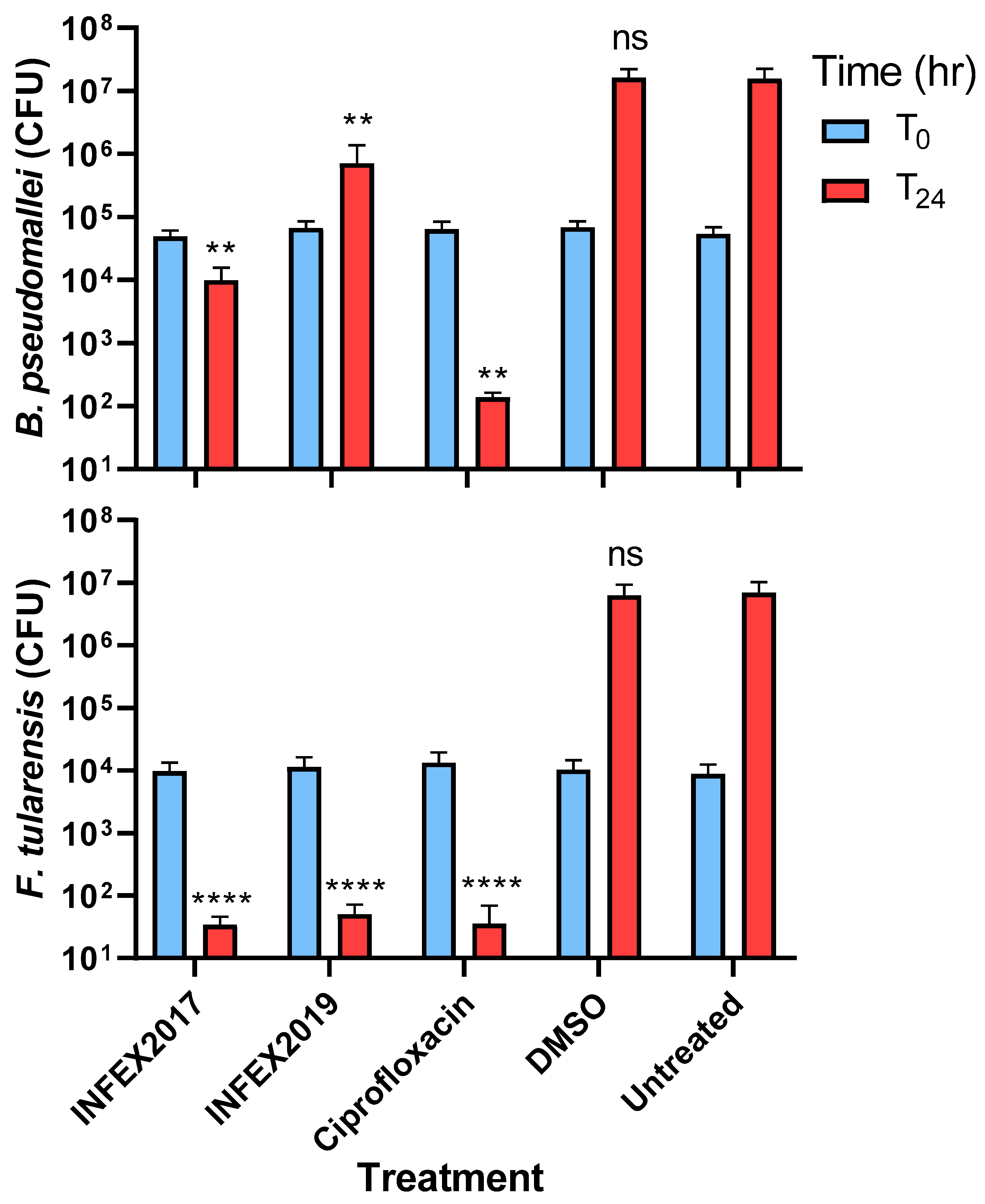
The intracellular activity of INFEX2017 and INFEX2019 (used at 4 X MIC) were compared with ciprofloxacin in J774.2 murine macrophage-like cells. A pair-wise analysis demonstrated a significant reduction in the intracellular concentration of B. pseudomallei and F. tularensis in cells incubated with INFEX2017, INFEX2019, or ciprofloxacin 24 h post-infection when compared to the untreated controls (Figure 2). INFEX2017, INFEX2019, and ciprofloxacin reduced intracellular F. tularensis by approximately 99% when the concentrations at 0 h (T0) and 24 h (T24) were compared (Figure 2).

3. Materials and Methods

3.1. Reagents

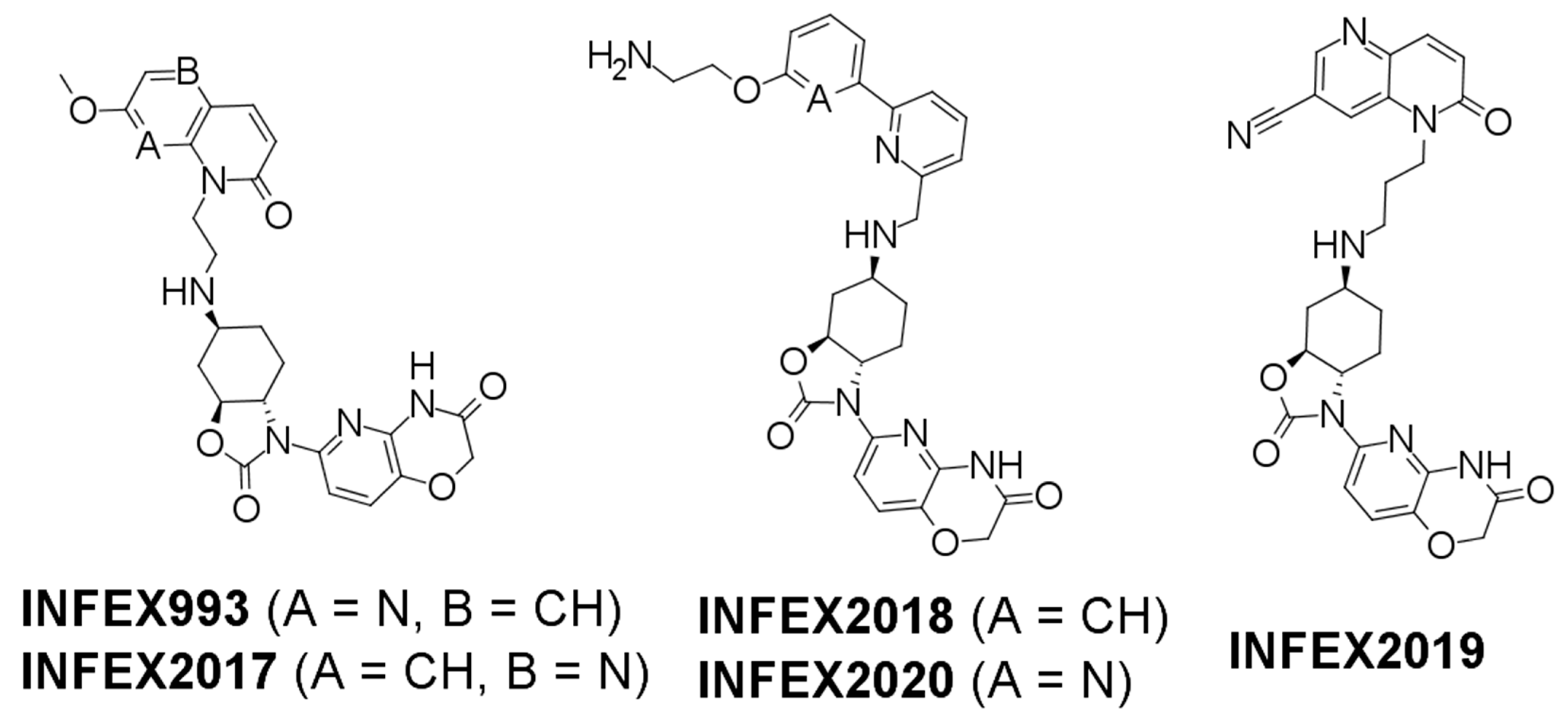
NTBI compounds were synthesized and supplied by Redx Anti-Infectives Ltd., Alderley Edge, UK, with subsequent transfer of intellectual property to Infex Therapeutics Ltd., Alderley Edge, UK. Stock vials of INFEX993, INFEX2017, INFEX2018, INFEX2019, and INFEX2020 (Figure 1), were solubilized in dimethyl sulfoxide (DMSO) at concentrations ranging from 5.3 to 10.3 mg/mL and subsequently diluted to the appropriate working concentration in the relevant media for the bacterium (as detailed below) (Figure 3). A 100 mg amount of ciprofloxacin (Sigma Aldrich Ltd.,Gillingham, UK) was added to 9 mL of sterile water and 1 mL of 1 M sodium hydroxide to form a working stock of 10 mg/mL. A 100 mg amount of doxycycline (Sigma Aldrich Ltd., UK) was added to 10 mL of distilled water. An equivalent concentration of sodium hydroxide or DMSO used to prepare the antibiotics was included as a control to ensure it could support growth of the bacteria.

3.2. Bacterial Strains and Cultures

All bacteriological procedures were carried out in accordance with the UK Advisory Committee on Dangerous Pathogens (ACDP) Containment Level 3 laboratory requirements. A 10 µL amount of a frozen stock of F. tularensis strain SCHU S4 [19] or B. pseudomallei strain K96243 [20] was added to 10 mL of Modified Cysteine Partial Hydrolysate (MCPH) broth supplemented with cysteine (100 µg/mL) and glucose (4%) or Luria Bertani (LB) broth (for F. tularensis or B. pseudomallei, respectively) and incubated at 37 °C with shaking at 180 rpm for 24 h. Bacterial cultures were then adjusted to optical densities of 0.1 or 0.25 at 590 nm in MCPH or Mueller–Hinton broth (for F. tularensis or B. pseudomallei, respectively). This provided an inoculum of approximately 108 CFU/mL. Further dilutions in MCPH or Mueller–Hinton broth were conducted according to the assay used. Bacterial enumeration was determined by plating serial dilutions of F. tularensis or B. pseudomallei onto blood cysteine glucose agar (BCGA) or L-agar (for F. tularensis or B. pseudomallei, respectively). Plates were incubated at 37 °C for 48 h prior to colony enumeration.
Antibiotic susceptibility was evaluated by determining (1) the minimum inhibitory concentration (MIC), (2) the kill kinetics over 24 h in time-kill assays, and (3) the intracellular activity in cell culture. All assay formats used triplicated assay wells (as a minimum), and data are reported from 3 independent experiments.

3.3. MIC Assays

Broth microdilution MIC assays were performed in accordance with Clinical and Laboratory Sciences Institute (CLSI) guidelines [21] with modification for F. tularensis SCHU S4 due to its more fastidious growth requirements. F. tularensis was cultured in MCPH broth supplemented with cysteine (100 µg/mL) and glucose (4% w/v) in 24-well microtiter plates. Assays were performed with bacteria at a final starting concentration of approximately 5 × 10⁵ CFU/mL and incubated for 24 h (B. pseudomallei) or approximately 2 × 10⁷ CFU/mL and incubated for 48 h (F. tularensis). Antibiotic concentrations of 64 µg/mL to 0.03 µg/mL were evaluated, and the MIC was determined as the concentration of antibiotic resulting in no visible bacterial growth following incubation.

3.4. Time-Kill Assays

Time-kill assays were performed at 4 X MIC in 10 mL of LB broth (B. pseudomallei) or MCPH with supplements (F. tularensis) [22]. Culture media were inoculated with bacteria at approximately 1 × 10⁴ CFU/mL (B. pseudomallei) or 1 × 10⁵ CFU/mL (F. tularensis) and incubated with shaking at 180 rpm and 37 °C. Samples were taken at 0, 2, 4, 6, and 24 h. A 10-fold serial dilution was performed in sterile phosphate-buffered saline (PBS), plated onto L-agar (B. pseudomallei) or BCGA (F. tularensis), and incubated at 37 °C for 48 h. A bactericidal effect was defined as a minimum 3 log10 reduction in CFU/mL; a bacteriostatic effect was defined as up to a 3 log10 reduction in CFU/mL [23] (compared with the original inoculum).

3.5. Intracellular Assays

To assess intracellular activity, triplicate wells of a 48-well plate were coated with 2 × 10⁵ J774.2 murine macrophage-like cells (ECACC, Porton Down, UK) cultured in Dulbecco’s Modified Eagle Medium (DMEM) supplemented with 2 mM L-glutamine and 10% v/v fetal bovine serum (FBS) (ThermoFisher Scientific, Basingstoke, UK) and incubated for 20 h at 37 °C/5% CO2. Following the removal of DMEM, B. pseudomallei or F. tularensis was added at a concentration of approximately 1 × 10⁶ CFU/mL in L-15 media supplemented with 10% v/v FBS at a multiplicity of infection (MOI) of 5 for 45 min at 37 °C. B. pseudomallei cells were washed twice with PBS and once with L-15. F. tularensis cells were washed once with PBS, incubated with 10 µg/mL of gentamicin (Sigma-Aldrich Ltd.) diluted in L-15 media for 45 min at 37 °C, and washed once with PBS. Antimicrobial compounds were added at 4 X MIC diluted in L-15. The concentration of intracellular bacteria was quantified by lysing the cells 24 h post-infection with water and plating on L-agar (B. pseudomallei) or BCGA (F. tularensis) as above.

3.6. Statistical Analysis

A comparison of the effect of the antimicrobial compounds on bacterial growth in time-kill experiments at multiple time points was determined by pair-wise analysis of the results from each experiment using a mixed-effects model with Dunnett’s multiple-comparison post-analysis test. Comparison of bacterial growth in cell infection assays 24 h post-infection was determined with a 2-way ANOVA with Tukey’s multiple comparisons post-analysis test. Each experiment was performed in triplicate, the untreated controls were compared with the treatment samples, and the magnitude of difference was reported (* p < 0.05, ** p < 0.01, and *** p < 0.001). All statistical analysis was performed using Graphpad Prism v.8 software.

4. Discussion

The identification of novel therapies to treat bacterial species (both public health and biodefence pathogens) is essential due to the increasing prevalence of antimicrobial resistance. NBTIs are a class of inhibitors that bind to DNA gyrase/topoisomerase IV at a different location to the quinolones, resulting in retention of activity against fluoroquinolone-resistant bacteria [12,24]. Several NBTIs are currently under investigation, including gepotidacin, which has been evaluated in a phase II clinical trial of urogenital gonorrhea and is currently in phase III clinical trials for the treatment of uncomplicated urinary tract infections (uUTIs). Gepotidacin, however, generally displays reduced activity against Gram-negative pathogens, meaning there is a need for improved generations of NBTI [15]. INFEX2017 and INFEX2019 were previously shown to display potent Gram-negative activity with promising hERG and in vitro DMPK profiles [18]. The data set detailed in the current study demonstrated that INFEX2017 and INFEX2019 also have promising in vitro activity against two Gram-negative biothreat pathogens—F. tularensis and B. pseudomallei—in multiple assays, in some instances demonstrating equivalence to traditional antibiotics. This activity for these compounds against both biothreat pathogens was improved when compared to data generated with an earlier generation of Gram-negative-focused NBTI (REDX07638) [25]. We hypothesized that the inhibitory mechanism of action of INFEX2017 and INFEX2019 against B. pseudomallei and F. tularensis is through targeting of DNA gyrase and topoisomerase IV, as recently reported for other Gram-negative pathogens by this compound series [18]. However, we acknowledge that this was not formally demonstrated in this study.
The promising in vitro activity observed for the two NBTIs proved the principle that inhibitors of this class have utility against two pathogens of biodefence interest: F. tularensis and B. pseudomallei. There are limited alternative therapies being developed that are non-traditional antibiotics but have antibacterial activity; where identified, these are primarily against B. pseudomallei. Some examples of novel candidates that have been identified as having similar levels of activity as INFEX2017 and INFEX2019 against B. pseudomallei include MMV688271 and MMV688179, both antiprotozoal agents identified from a compound screen that had MICs of 6 and 12.5 µg/mL, respectively (against strain K96243) [26]. A fatty acid synthesis inhibitor for β-ketoacyl-ACP synthases and a FabI-specific enoyl-ACP reductase inhibitor (PT01) were evaluated that had MICs for B. pseudomallei of 8 μg/mL (strain Bp400) and 1 μg/mL (strain 1026b), respectively [27,28]. Additional screening of a FabI1-specific inhibitor library identified an additional compound (PT52) that had an MIC against B. pseudomallei of 1 μg/mL (again using strain 1026b) [29].
In conclusion, we have provided proof-of-concept data to demonstrate the in vitro antibacterial activity of two novel NTBIs against the biothreat pathogens B. pseudomallei and F. tularensis. These data therefore highlighted their therapeutic potential against these biothreat pathogens. Since this study only reported activity against reference type strains, further work would expand the data set by evaluating the in vitro activity of these compounds against larger strain panels and additional bacterial pathogens. Confirmation of inhibitory mechanisms of action against these pathogens would also be warranted. Further study in vivo would determine whether the in vitro activity demonstrated above translates into efficacy and thereby warrants their further development as therapeutic candidates.

Author Contributions

S.V.H., K.B.B., M.I.R., I.C., M.C., A.O.W., D.O., K.B., J.K., A.-M.S., N.O. and A.L. conceived the concept and designed the experiments detailed in this manuscript; M.I.R. and A.O.W. conducted the experiments; K.B.B. and A.O.W. performed the data analysis; S.V.H., A.O.W., N.O. and A.-M.S. wrote the manuscript. All authors have read and agreed to the published version of the manuscript.

Funding

This research was funded by the Ministry of Defence, UK.

Institutional Review Board Statement

Not applicable.

Informed Consent Statement

Not applicable.

Data Availability Statement

Not applicable.

Acknowledgments

We would like to acknowledge the valuable input from Victoria Lee of Infex Therapeutics in the preparation of this manuscript The funders had no role in the study design, the data collection, the interpretation, or the decision to submit the work for publication.

Conflicts of Interest

The authors declare no conflict of interest.

References

  1. Cheng, A.C.; Currie, B.J. Melioidosis: Epidemiology, pathophysiology, and management. Clin. Microbiol. Rev. 2005, 18, 383–416. [Google Scholar] [CrossRef] [PubMed] [Green Version]
  2. Ellis, J.; Oyston, P.C.; Green, M.; Titball, R.W. Tularemia. Clin. Microbiol. Rev. 2002, 15, 631–646. [Google Scholar] [CrossRef] [PubMed] [Green Version]
  3. Rao, C.; Hu, Z.; Chen, J.; Tang, M.; Chen, H.; Lu, X.; Cao, L.; Deng, L.; Mao, X.; Li, Q. Molecular epidemiology and antibiotic resistance of Burkholderia pseudomallei isolates from Hainan. China Med. 2019, 98, 14461. [Google Scholar] [CrossRef]
  4. Narayanan, N.; Lacy, C.R.; Cruz, J.E.; Nahass, M.; Karp, J.; Barone, J.A.; Hermes-DeSantis, E.R. Disaster preparedness: Biological threats and treatment options. Pharmacotherapy 2018, 38, 217–234. [Google Scholar] [CrossRef] [PubMed] [Green Version]
  5. Sullivan, R.P.; Ward, L.; Currie, B.J. Oral eradication therapy for melioidosis; important but not without risks. Int. J. Infect. Dis. 2019, 80, 111–114. [Google Scholar] [CrossRef] [PubMed] [Green Version]
  6. Hooper, D.C. Emerging mechanisms of fluoroquinolone resistance. Emerg Infect Dis. 2001, 7, 337–341. [Google Scholar] [CrossRef] [Green Version]
  7. Blair, J.M.A.; Webber, M.A.; Baylay, A.J.; Ogbolu, D.O.; Piddock, L.J.V. Molecular mechanisms of antibiotic resistance. Nat. Rev. Microbiol. 2015, 13, 42–51. [Google Scholar] [CrossRef]
  8. Redgrave, L.S.; Sutton, S.B.; Webber, M.A.; Piddock, L.J.V. Fluoroquinolone resistance: Mechanisms, impact on bacteria, and role in evolutionary success. Trends Microbiol. 2014, 22, 438–445. [Google Scholar] [CrossRef]
  9. Wright, G.D. Molecular mechanisms of antibiotic resistance. Chem. Commun. 2011, 47, 4055–4061. [Google Scholar] [CrossRef]
  10. Gibson, E.G.; Bax, B.; Chan, P.F.; Osheroff, N. Mechanistic and structural basis for the actions of the antibacterial Gepotidacin against Staphylococcus aureus gyrase. ACS Infect. Dis. 2019, 5, 570–581. [Google Scholar] [CrossRef]
  11. Kolarič, A.; Anderluh, M.; Minovski, N. Two decades of successful SAR-grounded stories of the novel bacterial topoisomerase inhibitors (NBTIs). J. Med. Chem. 2020, 63, 5664–5674. [Google Scholar] [CrossRef] [PubMed]
  12. Jaswal, S.; Nehra, B.; Kumar, S.; Monge, V. Recent advancements in the medicinal chemistry of bacterial type II topoisomerase inhibitors. Bioorg. Chem. 2020, 104, 104266. [Google Scholar] [CrossRef] [PubMed]
  13. Reck, F.; Alm, R.A.; Brassil, P.; Newman, J.V.; Ciaccio, P.; McNulty, J.; Barthlow, H.; Goteti, K.; Breen, J.; Comita-Prevoir, J.; et al. Novel N-linked aminopiperidine inhibitors of bacterial topoisomerase type II with reduced p K a: Antibacterial agents with an improved safety profile. J. Med. Chem. 2012, 55, 6916–6933. [Google Scholar] [CrossRef]
  14. Bulter, A.; Helliwell, M.V.; Zhange, Y.; Hancox, J.C.; Dempsey, C.E. An update on the structure of hERG. Front. Pharmacol. 2020, 10, 1572. [Google Scholar]
  15. Biedenbach, D.J.; Bouchillon, S.K.; Hackel, M.; Miller, L.A.; Scangarella-Oman, N.E.; Jakielaszek, C.; Sahm, D.F. In vitro activity of gepotidacin, a novel triazaacenaphthylene bacterial topoisomerase inhibitor, against a broad spectrum of bacterial pathogens. Antimicrob. Agents. Chemother. 2016, 60, 1918–1923. [Google Scholar] [CrossRef] [PubMed] [Green Version]
  16. Jacobsson, S.; Golparian, D.; Scangarella-Oman, N.; Unemo, M. In vitro activity of the novel triazaacenaphthylene gepotidacin (GSK2140944) against MDR Neisseria gonorrhoeae. J. Antimicrob. Chemother. 2018, 73, 2072–2077. [Google Scholar] [CrossRef] [PubMed] [Green Version]
  17. Jakielaszek, C.; Hossain, M.; Qian, L.; Fishman, C.; Widdowson, K.; Hilliard, J.J.; Mannino, F.; Raychaudhuri, A.; Carniel, E.; Demons, S.; et al. Gepotidacin is efficacious in a nonhuman primate model of pneumonic plague. Sci. Transl. Med. 2022, 14, 1787. [Google Scholar] [CrossRef] [PubMed]
  18. Lyons, A.; Kirkham, J.; Blades, K.; Orr, D.; Dauncey, E.; Smith, O.; Dick, E.; Walker, R.; Matthews, T.; Bunt, A.; et al. Discovery and structure-activity relationships of a novel oxazolidinone class of bacterial type II topoisomerase inhibitors. Bioorg. Med. Chem. Lett. 2022, 65, 128648. [Google Scholar] [CrossRef]
  19. Larsson, P.; Oyston, P.C.F.; Chain, P.; Chu, M.C.; Duffield, M.; Fuxelius, H.-H.; Garcia, E.; Hälltorp, G.; Johansson, D.; E Isherwood, K.; et al. The complete genome sequence of Francisella tularensis, the causative agent of tularemia. Nat. Gen. 2005, 37, 153–159. [Google Scholar] [CrossRef] [Green Version]
  20. Holden, M.T.G.; Titball, R.W.; Peacock, S.J.; Cerdeño-Tárraga, A.M.; Atkins, T.; Crossman, L.C.; Pitt, T.; Churcher, C.; Mungall, K.; Bentley, S.D.; et al. Genomic plasticity of the causative agent of melioidosis, Burkholderia pseudomallei. Proc. Natl. Acad. Sci. USA 2004, 101, 14240–14245. [Google Scholar] [CrossRef] [Green Version]
  21. Methods for Dilution Antimicrobial Susceptibility Tests for Bacteria that Grow Aerobically; Approved Standard. CSLI Document M07-A10; Clinical and Laboratory Standards Institute: Wayne, PA, USA, 2015.
  22. Eyles, J.E.; Hartley, M.G.; Laws, T.R.; Oyston, P.C.F.; Griffin, K.F.; Titball, R.W. Protection afforded against aerosol challenge by systemic immunisation with inactivated Francisella tularensis live vaccine strain (LVS). Microb. Pathog. 2008, 44, 164–168. [Google Scholar] [CrossRef] [PubMed]
  23. Methods for Determining Bactericidal Activity of Antimicrobial Agents: Approved Guideline; CLSI Document M26-A; Clinical and Laboratory Standards Institute: Wayne, PA, USA, 1999.
  24. Bax, B.D.; Chan, P.F.; Eggleston, D.S.; Fosberry, A.; Gentry, D.R.; Gorrec, F.; Giordano, I.; Hann, M.M.; Hennessy, A.; Hibbs, M.; et al. Type IIA topoisomerase inhibition by a new class of antibacterial agents. Nature 2010, 466, 935–940. [Google Scholar] [CrossRef] [PubMed]
  25. Charrier, C.; Salisbury, A.M.; Savage, V.J.; Duffy, T.; Moyo, E.; Chaffer-Malam, N.; Ooi, N.; Newman, R.; Cheung, J.; Metzger, R.; et al. Novel bacterial topoisomerase inhibitors with potent broad-spectrum activity against drug-resistant bacteria. Antimicrob. Agents. Chemother. 2017, 61, e02100-16. [Google Scholar] [CrossRef] [Green Version]
  26. Ross, B.N.; Myers, J.N.; Muruato, L.A.; Tapia, D.; Torres, A.G. Evaluating new compounds to treat Burkholderia pseudomallei infections. Front. Cell. Infect. Microbiol. 2018, 8, 210. [Google Scholar] [CrossRef]
  27. Bommineni, G.R.; Kapilashrami, K.; Cummings, J.E.; Lu, L.; Knudson, S.E.; Gu, C.; Walker, S.G.; Slayden, S.A.; Tonge, P.J. Thiolactomycin-based inhibitors of bacterial β-Ketoacyl-ACP synthases with in vivo activity. J. Med. Chem. 2016, 59, 5377–5390. [Google Scholar] [CrossRef] [PubMed] [Green Version]
  28. Cummings, J.E.; Kingry, L.C.; Rholl, D.A.; Schweizer, H.P.; Tonge, P.J.; Slayden, R.A. The Burkholderia pseudomallei enoyl-acyl carrier protein reductase fabI1 is essential for in vivo growth and is the target of a novel chemotherapeutic with efficacy. Antimicrob. Agents. Chemother. 2014, 58, 931–935. [Google Scholar] [CrossRef] [Green Version]
  29. Cummings, J.E.; Beaupre, A.J.; Knudson, S.E.; Liu, N.; Yu, W.; Neckles, C.; Wang, H.; Khanna, A.; Bommineni, G.R.; Trunck, L.A.; et al. Substituted diphenyl ethers as a novel chemotherapeutic platform against Burkholderia pseudomallei. Antimicrob. Agents. Chemother. 2014, 58, 1646–1651. [Google Scholar] [CrossRef] [PubMed] [Green Version]
Figure 1. Time-kill assays. The activity of INFEX2017, INFEX2019, and ciprofloxacin against B. pseudomallei strain K96243 (upper panel) and F. tularensis strain SCHU S4 (lower panel) was assessed over 24 h. Time-kill assays were performed at 4 X MIC (16 µg/mL of INFEX2017 or INFEX2019 or 4 µg/mL of ciprofloxacin for B. pseudomallei and 2 µg/mL of INFEX2017 or INFEX2019 or 0.12 µg/mL of ciprofloxacin for F. tularensis). An untreated bacterial culture and a DMSO diluent control were also included. The dotted line represents a 3 log10 reduction in CFU/mL from the starting inoculum. Each data point is the mean of 3 independent replicated experiments, and the error bars represent the standard error. Statistical analysis was performed using a mixed-effects analysis with Dunnett’s multiple-comparison test. Statistically significant differences between the bacterial CFU of treated and untreated (media control) cultures at 24 h post-inoculation is reported (not significant (ns); p > 0.05; * p < 0.05; ** p < 0.01; *** p < 0.001).
Antibiotics 12 00983 g001
Figure 2. J774.2 cell infection assays. The intracellular activity of INFEX2017, INFEX2019, and ciprofloxacin against B. pseudomallei strain K96243 (upper panel) and F. tularensis strain SCHU S4 (lower panel) was assessed in a J774.2 cell infection assay. J774.2s cells were infected with bacteria at a target MOI of 5. Extracellular bacteria were removed by washing (and for F. tularensis by incubation with gentamicin (10 µg/mL)). Infected J774.2 cells were incubated for 24 h with 4 X MIC (16 µg/mL of INFEX2017 or INFEX2019 or 4 µg/mL ciprofloxacin for B. pseudomallei and 2 µg/mL of INFEX2017 or INFEX2019 or 0.12 µg/mL ciprofloxacin for F. tularensis). An untreated culture and a DMSO diluent control were included. The mean CFU of intracellular bacteria at 0 (T0) and 24 h (T24) is presented from 3 independent experiments with error bars representing the standard error. To determine any statistical difference between the intracellular bacterial burden of test compound cultures compared with the untreated (media only) control cultures, a mixed-effects analysis was performed with Dunnett’s multiple-comparison test. Significant differences at 24 h post-infection (T24) are reported (not significant (ns); p > 0.05; ** p < 0.001; **** p < 0.0001).
Antibiotics 12 00983 g002
Figure 3. Chemical structures of test compounds. The chemical structures of the NBTI compounds evaluated.
Antibiotics 12 00983 g003
Table 1. MICs for B. pseudomallei and F. tularensis.
Inhibitor/AntibioticMIC (μg/mL)
B. pseudomalleiF. tularensis
INFEX99320.06–0.125
INFEX201740.5–1
INFEX201888–16
INFEX201940.5–4
INFEX2020816–32
Ciprofloxacin0.5–20.03–0.5
Doxycycline0.5–42–4
Disclaimer/Publisher’s Note: The statements, opinions and data contained in all publications are solely those of the individual author(s) and contributor(s) and not of MDPI and/or the editor(s). MDPI and/or the editor(s) disclaim responsibility for any injury to people or property resulting from any ideas, methods, instructions or products referred to in the content.