Article

Occurrence of Carbapenemases, Extended-Spectrum Beta-Lactamases and AmpCs among Beta-Lactamase-Producing Gram-Negative Bacteria from Clinical Sources in Accra, Ghana

1
Department of Bacteriology, Noguchi Memorial Institute for Medical Research, University of Ghana, Accra 00233, Ghana
2
Department of Medical Laboratory Sciences, School of Biomedical and Allied Health Sciences, University of Ghana, Accra 00233, Ghana
3
Department of Microbiology, Korle-Bu Teaching Hospital, Accra 00233, Ghana
4
University of Ghana Hospital, Accra 00233, Ghana
5
Department of Medical Microbiology, University of Ghana Medical School, University of Ghana, Accra 00233, Ghana
*
Author to whom correspondence should be addressed.
Antibiotics 2023, 12(6), 1016; https://doi.org/10.3390/antibiotics12061016
Submission received: 25 March 2023 / Revised: 18 May 2023 / Accepted: 22 May 2023 / Published: 5 June 2023

Abstract

:
Beta-lactamase (β-lactamase)-producing Gram-negative bacteria (GNB) are of public health concern due to their resistance to routine antimicrobials. We investigated the antimicrobial resistance and occurrence of carbapenemases, extended-spectrum β-lactamases (ESBLs) and AmpCs among GNB from clinical sources. GNB were identified using matrix-assisted laser desorption/ionization time of flight–mass spectrometry (MALDITOF-MS). Antimicrobial susceptibility testing was performed via Kirby–Bauer disk diffusion and a microscan autoSCAN system. β-lactamase genes were determined via multiplex polymerase chain reactions. Of the 181 archived GNB analyzed, Escherichia coli and Klebsiella pneumoniae constituted 46% (n = 83) and 17% (n = 30), respectively. Resistance to ampicillin (51%), third-generation cephalosporins (21%), and ertapenem (21%) was observed among the isolates, with 44% being multi-drug resistant (MDR). β-lactamase genes such as AmpCs ((blaFOX-M (64%) and blaDHA-M and blaEDC-M (27%)), ESBLs ((blaCTX-M (81%), other β-lactamase genes blaTEM (73%) and blaSHV (27%)) and carbapenemase ((blaOXA-48 (60%) and blaNDM and blaKPC (40%)) were also detected. One K. pneumoniae co-harbored AmpC (blaFOX-M and blaEBC-M) and carbapenemase (blaKPC and blaOXA-48) genes. blaOXA-48 gene was detected in one carbapenem-resistant Acinetobacter baumannii. Overall, isolates were resistant to a wide range of antimicrobials including last-line treatment options. This underpins the need for continuous surveillance for effective management of infections caused by these pathogens in our settings.

1. Introduction

Gram-negative bacteria (GNB) are widespread in nature and cause life-threatening infections in humans [1]. The expression of beta-lactamase (β-lactamase) enzymes such as class C cephalosporinases (AmpCs), extended-spectrum β-lactamase (ESBL) and carbapenemase in GNB has been linked to increased antibiotic resistance resulting in therapeutic failure, prolonged hospital stays, increased healthcare cost and mortality [2].
Antimicrobial resistance (AMR) observed among GNB due to the expression of β-lactamase is a global public health concern [3]. In Ghana and other parts of the world, there is over-dependence on cephalosporin beta-lactam antibiotics for the treatment of infections due to their low toxicity and high potency, which have contributed to the phenomenon of resistance [4]. β-lactamases act by disrupting the active amide bond of the beta-lactam (β-lactam) ring and can be chromosomally encoded or acquired via mobile genetic elements (MGEs) such as plasmids, integrons, and transposons among bacteria species. The MGEs serve as vectors in transmitting β-lactamase (bla) and non-betalactam AMR genes among bacteria species, resulting in limited therapeutic options for the treatment of infections [5].
Plasmid-mediated AmpC β-lactamases (including blaFOX-M, blaDHA-M, blaCMY-M and blaACC-M) are clinically relevant cephalosporinases that mediate resistance to cephalothin, cefazolin, cefoxitin, most penicillins and β-lactamase inhibitor–beta-lactam combinations. The hyperproduction of AmpC confers resistance to extended-spectrum cephalosporins including cefotaxime, ceftazidime and ceftriaxone [6]. AmpC production has been frequently reported in Enterobacterales [7]. Of concern is the hyperproduction of AmpC β-lactamases and the upregulation of efflux pumps, which may synergistically confer additional resistance to carbapenems in GNB [8]. Most importantly, AmpC-producing bacteria are a cause of healthcare-associated infections leading to strained therapeutic options [9].
The ESBL group of the β-lactamases mediates a wide range of resistance activities against frequently administered antibiotics such as penicillins, oxyimino-cephalosporins and aztreonam, but they are inactive against beta-lactam inhibitors, cephamycins (cefoxitin) and carbapenems [10]. They are predominant among Enterobacterales strains and have been associated with multi-drug-resistant (MDR) phenotypes responsible for severe and fatal infection outcomes as well as hospital and community-linked outbreaks [11]. ESBL-producing Enterobacterales are part of the World Health Organization critical priority pathogens for the research and development of novel antibiotics [12]. The most disseminated genotypes of ESBL are blaTEM, blaSHV and blaCTX-M [13], which are mostly borne on plasmids, and these plasmids often carry genes that mediate resistance to other classes of antibiotics such as aminoglycosides, fluoroquinolones and trimethoprim-sulfamethoxazole, further reducing options for the treatment of ESBL-associated infections [14].
Carbapenems are a drug of choice for the treatment of infections caused by ESBL- and AmpC-producing pathogens [15]. However, the emergence of carbapenemase enzymes [16] has further reduced the treatment options available for infections caused by these pathogens [17]. These carbapenemase enzymes have been associated with clinically relevant pathogens of the Enterobacterales family, Pseudomonas aeruginosa (P. aeruginosa) and Acinetobacter baumannii (A. baumannii) strains [18].
Many variants of carbapenemases have been detected, with the most predominant being KPC (class A carbapenemases), NDM and VIM (class B metallo-beta-lactamases) and the OXA-48-type (class D carbapenemases) enzymes [19]. They have been reported globally as significant contributors to healthcare-associated infections [20]. The class A enzymes efficiently resist carbapenems and inhibit the actions of clavulanic acid, albeit poorly, whilst the class B enzymes inhibit the activities of β-lactamase inhibitors and carbapenems but not aztreonam [21]. On the other hand, class D carbapenemases, which are mostly isolated from A. baumannii, P. aeruginosa and less commonly in the Enterobacterales family, partially inhibit clavulanates and weakly hydrolyze carbapenems [22].
In Ghana, although GNB-harboring β-lactamases are established as widespread, most studies have only investigated the presence of ESBLs and carbapenemases in Enterobacterales with emphasis on Escherichia coli (E. coli) and Klebsiella pneumoniae (K. pneumoniae) [4,23]. There is therefore a paucity of studies that have reported the concurrent presence of β-lactamases such as AmpC, ESBL and carbapenemases in an array of GNB from clinical sources. Data on the antimicrobial resistance of β-lactamase-producing organisms are essential in guiding antimicrobial therapy, effective surveillance and infection prevention and control. This study therefore investigated the occurrence and antimicrobial resistance of AmpCs-, ESBLs- and carbapenemases among a collection of GNB recovered from clinical sources.

2. Results

2.1. Spectrum of Gram-Negative Bacteria

Out of the 181 GNB processed, 89% (n = 161/181) were Enterobacterales, comprising E. coli (n = 83/161; 52%), K. pneumoniae (n = 30/161; 19%), Proteus mirabilis (n = 18/161; 11%), Enterobacter spp. (n = 16/161; 10%), Salmonella spp. (n = 8/161; 5%), Citrobacter spp. (n = 3/161; 2%), Providencia spp. (n = 2/161; 1.2%) and Klebsiella oxytoca (n = 1/161; 0.6%). Pseudomonas aeruginosa (n = 8/181; 4.4%), Acinetobacter spp. (n = 4/181; 2.2%), Neisseria spp. (n = 3/181; 1.7%), Pseudomonas stutzeri (n = 1/181; 0.5%) and others (n = 4/181; 2.2%) were also detected. Table 1 shows bacteria species identified and clinical sources.

2.2. Antimicrobial Resistance Pattern

The top three antibiotics with the highest susceptibility were cefoxitin (n = 144/168; 86%), ertapenem and norfloxacin (n = 133/168; 79%). Cefuroxime and cefotaxime resistance was detected among 29% (n = 48/168) of the isolates. Ceftazidime (n = 47/168; 28%) and ampicillin (n = 86/168; 51%) resistance were also detected. For ertapenem, E. coli isolates recorded the highest level of resistance (n = 18/83; 22%). Pseudomonas aeruginosa (n = 8) and Pseudomonas stutzeri (n = 1) were susceptible to all antibiotics tested. Among the Acinetobacter spp. (n = 4), Acinetobacter nosocomialis (n = 1/4; 25%) showed resistance to trimethoprim/sulfamethozaxole with an MIC range >2/38 µg/mL. Ampicillin was the least effective antibiotic against all strains, with a resistance prevalence of 51%. Overall, 44%, (n = 74/168) of the isolates were multi-drug resistant (MDR). The three isolates with the highest MDR proportions were E. coli (n = 40/74; 54%), K. pneumoniae (n = 11/74; 15%) and Enterobacter spp. (n = 11/74; 15%). The MDR isolates were susceptible to cefoxitin (n = 52/74; 70%) and ertapenem (n = 42/74; 57%). Table 2 shows the antimicrobial resistance profiles of the isolates.

2.3. Phenotype and Resistance Gene Markers for AmpC, ESBL and Carbapenemase

All isolates except P. aeruginosa (n = 8), Acinetobacter spp. (n = 4), and P. stutzeri (n = 1), were analyzed for the production of AmpC and ESBL, phenotypically and via polymerase chain reaction (PCR). Twenty-eight (17%) isolates were phenotypically resistant to cefoxitin (presumptive AmpC producers) and were screened via PCR for the presence of plasmid-mediated AmpC β-lactamase genes. Eleven (39%) isolates were positive for AmpC gene variants. Thirty-six isolates (36/168; 21%) were phenotypically positive for ESBL production; of these, 29 (81%) contained blaCTX-M genes. E. coli were the predominant carriers of ESBL genes (n = 22/29; 76%), followed by K. pneumoniae 24% (n = 7/29).
Of the thirty-five ertapenem-resistant isolates, five were positive for carbapenemase genes, and these included two K. pneumoniae, one E. coli, an A. baumannii and a Providencia vermicola (P. vermicola). Table 3 shows the phenotypic and genotypic distribution of β-lactamase amongst the isolates.

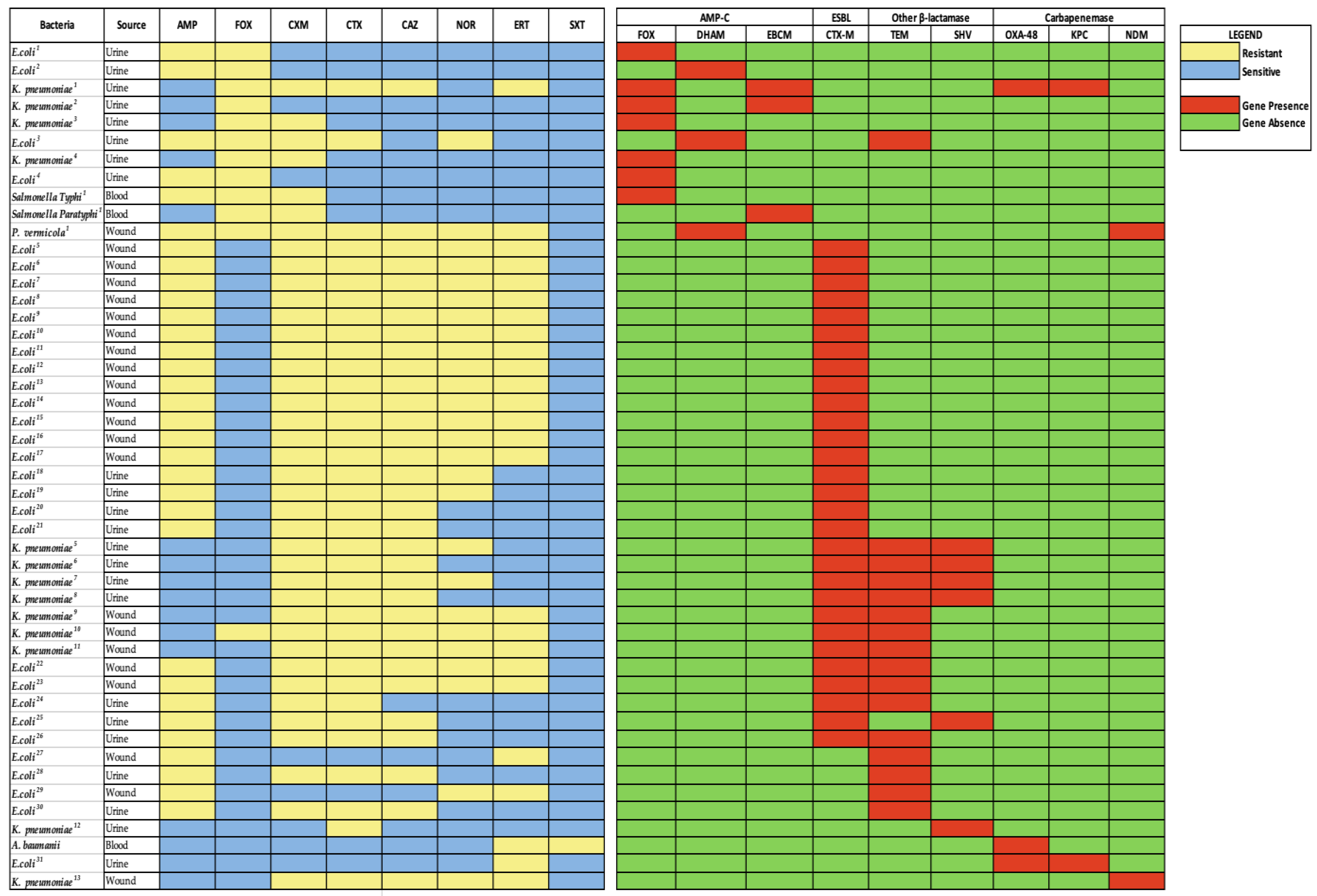
2.4. Resistance Gene Distribution among Isolates

AmpC genes were found among E. coli (n = 4/11; 36%), K. pneumoniae (n = 4/11; 36%), Salmonella spp. (n = 2/11; 18%) and P. vermicola (n = 1/11; 9%) isolates. These isolates originated from urine (n = 8), blood (n = 2) and wound (n = 1) samples. The most frequently identified AmpC gene was blaFOX-M (n = 7/11; 64%), observed in four K. pneumoniae (36%), two E. coli (18%) and one Salmonella Typhi (9%). Other AmpC genes were blaEBC-M (n = 3/11; 27%), identified in two K. pneumoniae, and one Salmonella paratyphi. The blaDHA-M gene (n = 3/11; 27%) was found in two E. coli and one P. vermicola isolates. Two out of four K. pneumoniae co-harbored blaFOX-M and blaEBC-M genes. The 29 isolates harboring the ESBL- blaCTX-M gene were isolated from urine (n = 11) and wound (n = 18) samples. Other β-lactamase genes, such as blaTEM (n = 16/22; 73%) and blaSHV (n = 6/22; 27%), were also observed among the phenotypically positive ESBL producing isolates. blaTEM gene was found in E. coli (n = 9/16; 56%) and K. pneumoniae (n = 7/16; 44%). The blaSHV gene was harbored by K. pneumoniae (n = 5/6; 83%) and E. coli (n = 1/6; 17%). In all, four (11%) isolates harbored a blaCTX-M, blaTEM and blaSHV β-lactamase genes whereas eleven (31%) contained both blaCTX-M and blaTEM genes. Five isolates (14%) possessed both blaCTX-M and blaSHV genes. The blaOXA-48 gene was the most common carbapenemase gene identified and was carried by three of the five carbapenem-resistant GN isolates (K. pneumoniae, E. coli and A. baumannii). Two of the isolates with blaOXA-48 (E. coli and K. pneumoniae) were from urine samples and each co-harbored a blaKPC gene. A. baumanni carried blaOXA-48 and was recovered from a blood sample. blaNDM was identified in K. pneumoniae (n = 1) and P. vermicola (n = 1) isolates, both from wound samples. Figure 1 shows beta lactamase distribution and antibiotypes.

3. Discussion

This study found high levels of β-lactamase genes among GNB. The finding that E. coli and K. pneumoniae were the predominant GNB is similar to previous studies conducted in the country [4] as well as other parts of the world [3]. These organisms have been associated with antibiotic resistance and implicated in series of infections associated with high morbidity and mortality rates [24].
The most effective antibiotic in the study was cefoxitin. Cefoxitin, a cephamycin/second-generation cephalosporin, is known to exhibit bactericidal actions against GNB and is very potent in vitro against ESBL producers [25]. All the ESBL-producing GNB in our study, except one, were susceptible to cefoxitin. It is known to be a suitable alternative for carbapenems in infection treatment [25]. A substantial proportion of the study isolates were susceptible to fluoroquinolone, contrary to other studies in the country [26] and elsewhere (Nigeria, Tanzania and Rwanda) [27,28,29]. Our findings could be an indication that such a class of antibiotic might still be relevant in the treatment of GNB-associated infections in Ghana.
The proportion (22%) of ESBL phenotypes observed among the Enterobacterales is in line with studies in the country [30] and in other parts of the world [31,32], with E. coli and K. pneumoniae as frequent ESBL producers. The most predominant ESBL gene was blaCTX-M (81%), which is the case globally [33,34,35]. On the contrary, blaTEM was the predominant gene in a recent study in Ghana [30] and Nigeria [36]. The presence of blaCTX-M represents a significant public health threat since isolates harboring this gene co-harbor extra antimicrobial resistance genes making them resistant to a wide range of antimicrobials [37]. This is particularly worrying since these drugs are part of treatment regimens for the management of infections in Ghana [8]. The co-harboring of blaCTX-M and other genes could foster the growth and dissemination pathogens with multiple resistance genes in the country [37].
The finding of greater proportions of AmpC-producing GNB being MDRs was expected since the presence of AmpC β-lactamases is accompanied with resistance to first to third-generation cephalosporins, including cephymacins. It is known that AmpC enzymes may disguise the true effect of ESBLs and their identification in a bacteria strain, thus complicating the treatment of infections caused by bacteria co-harboring both genes [38]. In this study, E. coli co-harbored AmpC and ESBL (blaTEM and blaDHA-M) genes; likewise, K. pneumoniae co-harbored AmpC and carbapenemase genes ((blaFOX-M + blaEBC-M) + (blaKPC + blaOXA-48)), which has also been observed in studies in Egypt [38] and Nigeria [39].
Carbapenems are the drug of choice for the treatment of severe infections [39]. In our study, the carbapenem-tested ertapenem was one of the most effective antibiotics, and this is an assurance that this drug class is still effective. However, the blaKPC gene, which is known to hydrolyze carbapenems and was initially identified in the north-eastern part of the US, has now spread throughout the US and most of the world [40]. Our study isolated carbapenem-resistant K. pneumoniae co-harboring blaKPC and blaOXA-48. To the best of our knowledge, this is one of two studies in the country to record a dual detectable carbapenemase gene (blaKPC and blaOXA-48) in a single carbapenem-resistant K. pneumoniae strain [41]. The presence of blaOXA-48 and blaNDM among GNB has been reported in other studies in Ghana [42]. Furthermore, we observed that the carbapenem-resistant A. baumanii (crAb) isolated from a blood culture harbored a blaOXA-48 gene in this study. In Ghana, Monheimer and colleagues identified crAb (77%) that harbored blaOXA-23, and blaNDM genes [43]. Carbapenem-resistant A. baumannii are among the WHO-prioritized organisms under surveillance worldwide since they are associated with life-threatening infections and the frequent carriage of multi-drug resistance [17]. Their isolation from a blood sample means that such a patient will not respond to the routinely used antimicrobials. Meanwhile, data are still limited in this part of Africa on the characteristics of this pathogen [44]. As far as we know, this is the first report of a blaOXA-48 crAb recovered from a blood culture in the country. In this study, there was one K. pneumoniae strain that co-harbored carbapenemase (blaKPC + blaOXA-48) and AmpC genes (blaFOX-M + blaEBC-M). Kaizeman in Iran had a similar result, but his K. pneumoniae isolates harbored ESBL genes (blaCTX-M, blaTEM and blaSHV) and a blaVIM carbapenemase gene [45].
The ability to harbor multiple resistance genes by GNB gives them the advantage to thrive longer in healthy individuals or the environment or cause infections in immune-compromised victims. The resistant genes identified in this study fail infection control systems and subsquently lead to dire outcomes [17]. There is the need to improve infection control measures, strengthen antimicrobial resistance surveillance and identify new treatment strategies in order to mitigate this problem.

4. Materials and Methods

4.1. Identification of Bacteria

This study analyzed 181 archived GNB recovered from clinical sources from April 2017 to April 2018 as a form of laboratory surveillance of antimicrobial resistance and β-lactamase production. Isolates originated from urine (n = 114), wounds (n = 38), blood (n = 21), throat swabs (n = 4), stools (n = 3), and an ear swab from humans (outpatients and inpatients) with diverse infections. The isolates were sub-cultured on blood and MacConkey agar. The identification and confirmation of bacteria species was achieved using matrix-assisted laser desorption/ionization time of flight (MALDI-TOF)–mass spectrometry (Bruker Daltonics, Bremen, Germany) using Biotyper™ system 2.0 software at the genus (log(score) 1.7–2.0)) and species (log(score) ≥ 2.0) level.

4.2. Antimicrobial Susceptibility Testing

Kirby–Bauer disk diffusion was performed using the 0.5 McFarland standard of bacteria suspension seeded into Mueller–Hinton agar plates. The antibiotics tested included ampicillin (10 µg), cefotaxime (30 µg), norfloxacin (10 µg), ceftazidime (30 µg), ertapenem (10 µg), cefoxitin (30 µg) and cefuroxime (30 µg). The measured zone sizes were interpreted according to the Clinical and Laboratory Standard (CLSI, M100, 26th ed, 2018) guidelines (CLSI 2018). P. aeruginosa was tested against gentamicin (10 µg), cefepime (30 µg), ceftazidime (30 µg), ciprofloxacin (5 µg), piperacillin tazobactam (110 µg), amikacin (30 µg) and meropenem (10 µg). Pseudomonas stutzeri and Acinetobacter spp. were tested against 11 antibiotics in a microbroth dilution using the MicroScan autoSCAN-4-System with the NC 66 panel (Beckman Coulter Life Sciences, Indianapolis, IN, USA) following the manufacturer’s instructions. K. pneumoniae ATCC 700603 and E. coli ATCC 25922 were used as control strains in the evaluation of the performance of the tests. An MDR phenotype was defined as non-susceptibility to ≥1 agent in ≥3 antimicrobial categories [46]. Isolates with non-susceptibility to 3rd-generation cephalosporins were examined for the possible presence of AmpCs and ESBLs according to the CLSI guideline.

4.3. Phenotypic Screening for AmpC, ESBL and Carbapenem Resistance

Investigations for AmpC, ESBL and carbapenemases were only carried out for Enterobacterales. Other GNB were screened for carbapenemase production. Using the disk diffusion assay, the presumptive detection of AmpC was performed by observing reduced susceptibility (inhibition zone < 18 mm) to cefoxitin (30 µg). The expression of ESBLs was ascertained via the combined double-disk method using cefotaxime (30 µg) and ceftazidime (30 µg) alone and in combination with clavulanic acid (10 µg). An inhibition zone difference of ≥5 mm between the single and the clavulanic acid combination disks for cefotaxime and ceftazidime confirmed ESBL expression. Isolates resistant to ertapenem (10 µg) (inhibition zone ≤ 15 mm) were deemed possible carbapenemase producers. These isolates were subjected to a confirmatory test using the Modified Hodges Test (MHT) and the modified Carbapenem Inactivation Method (mCIM) according to the CLSI guidelines. K. pneumoniae ATCC BAA 1705 and E. coli ATCC 25922 strains were used as positive and negative controls, respectively.

4.4. Molecular Detection of Antimicrobial-Resistant Gene Markers of Beta-Lactamases

Isolates that tested positive for AmpC, ESBL, or carbapenemase phenotypes were subjected to PCR to confirm the genes encoding for AmpC (blaMOX-M, blaACC-M, blaEBC-M, blaFOX-M, blaCIT-M and blaDHA-M) [47], ESBLs (blaCTX-M) [48], carbapenemases (blaKPC, blaNDM, blaVIM, blaIMP and blaOXA-48) [49], and other β-lactamase genes (blaTEM and blaSHV) [48] with slight modifications. Crude DNA (10 µL) was extracted from pure overnight cultures and suspended in 200μL of molecular-grade nuclease-free water, heated for 10 min at 98 °C, and centrifuged for 5 min at 4 °C and 20,000 g, as previously suggested by Quansah [42]. The supernatant was transferred into sterile 1.5 mL Eppendorf® tubes and used as a template for the PCR. For the PCR amplification, each reaction mix of 25 µL consisted of 12.5 uL of Green PCR Master Mix (2×) (DreamTaq, Thermo Scientific, Waltham, MA, USA), 4.5 µL of primer mix, 6 µL of molecular-grade nuclease-free water and 2 µL of crude DNA template, as previously demonstrated by Khurana et al., 2018 [49]. The primers used for PCR amplification and cycling conditions are listed in Table 4. All PCR amplicons were analyzed via horizontal gel-electrophoresis in a 2% (weight/volume) agarose gel (SeaKem®GTG®Agarose, Lonza, Basel, Switzerland) using Tris/Acetate/EDTA 50× concentrate buffer.

4.5. Data Analysis

Data from laboratory investigations were entered into a Microsoft Excel (version 2304) database for editing and analyses. Categorical variables were summarized with frequencies and percentages.

5. Conclusions

We show in this study that GNB are frequently resistant to antimicrobials and harbor genes for β-lactamases such as AmpCs, ESBLs and carbapenemases which are significant determinants of antimicrobial resistance. The finding of blaKPC and blaOXA-48 producing GNB is worrying, especially in the absence of the routine detection of these pathogens in most of the clinical microbiology laboratories in the country. The study results further support the need for efficient AMR surveillance systems at local and national levels to monitor the rise and spread of these beta-lactamase-producers and other multi-drug resistant pathogens in the country.

6. Limitations

Not all the classes of AmpC, ESBLs and carbapenemase genes were screened using PCR. Thus, resistant isolates harboring other relevant resistance genes were not identified. Because the genomes of the isolates were not sequenced, the specific types of TEM and SHV β-lactamase, as well as the OXA-48 identified in A. baumannii, were not determined.

Author Contributions

Conceptualization, B.E., N.O.-N. and F.A.O.; methodology, F.A.O., B.E. and N.O.-N.; validation, B.E., F.A.O., E.G., N.O.-N., N.T.K.D.D. and S.K.; formal analysis, F.A.O., B.E. and N.O.-N.; investigation, F.A.O., E.G., S.K., B.K.A.T., R.T., N.T.K.D.D., N.O.-N., J.A.O. and B.E.; resources, B.E. and N.O.-N.; data curation, F.A.O., E.G., B.E., N.O.-N. and S.K.; writing—original draft preparation, F.A.O. and B.E.; writing—review and editing, B.E., F.A.O., E.G., S.K., N.O.-N., J.A.O. and N.T.K.D.D.; visualization, F.A.O., N.O.-N. and B.E.; supervision, B.E., J.A.O. and N.O.-N.; project administration, B.E.; funding acquisition, B.E. All authors have read and agreed to the published version of the manuscript.

Funding

Funding (project code: RY89) to cover article processing fee was received from the Carnegie Corporation of New York. This support was provided to Beverly Egyir, a Fellow under the Cambridge Africa Partnership for Research Excellence (CAPREx).

Institutional Review Board Statement

Ethical approval for the study was received from the Institutional Review Board (IRB) of Noguchi Memorial Institute for Medical Research (Protocol number: 109/15-16) and University of Ghana Ethical and Protocol Review Committee, College of Health Sciences (Protocol number: CHS-Et/M.I-P.2.13/2017-2018).

Informed Consent Statement

This study investigated archived isolates previously collected and de-identified and therefore obtaining an informed consent was not applicable.

Data Availability Statement

The data used/analyzed in this study can be made available by the corresponding author on request.

Acknowledgments

The authors are grateful to the staff of Africa AMR Hub at Noguchi Memorial Institute for Medical Research, University of Ghana, for their contribution to/support of the study.

Conflicts of Interest

The authors declare no conflict of interest.

References

  1. De Oliveira DM, P.; Forde, B.M.; Kidd, T.J.; Harris PN, A.; Schembri, M.A.; Beatson, S.A.; Paterson, D.L.; Walker, M.J. Antimicrobial Resistance in ESKAPE Pathogens. Clin. Microbiol. Rev. 2020, 33, e00181-19. [Google Scholar] [CrossRef] [PubMed]
  2. Habeeb, M.A.; Sarwar, Y.; Ali, A.; Salman, M.; Haque, A. Rapid emergence of ESBL producers in E. coli causing urinary and wound infections in Pakistan. Pak. J. Med. Sci. 2013, 29, 540. [Google Scholar] [CrossRef] [PubMed]
  3. Ibrahim, M.E.; Abbas, M.; Al-Shahrai, A.M.; Elamin, B.K. Phenotypic Characterization and Antibiotic Resistance Patterns of Extended-Spectrum β-Lactamase- and AmpC β-Lactamase-Producing Gram-Negative Bacteria in a Referral Hospital, Saudi Arabia. Can. J. Infect. Dis. Med. Microbiol. 2019, 2019, 6054694. [Google Scholar] [CrossRef] [Green Version]
  4. Obeng-Nkrumah, N.; Twum-Danso, K.; Krogfelt, K.A.; Newman, M.J. High Levels of Extended-Spectrum Beta-Lactamases in a Major Teaching Hospital in Ghana: The Need for Regular Monitoring and Evaluation of Antibiotic Resistance. Am. J. Trop. Med. Hyg. 2013, 89, 960–964. [Google Scholar] [CrossRef]
  5. Bush, K. Proliferation and significance of clinically relevant β-lactamases. Ann. N.Y. Acad. Sci. 2013, 1277, 84–90. [Google Scholar] [CrossRef] [PubMed]
  6. Abdalhamid, B.; Albunayan, S.; Shaikh, A.; Elhadi, N.; Aljindan, R. Prevalence study of plasmid-mediated AmpC β-lactamases in Enterobacteriaceae lacking inducible ampC from Saudi hospitals. J. Med. Microbiol. 2017, 66, 1286–1290. [Google Scholar] [CrossRef]
  7. Mata, C.; Miró, E.; Rivera, A.; Mirelis, B.; Coll, P.; Navarro, F. Prevalence of acquired AmpC β-lactamases in Enterobacteriaceae lacking inducible chromosomal ampC genes at a Spanish hospital from 1999 to 2007. Clin. Microbiol. Infect. 2010, 16, 472–476. [Google Scholar] [CrossRef] [Green Version]
  8. Codjoe, F.; Donkor, E. Carbapenem Resistance: A Review. Med. Sci. 2017, 6, 1. [Google Scholar] [CrossRef] [Green Version]
  9. Reuland, E.A.; al Naiemi, N.; Raadsen, S.A.; Savelkoul, P.H.M.; Kluytmans, J.A.J.W.; Vandenbroucke-Grauls, C.M.J.E. Prevalence of ESBL-producing Enterobacteriaceae in raw vegetables. Eur. J. Clin. Microbiol. Infect. Dis. 2014, 33, 1843–1846. [Google Scholar] [CrossRef] [Green Version]
  10. Ghafourian, S.; Sadeghifard, N.; Soheili, S.; Sekawi, Z. Extended Spectrum Beta-lactamases: Definition, Classification and Epidemiology. Curr. Issues Mol. Biol. 2015, 17, 11–21. [Google Scholar] [CrossRef] [Green Version]
  11. Hijazi, S.M.; Fawzi, M.A.; Ali, F.M.; Abd El Galil, K.H. Multidrug-resistant ESBL-producing Enterobacteriaceae and associated risk factors in community infants in Lebanon. J. Infect. Dev. Ctries. 2016, 10, 947–955. [Google Scholar] [CrossRef] [PubMed]
  12. WHO. Prioritization of Pathogens to Guide Discovery, Research and Development of New Antibiotics for Drug-Resistant Bacterial Infections, Including Tuberculosis; WHO: Geneva, Switzerland, 2017; pp. 1–88. [Google Scholar]
  13. Bradford, P.A. Extended-Spectrum β-Lactamases in the 21st Century: Characterization, Epidemiology, and Detection of This Important Resistance Threat. Clin. Microbiol. Rev. 2001, 14, 933–951. [Google Scholar] [CrossRef] [PubMed] [Green Version]
  14. Lukac, P.J.; Bonomo, R.A.; Logan, L.K. Extended-Spectrum-Lactamase-Producing Enterobacteriaceae in Children: Old Foe, Emerging Threat. Clin. Infect. Dis. 2015, 60, 1389–1397. [Google Scholar] [CrossRef] [PubMed] [Green Version]
  15. Paterson, D.L.; Ko, W.-C.; von Gottberg, A.; Mohapatra, S.; Casellas, J.M.; Goossens, H.; Mulazimoglu, L.; Trenholme, G.; Klugman, K.P.; Bonomo, R.A.; et al. Antibiotic Therapy for Klebsiella pneumoniae Bacteremia: Implications of Production of Extended-Spectrum-Lactamases. Clin. Infect. Dis. 2004, 39, 31–37. [Google Scholar] [CrossRef] [PubMed] [Green Version]
  16. Meletis, G. Carbapenem resistance: Overview of the problem and future perspectives. Ther. Adv. Infect. Dis. 2016, 3, 15–21. [Google Scholar] [CrossRef] [Green Version]
  17. Papp-Wallace, K.M.; Endimiani, A.; Taracila, M.A.; Bonomo, R.A. Carbapenems: Past, Present, and Future. Antimicrob. Agents Chemother. 2011, 55, 4943–4960. [Google Scholar] [CrossRef] [Green Version]
  18. Livermore, D.M.; Woodford, N. The β-lactamase threat in Enterobacteriaceae, Pseudomonas and Acinetobacter. Trends Microbiol. 2006, 14, 413–420. [Google Scholar] [CrossRef]
  19. Halat, D.H.; Moubareck, C.A. The current burden of Carbapenemases: Review of significant properties and dissemination among gram-negative bacteria. Antibiotics 2020, 9, 186. [Google Scholar] [CrossRef]
  20. Schultsz, C.; Geerlings, S. Plasmid-Mediated Resistance in Enterobacteriaceae. Drugs 2012, 72, 1–16. [Google Scholar] [CrossRef]
  21. Bedenić, B.; Plečko, V.; Sardelić, S.; Uzunović, S.; Godič Torkar, K. Carbapenemases in Gram-Negative Bacteria: Laboratory Detection and Clinical Significance. BioMed Res. Int. 2014, 2014, 841951. [Google Scholar] [CrossRef]
  22. Moquet, O.; Bouchiat, C.; Kinana, A.; Seck, A.; Arouna, O.; Bercion, R.; Breurec, S.; Garin, B. Class D OXA-48 Carbapenemase in Multidrug-Resistant Enterobacteria, Senegal. Emerg. Infect. Dis. 2011, 17, 143–144. [Google Scholar] [CrossRef] [PubMed]
  23. Osei, M.-M.; Dayie, N.T.K.D.; Azaglo, G.S.K.; Tettey, E.Y.; Nartey, E.T.; Fenny, A.P.; Manzi, M.; Kumar, A.M.v.; Labi, A.-K.; Opintan, J.A.; et al. Alarming Levels of Multidrug Resistance in Aerobic Gram-Negative Bacilli Isolated from the Nasopharynx of Healthy Under-Five Children in Accra, Ghana. Int. J. Environ. Res. Public Health 2022, 19, 10927. [Google Scholar] [CrossRef] [PubMed]
  24. Donkor, E.S.; Horlortu, P.Z.; Dayie, N.T.; Obeng-Nkrumah, N.; Labi, A.-K. Community acquired urinary tract infections among adults in Accra, Ghana. Infect. Drug Resist. 2019, 12, 2059–2067. [Google Scholar] [CrossRef] [PubMed] [Green Version]
  25. Mambie, A.; Vuotto, F.; Poitrenaud, D.; Weyrich, P.; Cannesson, O.; Dessein, R.; Faure, K.; Guery, B.; Galpérine, T. Cefoxitin: An alternative to carbapenems in urinary tract infections due to extended-spectrum beta-lactamase-producing Enterobacteriaceae. Med. Et Mal. Infect. 2016, 46, 215–219. [Google Scholar] [CrossRef]
  26. Aldrich, C.; Hartman, H.; Feasey, N.; Chattaway, M.A.; Dekker, D.; Al-Emran, H.M.; Larkin, L.; McCormick, J.; Sarpong, N.; le Hello, S.; et al. Emergence of phylogenetically diverse and fluoroquinolone resistant Salmonella Enteritidis as a cause of invasive nontyphoidal Salmonella disease in Ghana. PLoS Negl. Trop. Dis. 2019, 13, e0007485. [Google Scholar] [CrossRef] [Green Version]
  27. Leopold, S.J.; van Leth, F.; Tarekegn, H.; Schultsz, C. Antimicrobial drug resistance among clinically relevant bacterial isolates in sub-Saharan Africa: A systematic review. J. Antimicrob. Chemother. 2014, 69, 2337–2353. [Google Scholar] [CrossRef] [Green Version]
  28. Kumburu, H.H.; Sonda, T.; Mmbaga, B.T.; Alifrangis, M.; Lund, O.; Kibiki, G.; Aarestrup, F.M. Patterns of infections, aetiological agents and antimicrobial resistance at a tertiary care hospital in northern Tanzania. Trop. Med. Int. Health 2017, 22, 454–464. [Google Scholar] [CrossRef] [Green Version]
  29. Ntirenganya, C.; Muvunyi, C.M.; Manzi, O.; Ogbuagu, O. High Prevalence of Antimicrobial Resistance Among Common Bacterial Isolates in a Tertiary Healthcare Facility in Rwanda. Am. J. Trop. Med. Hyg. 2015, 92, 865–870. [Google Scholar] [CrossRef]
  30. Dela, H.; Egyir, B.; Majekodunmi, A.O.; Behene, E.; Yeboah, C.; Ackah, D.; Bongo, R.N.A.; Bonfoh, B.; Zinsstag, J.; Bimi, L.; et al. Diarrhoeagenic E. coli occurrence and antimicrobial resistance of Extended Spectrum Beta-Lactamases isolated from diarrhoea patients attending health facilities in Accra, Ghana. PLoS ONE 2022, 17, e0268991. [Google Scholar] [CrossRef]
  31. Ouedraogo, A.-S.; Sanou, M.; Kissou, A.; Sanou, S.; Solaré, H.; Kaboré, F.; Poda, A.; Aberkane, S.; Bouzinbi, N.; Sano, I.; et al. High prevalence of extended-spectrum ß-lactamase producing enterobacteriaceae among clinical isolates in Burkina Faso. BMC Infect. Dis. 2016, 16, 326. [Google Scholar] [CrossRef] [Green Version]
  32. Nakayama, T.; Ueda, S.; Huong BT, M.; Tuyen, L.D.; Komalamisra, C.; Kusolsuk, T.; Hirai, I.; Yamamoto, Y. Wide dissemination of extended-spectrum&beta;-lactamase-producing Escherichia coli in community residents in the Indochinese peninsula. Infect. Drug Resist. 2015, 12, 3317–3325. [Google Scholar] [CrossRef] [Green Version]
  33. Duval, V.; Maiga, I.; Maiga, A.; Guillard, T.; Brasme, L.; Forte, D.; Madoux, J.; Vernet-Garnier, V.; de Champs, C. High prevalence of CTX-M-type beta-lactamases among clinical isolates of enterobacteriaceae in Bamako, Mali. Antimicrob Agents Chemother 2009, 53, 4957–4958. [Google Scholar] [CrossRef] [PubMed] [Green Version]
  34. Doi, Y.; Park, Y.S.; Rivera, J.I.; Adams-Haduch, J.M.; Hingwe, A.; Sordillo, E.M.; Lewis, J.S.; Howard, W.J.; Johnson, L.E.; Polsky, B.; et al. Community-Associated Extended-Spectrum β-Lactamase–Producing Escherichia coli Infection in the United States. Clin. Infect. Dis. 2013, 56, 641–648. [Google Scholar] [CrossRef] [PubMed]
  35. Peirano, G.; van Greune, C.H.J.; Pitout, J.D.D. Characteristics of infections caused by extended-spectrum β-lactamase–producing Escherichia coli from community hospitals in South Africa. Diagn. Microbiol. Infect. Dis. 2011, 69, 449–453. [Google Scholar] [CrossRef] [PubMed]
  36. Chukwu, E.E.; Awoderu, O.B.; Enwuru, C.A.; Afocha, E.E.; Lawal, R.G.; Ahmed, R.A.; Olanrewaju, I.; Onwuamah, C.K.; Audu, R.A.; Ogunsola, F.T. High prevalence of resistance to third-generation cephalosporins detected among clinical isolates from sentinel healthcare facilities in Lagos, Nigeria. Antimicrob. Resist. Infect. Control 2022, 11, 134. [Google Scholar] [CrossRef] [PubMed]
  37. Agyekum, A.; Fajardo-Lubián, A.; Ansong, D.; Partridge, S.R.; Agbenyega, T.; Iredell, J.R. blaCTX-M-15 carried by IncF-type plasmids is the dominant ESBL gene in Escherichia coli and Klebsiella pneumoniae at a hospital in Ghana. Diagn. Microbiol. Infect. Dis. 2016, 84, 328–333. [Google Scholar] [CrossRef]
  38. Shaaban, M.; Elshaer, S.L.; Abd El-Rahman, O.A. Prevalence of extended-spectrum β-lactamases, AmpC, and carbapenemases in Proteus mirabilis clinical isolates. BMC Microbiol. 2022, 22, 247. [Google Scholar] [CrossRef] [PubMed]
  39. Yusuf, I.; Haruna, M.; Yahaya, H. Prevalence and antibiotic susceptibility of AmpC and ESBLs producing clinical isolates at a tertiary health care center in Kano, north-west Nigeria. Afr. J. Clin. Exp. Microbiol. 2013, 14, 109–119. [Google Scholar] [CrossRef] [Green Version]
  40. Arnold, R.S.; Thom, K.A.; Sharma, S.; Phillips, M.; Kristie Johnson, J.; Morgan, D.J. Emergence of Klebsiella pneumoniae Carbapenemase-Producing Bacteria. South. Med. J. 2011, 104, 40–45. [Google Scholar] [CrossRef] [Green Version]
  41. Codjoe, F.S. Detection and Characterisation of Carbapenem-Resistant Gram-Negative Bacilli Infections in Ghana. Ph.D. Thesis, Sheffield Hallam University, Sheffield, UK, 2016. [Google Scholar]
  42. Quansah, E.; Amoah Barnie, P.; Omane Acheampong, D.; Obiri-Yeboah, D.; Odarkor Mills, R.; Asmah, E.; Cudjoe, O.; Dadzie, I. Geographical Distribution of β-Lactam Resistance among Klebsiella spp. from Selected Health Facilities in Ghana. Trop. Med. Infect. Dis. 2019, 4, 117. [Google Scholar] [CrossRef] [Green Version]
  43. Monnheimer, M.; Cooper, P.; Amegbletor, H.K.; Pellio, T.; Groß, U.; Pfeifer, Y.; Schulze, M.H. High Prevalence of Carbapenemase-Producing Acinetobacter baumannii in Wound Infections, Ghana, 2017/2018. Microorganisms 2021, 9, 537. [Google Scholar] [CrossRef] [PubMed]
  44. Codjoe, F.S.; Brown, C.A.; Smith, T.J.; Miller, K.; Donkor, E.S. Genetic relatedness in carbapenem-resistant isolates from clinical specimens in Ghana using ERIC-PCR technique. PLoS ONE 2019, 14, e0222168. [Google Scholar] [CrossRef] [PubMed] [Green Version]
  45. Kazemian, H.; Heidari, H.; Ghanavati, R.; Ghafourian, S.; Yazdani, F.; Sadeghifard, N.; Valadbeigi, H.; Maleki, A.; Pakzad, I. Phenotypic and Genotypic Characterization of ESBL-, AmpC-, and Carbapenemase-Producing Klebsiella pneumoniae and Escherichia coli Isolates. Med. Princ. Pract. 2019, 28, 547–551. [Google Scholar] [CrossRef] [PubMed]
  46. Magiorakos, A.P.; Srinivasan, A.; Carey, R.B.; Carmeli, Y.; Falagas, M.E.; Giske, C.G.; Harbarth, S.; Hindler, J.F.; Kahlmeter, G.; Olsson-Liljequist, B.; et al. Multidrug-resistant, extensively drug-resistant and pandrug-resistant bacteria: An international expert proposal for interim standard definitions for acquired resistance. Clin. Microbiol. Infect. 2012, 18, 268–281. [Google Scholar] [CrossRef] [Green Version]
  47. Obeng-Nkrumah, N.; Labi, A.K.; Blankson, H.; Awuah-Mensah, G.; Oduro-Mensah, D.; Anum, J.; Teye, J.; Kwashie, S.D.; Bako, E.; Ayeh-Kumi, P.F.; et al. Household cockroaches carry CTX-M-15-, OXA-48- And NDM-1-producing enterobacteria, and share beta-lactam resistance determinants with humans. BMC Microbiol. 2019, 19, 1–11. [Google Scholar] [CrossRef] [PubMed]
  48. Sharma, M. Prevalence and antibiogram of Extended Spectrum β-Lactamase (ESBL) producing Gram negative bacilli and further molecular characterization of ESBL producing Escherichia coli and Klebsiella spp. J. Clin. Diagn. Res. 2013, 7, 2173–2177. [Google Scholar] [CrossRef]
  49. Khurana, S.; Mathur, P.; Kapil, A.; Valsan, C.; Behera, B. Molecular epidemiology of beta-lactamase producing nosocomial Gram-negative pathogens from North and South Indian hospitals. J. Med. Microbiol. 2017, 66, 999–1004. [Google Scholar] [CrossRef] [PubMed]
Figure 1. β-lactamase gene distribution and antibiotypes of isolates. Yellow cells indicate GNB that are resistant to antibiotics, whilst blue shows susceptibility to antibiotics. The red-colored cells also depict which genes the isolate harbors, and the green-colored cells depict the absence of a gene in the isolate. NOTE: 1–31—Non duplicate isolates. AMP—ampicillin; CTX—cefotaxime; NOR—norfloxacin; CAZ—ceftazidime; ERT—ertapenem; FOX—cefoxitin; CXM—cefuroxime, SXT—trimethoprim sulphamethozaxole.
Antibiotics 12 01016 g001
Table 1. Gram-negative bacteria and their clinical sources.
IsolatesUrineWoundBloodThroatStoolEar
n (%)n (%)n (%)n (%)n (%)n (%)
Escherichia coli (n = 83)61 (73)18 (22)3 (4)__1 (1.2)
Klebsiella pneumoniae (n = 30)25 (83)3 (10)2 (7)___
Proteus mirabilis (n = 18)8 (44)2 (11)7 (39)_1 (6)_
Pseudomonas aeruginosa (n = 8)_8 (100)____
Pseudomonas stutzeri (n = 1)__1 (100)___
Enterobacter cloacae (n = 7)5 (71)_2 (29)___
Enterobacter asburiae (n = 4)4 (100)_____
Enterobacter kobei (n = 4)3 (75)1 (25)____
Enterobacter aerogenes (n = 1)1 (100)_____
Salmonella paratyphi (n = 3)1 (33)_1 (33)1 (33)__
Salmonella typhi (n = 3)__3 (100)___
Salmonella enterica (n = 2)__1 (50)_1 (50)_
Acinetobacter baumannii (n = 2)1 (50)_1 (50)___
Acinetobacter nosocomialis (n = 2)2 (100)_____
Neisseria subflava (n = 2)___2 (100)__
Klebsiella oxytoca (n = 1)_1 (100)____
Providencia stuartii (n = 1)_1 (100)____
Providencia vermicola (n = 1)_1 (100)____
Citrobacter koseri (n = 1)1 (100)_____
Citrobacter youngae (n = 1)____1 (100)_
Citrobacter freundi (n = 1)1 (100)_____
Neisseria meningiditis (n = 1)___1 (100)__
Kerstersia gyiorum (n = 1)_1 (100)____
Achromobacter xylososidans (n = 1)_1 (100)____
Alcaligenes faecalis (n = 1)_1 (100)____
Cupriavidus gilardii (n = 1)1 (100)_____
Total (181)114 (63)38 (21)21 (12)4 (2.2)3 (1.6)1 (0.5)
Note: n—number.
Table 2. Antimicrobial resistance profile of isolates recovered.
Bacterial IsolatesAMP
N (%)
CTX
N (%)
NOR
N (%)
CAZ
N (%)
ERT
N (%)
FOX
N (%)
CXM
N (%)
MDR
N (%)
E. coli (n = 83)76 (92)27 (33)23 (28)23 (28)18 (22)5 (6)29 (35)40 (48)
K. pneumoniae (n = 30)#_12 (40)5 (17)9 (30)6 (20)5 (17)10 (33)11 (37)
P. mirabilis (n = 18)4 (22)1 (6)06 (33)2 (11)01 (6)1 (6)
Enterobacter spp. (n = 16)#_3 (19)2 (13)4 (25)3 (19)8 (50)3 (19)11 (69)
Citrobacter spp. (n = 3)#_1 (33)0002 (67)02 (67)
Salmonella Typhi (n = 3)2 (67)00001 (33)$_1 (33)
Salmonella Paratyphi (n = 3)000001 (33)$_1 (33)
Neisseria spp. (n = 3)NA1 (33)NANANA0NA0
Salmonella enterica. (n = 2)0001 (50)00$_0
K. oxytoca (n = 1)#_0001 (100)01 (100)1 (100)
Providencia vermicola (n = 1)1 (100)1 (100)1 (100)1 (100)1 (100)1 (100)1 (100)1 (100)
Providencia stuartii (n = 1)1 (100)01 (100)01 (100)001 (100)
Kerstersia gyiorum (n = 1)001 (100)0001 (100)1 (100)
Achromobacter xylososidans (n = 1)1 (100)1 (100)1 (100)1 (100)1 (100)1 (100)1 (100)1 (100)
Alcaligenes faecalis (n = 1)1 (100)1 (100)1 (100)1 (100)1 (100)01 (100)1 (100)
Cupriavidus gilardii (n = 1)NA001 (100)1 (100)0NA1 (100)
Total (n = 168)86 (51)48 (29)35 (21)47 (28)35 (21)24 (14)48 (29)74 (44)
P. aeruginosa (n = 8) were susceptible to gentamicin (≥15 mm), cefepime (≥18 mm), ceftazidime (≥21 mm), ciprofloxacin (≥25 mm), piperacillin tazobactam (≥21 mm), amikacin (≥17 mm) and meropenem (≥19 mm). Acinetobacter sp. (n = 4)—were susceptible to amikacin (MIC ≤ 16 µg/mL), ampicillin/sulbactam (MIC ≤ 8/4 µg/mL), cefepime (MIC ≤ 1, 4–8 µg/mL), cefotaxime (MIC 8 µg/mL), ceftazidime (MIC 4 µg/mL), ciprofloxacin (MIC ≤ 1 µg/mL), gentamicin (MIC ≤ 4 µg/mL), levofloxacin (MIC ≤ 2 µg/mL), meropenem (MIC ≤ 1 µg/mL), tobramycin (MIC ≤ 4 µg/mL) and trimethoprim/sulfamethozaxole (MIC < 2/38 µg/mL). Acinetobacter nosocomialis (n = 1) was resistant to trimethoprim/sulfamethozaxole with MIC > 2/38 µg/mL. Pseudomonas stutzeri (n = 1) was susceptible to cefepime (MIC ≤ 1 µg/mL), cefotaxime (MIC ≤ 1 µg/mL), ceftazidime (MIC ≤ 1 µg/mL) and piperacillin/tazobactam (MIC ≤ 16 µg/mL). Note: NA—not applicable; #_—isolates were not tested because they are intrinsically resistant to ampicillin. $_—results were not reported because 1st- and 2nd-generation cephalosporins may appear active in vitro against Salmonellae but are clinically inactive. AMP—ampicillin; CTX—cefotaxime; NOR—norfloxacin; CAZ—ceftazidime; ERT—ertapenem; FOX—cefoxitin; CXM—cefuroxime; MDR—multi-drug resistance.
Table 3. Phenotypic and genotypic distribution of AmpC, ESBL and carbapenemases among isolates.
IsolateAmpCESBLCarbapenemase
Phenotypic
(n = 28)
Genotypic
(n = 11)
Phenotypic
(n = 36)
Genotypic
(n = 29)
Phenotypic
(n = 35)
Genotypic
(n = 5)
E. coli (n = 83)54 (80%)2722 (81%)181 (6%)
K. pneumoniae (n = 30)54 (80%)87 (88%)62 (33%)
Proteus mirabilis (n = 18)--1-2-
Enterobacter spp. (n = 16)8---3-
Salmonella spp. (n = 8)22 (100%)----
Acinetobacter spp. (n = 4)4---31 (33%)
Citrobacter spp. (n = 3)2-----
K. oxytoca (n = 1)----1-
Providencia vermicola (n = 1)11 (100%)--11 (100%)
Achromobacter xylososidans (n = 1)1-----
Cupriavidus gilardii (n = 1)----1-
P. aeruginosa (n = 8), Neisseria spp. (n = 3), Kerstersia gyiorum (n = 1), Alcaligenes faecalis (n = 1), Providencia stuartii (n = 1) and Pseudomonas stutzeri (n = 1) were negative for AmpC, ESBL and carbapenemase production.
Table 4. PCR amplification: primer sequences and cycling conditions.
GenePrimers (5′-3′)Size (Bp)Cycling ConditionsReferences
ESBL
CTX-MFP: GAAGGTCATCAAGAAGGTGCG
RP: GCATTGCCACGCTTTTCATAG
560Initial denaturation at 95 °C for 5 min, followed by 30 cycles of denaturation at 95 °C for 30 s, primer annealing at 60 °C for 30 s, extension at 72 °C for 2 min and a final elongation temperature at 72 °C for 10 min. [48]
OTHER BETA-LACTAMASE
SHVFP: GTCAGCGAAAAACACCTTGCC
RP: GTCTTATCGGCGATAAACCAG
383
TEMFP: GAGACAATAACCCTGGTAAAT
RP: AGAAGTAAGTTGGCAGCAGTG
420
     
CARBAPENEMASE
KPCFP: ATGTCACTGTATCGCCGTC
RP: AATCCCTCCGAGCGCGAG
863Amplification was carried out at 94 °C for 3 min as the initial step for denaturation, followed by 35 cycles of denaturation at 94 °C for 30 s,
annealing at 61.6 °C for 30 s and extension at 72 °C for 1 min. Final elongation was at 72 °C for 7 min.
[49]
OXA-48FP: GCTTGATCGCCCTCGATT
RP: GATTTGCTCCGTGGCCGAAA
281
IMPFP: GGCAGTCGCCCTAAAACAAA
RP: TAGTTACTTGGCTGTGATGG
737
VIMFP: AAAGTTATGCCGCACTCACC
RP: TGCAACTTCATGTTATGCCG
865
NDMFP: GGTGCATGCCCGGTGAAATC
RP: ATGCTGGCCTTGGGGAACG
660
AMPC
MOXMFR: GCTGCTCAAGGAGCACAGGAT
RP: CACATTGACATAGGTGTGGTGC
520Amplification was carried out at 94 °C for 15 min as the initial step for denaturation, 25 cycles of denaturation at 94 °C for 30 s, annealing at 64 °C for 90 s and extension at 72 °C for 60 s. Final elongation was at 72 °C for 10 min.[47]
CITMFR: TGGCCAGAACTGACAGGCAAA
RP: TTT CTC CTG AAC GTG GCT GGC
462
DHAMFR: AACTTTCACAGGTGTGCTGGGT
RP: CCGTACGCATACTGGCTTTGC
405
FOXMFP: AACATGGGGTATCAGGGAGATG
RP: CAAAGCGCGTAACCGGATTGG
190
ACCMFP: AACAGCCTCAGCAGCCGGTTA
RP: TTCGCCGCAATCATCCCTAGC
346
EBCMFR: TCGGTAAAGCCGATGTTGCGG
RP: CTTCCACTGCGGCTGCCAGTT
302
Disclaimer/Publisher’s Note: The statements, opinions and data contained in all publications are solely those of the individual author(s) and contributor(s) and not of MDPI and/or the editor(s). MDPI and/or the editor(s) disclaim responsibility for any injury to people or property resulting from any ideas, methods, instructions or products referred to in the content.

