Article

Co-Existence of Oxazolidinone Resistance Genes cfr(D) and optrA on Two Streptococcus parasuis Isolates from Swine

1
Guangdong Provincial Key Laboratory of Veterinary Pharmaceutics Development and Safety Evaluation, College of Veterinary Medicine, South China Agricultural University, Guangzhou 510642, China
2
National Laboratory of Safety Evaluation (Environmental Assessment) of Veterinary Drugs, College of Veterinary Medicine, South China Agricultural University, Guangzhou 510642, China
3
National Risk Assessment Laboratory for Antimicrobial Resistance of Animal Original Bacteria, College of Veterinary Medicine, South China Agricultural University, Guangzhou 510642, China
*
Authors to whom correspondence should be addressed.
Antibiotics 2023, 12(5), 825; https://doi.org/10.3390/antibiotics12050825
Submission received: 20 March 2023 / Revised: 13 April 2023 / Accepted: 19 April 2023 / Published: 28 April 2023

Abstract

:
This study was performed to investigate the presence and characteristics of the oxazolidinone resistance genes optrA and cfr(D) in Streptococcus parasuis. In total, 36 Streptococcus isolates (30 Streptococcus suis isolates, 6 Streptococcus parasuis isolates) were collected from pig farms in China in 2020–2021, using PCR to determine the presence of optrA and cfr. Then, 2 of the 36 Streptococcus isolates were further processed as follows. Whole-genome sequencing and de novo assembly were employed to analyze the genetic environment of the optrA and cfr(D) genes. Conjugation and inverse PCR were employed to verify the transferability of optrA and cfr(D). The optrA and cfr(D) genes were identified in two S. parasuis strains named SS17 and SS20, respectively. The optrA of the two isolates was located on chromosomes invariably associated with the araC gene and Tn554, which carry the resistance genes erm(A) and ant(9). The two plasmids that carry cfr(D), pSS17 (7550 bp) and pSS20-1 (7550 bp) have 100% nucleotide sequence identity. The cfr(D) was flanked by GMP synthase and IS1202. The findings of this study extend the current knowledge of the genetic background of optrA and cfr(D) and indicate that Tn554 and IS1202 may play an important role in the transmission of optrA and cfr(D), respectively.

1. Introduction

Oxazolidinones, such as linezolid and tedizolid, are regarded as the last-resort antibacterial compounds used to treat serious clinical infections caused by multidrug-resistant (MDR) Gram-positive bacteria, specifically methicillin-resistant strains of Staphylococcus aureus (MRSA) and vancomycin-resistant Enterococci (VRE) infections [1,2]. Statistics show that MRSA causes approximately 95,000 invasive infections and 19,000 deaths each year in the United States, which is a higher mortality rate than human immunodeficiency virus, viral hepatitis, tuberculosis and influenza combined [3,4]. Enterococci are of major importance in central line-associated bloodstream infections, catheter-associated urinary tract infections, ventilator-associated pneumonia and surgical site infections [5]. The target site of linezolid is the 50S large subunit of ribosomal proteins, especially the ribosomal proteins L3 and L4 that are encoded by the rplC and rplD genes, respectively [6]. However, with the widespread use of oxazolidinone antibiotics in livestock and poultry, many oxazolidinone-resistant bacteria have recently been reported [7,8,9,10]. The existence of transferable resistance genes, such as optrA, poxtA, cfr, and cfr-like genes, is considered to be one of the causes of oxazolidinone resistance [11,12].
The transferable oxazolidinone resistance genes, optrA and cfr, have been identified in considerable bacterial species worldwide. Since the optrA gene was first discovered in the Enterococcus spp. of human and animal origin in 2015 in China, numerous reports have indicated that the optrA genes exist in Gram-positive bacteria, such as Enterococcus faecalis and Staphylococcus sciuri [13]. The optrA gene confers transferable resistance to oxazolidinones and phenols by encoding an ATP-binging cassette (ABC-F) protein [14]. The cfr gene, initially isolated from S. sciuri, mainly confers multidrug resistance to lincosamides, oxazolidinones, phenols, and pleuromutilins by mediating methylation at position 2503 of the 23S rRNA gene [15]. The cfr and cfr-like genes (e.g., cfr(B), cfr(C) and cfr(D)) have been discovered in various Gram-positive and Gram-negative pathogens, such as Staphylococcus, Enterococcus, Streptococcus suis, Escherichia coli, and Micrococcus caseolyticus [16,17].
Streptococcus parasuis, once taxonomically classified as serotypes 20, 22, and 26 of S. suis, is rarely reported in clinics compared with S. suis because of the lack of appropriate detection methods that can be used to distinguish it from S. suis [18]. Given the lack of clinical isolates, the significance of S. parasuis for public health is underestimated [19]. The presence of S. parasuis has recently been reported in a few countries, such as China, Japan, Canada, and Switzerland [20,21,22,23]. The S. parasuis strain that harbors optrA and cfr(D) genes was first discovered in Qinghai Province, China, in 2018 [21]. Then, we accidentally isolated two S. parasuis strains that carry a chromosomal optrA gene and a plasmid-borne cfr(D) gene during drug-resistance monitoring on a pig farm in Guangdong Province, China, in 2021. Furthermore, the two S. parasuis isolates showed different sequence types. The strain has spread across a large geographic area among livestock and poultry. Here, we report two S. parasuis strains with cfr(D) and optrA from a Chinese pig farm.

2. Materials and Methods

2.1. Sample Collection and Bacterial Strains

A total of 912 samples (762 pig lung samples and 150 pig nasal swab samples) were collected from abattoir and pig farms in three provinces of China (i.e., Guangdong, Jiangxi, and Hunan) during 2020–2021. All samples were incubated in tryptic soy broth (TSB, 5% fetal bovine serum) and then streaked onto tryptic soy agar (TSA) with 5% defibrinated sheep blood. The rifampicin-resistant E. faecalis JH2-2 served as the recipient strain in the transfer experiments.

2.2. Antimicrobial Susceptibility Testing

Antimicrobial susceptibility testing for meropenem, rifampicin, linezolid, florfenicol, tetracycline, erythromycin, clindamycin, penicillin, ceftiofur, ampicillin, amoxicillin, enrofloxacin, cotrimoxazole, and vancomycin was performed using the broth microdilution method according to the guidelines of the Clinical and Laboratory Standards Institute (VET01-S2 and M100-S26) [24,25]. Streptococcus pneumoniae ATCC 49,619 was used as a quality control strain to determine the minimum inhibitory concentration.

2.3. PCR Analysis

The optrA and cfr genes were detected in all strains by using polymerase chain reaction (PCR), as described previously [26,27]. The presence of circular intermediates was detected by inverse PCR using the primers in-optrA-F, GGGAACAGTTGATGAGAGAA, and in-optrA-R, CCAACACCATATTACCATCAT (annealing temperature of 51 °C). Sanger sequencing was used for all PCR products.

2.4. Whole-Genome Sequencing (WGS) and Analysis

The genomic DNA of the S. parasuis strains SS17 and SS20 that carry both optrA and cfr genes was extracted using a HiPure Bacterial DNA Kit (Magen, Shanghai, China). The WGS was performed on an Illumina HiSeq TM2000 sequence platform (Novogene, Beijing, China) and PacBio RS Ⅲ sequencing platform (Tianjin Biochip Company, Tianjin, China). Draft genomes of Illumina HiSeq sequences were assembled using the CLC Genomics Workbench 10.0.1 (CLC Bio, Aarhus, Denmark) [28]. HGAP4 analysis was used to generate assemblies de novo and data statistics for the original genome data that was measured using Pacbio sequel technology [29]. The Rapid Annotation using the Subsystem Technology (RAST) (https://rast.nmpdr.org/, accessed on 7 August 2021) server was accessed for genome annotation. Antimicrobial resistance genes, virulence genes, and mobile genetic elements were identified using CGE ResFinder 3.2 (https://cge.cbs.dtu.dk/services/ResFinder/, accessed on 7 August 2021), Virulence Finder 2.0 (https://cge.cbs.dtu.dk/services/VirulenceFinder/accessed on 7 August 2021), and MGE (https://cge.cbs.dtu.dk/services/MobileElementFinder/, accessed on 7 August 2021), respectively. The genetic environments of optrA and cfr were analyzed using the BLAST program (http://blast.ncbi.nlm.nih.gov/Blast.cgi, accessed on 7 August 2021) and Easyfig 2.2.5 (developed by the Beatson Microbial Genomics Lab, Brisbane, Australia) [30].

2.5. Phylogenetic Analyses of S. parasuis Isolates

To construct the phylogeny of S. parasuis, all of the genomes of S. parasuis (n = 9, collected from NCBI) were extracted compared with the genomes of the S. parasuis isolates in this study. The phylogenic tree was constructed by RAxML, with the genome of SS20 used as a reference [31]. Snippy was employed to calculate the single nucleotide polymorphism (SNP) among the various genomes. The phylogenic tree was illustrated using iTOL [32].

2.6. Transfer Experiments

Conjugation experiments were performed by filter mating using rifampicin-resistant E. faecalis JH2-2 as the recipient strain and the two isolates, which carried the cfr gene on the plasmids and optrA in the chromosome, as donors [33]. Transconjugants were selected on brain–heart infusion agar plates containing 100 mg/L rifampicin and 32 mg/L florfenicol or 10 mg/L chloramphenicol, 20 mg/L chloramphenicol, and 30 mg/L chloramphenicol [1].

2.7. Nucleotide Sequence Accession Numbers

The complete genomes of S. parasuis SS17 and SS20 have been deposited in GenBank and assigned the nucleotide sequence accession numbers CP090522 and CP086728.

3. Results

3.1. Identification of cfr(D) and optrA in the Streptococcus Isolates

A total of 30 strains of S. suis (3.29%, 30/912) and 6 strains of S. parasuis (0.66%, 6/912) were isolated and identified from 912 samples. Among the pig lung samples, 26 S. suis isolates (3.41%, 26/762) and 4 S. parasuis isolates (0.52%, 4/762) were isolated from 762 samples, but none of these were positive for optrA or cfr(D). From the 150 pig nasal swab samples, 4 S. suis and 2 S. parasuis strains were isolated. The coexistence of optrA and cfr(D) in the two S. parasuis isolates, SS17 and SS20, was detected.

3.2. Antibiotic Resistance and Resistance Determinants

The resistance rate to erythromycin and clindamycin was over 90% among 36 Streptococcus strains, followed by tetracycline. Most strains remain susceptible to florfenicol, meropenem, and enrofloxacin. Antibiotic susceptibility tests showed that S. parasuis SS17 and SS20 demonstrated a multidrug resistance profile. They were resistant to florfenicol, erythromycin, clindamycin, tetracycline, penicillin, ampicillin, and enrofloxacin, but remained susceptible to meropenem, vancomycin, linezolid, amoxicillin, and ceftiofur. Acquired drug resistance gene test results showed that S. parasuis SS17 contained ant(6)-la, optrA, aac(6′)-aph(2″), erm(B), tet(M), and cfr(D), while S. parasuis SS20 contained ant(6)-la, optrA, erm(B), tet(M), msr(D), mef(A), and cfr(D) (Table 1).

3.3. WGS Analyses

The whole-genome sequence analysis showed that the chromosomes of SS17 and SS20 were 1,959,737 bp and 2,083,983 bp in size with GC contents of 39.6% and 39.5%, respectively. The genome of SS17 contained 2066 coding sequences and 46 RNA genes shown by RAST, while the SS20 contained 2275 coding sequences and 46 RNA genes. S. parasuis SS17 harbored a chromosomal optrA and a cfr(D)-carrying plasmid named pSS17. S. parasuis SS20 contained a chromosomal optrA and two plasmids named pSS20-1 and pSS20-2. The plasmid pSS20-1 carried a cfr(D) gene, while another plasmid, pSS20-1, was associated with no antimicrobial resistance gene.

3.4. Characterization of Plasmids Carrying cfr(D)

The plasmids pSS17 and pSS20-1, which carry cfr(D), had a 100% nucleotide sequence identity. They were 7550 bp in length with 37.8% GC content and have nine coding sequences and no RNAs. Except for the four open reading frames (ORFs) that encode hypothetical proteins, the remaining five ORF coding proteins were identified as cfr(D), GMP synthase, replication protein, IS1202, and IS431mec (Figure 1a,b). The nucleotide sequences of cfr(D) in pSS17 and pSS20-1 showed 100% (1074 of 1074) identity with the corresponding cfr(D) sequence from plasmid pH35-cfrD, which was from the S. parasuis strain H35 (GenBank accession no.CP076722.1) of porcine origin. The cfr(D) genes of pSS17 and pSS20-1 were flanked by GMP synthase, IS1202, and IS431mec. BLASTn analysis showed that pSS17 and pSS20-1 shared 99% (5334 of 5335) nucleotide sequence identity with the pH35-cfrD plasmid of the S. parasuis strain H35 isolated from a lung sample of a pig in Qinghai province, China, in 2018. In addition, SS17, SS20 and H35 had 100% identity in the cfr(D) gene sequences. This result indicated the possibility of a similar origin for pSS17, pSS20-1, and pH35-cfrD [21]. To identify the ability of the cfr(D) plasmid to conjugate, filter matings were performed using graded levels of chloramphenicol (10, 20, and 30 mg/L) or 32 mg/L florfenicol with the E. faecalis JH2-2 as the recipient strain. Nevertheless, no transconjugant was obtained in the triplicate assays.

3.5. Genetic Environment of optrA in the Chromosomal DNA

The optrA genes from SS17 and SS20 had 100% nucleotide sequence identity. Genetic environment analysis indicated that the optrA genes of SS17 and SS20 located on the chromosome were associated with the Tn554 and araC genes. Tn554 carried the resistance genes erm(A) and ant(9). The araC gene was a transcriptional regulator gene, forming a core segment of 3453 bp with optrA. However, the optrA of S. parasuis H35 was flanked by IS1216E elements. The optrA of SS17 and SS20 had three SNPs (791, T→C; 1120, G→A; 1729, T→C) compared with S. parasuis H35 from the same host in the same country. BLASTn analysis revealed that the optrA-carrying fragment exhibited high similarity with the corresponding region in the chromosomal DNA of the E. faecium strain GJA5 (GenBank accession no. MK251151.1), Staphylococcus sp. MZ7 (GenBank accession no. CP076027.1), Staphylococcus sp. MZ1 (GenBank accession no. CP076025.1), and the plasmid pL15 of the E. faecalis strain L15 (GenBank accession no. CP042214.1) (Figure 1c).
The transposon Tn554 containing erm(A) and ant(9) was detected upstream of araC–optrA of SS17 and SS20, sharing 100% nucleotide sequence identity with the E. faecalis strain L15 plasmid (GenBank accession no. CP042214.1), the E. faecium strain GJA5, Staphylococcus sp. MZ7, and Staphylococcus sp. MZ1 (Figure 1c). Tn554 may play an important role in the horizontal transmission of optrA in Gram-positive bacteria [34,35]. The araC–optrA gene clusters in SS17 and SS20 had 100% nucleotide sequence identity with the E. faecium strain GJA5, the E. faecalis strain L15 plasmid, and the S. parasuis strain H35 (Figure 1c). The ability of the araC- optrA gene clusters in E. faecalis to form circular intermediates and spread horizontally has been confirmed [1]. The ability of the gene to form a covalent closure circle appeared to enhance its ability to excise and integrate into the chromosome or other mobile genetic elements, as previously characterized [36,37,38]. Therefore, here, we used inverse PCR to determine the ability of optrA to form circular intermediates by using genomes of SS17 and SS20 as templates with the primers designed at both ends of the optrA gene. The result revealed that optrA formed a circle with its flanking 269 base pairs among the two strains. However, no transconjugant harboring optrA was obtained in the transfer experiments. It is still necessary to confirm the optrA’s transferability.

3.6. Phylogenetic Relatedness of Streptococcus parasuis Strains

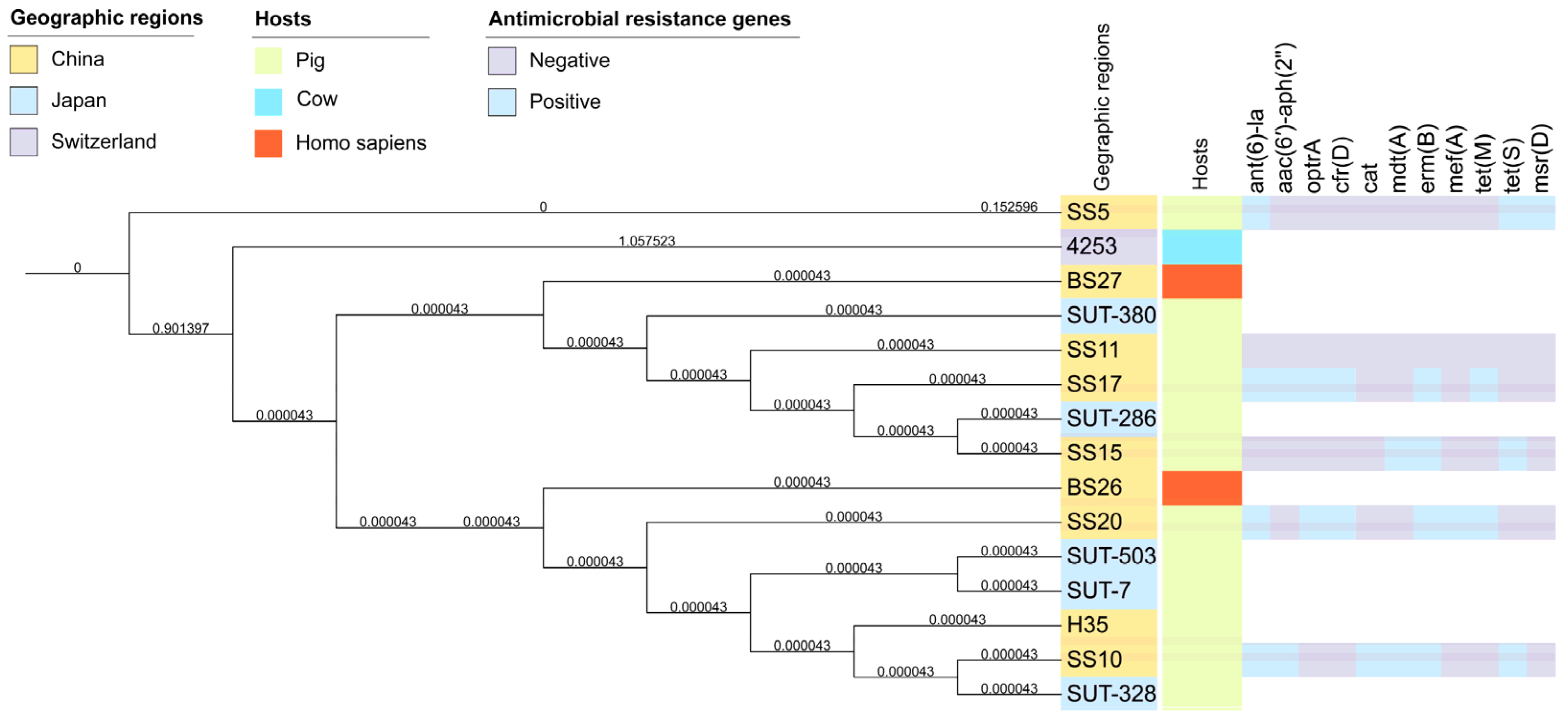
A phylogenic tree was produced to analyze the evolution of S. parasuis. The phylogenic tree demonstrated that all S. parasuis strains were closely related. No SNPs were identified in SS17 and SS20. Therefore, it is clear that S. parasuis has not evolved significantly among different hosts or geographical locations. (Figure 2). These strains carried only a few ARGs, e.g., ant(6)-la and erm(B), and some did not contain any ARGs. At the same time, the distribution of ARGs explained the results of the antibiotic resistance testing.

4. Discussion

A major factor in the prevalence of antibiotic resistance genes in the food chain is the presence of antibiotic-resistant bacteria in food animals, which poses a potential risk to public health and safety [39]. Streptococcus suis is an important zoonotic pathogen that can be transmitted to humans through contact with contaminated animal products or sick animals [40]. The ST25 and ST28 strains were dominant (9/30) among the isolates in this study. ST1 was identified as having a significantly higher virulence than ST25/28, and the remaining strains were ST242, ST27, ST1, ST7, and so on [41]. In addition to being extremely virulent, S. suis is recognized as a reservoir of ARGs, where transposons or integration and binding elements play a crucial role in the propagation of the organism [42]. S. parasuis is a close relative of S. suis, which may be an opportunistic pathogen, and it has been reported that it infects pigs, cattle, and humans [22]. The resistance of S. parasuis should also be taken into consideration. In this investigation, S. suis isolates only carried one to three ARGs, primarily erm(B) and tet(O), but S. parasuis isolates carried ant(6)-la, aac(6′)-aph(2″), mdt(A), lsa(E), cat, tet(S) and other ARGs, which carry far more resistance genes. The detection rate of tet(O/M), ant(6)-la, and erm(B) in S. parasuis is higher, according to NCBI public data [43]. Based on these findings, S. parasuis is a potentially opportunistic zoonotic pathogen that may serve as a reservoir of resistance genes.
The spread of antibiotic resistance is significantly aided by both [44]. Since the optrA gene was discovered in China in 2015, it has been widely spread and detected in bacterial genera with diverse origins, including Enterococcus and Staphylococcus, in many different nations [45]. Platforms carrying the optrA gene can be categorized into three groups: those carrying optrA on chromosomally borne integration and conjugation elements (ICE), such as Tn6674, Tn558, and IS1216E; those on which the optrA gene is located on medium-sized plasmids (30–60 Kb) from the RepA_N, Inc18, and Rep_3 plasmid families to form impB–fexA–optrA; and those carrying optrA–araC on the chromosome or plasmids [46,47]. In this study, it was found that optrA was located in chromosomally borne Tn554 carrying erm(A) and ant(9). The cfr gene was originally discovered on the multi-resistant pSCFS1 (16.5 kb) and pSCFS3 (35.7 kb) plasmids of Staphylococcus sciuri [48,49]. The cfr-carrying segment (IS21-558-cfr) was reported initially, which could form in tandem [50]. Mobile elements play an important role in the horizontal transfer of drug resistance genes. Subsequently, another multidrug-resistant plasmid (50 kb) carrying cfr was found in Staphylococcus, and a circular plasmid with five ORFs (rep-Deltapre/mob-cfr-pre/mob-ermC) was found in its transformants (7057 bp) [51]. Here, we found a new plasmid carrying the cfr gene in S. parasuis, 7550 bp, which is composed of 5 ORFs and rep-IS431mec-IS1202-cfr-GME synthase. Unfortunately, the transformants of cfr and optrA could not be obtained, and their transferability between different genera has yet to be confirmed. Interestingly, SS17 and SS20 remained sensitive to linezolid despite containing two oxazolidinone resistance mechanisms, cfr(D) and optrA. A study showed that cfr(D) did not produce any resistance when overexpressed in E. faecalis and E. faecium, but it was responsible for the phenicol resistance phenotype of Escherichia coli [52]. Meanwhile, the lack of cfr and cfr-like gene-mediated resistance to phenol and oxazolidinone in E. faecalis and E. faecium has been much reported [53,54]. Perhaps this phenomenon does not only occur in a single species. In contrast to the ubiquity of resistance in Gram-negative bacteria, Gram-positive bacteria still seem to preserve a defense against drug resistance genes. Since optrA was first identified, at least 69 variants have been identified; it is inferred that there are 1 to 20 amino acid differences. The different optrA variants may have an effect on the MIC of the oxazolidine of the corresponding isolates that show sensitivity/resistance [7].
In conclusion, this study reported the co-occurrence of optrA and cfr(D) operons in S.parasuis. The presence of Tn554–optrA in Enterococcus spp., Staphylococcus spp., and Streptococcus spp. with various genetic and source backgrounds demonstrated that the Tn554 element plays an important role in the dissemination of optrA. Attention should be paid to the potential risks of plasmid-borne cfr(D) transference from streptococcus to other Gram-positive bacteria. Meanwhile, the existence of additional resistance genes, erm(A) and ant(9), and the use of various types of antibiotics may contribute to the prevalence of optrA and cfr(D). Therefore, it is urgently necessary to continuously monitor the spread of optrA and cfr(D) among Gram-positive bacteria, and monitor the prudent use of antibiotics in food animals.

Author Contributions

Conceptualization, N.H.; methodology, N.H. and J.L.; software, N.H. and Y.P.; validation, N.H., J.L. and P.W., formal analysis, N.H.; resources, N.H. and P.W.; data curation, N.H.; writing—original draft preparation, N.H.; writing—review and editing, Z.Z., W.X. and T.X.; visualization, N.H.; supervision, Z.Z.; project administration, Z.Z.; funding acquisition, Z.Z. All authors have read and agreed to the published version of the manuscript.

Funding

This study was supported by the Local Innovative and Research Teams Project of Guangdong Pearl River Talents Program (2019BT02N054) and the Natural Science Foundation of Guangdong Province of China (2021A1515011159).

Data Availability Statement

The data presented in this study are available in article.

Acknowledgments

This study was supported by the Local Innovative and Research Teams Project of Guangdong Pearl River Talents Program (2019BT02N054) and the Natural Science Foundation of Guangdong Province of China (2021A1515011159).

Conflicts of Interest

The authors declare no conflict of interest.

References

  1. Almeida, L.M.; Lebreton, F.; Gaca, A.; Bispo, P.M.; Saavedra, J.T.; Calumby, R.N.; Grillo, L.M.; Nascimento, T.G.; Filsner, P.H.; Moreno, A.M.; et al. Transferable Resistance Gene in Enterococcus faecalis from Swine in Brazil. Antimicrob. Agents Chemother. 2020, 64, e00142-20. [Google Scholar] [CrossRef] [PubMed]
  2. Leach, K.L.; Brickner, S.J.; Noe, M.C.; Miller, P.F. Linezolid, the first oxazolidinone antibacterial agent. Ann. N. Y. Acad. Sci. 2011, 1222, 49–54. [Google Scholar] [CrossRef] [PubMed]
  3. Klevens, R.M.; Morrison, M.A.; Nadle, J.; Petit, S.; Gershman, K.; Ray, S.; Harrison, L.H.; Lynfield, R.; Dumyati, G.; Townes, J.M.; et al. Invasive methicillin-resistant Staphylococcus aureus infections in the United States. JAMA 2007, 298, 1763–1771. [Google Scholar] [CrossRef]
  4. Hoyert, D.L.; Xu, J. Deaths: Preliminary data for 2011. Natl. Vital. Stat. Rep. 2012, 61, 1–65. [Google Scholar] [PubMed]
  5. Vehreschild, M.J.G.T.; Haverkamp, M.; Biehl, L.M.; Lemmen, S.; Fätkenheuer, G. Vancomycin-resistant enterococci (VRE): A reason to isolate? Infection 2019, 47, 7–11. [Google Scholar] [CrossRef]
  6. Locke, J.B.; Hilgers, M.; Shaw, K.J. Novel ribosomal mutations in Staphylococcus aureus strains identified through selection with the oxazolidinones linezolid and torezolid (TR-700). Antimicrob. Agents Chemother. 2009, 53, 5265–5274. [Google Scholar] [CrossRef] [PubMed]
  7. Schwarz, S.; Zhang, W.; Du, X.-D.; Krüger, H.; Feßler, A.T.; Ma, S.; Zhu, Y.; Wu, C.; Shen, J.; Wang, Y. Mobile Oxazolidinone Resistance Genes in Gram-Positive and Gram-Negative Bacteria. Clin. Microbiol. Rev. 2021, 34, e0018820. [Google Scholar] [CrossRef]
  8. Jung, Y.-H.; Cha, M.-H.; Woo, G.-J.; Chi, Y.-M. Characterization of oxazolidinone and phenicol resistance genes in non-clinical enterococcal isolates from Korea. J. Glob. Antimicrob. Resist. 2021, 24, 363–369. [Google Scholar] [CrossRef]
  9. Meka, V.G.; Gold, H.S. Antimicrobial resistance to linezolid. Clin. Infect. Dis. 2004, 39, 1010–1015. [Google Scholar] [CrossRef]
  10. Aarestrup, F.M. The livestock reservoir for antimicrobial resistance: A personal view on changing patterns of risks, effects of interventions and the way forward. Philos. Trans. R. Soc. Lond. B Biol. Sci. 2015, 370, 20140085. [Google Scholar] [CrossRef]
  11. Liu, B.G.; Yuan, X.L.; He, D.D.; Hu, G.Z.; Miao, M.S.; Xu, E.P. Research progress on the oxazolidinone drug linezolid resistance. Eur. Rev. Med. Pharm. Sci. 2020, 24, 9274–9281. [Google Scholar] [CrossRef]
  12. Deshpande, L.M.; Castanheira, M.; Flamm, R.K.; Mendes, R.E. Evolving oxazolidinone resistance mechanisms in a worldwide collection of enterococcal clinical isolates: Results from the SENTRY Antimicrobial Surveillance Program. J. Antimicrob. Chemother. 2018, 73, 2314–2322. [Google Scholar] [CrossRef] [PubMed]
  13. Wang, Y.; Gong, S.; Dong, X.; Li, J.; Grenier, D.; Yi, L. Mixed Biofilm of and Impacts Antibiotic Susceptibility and Modulates Virulence Factor Gene Expression. Front. Microbiol. 2020, 11, 507. [Google Scholar] [CrossRef] [PubMed]
  14. Sharkey, L.K.R.; O’Neill, A.J. Antibiotic Resistance ABC-F Proteins: Bringing Target Protection into the Limelight. ACS Infect. Dis. 2018, 4, 239–246. [Google Scholar] [CrossRef]
  15. Locke, J.B.; Finn, J.; Hilgers, M.; Morales, G.; Rahawi, S.; Kedar, G.C.; Picazo, J.J.; Im, W.; Shaw, K.J.; Stein, J.L. Structure-activity relationships of diverse oxazolidinones for linezolid-resistant Staphylococcus aureus strains possessing the cfr methyltransferase gene or ribosomal mutations. Antimicrob. Agents Chemother. 2010, 54, 5337–5343. [Google Scholar] [CrossRef]
  16. Stojkovic, V.; Ulate, M.F.; Hidalgo-Villeda, F.; Aguilar, E.; Monge-Cascante, C.; Pizarro-Guajardo, M.; Tsai, K.; Tzoc, E.; Camorlinga, M.; Paredes-Sabja, D.; et al. cfr(B), cfr(C), and a New cfr-Like Gene, cfr(E), in Clostridium difficile Strains Recovered across Latin America. Antimicrob. Agents Chemother. 2019, 64, e01074-19. [Google Scholar] [CrossRef]
  17. Huang, J.; Sun, J.; Wu, Y.; Chen, L.; Duan, D.; Lv, X.; Wang, L. Identification and pathogenicity of an XDR Streptococcus suis isolate that harbours the phenicol-oxazolidinone resistance genes optrA and cfr, and the bacitracin resistance locus bcrABDR. Int. J. Antimicrob. Agents 2019, 54, 43–48. [Google Scholar] [CrossRef]
  18. Nomoto, R.; Maruyama, F.; Ishida, S.; Tohya, M.; Sekizaki, T.; Osawa, R. Reappraisal of the taxonomy of Streptococcus suis serotypes 20, 22 and 26: Streptococcus parasuis sp. nov. Int. J. Syst. Evol. Microbiol. 2015, 65, 438–443. [Google Scholar] [CrossRef]
  19. Wang, J.; Yi, X.; Liang, P.; Tao, Y.; Wang, Y.; Jin, D.; Luo, B.; Yang, J.; Zheng, H. Investigation of the Genomic and Pathogenic Features of the Potentially Zoonotic. Pathogens 2021, 10, 834. [Google Scholar] [CrossRef]
  20. Nomoto, R.; Ishida-Kuroki, K.; Nakagawa, I.; Sekizaki, T. Complete Genome Sequences of Four Streptococcus parasuis Strains Obtained from Saliva of Domestic Pigs in Japan. Microbiol. Resour. Announc. 2022, 11, e0124521. [Google Scholar] [CrossRef]
  21. Zhu, Y.; Yang, Q.; Schwarz, S.; Yang, W.; Xu, Q.; Wang, L.; Liu, S.; Zhang, W. Identification of a Streptococcus parasuis isolate co-harbouring the oxazolidinone resistance genes cfr(D) and optrA. J. Antimicrob. Chemother. 2021, 76, 3059–3061. [Google Scholar] [CrossRef]
  22. Yamada, R.; Tien, L.H.T.; Arai, S.; Tohya, M.; Ishida-Kuroki, K.; Nomoto, R.; Kim, H.; Suzuki, E.; Osawa, R.; Watanabe, T.; et al. Development of PCR for identifying Streptococcus parasuis, a close relative of Streptococcus suis. J. Vet. Med. Sci. 2018, 80, 1101–1107. [Google Scholar] [CrossRef] [PubMed]
  23. Stevens, M.J.A.; Cernela, N.; Corti, S.; Stephan, R. Draft Genome Sequence of Streptococcus parasuis 4253, the First Available for the Species. Microbiol. Resour. Announc. 2019, 8, e00203-19. [Google Scholar] [CrossRef] [PubMed]
  24. Clinical and Laboratory Standards Institute. Performance Standards for an Timicrobial Susceptibility Testing; Twenty-Sixth Informational Supplement M100-S26; CLSI: Wayne, PA, USA, 2016. [Google Scholar]
  25. Clinical and laboratory Standards Institute. Performance Standards for Antimi Crobial Disk and Diffusion Susceptibility Tests for Bacteria Isolated from Animals; Second Informational Supplement VET01-S2; CLSI: Wayne, PA, USA, 2013. [Google Scholar]
  26. Wang, Y.; Lv, Y.; Cai, J.; Schwarz, S.; Cui, L.; Hu, Z.; Zhang, R.; Li, J.; Zhao, Q.; He, T.; et al. A novel gene, optrA, that confers transferable resistance to oxazolidinones and phenicols and its presence in Enterococcus faecalis and Enterococcus faecium of human and animal origin. J. Antimicrob. Chemother. 2015, 70, 2182–2190. [Google Scholar] [CrossRef] [PubMed]
  27. Wang, Y.; Zhang, W.; Wang, J.; Wu, C.; Shen, Z.; Fu, X.; Yan, Y.; Zhang, Q.; Schwarz, S.; Shen, J. Distribution of the multidrug resistance gene cfr in Staphylococcus species isolates from swine farms in China. Antimicrob. Agents Chemother. 2012, 56, 1485–1490. [Google Scholar] [CrossRef] [PubMed]
  28. Chin, C.-S.; Alexander, D.H.; Marks, P.; Klammer, A.A.; Drake, J.; Heiner, C.; Clum, A.; Copeland, A.; Huddleston, J.; Eichler, E.E.; et al. Nonhybrid, finished microbial genome assemblies from long-read SMRT sequencing data. Nat. Methods 2013, 10, 563–569. [Google Scholar] [CrossRef]
  29. Bitar, I.; Moussa, J.; Abboud, E.; Hrabak, J.; Tokajian, S. Integration of two pKPX-2-derived antibiotic resistance islands in the genome of an ESBL-producing Klebsiella pneumoniae ST3483 from Lebanon. J. Glob. Antimicrob. Resist. 2019, 18, 257–259. [Google Scholar] [CrossRef]
  30. Zankari, E.; Hasman, H.; Cosentino, S.; Vestergaard, M.; Rasmussen, S.; Lund, O.; Aarestrup, F.M.; Larsen, M.V. Identification of acquired antimicrobial resistance genes. J. Antimicrob. Chemother. 2012, 67, 2640–2644. [Google Scholar] [CrossRef]
  31. Stamatakis, A. RaxML version 8: A tool for phylogenetic analysis and post-analysis of large phylogenies. Bioinformatics 2014, 30, 1312–1313. [Google Scholar] [CrossRef]
  32. Letunic, I.; Bork, P. Interactive Tree Of Life (iTOL) v5: An online tool for phylogenetic tree display and annotation. Nucleic Acids Res. 2021, 49, W293–W296. [Google Scholar] [CrossRef]
  33. Yu, R.; Zhang, Y.; Xu, Y.; Schwarz, S.; Li, X.-S.; Shang, Y.-H.; Du, X.-D. Emergence of a (M) Variant Conferring Resistance to Tigecycline in. Front. Vet. Sci. 2021, 8, 709327. [Google Scholar] [CrossRef] [PubMed]
  34. Li, D.; Li, X.Y.; Schwarz, S.; Yang, M.; Zhang, S.M.; Hao, W.; Du, X.D. Tn6674 Is a Novel Enterococcal optrA-Carrying Multiresistance Transposon of the Tn554 Family. Antimicrob. Agents Chemother. 2019, 63, e00142-20. [Google Scholar] [CrossRef]
  35. Yao, T.G.; Li, B.Y.; Luan, R.D.; Wang, H.N.; Lei, C.W. Whole genome sequence of Enterococcus gallinarum EG81, a porcine strain harbouring the oxazolidinone-phenicol resistance gene optrA with chromosomal and plasmid location. J. Glob. Antimicrob. Resist. 2020, 22, 598–600. [Google Scholar] [CrossRef]
  36. He, Y.Z.; Li, X.P.; Miao, Y.Y.; Lin, J.; Sun, R.Y.; Wang, X.P.; Guo, Y.Y.; Liao, X.P.; Liu, Y.H.; Feng, Y.; et al. The ISApl1 2 Dimer Circular Intermediate Participates in mcr-1 Transposition. Front. Microbiol. 2019, 10, 15. [Google Scholar] [CrossRef] [PubMed]
  37. Lyras, D.; Rood, J.I. Transposition of Tn4451 and Tn4453 involves a circular intermediate that forms a promoter for the large resolvase, TnpX. Mol. Microbiol. 2000, 38, 588–601. [Google Scholar] [CrossRef]
  38. Manganelli, R.; Romano, L.; Ricci, S.; Zazzi, M.; Pozzi, G. Dosage of Tn916 circular intermediates in Enterococcus faecalis. Plasmid 1995, 34, 48–57. [Google Scholar] [CrossRef]
  39. García-Sánchez, L.; Melero, B.; Diez, A.M.; Jaime, I.; Rovira, J. Characterization of Campylobacter species in Spanish retail from different fresh chicken products and their antimicrobial resistance. Food Microbiol. 2018, 76, 457–465. [Google Scholar] [CrossRef]
  40. Susilawathi, N.M.; Tarini, N.M.A.; Fatmawati, N.N.D.; Mayura, P.I.B.; Suryapraba, A.A.A.; Subrata, M.; Sudewi, A.A.R.; Mahardika, G.N. Streptococcus suis-Associated Meningitis, Bali, Indonesia, 2014–2017. Emerg. Infect. Dis. 2019, 25, 2235–2242. [Google Scholar] [CrossRef] [PubMed]
  41. Segura, M.; Fittipaldi, N.; Calzas, C.; Gottschalk, M. Critical Streptococcus suis Virulence Factors: Are They All Really Critical? Trends Microbiol. 2017, 25, 585–599. [Google Scholar] [CrossRef]
  42. Haenni, M.; Lupo, A.; Madec, J.-Y. Antimicrobial Resistance in spp. Microbiol. Spectr. 2018, 6, 585–599. [Google Scholar] [CrossRef] [PubMed]
  43. Guo, G.; Wang, Z.; Li, Q.; Yu, Y.; Li, Y.; Tan, Z.; Zhang, W. Genomic characterization of Streptococcus parasuis, a close relative of Streptococcus suis and also a potential opportunistic zoonotic pathogen. BMC Genom. 2022, 23, 469. [Google Scholar] [CrossRef]
  44. Mishra, S.; Klümper, U.; Voolaid, V.; Berendonk, T.U.; Kneis, D. Simultaneous estimation of parameters governing the vertical and horizontal transfer of antibiotic resistance genes. Sci. Total Environ. 2021, 798, 149174. [Google Scholar] [CrossRef]
  45. Biggel, M.; Nüesch-Inderbinen, M.; Jans, C.; Stevens, M.J.A.; Stephan, R. Genetic Context of and in Florfenicol-Resistant Enterococci Isolated from Flowing Surface Water in Switzerland. Antimicrob. Agents Chemother. 2021, 65, e0108321. [Google Scholar] [CrossRef] [PubMed]
  46. Freitas, A.R.; Tedim, A.P.; Novais, C.; Lanza, V.F.; Peixe, L. Comparative genomics of global optrA-carrying uncovers a common chromosomal hotspot for acquisition within a diversity of core and accessory genomes. Microb. Genom. 2020, 6, AAC0108321. [Google Scholar] [CrossRef] [PubMed]
  47. Fan, R.; Li, D.; Feßler, A.T.; Wu, C.; Schwarz, S.; Wang, Y. Distribution of optrA and cfr in florfenicol-resistant Staphylococcus sciuri of pig origin. Vet. Microbiol. 2017, 210, 43–48. [Google Scholar] [CrossRef] [PubMed]
  48. Schwarz, S.; Werckenthin, C.; Kehrenberg, C. Identification of a plasmid-borne chloramphenicol-florfenicol resistance gene in Staphylococcus sciuri. Antimicrob. Agents Chemother. 2000, 44, 2530–2533. [Google Scholar] [CrossRef]
  49. Kehrenberg, C.; Ojo, K.K.; Schwarz, S. Nucleotide sequence and organization of the multiresistance plasmid pSCFS1 from Staphylococcus sciuri. J. Antimicrob. Chemother. 2004, 54, 936–939. [Google Scholar] [CrossRef]
  50. Kehrenberg, C.; Aarestrup, F.M.; Schwarz, S. IS21-558 insertion sequences are involved in the mobility of the multiresistance gene cfr. Antimicrob. Agents Chemother. 2007, 51, 483–487. [Google Scholar] [CrossRef]
  51. Li, S.-M.; Zhou, Y.-F.; Li, L.; Fang, L.-X.; Duan, J.-H.; Liu, F.-R.; Liang, H.-Q.; Wu, Y.-T.; Gu, W.-Q.; Liao, X.-P.; et al. Characterization of the Multi-Drug Resistance Gene in Methicillin-Resistant (MRSA) Strains Isolated from Animals and Humans in China. Front. Microbiol. 2018, 9, 2925. [Google Scholar] [CrossRef]
  52. Guerin, F.; Sassi, M.; Dejoies, L.; Zouari, A.; Schutz, S.; Potrel, S.; Auzou, M.; Collet, A.; Lecointe, D.; Auger, G.; et al. Molecular and functional analysis of the novel cfr(D) linezolid resistance gene identified in Enterococcus faecium. J. Antimicrob. Chemother. 2020, 75, 1699–1703. [Google Scholar] [CrossRef]
  53. Brenciani, A.; Morroni, G.; Vincenzi, C.; Manso, E.; Mingoia, M.; Giovanetti, E.; Varaldo, P.E. Detection in Italy of two clinical Enterococcus faecium isolates carrying both the oxazolidinone and phenicol resistance gene optrA and a silent multiresistance gene cfr. J. Antimicrob. Chemother. 2016, 71, 1118–1119. [Google Scholar] [CrossRef] [PubMed]
  54. Fang, L.-X.; Duan, J.-H.; Chen, M.-Y.; Deng, H.; Liang, H.-Q.; Xiong, Y.Q.; Sun, J.; Liu, Y.-H.; Liao, X.-P. Prevalence of cfr in Enterococcus faecalis strains isolated from swine farms in China: Predominated cfr-carrying pCPPF5-like plasmids conferring “non-linezolid resistance” phenotype. Infect. Genet. Evol. 2018, 62, 188–192. [Google Scholar] [CrossRef] [PubMed]
Figure 1. (a,b) are the structure and organization of the plasmids of pSS17 and pSS20-1, respectively. The circles (from the outside to inside) indicate the predicated coding sequences, GC-skew [(G + C)/(G + C)], GC content and scale in bp. The coding sequences with different functions are shown in different colors. Arrows indicate the direction of transcription of the genes. (c) Genetic environment of optrA in the chromosomal DNA of S.parasuis SS17 and SS20 compared to other plasmids and genomes. Resistance genes are indicated with pink arrows. Transposases are shown with blue arrows labeled by their name. Other elements are highlighted with green arrows. Shared regions with >99% identity are denoted by nattier blue shading.
Antibiotics 12 00825 g001
Figure 2. The phylogenetic analysis of S. parasuis isolates. The geographic regions of the sources of these isolates and the hosts are displayed in different colors. The antimicrobial resistance genes are indicated using a heatmap; light blue is positive, and purple is negative.
Antibiotics 12 00825 g002
Table 1. Minimum inhibitory concentrations of 13 antimicrobial agents and ARGs among 2 Streptococcus parasuis isolates carrying both optrA and a cfr(D).
IsolateSpeciesSourceMLSTMIC (mg/L)
ERYCLIMEMCEFLZDVANTETPENAMPENRSXTFFCAMOResistance Genes
SS17S. parasuisnasal swabNA32>32≤0.030.510.126421432640.5ant(6)-la, aac(6′)-aph(2″), optrA, erm(B), tet(M), cfr(D)
SS20S. parasuisnasal swabNA32>32≤0.030.510.126421464>641erm(B), ant(6)-la, optrA, mef(A), tet(M), cfr(D)
MLST, multi-locus sequence type; MIC, minimum inhibitory concentration; ERY, erythromycin; CLI, clindamycin; MEM, meropenem; CEF, ceftiofur; LZD, linezolid; VAN, vancomycin; TET, tetracycline; PEN, penicillin; AMP, ampicillin; ENR, enrofloxacin; SXT, cotrimoxazole; FFC, florfenicol; AMO, amoxicillin; NA, not available.
Disclaimer/Publisher’s Note: The statements, opinions and data contained in all publications are solely those of the individual author(s) and contributor(s) and not of MDPI and/or the editor(s). MDPI and/or the editor(s) disclaim responsibility for any injury to people or property resulting from any ideas, methods, instructions or products referred to in the content.