Article

Effect of Quorum Sensing Molecule Farnesol on Mixed Biofilms of Candida albicans and Staphylococcus aureus

1
Department of Microbiology and Virology, Faculty of Natural Sciences, Comenius University in Bratislava, Ilkovičova 6, 842 15 Bratislava, Slovakia
2
Department of Environmental Health, Research and Development Unit, National Institute of Health Dr. Ricardo Jorge (INSA), Av. Padre Cruz, 1649-016 Lisboa, Portugal
3
Biofilmcenter, Institute of Microbiology, Infectious Diseases and Immunology, Charité—Universitätsmedizin Berlin, Hindenburgdamm 30, 12203 Berlin, Germany
4
MoKi Analytics GmbH, Charité-Universitätsmedizin Berlin, Hindenburdamm 30, 12203 Berlin, Germany
5
Moter Diagnostics, Marienplatz 9, 12207 Berlin, Germany
*
Author to whom correspondence should be addressed.
Antibiotics 2023, 12(3), 441; https://doi.org/10.3390/antibiotics12030441
Submission received: 1 February 2023 / Revised: 19 February 2023 / Accepted: 21 February 2023 / Published: 23 February 2023
(This article belongs to the Special Issue Microbial Biofilms, Antimicrobials, and Virulence Determinants)

Abstract

:
The natural bioactive molecule farnesol (FAR) is widely studied mainly for its antibiofilm and antimicrobial properties. In addition, it increases the effectiveness of some antimicrobial substances, which makes it interesting for the development of combined therapy. In the present work, the effect of FAR either alone or in combination with oxacillin (OXA) on mixed biofilms formed by clinically relevant pathogens, Candida albicans and Staphylococcus aureus, was studied. S. aureus isolates used for biofilm formation originated from blood cultures and central venous catheters (CVC) were characterized in terms of antimicrobial resistance. The minimal biofilm inhibitory concentration (MBIC50) for FAR of 48 h mixed biofilms formed by the C. albicans and methicillin-sensitive S. aureus (MSSA) was determined to be 125 μM, and for the mixed biofilms with methicillin-resistant S. aureus (MRSA) was determined to be 250 μM. Treatment of mixed biofilms with OXA (2 mg/mL) showed ≤4% inhibition; however, the combination of OXA (2 mg/mL) and FAR (300 μM) resulted in 80% inhibition of biofilms. In addition, planktonic cells of S. aureus exhibited an increased susceptibility to OXA, cefoxitin and kanamycin in the presence of FAR (150 and 300 μM). Scanning electron microscopy (SEM) micrographs confirmed patchy biofilm and lack of candidal hyphae in the samples treated with FAR and FAR/OXA in comparison to control and mixed biofilms treated only with OXA. Intriguingly, in a pilot experiment using fluorescence in situ hybridization (FISH), considerable differences in activity (as indicated by ribosome content) of staphylococcal cells were detected. While the activity rate of the staphylococci in mixed biofilms treated with FAR was high, no FISH-positive signal for staphylococcal cells was found in the biofilm treated with FAR/OXA.

1. Introduction

Candida albicans represents one of the clinically most important opportunistic pathogens, usually surviving in mixed biofilms. Most often, Candida coexists with a grampositive bacterium Staphylococcus aureus [1]. Among resistant strains, methicillin-resistant S. aureus (MRSA) is highly linked to biofilm production, mostly in bloodstream infections (BSI) [2]. Infections caused by C. albicans and S. aureus biofilms are usually associated with usage of temporary indwelling medical devices, particularly central venous catheters (CVC), urinary catheters or the application of cardiovascular devices [3,4]. These infections cause high morbidity and mortality, especially in hospitals in intensive care units [5,6]. Several advantages arise from the inter-kingdom relationship that helps to protect the microorganisms from external factors and host immune response [7]. Mixed biofilms formed by Candida and Staphylococcus are significantly more tolerant to antimicrobial agents than mono-species biofilms. Inhibition of biofilms requires 100 to 1000 folds higher concentrations of antimicrobial agents than planktonic microorganisms and usually requires combined therapy [8,9,10,11,12]. Currently, several systemic and local therapeutic schemes, such as lock solutions, are available for the treatment of infections caused by biofilms [13,14]. Many investigators focus on the development of new antibiofilm and biocompatible materials with incorporated antimicrobial agents, peptides or nanomaterials [15,16,17,18]. Nowadays, there is a strong trend towards the development of hybrid systems of known antimicrobial compounds with more than one mode of action [19].
Quorum sensing (QS) molecules seem to be ideal candidates for developing a combination therapy because of their potential to control pathogens [20,21]. Bacteria and fungi, even higher organisms, naturally produce small diffusible signaling molecules for intra- and inter-species communication. In bacteria, they are mainly involved in the coordination of population density, which is associated with gene regulation related to the production of different virulence factors, such as enzymes or toxins, biofilm formation, the production of antimicrobials or the mediation of conjugation [22]. In fungi, QS molecules mainly affect cell morphology, population growth, virulence control, reproduction, secondary metabolites and pigment production [23]. Tetraprenoid farnesol (FAR) is synthesized as a byproduct of the ergosterol pathway of the polymorphic fungus C. albicans. FAR plays a central role in both fungal cells and biofilm physiology in a concentration-dependent manner [24,25]. Concentrations of FAR higher than 100 µM block the morphological shift from the yeast to the hyphal form. This phenomenon is important in mixed biofilms of C. albicans-S. aureus because the bacteria preferentially bind into the hyphal filaments through candidal adhesin Als3p, which is not expressed in the yeast form [1]. The antimicrobial and antibiofilm effect of FAR in high concentrations was confirmed also in bacteria [26,27]. In addition, the synergistic effect of FAR with antifungal agents [28,29] and antibiotics [30,31] has been studied. On the other hand, Kong et al. (2017) found that low concentrations of FAR (40–50 µM), considered to be physiological in mixed biofilms of C. albicans-S. aureus, resulted in enhanced tolerance of S. aureus to antimicrobials [32].
We assume that FAR, as a naturally produced bioactive molecule, could be a promising adjuvant to enhance the effect of antimicrobial agents or for further development of drug delivery materials to prevent biofilm and microbial infection. The aim of the present study is to investigate the inhibiting effect of the combination of FAR and OXA on mixed biofilms formed by C. albicans and S. aureus with different antibiotic susceptibility profiles.

2. Results

2.1. Characterization of Strains in Terms of Identification, Resistance and Biofilm Formation

The yeast C. albicans SC 5314 [33] and the bacterium S. aureus CCM 3953–ATCC 25923 (Czech Collection of Microorganisms, Brno, Czech Republic) were used as standard strains in the experiments. Three clinical isolates of S. aureus were used: one from a blood culture and two from CVC. The identity of the microorganisms was verified by the growth on specific cultivating media (CHROM agar Candida, Mannitol Salt Agar). In all of S. aureus isolates, the femA gene was confirmed [34]. In two of the S. aureus isolates, namely DHN 21 528 (further referred to as MRSA1) and L18 (referred to as MRSA2), the methicillin-resistant genotype was confirmed. Standard strain S. aureus CCM 3953 (referred to as MSSA1) and the DRA 13 541 isolate (referred to as MSSA2) were evaluated as methicillin-sensitive strains. We observed slower growth of MRSA isolates compared to others (results are summarized in Supplementary Materials Figures S1–S3).
The MRSA isolates were resistant to tested penicillins, cephalosporins, ertapenem, fluoroquinolones, tobramycin, erythromycin and clindamycin. All isolates were susceptible to vancomycin, gentamicin, tetracycline and quinupristin-dalphopristin. The standard strain S. aureus MSSA1, was susceptible to all tested antibiotics (Table 1).
All clinical isolates were beta-lactamase producers. Resistance to fluoroquinolones can be associated with the expression of several types of efflux pumps. The norA, norB and norC genes were detected in all of the isolates, but their expression rates were not studied. From the group of aminoglycoside modification enzymes, the aminoglycozide O-nucleozidtransferase (ANTs) was confirmed in both MRSA isolates. Macrolide and lincosamide resistance can be associated with ribosomal binding site modification by methylation or mutation in the 23S rRNA gene encoded by erythromycin ribosome methylases (erm) genes. This phenomenon leads to cross-resistance to these antibiotics. In this study, only ermA was confirmed in both MRSA isolates. Efflux pumps encoded by the msrA and msrB genes responsible for pumping macrolides out of the cell were not found in the studied isolates (Table 2; Figures S4 and S5). The standard strain C. albicans has been previously characterized in another study by Černáková et al. (2019) [35] and Kucharíková et al. (2011) [36] in terms of antifungal susceptibilities. The strain was susceptible to all tested antifungals used in medical practice, including fluconazole, caspofungin and anidulafungin.
All microbial isolates used in this study showed a moderate ability to form biofilm, when tested as a single species. A slightly weaker biofilm was produced by the two MRSA isolates. This phenomenon was more clearly visible when studying mixed biofilms of C. albicans-MRSA (Table 3).

2.2. Effect of FAR and the Combination of FAR with Antibiotics to S. aureus Isolates

The effectiveness of FAR was evaluated as MIC50, which is the concentration inhibiting 50% of cell growth. At first, planktonic cells of S. aureus were tested using the broth microdilution method. The MIC50 for the standard strain MSSA1 was 125 µM while for the other isolates, it was one fold higher at 250 µM (Figure 1). The concentration that resulted in 50% inhibition for C. albicans was established on 1 mM in a previous study by Černáková et al. (2019) [35].
Two concentrations of FAR (150 and 300 µM) were chosen to monitor its possible potentiating effect in combination with antibiotics on S. aureus isolates. Four antibiotics were tested using the E-test: two from the group of beta-lactams, one from the group of aminoglycosides and one from the group of fluoroquinolones. According to previous screening by disc diffusion method, the tested antibiotics were effective against MSSA, while MRSA was resistant to them. The experiment showed that both concentrations of FAR enhanced the effect of oxacillin, cefoxitin and kanamycin; however, the effect was more noticeable in MRSA isolates. Of note, MRSA2 in the presence of FAR (300 µM) reached the MIC for cefoxitin (4 mg/mL). On the other hand, no enhancing effect in the presence of FAR was observed for ciprofloxacin in any of the tested isolates (Table 4). The mode of action of antibiotics can be one of the factors that affects the synergistic effect of FAR with the antibiotics. As FAR acts on the level of cell wall, the effect is most evident using beta-lactams.

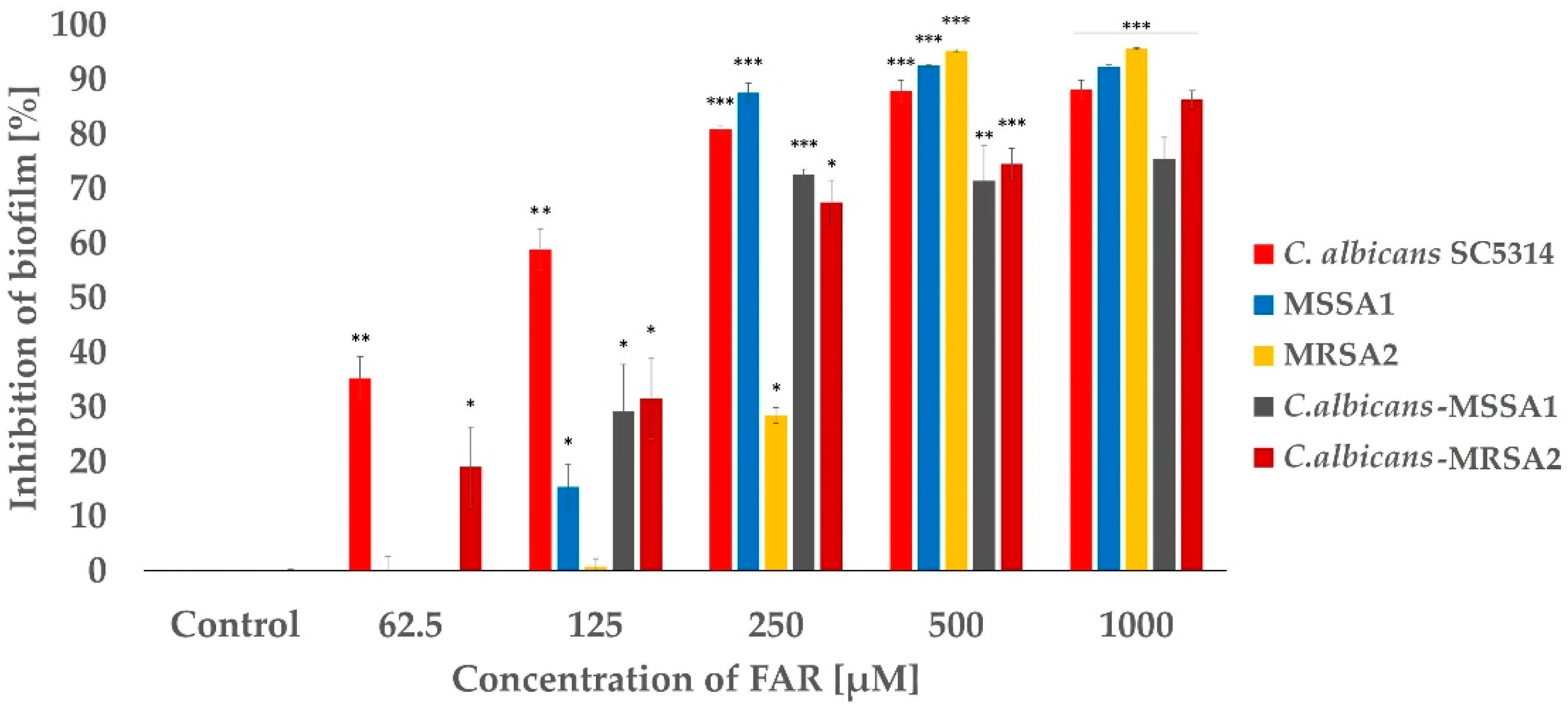
2.3. Study of the Effect of FAR and the Combination of FAR and OXA on Mono-Species and Mixed Biofilms Formed by Candida albicans-MSSA or MRSA

Since resistant and susceptible isolates showed similar characteristics, we selected one representative of each group for further studies. First, the 50% of biofilm inhibition, the MBIC50 of FAR, was determined for mono-species and mixed biofilms of C. albicans-MSSA1 and C. albicans-MRSA2. The MBIC50 of FAR for mixed biofilms of C. albicans-MSSA1 and C. albicans-MRSA2 was established as 125 and 250 µM, respectively (Figure 2). Moreover, inhibition did not differ much at higher concentrations (≥250 µM) in mixed biofilms. Comparing single-species and mixed biofilms, the single-species biofilms were more susceptible to FAR than their mixed counterparts. The only exception was the biofilm of MRSA2, the MBIC50 value of which was significantly higher, between concentrations of 250 and 500 µM. Thus, the biofilm formed by MSSA1 was more sensitive than those of the MRSA2 strain. A mono-species C. albicans biofilm achieved the MBIC50 at a concentration of 125 µM (Figure 2).
Based on the previous experiment, two concentrations of FAR, 150 and 300 µM, were selected. A concentration of 150 µM was closest to the MBIC50 for mixed biofilms of C. albicans-S. aureus, and the second concentration was chosen to monitor a possible synergistic effect of FAR. The concentration of OXA, 2 mg/mL, represents the MIC for S. aureus according to the EUCAST (version 12.0, 2022) [37]. Figure 3 shows the efficacy of FAR and OXA separately and in combination on mixed biofilms, as assessed by the XTT assay. Treatment of mixed biofilms with OXA showed an inhibition of 4% when C. albicans was mixed with MSSA and 2% in C. albicans-MRSA biofilms. The combination of OXA/FAR (300 μM) represented 80% inhibition of mixed biofilms. However, the expected synergistic effect, particularly in C. albicans-MSSA biofilms, was not as significant compared to the biofilm samples treated with FAR alone. Although FAR and the combination of FAR (150 µM)/OXA showed lower efficacy on C. albicans-MRSA2 biofilm compared to C. albicans-MSSA1 biofilm, the inhibition rate reached the same value of 80% when the concentration of FAR was increased to 300 µM. In single biofilms, the differences in inhibition after FAR treatment compared to FAR/OXA treatment were significantly higher than those observed under the same conditions for mixed biofilms. Moreover, the inhibition rate of OXA (2 mg/L) to the single biofilm formed by MSSA1 isolate showed higher inhibition (≥50%) than when mixed with C. albicans.

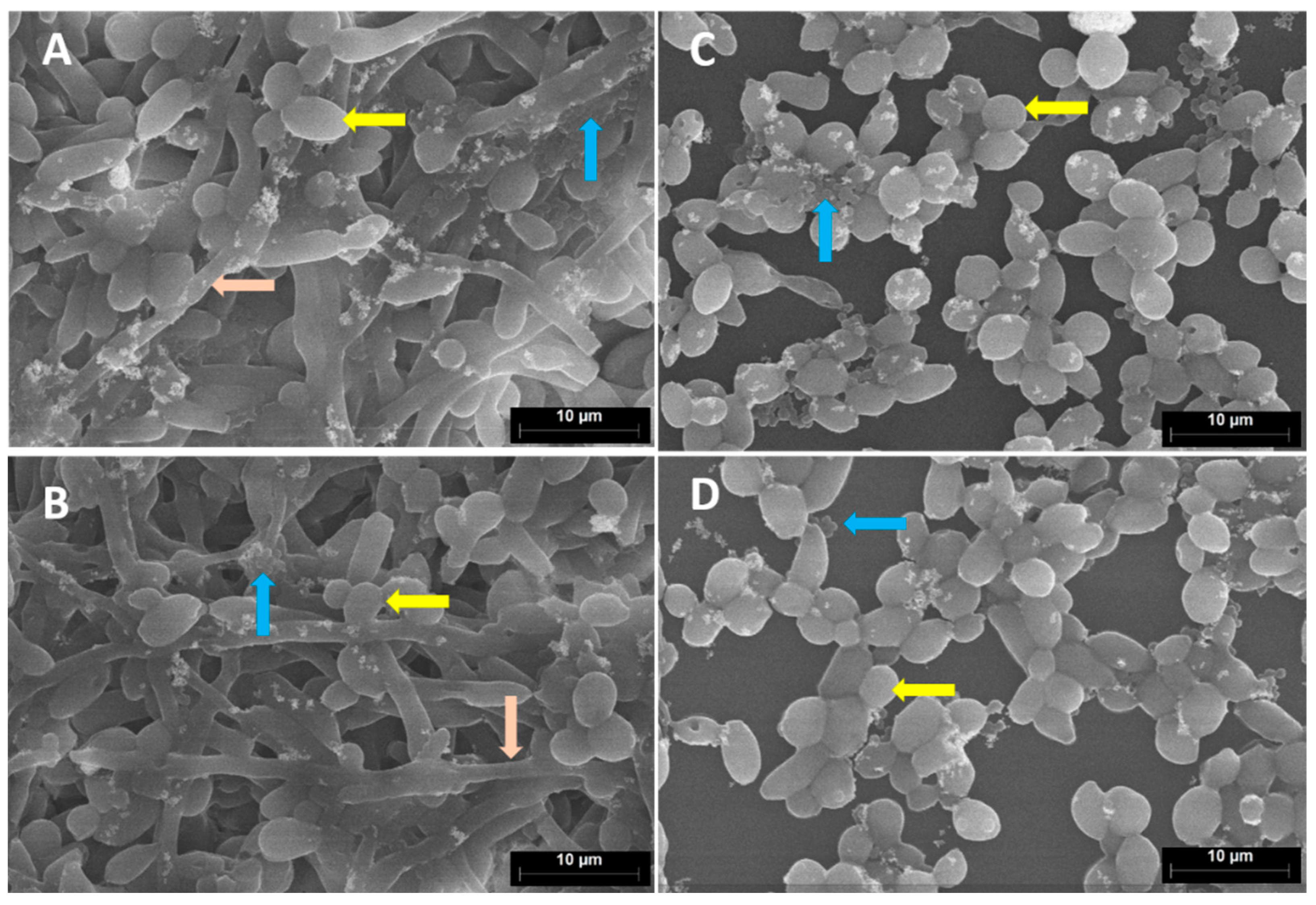
2.4. Microscopic Evaluation of the Effect of FAR and the Combination of FAR with OXA on Mixed Biofilms

Since the influence of FAR (300 µM) and OXA (2 mg/mL) was similar in both studied biofilms (C. albicans-MSSA1 and MRSA2), we decided to choose the C. albicans-MRSA2 biofilms for microscopic analysis. SEM was used to characterize the biofilms with regard to biofilm mass, distribution of bacterial and fungal cell forms and the impact of the studied antimicrobial agents on biofilms. SEM micrographs (Figure 4) confirmed damaged biofilm after using FAR (300 µM) and the combination of FAR (300 µM)/OXA (2 mg/mL) in mixed biofilms. Significantly less candidal hyphae were observed in the images of biofilms treated with FAR (Figure 4C,D) in comparison to the control (Figure 4A) and the biofilm treated only with OXA (2 mg/mL) (Figure 4B).
In order to better elucidate the metabolic activity of fungal and bacterial cells based on ribosome content upon treatment with FAR or FAR/OXA, FISH was applied in a pilot study. Representative epifluorescence images are shown in Figure 5. Hybridization was carried out with specific probes for Candida sp. (CAND10) [38] and Staphylococcus sp. (STAPHY) [39], in combination with the panbacterial probe EUB338 [40]. In the untreated control sample, thin biofilms and patchy groups of microorganisms were detected. All microbial cells (Candida and staphylococci) within the biofilm were FISH-positive and therefore presumably metabolically active. A similar result was obtained after 24 h of incubation, however, the biofilm was thicker (upper row). The sample treated with FAR showed thinner biofilms and smaller groups of microorganisms. FISH-positive signals were detected for C. albicans; however, part of the staphylococci were FISH-negative. No differences were observed concerning the activity of the microorganism at the two monitored time points (middle row). Microscopic images of mixed biofilms (12 h incubation) treated with the combination of FAR/OXA revealed single cells or small groups of microorganisms. Candida showed FISH-positive signals, whereas staphylococcal cells did not show any FISH-signal, indicating their inactive form (lower row).

3. Discussion

The yeast C. albicans and the bacterium S. aureus represent a harmonious combination, often organized in biofilms. In this form, they usually colonize medical devices and become the main cause of healthcare-associated infections (HAIs). According to the ECDC European prevalence study [41], the composite index of antimicrobial resistance of bacteria from HAIs in Slovakia reached 34.8%. By comparison, the overall index of the 29 countries that participated in this survey was 31.6%. On the other hand, the work of Černáková et al. (2022) [42] shows that, compared with bacteria, yeasts are still isolated in a lower number in hospitalized patients in Slovakia. Nonetheless, and critically, there is a high rate of antifungal resistance to at least one antifungal drug (particularly to azoles and 5′-FC—around 82%), which is an important clinical finding. The increased recalcitrance of biofilms to antimicrobial agents and the lack of guidelines for their treatment make these infections riskier and can lead to chronic conditions [43,44]. Microorganisms communicate in biofilms through QS molecules, the use or modification of which could be a promising tool for their eradication [45,46,47].
In the present study, one MSSA and two MRSA isolates were characterized in terms of antimicrobial susceptibility. Both of the MRSA isolates originating from different hospitals showed the same resistance profiles. Based on the source and the phenotype, we can assume that the isolates belong to healthcare-associated MRSA (HA-MRSA). These MRSA isolates commonly harbour SCCmec types I, II, or III, which contain genes that confer resistance to non-beta-lactam antimicrobials [48,49]. In contrast to the community-associated MRSA (CA-MRSA) strains, which are more common but also more virulent, the HA-MRSA tend to cause serious infections such as pneumonia, bacteremia and invasive infections in patients who are exposed to the healthcare setting [50,51]. The third category of MRSA is livestock-associated MRSA (LA-MRSA), which is associated with animal contact [52,53]. In staphylococci, resistance caused by target methylation of ribosomes is relatively widespread. This mechanism confers cross-resistance to macrolides, lincosamides and streptogramins B, known as the MLSB phenotype. One of the methylases, encoded by the ErmA gene, was detected in both MRSA isolates. Since the effect of quinupristin-dalphopristin remains active even in methylase-producing staphylococci, testing for streptogramin B would be required to confirm the MLSB phenotype [54]. The MSSA2 also showed multidrug resistance (MDR), which is not rare worldwide [52,55]. However, none of the tested genes for MLSB resistance was detected. Since we did not study other mechanisms, we can only consider enzymatic inactivation, which occurs less frequently than efflux and ribosome-modifying genes in clinical isolates [56].
Biofilm-forming capacity has been described as a virulence factor in S. aureus [57]. All three isolates and the standard strain (MSSA1) showed moderate biofilm formation, and in addition, they were icaA (gene associated with cell adhesion) positive by PCR. If combined with C. albicans, MSSA strains formed stronger biofilms. This was correlated with growth rate (shown in Supplementary Materials Figure S2), where MRSA1 showed the slowest growth compared to other strains. These data are not in agreement with the study of Leshem et al. (2022), where MRSA strains showed higher biofilm-producing capacities when compared to MSSA strains [58]. However, several studies point to high variability between MSSA and MRSA regarding biofilm formation [59,60].
In polymicrobial biofilms, QS represents a key process in cell-cell communication [61]. One of the best-characterized QS molecules is FAR, naturally produced by C. albicans [62]. In this study, we showed that for planktonic S. aureus, the MIC50 of FAR was at concentrations of 125–250 μM (27.75–55.5 μg/mL), which is slightly higher than reported in the literature. Kuroda et al. (2007) reported the MIC for S. aureus obtained by broth dilution method reached 125 mg/L and by agar dilution method 2000 mg/L [63]. According to other studies dealing with FAR, the MIC for S. aureus ranged from 20 to 80 μg/mL [26,64,65]. The hydrophobic nature of FAR favors its accumulation in the membranes, causing membrane leakage. Inoue et al. (2004) proved significant leakage of K+ ions after exposure to FAR [26]. The study of Kaneko et al. (2011) described the inhibition of 3-hydroxy-3-methylglutaryl (HMG)-CoA reductase as an inhibition mechanism [64]. Kuroda et al. (2007) studied the synergistic effect of FAR with antibiotics and found out that FAR increased beta-lactam susceptibility of MRSA by inhibition of cell wall biosynthesis [63]. In our study, we observed a synergistic effect of FAR with OXA and CEF, a moderate effect with FAR/K and no effect with FAR/CIP tested on S. aureus strains. In general, FAR can help antibiotics to penetrate into the cell, but in case of beta-lactams, it interplays with the antibiotic. Therefore, OXA, as a representative of beta-lactams, was selected for further experiments.
The response of biofilms of S. aureus strains to FAR was not comparable to planktonic cells. Biofilms, as expected, were more recalcitrant to FAR than to other antimicrobial agents [66]. In addition, the study of Koo et al. (2003) proved that FAR can affect glucan synthesis and consequently reduce the accumulation and biomass of biofilms [67]. The synergistic relationship of S. aureus and C. albicans results in enhanced recalcitrance of biofilms formed by these pathogens to antimicrobial agents [9,11,68]. In our study, we observed that polymicrobial biofilm tolerated higher concentrations of FAR (≥500 μM) compared to single-species biofilms. The MBIC50 for mixed biofilms ranged between 125–250 μM. In single-species C. albicans biofilms, the MBIC50 was between 62.5–125 μM. Several studies have shown that concentrations higher than 100 μM inhibit mono-species as well as mixed biofilms of C. albicans- and S. aureus [31,32].
The synergistic effect of OXA (2 mg/mL) was tested in the presence of 150 and 300 μM FAR on mixed biofilms of C. albicans-MSSA1 or MRSA2. While OXA alone was not effective enough, 80% of mixed biofilm inhibition was achieved by the combination of 300 μM FAR/OXA. The increased susceptibility of MRSA to OXA in the presence of FAR is preserved in biofilms by the same mechanism described in planktonic cells by Kuroda et al. (2007). FAR inhibits cell wall synthesis through reduction in free C55 lipid carrier, resulting in a subsequent retardation of peptidoglycan monomer precursor transport across the cell membrane. In addition, they proved that FAR affects the secretion and activity of beta-lactamases [63]. Similarly, a reduction in the S. aureus population in biofilms was described by Jabra-Rizk et al. (2006) when they studied the combined effect of gentamicin (2.5× MIC) with FAR (200 μM) [31].
In order to examine the architecture and density of mixed biofilms of C. albicans-MRSA2 after treatment with FAR (300 μM), OXA (2 mg/mL) and their combination, we used SEM. The untreated biofilm was thicker and harbored hyphae, yeast cells and clusters of staphylococci. A similar result was observed in the samples treated only with OXA, indicating the lack of impact of the antibiotic on mixed biofilm. In samples treated with FAR, no hyphae were present, and microbial communities did not resemble mature biofilms. Ramage et al. (2002) described this phenomenon in Candida biofilms when they detected a scant biofilm predominantly composed of yeast cells and pseudohyphae in the presence of 300 μM FAR [24]. Décanis et al. (2011) also detected changes in the cell-wall shape or a visible disconnection between the cell wall and cytoplasm after the addition of FAR [69]. In our study, we observed a slight difference between biofilm treated with FAR and a combination of FAR/OXA, only in the reduced number of staphylococcal cells. Therefore, FISH was applied as a tool for monitoring the activity of the microbial cells within biofilms based on ribosome content. This method allows the visualization and identification of microorganisms by means of specific, 16S rRNA-targeted fluorescent probes based on the amount of ribosomes per cell, which is directly associated with the metabolic activity of microbial cells [70,71]. As a pilot study, the metabolic activity of the microbial cells in mixed biofilms treated with FAR (300 μM) and a combination of FAR (300 μM)/OXA (2 mg/mL) was monitored after 12 and 24 h of treatment. As we wanted to get closer to the clinical scenario, we used polyurethane (PU) as a biofilm carrier material, the material from which CVCs are made. In addition, the MRSA2 strain indeed originated from a CVC infection. Biofilms treated with FAR showed positive FISH signals for C. albicans cells and a partially positive signal for S. aureus. However, no changes were observed between the two time points studied regarding the activity of the microorganisms. Similar results were published by Koo et al. (2003), who studied 1.33 mM tt-farnesol on biofilms formed by Streptococcus mutans and showed only slightly lower numbers of viable cells after treatment [67]. In this study, the FISH-positive signal of C. albicans in mixed biofilms treated by the combination of antimicrobial agents remained unchanged. However, no FISH-positive signals and activity of S. aureus cells were detected in the sample fixed after 12 h. The results obtained by the XTT test showed that FAR and FAR/OXA have a similar effect on mixed biofilms. First results using FISH suggest that FAR/OXA causes a more pronounced reduction in FISH-signal and microbial activity in the staphylococcal cells. This suggests a synergistic effect of FAR that may sensitize these cells to OXA. More studies are required to determine if these cells are dead or just metabolically inactive in a resting stage. Despite the promising results, one of the main limitations of the study is the inclusion of a small number of isolates. However, the tested isolates were selected according to preliminary results from a collection originating from the hospitals of Slovak Republic. To support the presented results, it could be beneficial to extend the study with isolates from other regions with different characteristics.

4. Materials and Methods

4.1. Characterization of Microbial Strains

In this work, standard strains of C. albicans SC 5314 [33] and S. aureus CCM 3953-ATCC 25923 (Czech collection of microorganisms, Brno, CR), and 3 clinical isolates of S. aureus were used; DHN 21 528 isolated from a blood culture provided by the HPL laboratories in Bratislava (SK), DRA13 541 isolated from the tip of the CVC of a pediatric patient provided by the University Hospital in Bratislava (SK) and L18 strain acquired from CVC kindly provided by Prof. Lívia Slobodníková, Ph.D. from the Institute of Microbiology, Bratislava (SK). Mueller Hinton (MH) broth or agar was used for bacterial growth, and yeast extract–peptone–dextrose medium (YPD) was used for Candida cultivation. All cultivating media were purchased from Biolife, Milan, Italy. Microorganisms were preserved at −20 °C in an appropriate broth supplemented with 60% glycerol.

4.2. Antimicrobial Susceptibilities and Resistance Signatures of S. aureus Strains

The susceptibility profiles of the S. aureus isolates were determined using the disc diffusion method according to the protocol outlined by the European Committee on Antimicrobial Susceptibility Testing (EUCAST) (version 12.0, 2022) [37]. Briefly, the overnight cultures of bacteria were washed twice in a physiological solution of phosphate-buffered saline (PBS, 137 mM NaCl, 2.7 mM KCl, 10 mM Na2HPO4, 2 mM KH2PO4, pH 7.4, all chemicals from AppliChem, Darmstadt, Germany). Bacterial cells were adjusted to a concentration corresponding to 0.5 McFarland Standard turbidity (1.5 × 10⁵ bacteria/mL). A volume of 100 µL of bacterial culture was inoculated onto the MHA and antibiotic discs were distributed on top of the media (Oxacillin 10 µg/mL, Cefotaxime 30 µg, Cefoxitin 30 µg, Ceftazidime 30 µg, Ciprofloxacin 5 µg, Ofloxacin 5 µg, Gentamicin 10 µg, Tobramycin 10 µg, Vancomycin 30 µg, Erythromycin 15 µg and Tetracycline 30 µg). The media were incubated for 24 h at 37 °C. The measured susceptibility values were evaluated according to the EUCAST guidelines (EUCAST, version 12.0, 2022) [37].
BD BBL™ Cefinase™ disks were used for the determination of beta-lactamases production in S. aureus according to the manufacturer’s instructions (Becton Dickinson, Canaan, CT, USA). A few colonies were transferred to a disk, and after 1 h of incubation at 37 °C, the color change of the disk was read; yellow and red color represented a positive reaction, and no color change indicated no beta-lactamases production.
Polymerase chain reaction (PCR) was performed for the detection of genes related to different antimicrobial resistance profiles. Oligonucleotide primer sequences and their properties are listed in Table S1. Genomic DNA was isolated with HigherPurity™ Bacterial Genomic DNA Isolation Kit according to the manufacturer’s instructions (CanvaxBiotech, Córdoba, Spain). The total volume of the PCR was 20 μL and consisted of 4 μL 5× FIREPol® Master Mix (Solis BioDyne, Tartu, Estonia), 1 μL (0.01–10 ng/μL) of template DNA, 0.5 μL of 10 pM Forward primer, 0.5 μL of 10 pM reverse primer and 14 μL nuclease-free water. The PCR reaction was performed in an iCycler Thermal Cycler (BIORAD, USA). Nuclease-free water was used as a negative control. PCR programs were used with modifications according to primers described previously: mecA [72], norA, norB, norC genes [32], aph(3′)-IIIa, ant(4′)-Ia, aac(6′)-Ie/aph(2″) genes [73], ermA, ermB, ermC genes [74], msrA gene [75], msrB gene [76]. Visualization of PCR products was performed in 1.5% agarose gel in Tris-borate-EDTA buffer (TBE) with 4 μL of GoodView Nucleic Acid Stain-HGV-II (SBS Genetech, Beijing, China), and the DNA Ladder (Invitrogen, Carlsbad, CA, USA) was used to estimate the length of products. Electrophoresis was performed at 80 V for 90 min (PowerPac™, Bio-Rad Laboratories Inc., Hercules, CA, USA). After separation, DNA fragments were visualized using an UV-Transilluminator MUV 21-312-220 (Major Science, Sea Gull Way Saratoga, CA, USA) at a wavelength of 254 nm.

4.3. Biofilm Assay

Determination of the ability of S. aureus isolates, C. albicans and a combination of C. albicans-S. aureus to form biofilm was performed as described by Ramage et al. (2001) [77] and Harriott and Noverr (2009) [9] with modifications. Biofilms were classified based on absorbance (OD570) according to Stepanovic et al. (2000) [78]: OD ≤ 0.2—weak biofilm, 0.2 < OD ≤ 0.4—moderate biofilm, 0.4 < OD ≤ 0.6—strong biofilm and OD > 0.6—very strong biofilm. For mono-species biofilms, overnight cultures of S. aureus in MHB or C. albicans in YPD were prepared. Cells were pelleted by centrifugation and washed twice in PBS. Finally, the pellets were resuspended in an MHB medium with 2% glucose. C. albicans was adjusted to a concentration of 2 × 10⁶ cells/mL using a Bürker chamber. The S. aureus cell suspension was diluted to OD570 = 0.5 corresponding to 1 × 10⁸ cells/mL. One hundred microliters of cultures of C. albicans or S. aureus were added into a high-adherence 96-well microtitre plate in 3 parallel wells and supplemented with 100 μL of MHB with 2% glucose. After 90 min of static incubation at 37 °C, non-adherent cells of C. albicans were removed. Plates with microorganisms were then continuously incubated for 24 h.
For mixed biofilms, both microorganisms, C. albicans and S. aureus, were added together into the 96-well microtitre plate. One hundred microliters of C. albicans and 50 μL of S. aureus cultures were prepared in the same way as for mono-species biofilms, and wells were adjusted with MHB with 2% glucose to 200 μL. The plates were statically incubated for 24 h at 37 °C. The quantity of biomass was evaluated using a 0.1% crystal violet solution and measured spectrophotometrically at OD570 (Dynex MRX-TC Revelation, Dynex Technologies, Chantilly, VA, USA). Briefly, the biofilms were washed twice in PBS solution and air-dried for 10 min at room temperature (RT). Following that, 110 µL of 0.1% crystal violet solution was added to each sample and incubated for 45 min. The samples were washed three times in distilled water, and then 200 µL of 96% ethanol was added to each the sample and incubated for 45 min. Finally, 110 µL of each sample was transferred into a new well and measured spectrophotometrically at OD570 (Dynex MRX-TC Revelation, Dynex Technologies, Chantilly, VA, USA), against ethanol.

4.4. Susceptibility Testing of FAR to Planktonic S. aureus

Susceptibility testing was carried out using the microdilution method according to the EUCAST protocol (version 12.0, 2022) [37]. Overnight cultures of S. aureus were washed twice in PBS and adjusted to 5 × 10⁵ bacteria/mL in MHB. The stock solution of FAR (75 mM, Sigma-Aldrich, Steinheim, Germany), prepared in 96% ethanol (Centralchem, Banská Bystrica, Slovakia), was diluted in MHB medium to obtain final concentrations in the wells, namely 1000, 500, 250, 125 and 62.5 µM (corresponding to 222, 111, 55.5, 27.75, 13.88 mg/mL). Next, 100 μL of the bacterial cell suspension and 100 μL of FAR at the appropriate concentration were added to the wells. Bacteria without treatment served as a positive control. From each sample, three parallel wells were prepared. The plates were incubated statically for 24 h at 37 °C. The effectiveness of FAR was determined in terms of MIC50. The intensity of bacterial growth was measured spectrophotometrically at OD570 (Dynex MRX-TC Revelation, Dynex Technologies, Chantilly, VA, USA) against the control with MHB.

4.5. Susceptibility Testing of FAR with Antibiotics on Planktonic S. aureus

The susceptibility of FAR in combination with Oxacillin, Cefoxitin, Kanamycin and Ciprofloxacin was tested using E-test strips (Liofilchem, Roseto Degli Abruzzi, Italy). Overnight cultures were washed twice in PBS and adjusted to a concentration corresponding to 1 McFarland Standard turbidity (3 × 10⁵ bacteria/mL) in MHB. One hundred microliters of the bacterial solution was spread onto the MHA and MHA supplemented with 150 and 300 µM FAR. The FAR stock solutions in MHB were prepared as described in Section 4.4. After drying the media with inoculum, antibiotic strips were carefully placed on the agar surface and incubated for 24 h at 37 °C. E-tests were evaluated in terms of MIC and compared to the tables of EUCAST (version 12.0, 2022) [37].

4.6. Susceptibility Testing of FAR on Mono-Species and Mixed Biofilms

For determination of the effect of FAR, 48 h old biofilms were grown. Overnight cultures of S. aureus were washed twice in PBS and diluted to OD570 = 0.5, corresponding to 1 × 10⁸ bacteria/mL in MHB medium supplemented with 2% glucose. One hundred microliters of inoculum and 100 μL of MHB medium with 2% glucose or medium with FAR in the appropriate concentration were added into a 96-well plate. The range of tested FAR concentrations was the same as described in Section 4.4 (62.5, 125, 150, 250, 300, 500, or 1000 µM). After 24 h of incubation, the biofilms were carefully washed in PBS and the old medium was exchanged for fresh medium including the appropriate concentration of FAR. The samples were then incubated for an additional 24 h. The metabolic activity of biofilm samples was measured by the reduction in XTT [2,3-bis(2-methoxy-4-nitro-5-sulfophenyl)-2H tetrazolium-5-carboxanilide] as described by Ramage et al. (2001) [77]. Briefly, the biofilms were washed twice in PBS, and then 110 μL of XTT (0.5 mg/mL) in PBS and menadione were added. The samples were incubated in the dark for 90 min at 37 °C. The absorbance was measured at OD490 against the XTT with menadione.
To prepare a 48 h old biofilm of C. albicans, the overnight culture was washed with PBS twice and adjusted to 4 × 10⁶ cells/mL in MHB with 2% glucose using the Bürker chamber. One hundred microliters of inoculum and 100 μL of MHB with 2% glucose or medium containing FAR at the appropriate concentration were added to a 96-well plate. After 24 h of incubation, the biofilms were carefully washed in PBS and a new medium (with FAR) was added. After 24 h of incubation, the samples were evaluated by the XTT method as described above.
The preparation of 48 h old mixed biofilms of C. albicans-S. aureus initially followed the same process as in the case of single-species biofilms. First, the C. albicans biofilm was assembled and after 24 h of incubation, 50 μL of S. aureus suspension was added to the pre-formed biofilm. MHB with 2% glucose or medium including FAR at the appropriate concentration was adjusted to 200 μL and incubated for 24 h at 37 °C. The metabolic activity of the samples was evaluated using the XTT method.

4.7. Susceptibility Testing of FAR/OXA Combination on Mono-Species and Mixed Biofilms

The procedure and conditions for the preparation of 48 h old biofilms for this experiment were the same as described in Section 4.6. Two concentrations of FAR, 150 and 300 µM, were prepared from the stock solutions of FAR (75 mM, Sigma-Aldrich, Steinheim, Germany) as described in Section 4.4. The final concentration of OXA (Sigma-Aldrich, Steinheim, Germany) was 2 mg/mL, representing the MIC for S. aureus (EUCAST, version 12.0, 2022). The antimicrobial agents were added at t=0 and after 24 h of the sample’s incubation.

4.8. Microscopic Analysis of Mixed Biofilms

For the SEM analysis, 48 h old biofilms of C. albicans-S. aureus were prepared in 24-well plates as described in Section 4.6. Biofilms were treated with FAR (300 μM), OXA (2 mg/mL) or with a combination of FAR/OXA. After 48 h of incubation at 37 °C, the biofilms were fixed with 4% paraformaldehyde (Sigma-Aldrich, Steinheim, Germany) in PBS and incubated for 1 h in the dark at RT. The fixative was removed, and the samples were washed twice in PBS for 10 min. Samples were post-fixated with 1% osmium tetroxide (Sigma-Aldrich, Steinheim, Germany) in PBS for 1 h in the dark and on ice. Then, the samples were washed twice in PBS and deionized water for 10 min each at RT. The samples were dehydrated using a serial dilution of ethanol: 25%, 50%, 70% and 95%, each step for 10 min in the dark and on ice. Finally, 100% ethanol was added for 15 min, and this step was repeated one more time. After complete drying, the biofilm samples formed on the bottom of the 24-well plate were carefully cut from the plate using heat. Sputter-coated samples with carbon (20 nm) using a Sputter Coater QISOT ES (Quorum Technologies, Lewes, UK) were mounted on the SEM sample holder with carbon tape and analyzed under an electron microscope, Hitachi S2400 (Hitachi, Tokyo, Japan) using a secondary electron detector.
For FISH analysis, C. albicans-MRSA2 biofilms were grown on polyurethane (PU) carriers (VARNISH-PU 2 KW of Isomat S.A., Thessaloniki, Greece) to mimic catheter material. The PU carriers used in this study were prepared according to Dadi et al. (2021b) [79]. The 48 h biofilms were prepared as was described in the Section 4.6. Untreated control, biofilm treated with FAR (300 µM) and biofilm treated with a combination of FAR (300 µM)/OXA (2 mg/mL) were fixed at two time points, 12 h and 24 h after the addition of S. aureus to the pre-formed C. albicans biofilms. The PU carriers were carefully washed in PBS and fixed in FISH fixation solution FISHopt (MoKi Analytics, Berlin, Germany) overnight. After removal of the fixative, the samples were washed in PBS and dehydrated with 100% acetone for 1 h at 4 °C. Then, the infiltration solution of Technovit® 8100 (Kulzer KmbH, Wehrheim, Germany) was added into the samples and incubated for 10 h at 4 °C. The samples were carefully transferred into Eppendorf tubes filled with the polymerization solution of Technovit® 8100 and allowed to harden at 4 °C. Samples were sectioned into 2 µm sections. Hybridization was carried out with specific FISH probes targeting ribosomes of C. albicans (CAND10) [38], labeled with FITC (green), the panbacterial probe EUB338 [40], labeled with Cy5 (magenta) and the Staphylococcus sp.-specific probe STAPHY [39], labeled with Cy3 (yellow). The nucleic acid stain DAPI was used to visualize all microbial nucleic acids in the biofilm samples.

4.9. Statistical Analysis

Results were evaluated by statistical analysis using a one-way t-test. Differences were considered statistically significant at p < 0.05 (*), p < 0.01 (**) and p < 0.001 (***).

5. Conclusions

We have demonstrated a sensitizing effect of FAR on planktonic MSSA and MRSA isolates treated with beta-lactams and kanamycin, but not ciprofloxacin. This effect was not significant in mixed biofilms of C. albicans-MSSA or MRSA, although staphylococcal cells became inactive after the combined treatment of FAR and OXAllin. Thus, we may conclude that FAR acts on several levels. By blocking hyphae, it prevents the formation of a compact biofilm and, at the same time, increases the sensitivity of MSSA or MRSA to beta-lactam antibiotics. Therefore, FAR could be a promising adjuvant for the treatment of mixed biofilms of C. albicans-MSSA as well as MRSA.

Supplementary Materials

The following supporting information can be downloaded at: https://www.mdpi.com/article/10.3390/antibiotics12030441/s1, Figure S1 Specific growth of C. albicans SC 5314 on CHROMagar Candida; Figure S2: Phenotype identification and growth curves of S. aureus isolates; Figure S3: PCR detection of mecA and femA genes in isolates of S. aureus; Figure S4: PCR detection of norA,B,C genes in isolates of S. aureus; Figure S5: PCR detection of ant(4′)-Ia and ermA genes in isolates of S. aureus; Table S1: List of oligonucleotide sequences. References [32,34,72,73,74,75,76] are cited in the supplementary materials.

Author Contributions

Conceptualization, B.G.-R., L.J. and H.B.; methodology, all authors.; investigation, S.K., B.G.-R., L.K. and L.J.; writing—original draft preparation, B.G.-R. and S.K.; writing—review and editing, L.J., A.M., J.K., L.K., B.G.-R. and S.K. All authors have read and agreed to the published version of the manuscript.

Funding

This research was funded by the Slovak Research and Development Agency under contracts of SK-PT-18-0006 as part of the Bilateral Cooperation Program (2019–2022), APVV-21-0302 and APVV-18-0075. This work was also supported by the EU Grant number 952398—CEMBO, Call: H2020-WIDESPREAD-05-2020—Twinning.

Institutional Review Board Statement

Not applicable.

Informed Consent Statement

Not applicable.

Data Availability Statement

The data presented in this study are available on https://zenodo.org/record/7595525 (accessed on 1 February 2023).

Conflicts of Interest

The authors declare no conflict of interest.

References

  1. Peters, B.M.; Ovchinnikova, E.S.; Krom, B.P.; Schlecht, L.M.; Zhou, H.; Hoyer, L.L.; Busscher, H.J.; van der Mei, H.C.; Jabra-Rizk, M.A.; Shirtliff, M.E. Staphylococcus aureus adherence to Candida albicans hyphae is mediated by the hyphal adhesin Als3p. Microbiology 2012, 158, 2975–2986. [Google Scholar] [CrossRef] [PubMed] [Green Version]
  2. Pinto, H.; Simões, M.; Borges, A. Prevalence and Impact of Biofilms on Bloodstream and Urinary Tract Infections: A Systematic Review and Meta-Analysis. Antibiotics 2021, 10, 825. [Google Scholar] [CrossRef] [PubMed]
  3. Edmiston, C.E.; McBain, A.J.; Roberts, C.; Leaper, D. Clinical and Microbiological Aspects of Biofilm-Associated Surgical Site Infections. In Biofilm-Based Healthcare-Associated Infections; Donelli, G., Ed.; Advances in Experimental Medicine and Biology; Springer International Publishing: Cham, Switzerland, 2015; pp. 47–67. ISBN 978-3-319-11038-7. [Google Scholar]
  4. Vandecandelaere, I.; Matthijs, N.; van Nieuwerburgh, F.; Deforce, D.; Vosters, P.; de Bus, L.; Nelis, H.J.; Depuydt, P.; Coenye, T. Assessment of Microbial Diversity in Biofilms Recovered from Endotracheal Tubes Using Culture Dependent and Independent Approaches. PLoS ONE 2012, 7, e38401. [Google Scholar] [CrossRef] [PubMed]
  5. European Centre for Disease Prevention and Control. Surveillance of Healthcare-Associated Infections and Prevention Indicators in European Intensive Care Units Year: Hai-Net Icu Protocol; Version 2.2; ECDC: Stockholm, Sweden, 2017; Available online: https://www.ecdc.europa.eu/en/publications-data/surveillance-healthcare-associated-infections-and-prevention-indicators-european (accessed on 23 September 2021).
  6. Carolus, H.; Van Dyck, K.; Van Dijck, P. Candida albicans and Staphylococcus species: A threatening twosome. Front. Microbiol. 2019, 10, 2162. [Google Scholar] [CrossRef] [PubMed]
  7. Todd, O.A.; Peters, B.M. Candida albicans and Staphylococcus aureus pathogenicity and polymicrobial interactions: Lessons beyond koch’s postulates. J. Fungi 2019, 5, 81. [Google Scholar] [CrossRef] [Green Version]
  8. Kean, R.; Rajendran, R.; Haggarty, J.; Townsend, E.M.; Short, B.; Burgess, K.E.; Lang, S.; Millington, O.; Mackay, W.G.; Williams, C.; et al. Candida albicans Mycofilms Support Staphylococcus aureus Colonization and Enhances Miconazole Resistance in Dual-Species Interactions. Front. Microbiol. 2017, 23, 258. [Google Scholar] [CrossRef] [Green Version]
  9. Harriott, M.M.; Noverr, M.C. Candida albicans and Staphylococcus aureus form polymicrobial biofilms: Effects on antimicrobial resistance. Antimicrob. Agents Chemother. 2009, 53, 3914–3922. [Google Scholar] [CrossRef] [Green Version]
  10. Harriott, M.M.; Noverr, M.C. Ability of Candida albicans mutants to induce Staphylococcus aureus vancomycin resistance during polymicrobial biofilm formation. Antimicrob Agents Chemother. 2010, 54, 3746–3755. [Google Scholar] [CrossRef] [Green Version]
  11. Kong, E.F.; Tsui, C.; Kucharíková, S.; Andes, D.; Van Dijck, P.; Jabra-Rizk, M.A. Commensal Protection of Staphylococcus aureus against Antimicrobials by Candida albicans Biofilm Matrix. MBio 2016, 7, e01365-16. [Google Scholar] [CrossRef] [Green Version]
  12. Adam, B.; Baillie, G.S.; Douglas, L.J. Mixed species biofilms of Candida albicans and Staphylococcus epidermidis. J. Med. Microbiol. 2002, 51, 344–349. [Google Scholar] [CrossRef] [Green Version]
  13. Tuon, F.F.; Suss, P.H.; Telles, J.P.; Dantas, L.R.; Borges, N.H.; Ribeiro, V.S.T. Antimicrobial Treatment of Staphylococcus aureus Biofilms. Antibiotics 2023, 12, 87. [Google Scholar] [CrossRef] [PubMed]
  14. Wang, Y.; Sun, X. Reevaluation of lock solutions for Central venous catheters in hemodialysis: A narrative review. Ren. Fail. 2022, 44, 1501–1518. [Google Scholar] [CrossRef] [PubMed]
  15. Vazquez-Rodriguez, J.A.; Shaqour, B.; Guarch-Pérez, C.; Choińska, E.; Riool, M.; Verleije, B.; Beyers, K.; Costantini, V.J.A.; Świeszkowski, W.; Zaat, S.A.J.; et al. A Niclosamide-releasing hot-melt extruded catheter prevents Staphylococcus aureus experimental biomaterial-associated infection. Sci. Rep. 2022, 12, 12329. [Google Scholar] [CrossRef] [PubMed]
  16. Aldilla, V.R.; Chen, R.; Kuppusamy, R.; Chakraborty, S.; Willcox, M.D.P.; Black, D.S.; Thordarson, P.; Martin, A.D.; Kumar, N. Hydrogels with intrinsic antibacterial activity prepared from naphthyl anthranilamide (NaA) capped peptide mimics. Sci. Rep. 2022, 12, 22259. [Google Scholar] [CrossRef]
  17. Griesser, S.S.; Jasieniak, M.; Vasilev, K.; Griesser, H.J. Antimicrobial Peptides Grafted onto a Plasma Polymer Interlayer Platform: Performance upon Extended Bacterial Challenge. Coatings 2021, 11, 68. [Google Scholar] [CrossRef]
  18. Shalom, Y.; Perelshtein, I.; Perkas, N.; Gedanken, A.; Banin, E. Catheters Coated with Zn-Doped CuO Nanoparticles Delay the Onset of Catheter-Associated Urinary Tract Infections. Nano Res. 2017, 10, 520–533. [Google Scholar] [CrossRef]
  19. Kuppusamy, R.; Browne, K.; Suresh, D.; Do Rosario, R.M.; Chakraborty, S.; Yang, S.; Willcox, M.D.; Black, D.S.; Chen, R.; Kumar, N. Transition Towards Antibiotic Hybrid Vehicles: The Next Generation Antibacterials. Curr. Med. Chem. 2022, 30, 104–125. [Google Scholar] [CrossRef]
  20. Nowacka, M.; Kowalewska, A.; Kręgiel, D. Farnesol-Containing Macromolecular Systems for Antibiofilm Strategies. Surfaces 2020, 3, 197–210. [Google Scholar] [CrossRef]
  21. Jiang, Q.; Chen, J.; Yang, C.; Yin, Y.; Yao, K. Quorum Sensing: A Prospective Therapeutic Target for Bacterial Diseases. Biomed Res. Int. 2019, 2019, 2015978. [Google Scholar] [CrossRef] [Green Version]
  22. Abisado, R.G.; Benomar, S.; Klaus, J.R.; Dandekar, A.A.; Chandler, J.R. Bacterial quorum sensing and microbial community interactions. MBio 2018, 9, e02331-17. [Google Scholar] [CrossRef] [Green Version]
  23. Mehmood, A.; Liu, G.; Wang, X.; Meng, G.; Wang, C.; Liu, Y. Fungal Quorum-Sensing Molecules and Inhibitors with Potential Antifungal Activity: A Review. Molecules 2019, 24, 1950. [Google Scholar] [CrossRef] [PubMed] [Green Version]
  24. Ramage, G.; Saville, S.P.; Wickes, B.L.; López-Ribot, J.L. Inhibition of Candida albicans biofilm formation by farnesol, a quorum-sensing molecule. Appl. Environ. Microbiol. 2002, 68, 5459–5463. [Google Scholar] [CrossRef] [PubMed] [Green Version]
  25. Hornby, J.M.; Jensen, E.C.; Lisec, A.D.; Tasto, J.J.; Jahnke, B.; Shoemaker, R.; Dussault, P.; Nickerson, K.W. Quorum sensing in the dimorphic fungus Candida albicans is mediated by farnesol. Appl. Environ. Microbiol. 2001, 67, 2982–2992. [Google Scholar] [CrossRef] [PubMed] [Green Version]
  26. Inoue, Y.; Shiraishi, A.; Hada, T.; Hirose, K.; Hamashima, H.; Shimada, J. The antibacterial effects of terpene alcohols on Staphylococcus aureus and their mode of action. FEMS Microbiol. Lett. 2004, 237, 325–331. [Google Scholar] [CrossRef] [Green Version]
  27. Akiyama, H.; Oono, T.; Huh, W.K.; Yamasaki, O.; Ogawa, S.; Katsuyama, M.; Ichikawa, H.; Iwatsuki, K. Actions of farnesol and xylitol against Staphylococcus aureus. Chemotherapy 2002, 48, 122–128. [Google Scholar] [CrossRef] [PubMed]
  28. Dekkerová, J.; Černáková, L.; Kendra, S.; Borghi, E.; Ottaviano, E.; Willinger, B.; Bujdáková, H. Farnesol Boosts the Antifungal Effect of Fluconazole and Modulates Resistance in Candida auris through Regulation of the CDR1 and ERG11 Genes. J. Fungi 2022, 8, 783. [Google Scholar] [CrossRef]
  29. Sharma, M.; Prasad, R. The Quorum-Sensing Molecule Farnesol Is a Modulator of Drug Efflux Mediated by ABC Multidrug Transporters and Synergizes with Drugs in Candida Albicans. Antimicrob. Agents Chemother. 2011, 55, 4834–4843. [Google Scholar] [CrossRef] [Green Version]
  30. Kim, C.; Hesek, D.; Lee, M.; Mobashery, S. Potentiation of the activity of β-lactam antibiotics by farnesol and its derivatives. Bioorg. Med. Chem. Lett. 2018, 28, 642–645. [Google Scholar] [CrossRef]
  31. Jabra-Rizk, M.A.; Meiller, T.F.; James, C.E.; Shirtliff, M.E. Effect of farnesol on Staphylococcus aureus biofilm formation and antimicrobial susceptibility. Antimicrob. Agents Chemother. 2006, 50, 1463–1469. [Google Scholar] [CrossRef] [Green Version]
  32. Kong, E.F.; Tsui, C.; Kucharíková, S.; Van Dijck, P.; Jabra-Rizk, M.A. Modulation of Staphylococcus aureus Response to Antimicrobials by the Candida albicans Quorum Sensing Molecule Farnesol. Antimicrob. Agents Chemother. 2017, 61, e01573-17. [Google Scholar] [CrossRef] [Green Version]
  33. Gillum, A.M.; Tsay, E.Y.; Kirsch, D.R. Isolation of the Candida albicans gene for orotidine-5’-phosphate decarboxylase by complementation of S. cerevisiae ura3 and E. coli pyrF mutations. Mol. Gen. Genet. 1984, 198, 179–182. [Google Scholar] [CrossRef]
  34. Vannuffel, P.; Heusterspreute, M.; Bouyer, M.; Vandercam, B.; Philippe, M.; Gala, J.L. Molecular characterization of femA from Staphylococcus hominis and Staphylococcus saprophyticus, and femA-based discrimination of staphylococcal species. Res. Microbiol. 1999, 150, 129–141. [Google Scholar] [CrossRef] [PubMed]
  35. Černáková, L.; Dižová, S.; Gášková, D.; Jančíková, I.; Bujdáková, H. Impact of Farnesol as a Modulator of Efflux Pumps in a Fluconazole-Resistant Strain of Candida albicans. Microb. Drug Resist. 2019, 25, 805–812. [Google Scholar] [CrossRef]
  36. Kucharíková, S.; Tournu, H.; Lagrou, K.; Van Dijck, P.; Bujdáková, H. Detailed comparison of Candida albicans and Candida glabrata biofilms under different conditions and their susceptibility to caspofungin and anidulafungin. J. Med. Microbiol. 2011, 60, 1261–1269. [Google Scholar] [CrossRef] [PubMed]
  37. European Committee On Antimicrobial Susceptibility Testing (EUCAST, Version 12.0, 2022), Clinical Breakpoints—Breakpoints and Guidance. Available online: https://www.eucast.org/eucast_news/news_singleview/?tx_ttnews%5Btt_news%5D=459&cHash=160a5b91371e598957e10178fb3aa143 (accessed on 2 September 2022).
  38. Schoenrath, F.; Kursawe, L.; Nersesian, G.; Kikhney, J.; Schmidt, J.; Barthel, F.; Kaufmann, F.; Knierim, J.; Knosalla, C.; Hennig, F.; et al. Fluorescence In Situ Hybridization and Polymerase Chain Reaction to Detect Infections in Patients With Left Ventricular Assist Devices. ASAIO J. 2021, 67, 536–545. [Google Scholar] [CrossRef] [PubMed]
  39. Trebesius, K.; Leitritz, L.; Adler, K.; Schubert, S.; Autenrieth, I.B.; Heesemann, J. Culture independent and rapid identification of bacterial pathogens in necrotising fasciitis and streptococcal toxic shock syndrome by fluorescence in situ hybridisation. Med. Microbiol. Immunol. 2000, 188, 169–175. [Google Scholar] [CrossRef] [PubMed]
  40. Amann, R.I.; Binder, B.J.; Olson, R.J.; Chisholm, S.W.; Devereux, R.; Stahl, D.A. Combination of 16S rRNA-targeted oligonucleotide probes with flow cytometry for analyzing mixed microbial populations. Appl. Environ. Microbiol. 1990, 56, 1919–1925. [Google Scholar] [CrossRef] [Green Version]
  41. Suetens, C.; Latour, K.; Kärki, T.; Ricchizzi, E.; Kinross, P.; Moro, M.L.; Jans, B.; Hopkins, S.; Hansen, S.; Lyytikäinen, O.; et al. Healthcare-Associated Infections Prevalence Study Group. Prevalence of healthcare-associated infections, estimated incidence and composite antimicrobial resistance index in acute care hospitals and long-term care facilities: Results from two European point prevalence surveys, 2016 to 2017. EuroSurveillance 2018, 23, 1800516. [Google Scholar] [CrossRef] [Green Version]
  42. Černáková, L.; Líšková, A.; Lengyelová, L.; Rodrigues, C.F. Prevalence and Antifungal Susceptibility Profile of Oral Candida spp. Isolates from a Hospital in Slovakia. Medicina 2022, 58, 576. [Google Scholar] [CrossRef]
  43. Hall-Stoodley, L.; Stoodley, P.; Kathju, S.; Høiby, N.; Moser, C.; Costerton, J.W.; Moter, A.; Bjarnsholt, T. Towards diagnostic guidelines for biofilm-associated infections. FEMS Immunol. Med. Microbiol. 2012, 65, 127–145. [Google Scholar] [CrossRef] [Green Version]
  44. Dadi, N.C.T.; Radochová, B.; Vargová, J.; Bujdáková, H. Impact of Healthcare-Associated Infections Connected to Medical Devices-An Update. Microorganisms 2021, 9, 2332. [Google Scholar] [CrossRef] [PubMed]
  45. Grandclément, C.; Tannières, M.; Moréra, S.; Dessaux, Y.; Faure, D. Quorum quenching: Role in nature and applied developments. FEMS Microbiol. Rev. 2016, 40, 86–116. [Google Scholar] [CrossRef] [PubMed]
  46. Kalia, V.C. Quorum Sensing vs. Quorum Quenching: A Battle With No End in Sight; Springer: New Delhi, India, 2015; pp. 23–37. [Google Scholar]
  47. Fetzner, S. Quorum quenching enzymes. J. Biotechnol. 2015, 201, 2–14. [Google Scholar] [CrossRef] [PubMed]
  48. Kateete, D.P.; Bwanga, F.; Seni, J.; Mayanja, R.; Kigozi, E.; Mujuni, B.; Ashaba, F.K.; Baluku, H.; Najjuka, C.F.; Källander, K.; et al. CA-MRSA and HA-MRSA coexist in community and hospital settings in Uganda. Antimicrob. Resist. Infect. Control 2019, 8, 94. [Google Scholar] [CrossRef] [PubMed]
  49. Ito, T.; Okuma, K.; Ma, X.X.; Yuzawa, H.; Hiramatsu, K. Insights on antibiotic resistance of Staphylococcus aureus from its whole genome: Genomic island SCC. Drug Resist. Updates 2003, 6, 41–52. [Google Scholar] [CrossRef]
  50. Kong, E.F.; Johnson, J.K.; Jabra-Rizk, M.A. Community-Associated Methicillin-Resistant Staphylococcus aureus: An Enemy amidst Us. PLoS Pathog. 2016, 12, e1005837. [Google Scholar] [CrossRef] [Green Version]
  51. David, M.Z.; Daum, R.S. Community-associated methicillin-resistant Staphylococcus aureus: Epidemiology and clinical consequences of an emerging epidemic. Clin. Microbiol. Rev. 2010, 23, 616–687. [Google Scholar] [CrossRef] [Green Version]
  52. Pardo, L.; Machado, V.; Cuello, D.; Aguerrebere, P.; Seija, V.; Braga, V.; Varela, G. Macrolide-lincosamide-streptogramin B resistance phenotypes and their associated genotypes in Staphylococcus aureus isolates from a tertiary level public hospital of Uruguay. Rev. Argent. Microbiol. 2020, 52, 202–210. [Google Scholar] [CrossRef]
  53. Voss, A.; Loeffen, F.; Bakker, J.; Klaassen, C.; Wulf, M. Methicillin-resistant Staphylococcus aureus in Pig Farming. Emerg. Infect Dis. 2005, 11, 1965–1966. [Google Scholar] [CrossRef]
  54. Clarebout, G.; Nativelle, E.; Bozdogan, B.; Villers, C.; Leclercq, R. Bactericidal activity of quinupristin-dalfopristin against strains of Staphylococcus aureus with the MLS(B) phenotype of resistance according to the erm gene type. Int. J. Antimicrob. Agents 2004, 24, 444–449. [Google Scholar] [CrossRef]
  55. Thapa, D.; Pyakurel, S.; Thapa, S.; Lamsal, S.; Chaudhari, M.; Adhikari, N.; Shrestha, D. Staphylococcus aureus with inducible clindamycin resistance and methicillin resistance in a tertiary hospital in Nepal. Trop. Med. Health 2021, 49, 99. [Google Scholar] [CrossRef] [PubMed]
  56. Alekshun, M.N.; Levy, S.B. Molecular mechanisms of antibacterial multidrug resistance. Cell 2007, 128, 1037–1050. [Google Scholar] [CrossRef] [PubMed] [Green Version]
  57. Archer, G.L. Staphylococcus aureus: A well–armed pathogen. Clin. Infect. Dis. 1998, 26, 1179–1181. [Google Scholar] [CrossRef] [PubMed] [Green Version]
  58. Leshem, T.; Schnall, B.S.; Azrad, M.; Baum, M.; Rokney, A.; Peretz, A. Incidence of biofilm formation among MRSA and MSSA clinical isolates from hospitalized patients in Israel. J. Appl. Microbiol. 2022, 133, 922–929. [Google Scholar] [CrossRef] [PubMed]
  59. Senobar Tahaei, S.A.; Stájer, A.; Barrak, I.; Ostorházi, E.; Szabó, D.; Gajdács, M. Correlation Between Biofilm-Formation and the Antibiotic Resistant Phenotype in Staphylococcus aureus Isolates: A Laboratory-Based Study in Hungary and a Review of the Literature. Infect. Drug Resist. 2021, 14, 1155–1168. [Google Scholar] [CrossRef]
  60. Ghasemian, A.; Najar Peerayeh, S.; Bakhshi, B.; Mirzaee, M. Comparison of Biofilm Formation between Methicillin-Resistant and Methicillin-Susceptible Isolates of Staphylococcus aureus. Iran Biomed. J. 2016, 20, 175–181. [Google Scholar] [CrossRef]
  61. Demuyser, L.; Jabra-Rizk, M.A.; Van Dijck, P. Microbial cell surface proteins and secreted metabolites involved in multispecies biofilms. Pathog. Dis. 2014, 70, 219–230. [Google Scholar] [CrossRef] [Green Version]
  62. Keller, L.; Surette, M.G. Communication in bacteria: An ecological and evolutionary perspective. Nat. Rev. Microbiol. 2006, 4, 249–258. [Google Scholar] [CrossRef]
  63. Kuroda, M.; Nagasaki, S.; Ohta, T. Sesquiterpene farnesol inhibits recycling of the C55 lipid carrier of the murein monomer precursor contributing to increased susceptibility to β-lactams in methicillin-resistant Staphylococcus aureus. J. Antimicrob. Chemother. 2007, 59, 425–432. [Google Scholar] [CrossRef] [Green Version]
  64. Kaneko, M.; Togashi, N.; Hamashima, H.; Hirohara, M.; Inoue, Y. Effect of farnesol on mevalonate pathway of Staphylococcus aureus. J. Antibiot. 2011, 64, 547–549. [Google Scholar] [CrossRef] [Green Version]
  65. Hada, T.; Shiraishi, A.; Furuse, S.; Inoue, Y.; Hamashima, H.; Matsumoto, Y.; Masuda, K.; Shiojima, K.; Shimada, J. Inhibitory effects of terpenes on the growth of Staphylococcus aureus. Nat. Med. 2003, 57, 64–67. [Google Scholar]
  66. Høiby, N.; Bjarnsholt, T.; Givskov, M.; Molin, S.; Ciofu, O. Antibiotic resistance of bacterial biofilms. Int. J. Antimicrob. Agents 2010, 35, 322–332. [Google Scholar] [CrossRef] [PubMed] [Green Version]
  67. Koo, H.; Hayacibara, M.F.; Schobel, B.D.; Cury, J.A.; Rosalen, P.L.; Park, Y.K.; Vacca-Smith, A.M.; Bowen, W.H. Inhibition of Streptococcus mutans biofilm accumulation and polysaccharide production by apigenin and tt-farnesol. J. Antimicrob. Chemother. 2003, 52, 782–789. [Google Scholar] [CrossRef] [PubMed]
  68. Peters, B.M.; Jabra-Rizk, M.A.; Scheper, M.A.; Leid, J.G.; Costerton, J.W.; Shirtliff, M.E. Microbial interactions and differential protein expression in Staphylococcus aureusCandida albicans dual-species biofilms. FEMS Immunol. Med. Microbiol. 2010, 59, 493–503. [Google Scholar] [CrossRef] [Green Version]
  69. Décanis, N.; Tazi, N.; Correia, A.; Vilanova, M.; Rouabhia, M. Farnesol, a fungal quorum-sensing molecule triggers Candida albicans morphological changes by downregulating the expression of different secreted aspartyl proteinase genes. Open Microbiol. J. 2011, 5, 119–126. [Google Scholar] [CrossRef]
  70. Santos Ferreira, I.; Kikhney, J.; Kursawe, L.; Kasper, S.; Gonçalves, L.M.D.; Trampuz, A.; Moter, A.; Bettencourt, A.F.; Almeida, A.J. Encapsulation in polymeric microparticles improves daptomycin activity against mature Staphylococci biofilms-a thermal and imaging study. AAPS Pharm. Sci. Tech. 2018, 19, 1625–1636. [Google Scholar] [CrossRef]
  71. Sutrave, S.; Kikhney, J.; Schmidt, J.; Petrich, A.; Wiessner, A.; Kursawe, L.; Gebhardt, M.; Kertzscher, U.; Gabel, G.; Goubergrits, L.; et al. Effect of daptomycin and vancomycin on Staphylococcus epidermidis biofilms: An in vitro assessment using fluorescence in situ hybridization. PLoS ONE 2019, 14, e0221786. [Google Scholar] [CrossRef]
  72. Martineau, F.; Picard, F.J.; Lansac, N.; Ménard, C.; Roy, P.H.; Ouellette, M.; Bergeron, M.G. Correlation between the resistance genotype determined by multiplex PCR assays and the antibiotic susceptibility patterns of Staphylococcus aureus and Staphylococcus epidermidis. Antimicrob. Agents Chemother. 2000, 44, 231–238. [Google Scholar] [CrossRef] [Green Version]
  73. Choi, S.M.; Kim, S.H.; Kim, H.J.; Lee, D.G.; Choi, J.H.; Yoo, J.H.; Kang, J.H.; Shin, W.S.; Kang, M.W. Multiplex PCR for the detection of genes encoding aminoglycoside modifying enzymes and methicillin resistance among Staphylococcus species. J. Korean Med. Sci. 2003, 18, 631–636. [Google Scholar] [CrossRef]
  74. Sutcliffe, J.; Grebe, T.; Tait-Kamradt, A.; Wondrack, L. Detection of erythromycin-resistant determinants by PCR. Antimicrob. Agents Chemother. 1996, 40, 2562–2566. [Google Scholar] [CrossRef] [Green Version]
  75. Ojo, K.K.; Striplin, M.J.; Ulep, C.C.; Close, N.S.; Zittle, J.; Luis, H.; Bernardo, M.; Leitao, J.; Roberts, M.C. Staphylococcus efflux msr(A) gene characterized in Streptococcus, Enterococcus, Corynebacterium, and Pseudomonas isolates. Antimicrob. Agents Chemother. 2006, 50, 1089–1091. [Google Scholar] [CrossRef] [Green Version]
  76. Rossato, A.M.; Primon-Barros, M.; Rocha, L.D.L.; Reiter, K.C.; Dias, C.A.G.; d’Azevedo, P.A. Resistance profile to antimicrobials agents in methicillin-resistant Staphylococcus aureus isolated from hospitals in South Brazil between 2014–2019. Rev. Soc. Bras. Med. Trop. 2020, 53, e20200431. [Google Scholar] [CrossRef] [PubMed]
  77. Ramage, G.; Van de Walle, K.; Wickes, B.L.; Lopez-Ribot, J.L. Standardized method for in vitro antifungal susceptibility testing of Candida albicans. Antimicrob. Agents Chemother. 2001, 45, 2475–2479. [Google Scholar] [CrossRef] [PubMed] [Green Version]
  78. Stepanovic, S.; Vukovic, D.; Dakic, I.; Savic, B.; Svabic-Vlahovic, M.A. Modified microtiter-plate test for quantification of staphylococcal biofilm formation. J. Microbiol. Methods 2000, 40, 175–179. [Google Scholar] [CrossRef] [PubMed]
  79. Dadi, N.C.T.; Bujdák, J.; Medvecká, V.; Pálková, H.; Barlog, M.; Bujdáková, H. Surface Characterization and Anti-Biofilm Effectiveness of Hybrid Films of Polyurethane Functionalized with Saponite and Phloxine B. Materials 2021, 14, 7583. [Google Scholar] [CrossRef] [PubMed]
Figure 1. Inhibitory effect of FAR on the planktonic cells of S. aureus determined by MIC50 in the presence of different concentrations of FAR; the control sample was without FAR. Percentage of growth inhibition was calculated from the OD570 values of samples compared to the inhibition of the control sample set to 0%. Data represent the average of 3 independent experiments performed in triplicate. A p < 0.01 (**) was considered statistically highly significant; p < 0.001 (***) was considered extremely significant.
Antibiotics 12 00441 g001
Figure 2. Inhibitory effect of FAR on single-species and mixed biofilms formed by C. albicans-MSSA1 and C. albicans-MRSA2 determined by MBIC50 using the XTT assay. Optical density (OD490) of the suspensions was determined after 48 h cultivation of biofilms in the presence of different concentrations of FAR; the control sample was without FAR. Percentage of growth inhibition was calculated from the OD490 values of samples compared to the inhibition of the control sample set to 0%. Data represent the average of 3 independent experiments performed in triplicate. A p < 0.05 (*) was considered statistically significant; p < 0.01 (**) was considered highly significant; p < 0.001 (***) was considered extremely significant.
Antibiotics 12 00441 g002
Figure 3. Inhibitory effect of FAR (150 and 300 µM) in combination with OXA (2 mg/mL) on mixed biofilms formed by C. albicans-MSSA1 and C. albicans-MRSA2 using the XTT assay. Optical density (OD490) of suspension was determined after 48 h cultivation of biofilms in the presence of different concentrations of FAR; the control sample was without FAR. Percentage of growth inhibition was calculated from the OD490 values of samples compared to the inhibition of the control sample set to 0%. Data represent the average of 3 independent experiments performed in triplicate. A p < 0.01 (**) was considered statistically highly significant; p < 0.001 (***) was considered extremely significant.
Antibiotics 12 00441 g003
Figure 4. SEM micrographs of mixed biofilms of C. albicans-MRSA2 after 48 h of incubation; (A) control without treatment, (B) treated with OXA (2 mg/mL), (C) treated with FAR (300 µM), (D) treated with FAR (300 µM)/OXA (2 mg/mL). The scale bars are 10 µm. Yellow arrows show yeast cells, pink arrows show hyphae and blue arrows show staphylococcal cells.
Antibiotics 12 00441 g004
Figure 5. FISH images of mixed biofilms of C. albicans-MRSA2 after 12 h of incubation; the upper row represents control biofilm, the middle row represents the biofilm treated with FAR (300 µM) and the lower row represents the biofilm treated with FAR (300 µM)/OXA (2 mg/mL). Candida was hybridized with the Candida-specific FISH probe CAND10, labeled with FITC (green), almost all bacteria were hybridized with the panbacterial probe EUB338, labeled with Cy5 (magenta), Staphylococcus sp. were hybridized with Staphylococcus sp.-specific probe (STAPHY), labeled with Cy3 (yellow). Nucleic acids of all cells were stained with DAPI (shown in black-and-white). The first image in each row shows an overlay of all fluorescence channels, followed by CAND10, EUB338, STAPHY and DAPI.
Antibiotics 12 00441 g005
Table 1. Antimicrobial susceptibility profiles of S. aureus isolates.
Antibiotic GroupAntibiotic/DoseStrain of S. aureus
MSSA1MSSA2MRSA1MRSA2
PenicillinsOxacillin 10 µgSSRR
Ampicillin 10 µgSRRR
CephalosporinsCefotaxime 30 µgSSRR
Cefoxitin 30 µgSSRR
CarbapenemsErtapenem 10 µgSSRR
GlycopeptidesVancomycin 30 µgSSSS
FluoroquinolonesCiprofloxacin 5 µgSSRR
Ofloxacin 5 µgSRRR
AminoglycosidesGentamicin 10 µgSSSS
Tobramycin 10 µgSSRR
MacrolidesErythromycin 15 µgSRRR
LincosamidesClindamycin 2 µgSRRR
StreptograminesQuinupristin/dalphopristin 15 µg SSSS
TetracyclinesTetracycline 30 µgSSSS
S: susceptible; R: resistant.
Table 2. Resistance signatures of S. aureus isolates.
Antibiotic GroupResistance SignaturesS. aureus Isolates
MSSA1MSSA2MRSA1MRSA2
Beta-lactamsmecA++
Beta-lactamases production+++
FluoroquinolonesnorA++++
norB++++
norC++++
Aminoglycosidesant(4′)-Ia++
aph(3′)-III
aac(6′)-aph(2″)
Macrolides, lincosamides and streptogramin BermA++
ermB
ermC
msrA
msrB
+: present, −: not present.
Table 3. The ability of biofilm formation of S. aureus isolates, C. albicans and the combination of C. albicans-S. aureus in mixed biofilms.
Microbial Strains OD570SDBiofilm Intensity
MSSA10.3240.034moderate
MSSA20.3370.012moderate
MRSA10.2280.01moderate
MRSA20.2910.013moderate
C. albicans0.3630.03moderate
C. albicans-MSSA10.4130.093strong
C. albinans-MSSA20.4760.036strong
C. albicans-MRSA10.340.03moderate
C. albicans-MRSA20.3760.094moderate
SD: standard deviation.
Table 4. Effect of FAR (150 and 300 µM) on the Antibiotic Susceptibility Profile of S. aureus isolates.
MSSA1MSSA2MRSA1MRSA2
MIC (mg/mL)
OXAControl0.380.754832
150 µM FAR0.19 0.52412
300 µM FAR0.190.3846
FOXControl MIC23256256
150 µM FAR1.5296256
300 µM FAR12162
KControl MIC1.53256256
150 µM FAR1396256
300 µM FAR0.50.752448
CIPControl MIC0.250.193232
150 µM FAR0.0940.0943232
300 µM FAR0.0120.0323232
OXA—oxacillin, FOX—cefoxitin, K—kanamycin, CIP—ciprofloxacin.
Disclaimer/Publisher’s Note: The statements, opinions and data contained in all publications are solely those of the individual author(s) and contributor(s) and not of MDPI and/or the editor(s). MDPI and/or the editor(s) disclaim responsibility for any injury to people or property resulting from any ideas, methods, instructions or products referred to in the content.

